Competencies
- AN78.2: Describe the development of trophoblast.
- AN78.4: Describe the formation of extraembryonic mesoderm and coelom, bilaminar disc, and prochordal plate.
INTRODUCTION
- Implantation starts at the end of the first week and is completed during the second week of development.
- The trophoblast proliferates and invades the endometrial stroma, allowing the blastocyst to gradually sink into the uterine wall.
- By about the 12th day after fertilization, the blastocyst becomes completely embedded within the endometrium.
- The surface defect created at the site of entry is initially closed by a fibrin clot. Subsequently, the endometrial epithelium regenerates and restores the continuity of the uterine lining.
CHANGES ON DAY 8
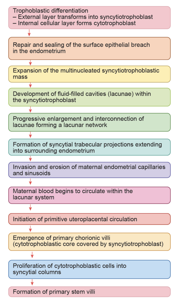
Changes in Trophoblasts
- By the eighth day after fertilization, the blastocyst is partially embedded in the endometrium.
- The trophoblast differentiates into two distinct layers.
- The inner layer is the cytotrophoblast, composed of mitotically active mononuclear cells.
- The outer layer is the syncytiotrophoblast, which consists of a multinucleated mass without clear cell boundaries.
- Cells from the cytotrophoblast proliferate and fuse with the outer layer, contributing to the growth of the syncytiotrophoblast.
- The syncytiotrophoblast secretes human chorionic gonadotropin. This hormone supports the corpus luteum during early pregnancy and maintains progesterone production. Elevated levels of this hormone are associated with early pregnancy symptoms such as nausea and vomiting.
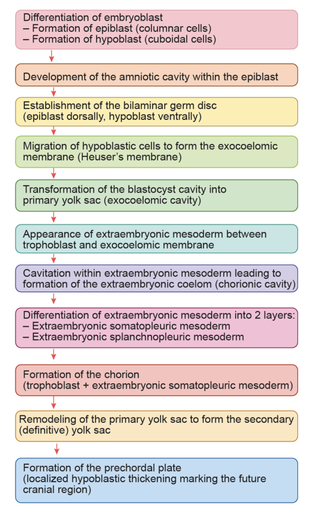
Changes in Embryoblast
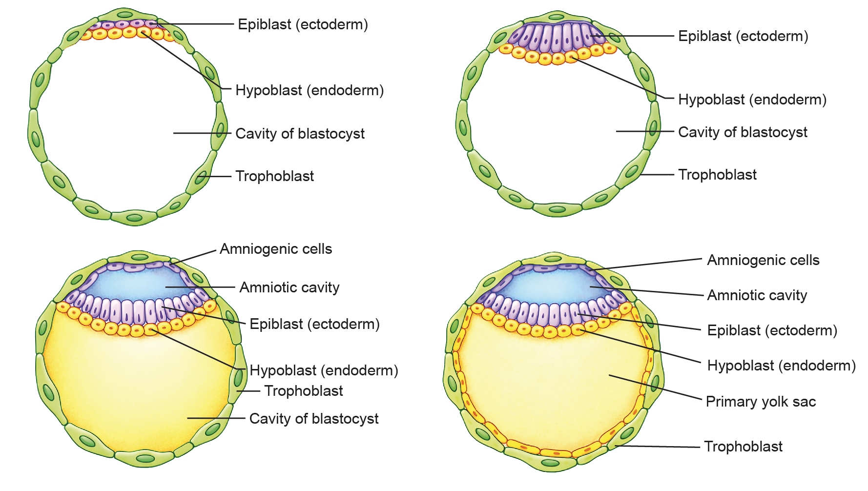
- The embryoblast or inner cell mass differentiates into two distinct layers.
- Hypoblast, which consists of flat to low cuboidal cells facing the blastocoel. It is the earliest layer to appear during this stage of development.
- Epiblast, formed by columnar cells located adjacent to the cytotrophoblast at the embryonic pole. The epiblast is also referred to as the primitive ectoderm.
- A space develops within the epiblast and enlarges to form the amniotic cavity. The epiblast cells lining the roof of this cavity flatten and are called amnioblasts, which contribute to formation of the amnion. The remaining epiblast cells form the floor of the cavity.
- With the formation of the amniotic cavity and differentiation of hypoblast and epiblast, the embryo becomes organized into a bilaminar germ disc. The junction between the amnioblasts and the epiblast is termed the amnioectodermal junction. Establishment of this bilaminar arrangement defines the dorsoventral axis of the developing embryo.
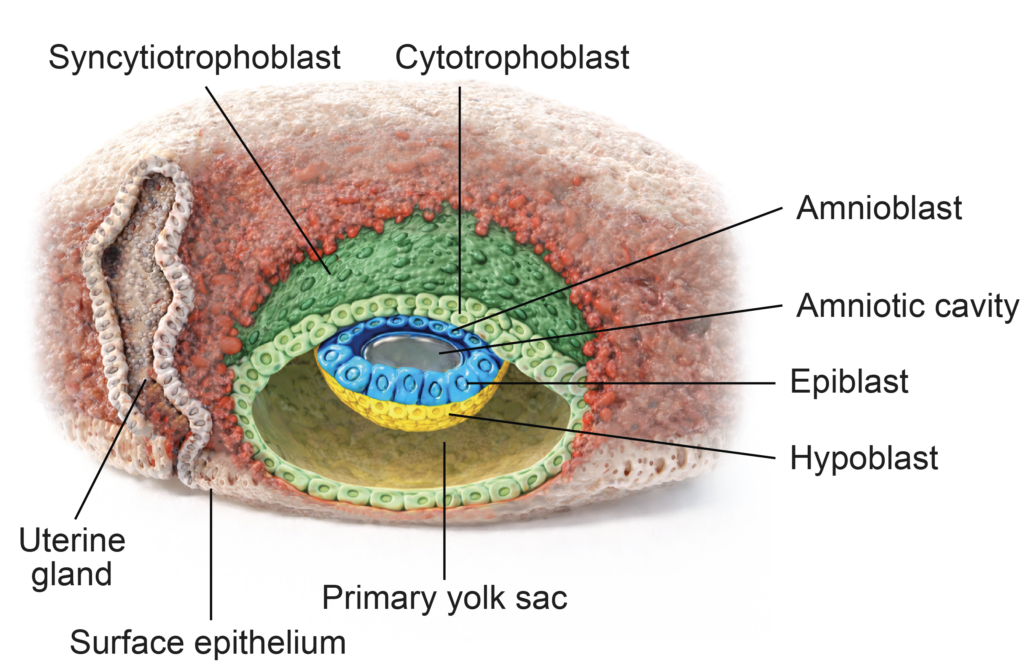
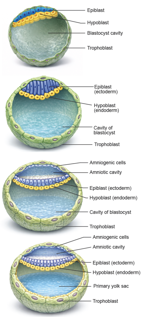
Figure 5.2a: Line diagram: Formation of ectoderm, endoderm, amniotic cavity and primary yolk sac (Click to see figure)
CHANGES ON DAY 9–10
Changes in Trophoblasts
- By the ninth and tenth day, the implanted blastocyst becomes more deeply embedded within the endometrium. The small surface defect created during implantation is sealed by a fibrin coagulum.
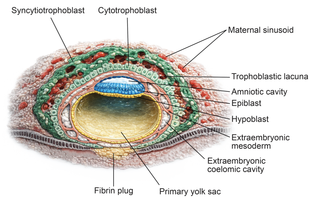
- The syncytiotrophoblast expands rapidly, particularly at the embryonic pole. Its cells proliferate and fuse, losing their distinct cell boundaries. This results in a continuous multinucleated mass without visible cell membranes.
- At this stage, multiple small cavities appear within the syncytiotrophoblast. These cavities, known as lacunae, enlarge and merge to form interconnected spaces. This phase is called the lacunar stage of trophoblast development and marks the early establishment of maternal blood circulation within the developing placenta.
Changes in Embryoblast
- At the abembryonic pole, cells of the hypoblast proliferate and migrate along the inner surface of the cytotrophoblast, facing the blastocyst cavity. These flattened cells form a thin lining called the exocoelomic membrane (also known as Heuser’s membrane).
- The hypoblast cells that remain adjacent to theepiblast retain a cuboidal shape and are termed visceral hypoblast cells.
- Following formation of the exocoelomic membrane, the original blastocyst cavity is transformed into the primary yolk sac, also called the exocoelomic cavity. At this stage, the bilaminar germ disc lies between the amniotic cavity above and the primary yolk sac below.
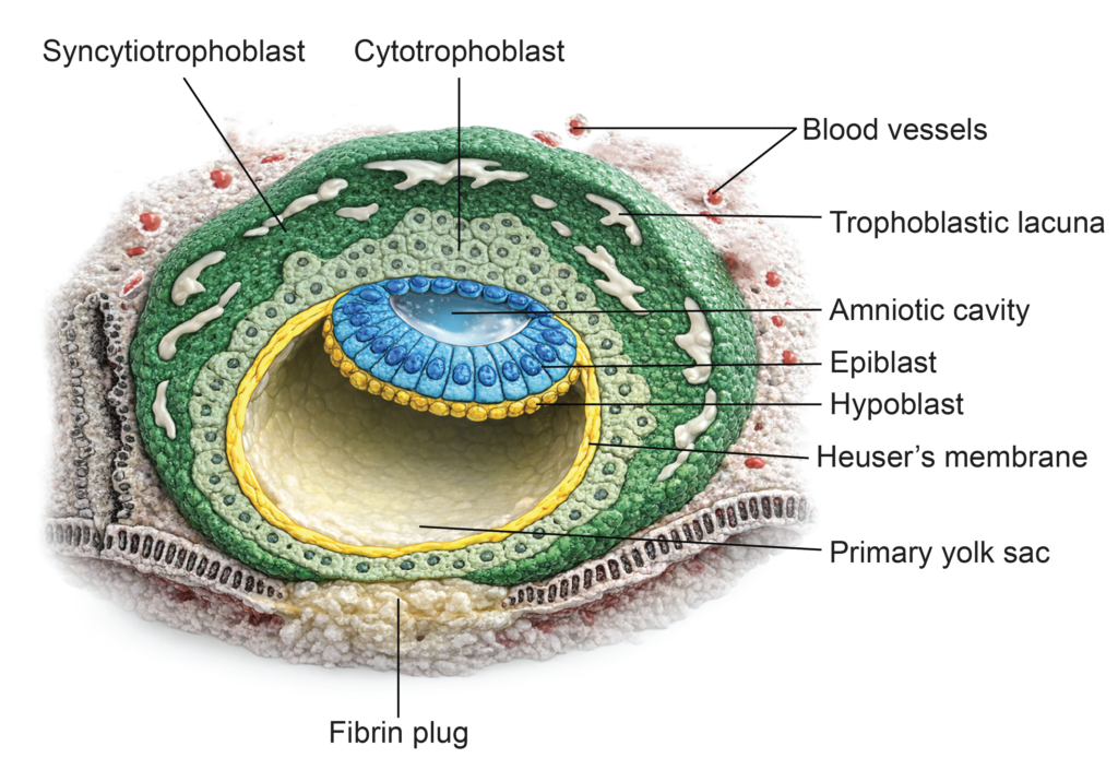

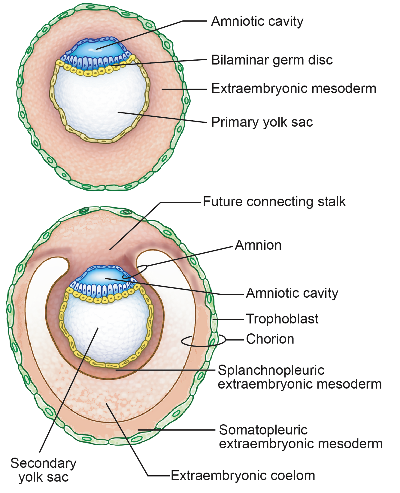
Figure 5.4a: Line diagram: Formation of extraembryonic mesoderm, extraembryonic coelom, amnion and chorion (Click to see figure)
CHANGES ON DAY 11–12
Changes in Trophoblasts
- By the eleventh and twelfth day, the surface endometrial epithelium has completely regenerated, and the implantation site is no longer visible.
- The lacunae within the syncytiotrophoblast enlarge further and become interconnected. These changes are more marked at the embryonic pole. The syncytiotrophoblast forms cellular strands known as trabeculae, which extend between the lacunar spaces.
- During this period, the syncytiotrophoblast invades and erodes maternal capillaries, venules, and later small arterioles. Maternal blood enters the lacunar system, filling these spaces. Erosion of veins generally precedes that of arteries. The presence of maternal blood within the lacunae establishes the early uteroplacental circulation, an essential step in placental development.
Changes in Embryoblast
- Cells associated with the primary yolk sac proliferate and form a layer of loosely arranged connective tissue between the cytotrophoblast and the amnion and yolk sac. This layer is called the extraembryonic mesoderm. It spreads around these structures and separates them from the cytotrophoblast.
- Small cavities then appear within the extraembryonic mesoderm. These spaces fuse to form a single large cavity known as the extraembryonic coelom, also called the chorionic cavity.
- As the chorionic cavity enlarges, it separates the amniotic cavity and yolk sac from the cytotrophoblast, except at the caudal end of the bilaminar germ disc. In this region, a band of extraembryonic mesoderm persists and forms the connecting stalk, which later develops into the umbilical cord.
- Formation of the chorionic cavity divides the extraembryonic mesoderm into two layers. The somatopleuric (somatic) extraembryonic mesoderm lines the cytotrophoblast and amnion. The splanchnopleuric (splanchnic) extraembryonic mesoderm surrounds the yolk sac.
CHANGES ON DAY 13–14
Changes in Trophoblasts
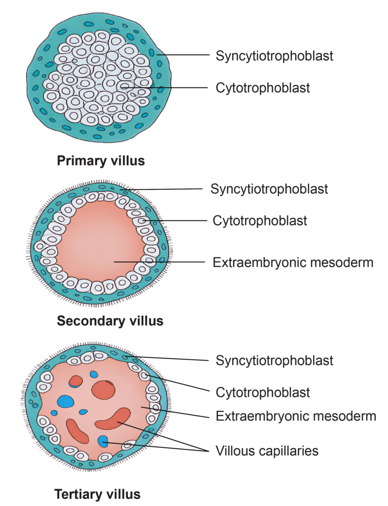
- The syncytiotrophoblast develops finger-like projections that extend into the surrounding tissue. These projections are called chorionic villi and are located between adjacent lacunar spaces. Cells of the cytotrophoblast grow into these projections, forming the primary villi.
- Each primary villus consists of a central core of cytotrophoblast covered externally by syncytiotrophoblast.
- By this stage, the surface defect in the endometrium caused by implantation has completely healed.
Changes in Trophoblasts
- As the chorionic cavity enlarges, part of the primary yolk sac is constricted and separated. The remaining smaller cavity is called the secondary yolk sac. The detached portions persist temporarily as small exocoelomic cysts within the chorionic cavity.
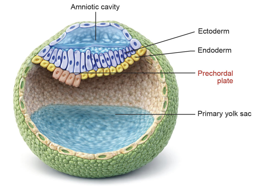
- At approximately 14 days, the embryo appears as a flat bilaminar germ disc, composed of epiblast and hypoblast. It lies between the floor of the amniotic cavity above and the roof of the secondary yolk sac below.
- Near the cranial end of the germ disc, a localized area of hypoblast cells becomes columnar and thickened. This region is known as the prechordal plate.
- The prechordal plate represents the earliest morphological sign of regional specialization. It establishes the cephalocaudal axis of the embryo and plays an essential role in the development of the future head region.

ENDOMETRIAL CHANGES
Implantation and Decidual Reaction
- During implantation, the blastocyst penetrates the endometrium through the invasive activity of the trophoblast. By approximately the 12th day of development, it becomes completely embedded within the endometrial stroma.
- The surface defect created during penetration is initially sealed by a fibrin clot. Subsequently, the endometrial epithelium regenerates and restores surface continuity.
- The implanted blastocyst lies within the stratum compactum of the endometrium. During pregnancy, the endometrium is termed the decidua because it is shed after delivery. The portion directly beneath the implanted blastocyst is called the decidua basalis. The trophoblast invades this region, which later contributes to the maternal component of the placenta.
Decidual Reaction
- The decidual reaction refers to structural and functional changes in the endometrium that facilitate implantation and early embryonic development.
- Under the influence of pregnancy hormones, especially progesterone, endometrial stromal cells enlarge and accumulate glycogen and lipid droplets. The amount of intracellular material increases, and the tissue becomes edematous due to fluid accumulation. In addition, the endometrial glands become enlarged and more tortuous.
- These changes create a supportive environment for the implanted embryo and early placental development.
Important Questions
- Write a short note on bilaminar germ disc or bilaminar blastocyst.
- Write a short note on prechordal plate.
- What is chorion?
- Explain decidual reaction.
- List the trophoblastic changes in the second week.
- Explain ‘why conceptus is not rejected by its mother’?