Competencies
- AN79.1: Describe the formation and fate of primitive streak.
- AN79.2: Describe the formation and fate of notochord.
- AN79.3: Describe the process of neurulation.
- AN79.4: Describe the development of somites and intraembryonic coelom.
- AN79.5: Explain embryological basis of congenital malformations, nucleus pulposus, sacrococcygeal teratomas, neural tube defects.
INTRODUCTION
- The third week of embryonic development corresponds to about five weeks after the first day of the last menstrual period in obstetric dating. This week marks the beginning of major structural organization of the embryo.
- This week is critical because it establishes the basic body plan and initiates the formation of major organ systems.
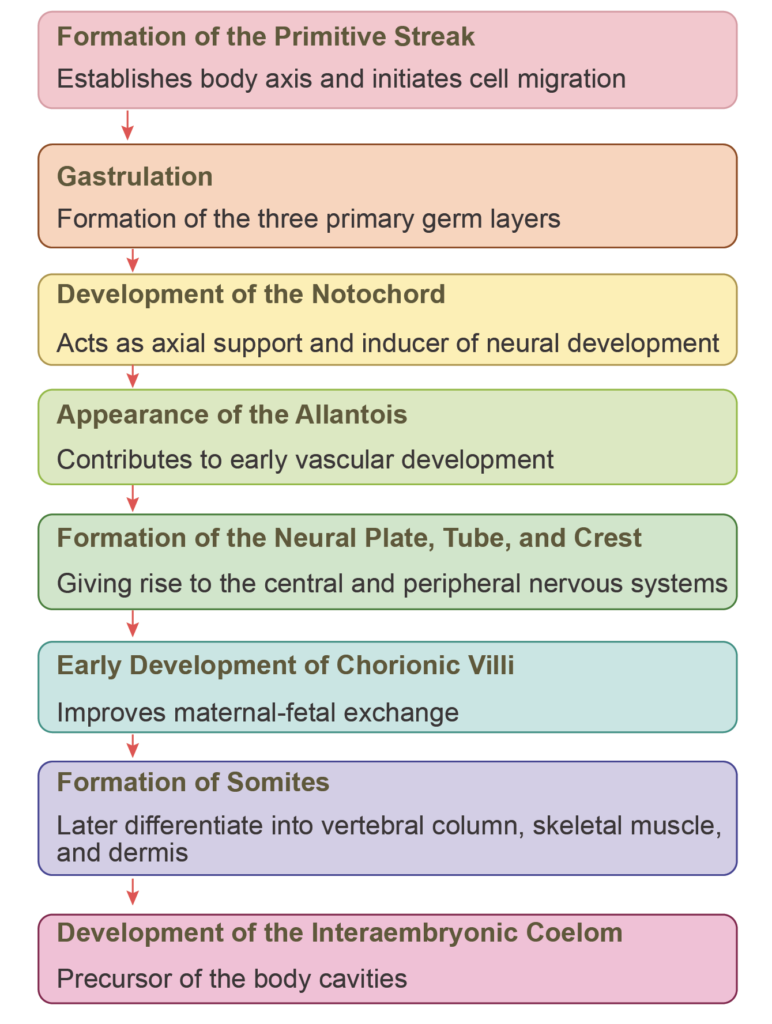
- The main events of the third week include:
- Formation of the primitive streak, which establishes the body axis and initiates cell migration
- Gastrulation, during which the three primary germ layers—ectoderm, mesoderm, and endoderm—are formed
- Development of the notochord, which acts as the axial support and an inducer of neural development
- Appearance of the allantois, contributing to early vascular development
- Formation of the neural plate, followed by the neural tube and neural crest cells, which give rise to the central and peripheral nervous systems
- Early development of chorionic villi, improving maternal–fetal exchange
- Formation of somites, which later differentiate into the vertebral column, skeletal muscle, and dermis
- Development of the intraembryonic coelom, the precursor of the body cavities
GASTRULATION (FORMATION OF GERM LAYERS)
Definition:
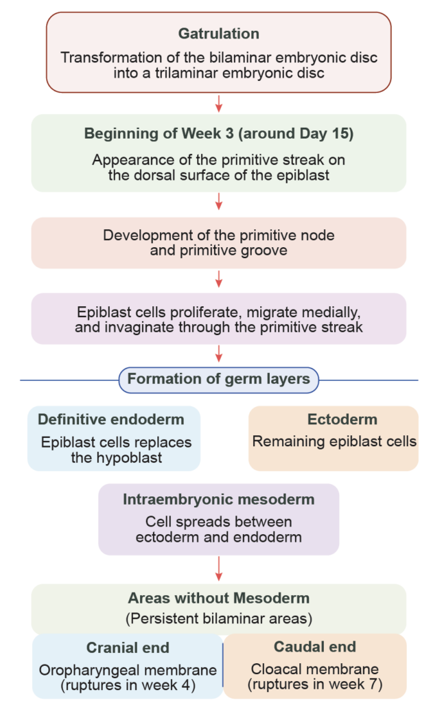
- Gastrulation is the process by which the bilaminar embryonic disc is transformed into a trilaminar disc during the third week of development. This process establishes the three primary germ layers and initiates body patterning.
Sequence of Events
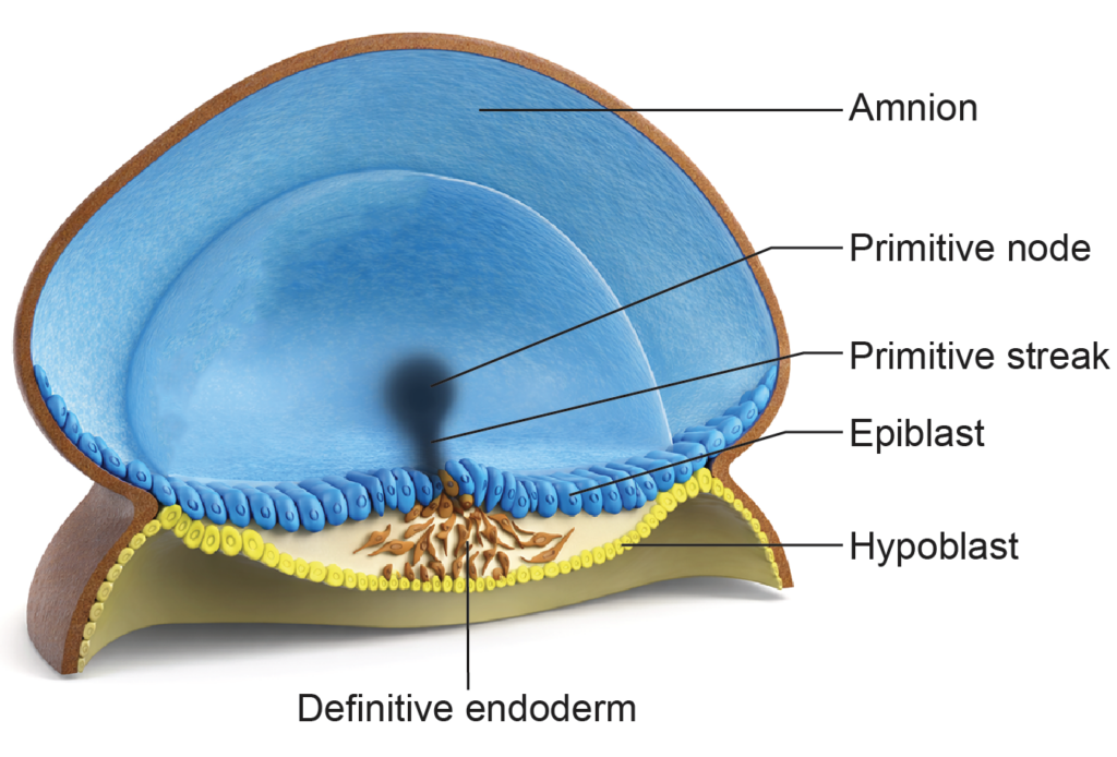
- The bilaminar disc of the second week continues to grow and reorganize.
- Around the 15th day, a linear thickening appears in the midline of the epiblast at the caudal end. This structure is the primitive streak. Its formation marks the beginning of gastrulation.
- The cranial end of the primitive streak enlarges to form the primitive node (also called Hensen’s node). A small central depression within it is the primitive pit. A shallow longitudinal depression along the streak is the primitive groove.
Establishment of Body Axes
- The appearance of the primitive streak defines the main body axes:
- Craniocaudal axis
- Mediolateral axis
- Dorsoventral axis
- As development proceeds, both the embryonic disc and the primitive streak elongate in the craniocaudal direction.
Formation of Germ Layers
- Cells of the epiblast migrate toward the primitive streak. They detach from neighboring cells, lose their epithelial connections, and undergo invagination.
- The first migrating cells displace the hypoblast and form the definitive endoderm.
- Subsequent migrating cells spread between the newly formed endoderm and the epiblast to form the intraembryonic mesoderm. This occurs through epithelial-to-mesenchymal transition (EMT).
- The remaining epiblast cells that do not migrate become the definitive ectoderm.
- Thus, the epiblast gives rise to all three germ layers: ectoderm, mesoderm, and endoderm. Gastrulation therefore establishes the basic structural framework of the embryo.
- During gastrulation, migrating intraembryonic mesoderm spreads laterally and merges with the extraembryonic mesoderm at the margins of the embryonic disc. As it expands, the mesoderm separates the ectoderm from the endoderm throughout most of the disc. However, two regions remain bilaminar, where mesoderm does not interpose between the two layers.
Areas Lacking Mesoderm
- Buccopharyngeal membrane (or oropharyngeal membrane): This membrane is located at the cranial end of the embryonic disc within the region of the prechordal plate. It appears as a small oval area where ectoderm and endoderm are firmly adherent, without intervening mesoderm.
It remains bilaminar. During the fourth week, this membrane ruptures, creating the opening of the primitive oral cavity (stomatodeum). - Cloacal membrane: This structure is present at the caudal end of the embryonic disc, near the termination of the primitive streak. Here again, ectoderm and endoderm remain directly attached, forming a circular bilaminar region.
With further development, it is subdivided into the anal membrane and the urogenital membrane. The cloacal membrane breaks down around the seventh week, forming the external openings of the anus, urethra, and parts of the genital tract.
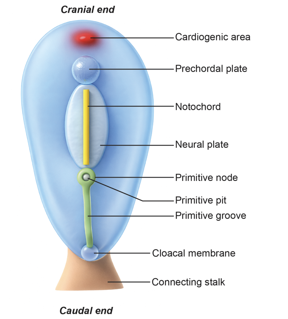
Pericardial bar
- The pericardial bar is a horseshoe-shaped condensation of mesoderm located in the midline, cranial to the buccopharyngeal membrane. It represents the early cardiogenic region and contributes to the formation of the primitive heart.
NOTOCHORD
Definition
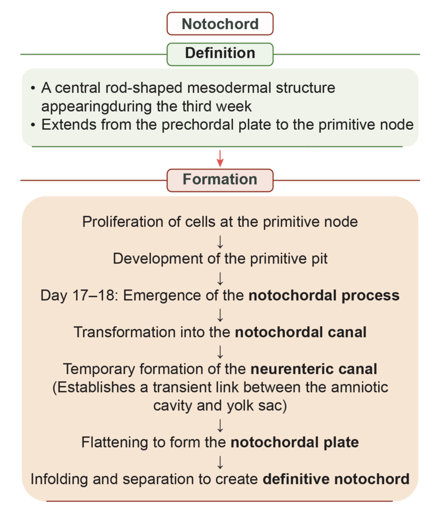
- The notochord is a midline embryonic structure formed during the third week of development. It arises from epiblast cells that migrate through the primitive node.
- It develops from the axial mesoderm, also called chordamesoderm, which lies along the central longitudinal axis of the embryo. The notochord serves as the primary axial support and plays an essential role in neural tube formation and vertebral column development.
Extent
- The notochord extends:
- From the cranial end of the primitive streak
- To the prechordal plate
Sequential Events in the Formation of Notochord
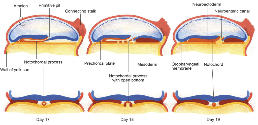
- Primitive pit formation: Cells of the primitive node proliferate and create a small central depression known as the primitive pit.
- Notochordal process: Around the 17th to 18th day, cells from the primitive node invaginate and migrate cranially between the ectoderm and endoderm. They form a hollow cellular tube called the notochordal process (also termed the head process), which extends up to the prechordal plate.
- Notochordal canal: The cavity of the primitive pit extends into the notochordal process, forming the notochordal canal. The ventral wall of this canal fuses temporarily with the underlying endoderm.
- Neurenteric canal: Degeneration of the fused ventral wall creates a transient communication between the amniotic cavity and the yolk sac. This passage is known as the neurenteric canal.
- Notochordal plate: The remaining dorsal part of the notochordal process flattens to form the notochordal plate, which lies in the roof of the yolk sac.
- Definitive notochord: The notochordal plate then folds inward and detaches from the endoderm, forming a solid rod of cells called the definitive notochord.
The definitive notochord defines the central axis of the embryo and contributes to the development of the vertebral column and intervertebral discs.
Significance of Notochord
- Its main functions are:
- Establishes the longitudinal body axis, including the craniocaudal, anteroposterior, and right–left axes.
- Provides the structural template for the axial skeleton, particularly contributing to the formation of the vertebral bodies.
- Acts as a primary inductive structure, stimulating the overlying ectoderm to form the neural plate and influencing surrounding mesodermal differentiation.
- In humans, most of the notochord degenerates as the vertebral column develops. Persistent remnants form the nucleus pulposus of the intervertebral discs. Rarely, residual notochordal cells may give rise to a tumor known as a chordoma.
Importance of the Neurenteric Canal
- The neurenteric canal is a temporary communication between the amniotic cavity and the yolk sac during early development.
- It may allow limited exchange of fluid and nutrients before the establishment of effective intraembryonic circulation, which is not yet fully developed during the third week. The canal normally disappears as development progresses.
ALLANTOIS
Definition
- The allantois, also called the allantoenteric diverticulum, is a small tubular outgrowth arising from the posterior wall of the yolk sac and extending into the connecting stalk during early embryonic development.
Development and Fate
- Around the 16th day of development, a finger-like diverticulum grows from the yolk sac into the connecting stalk to form the allantois. With formation of the tail fold, the intraembryonic part of the allantois becomes continuous with the cloaca, the terminal part of the hindgut.
- As development proceeds:
- The proximal portion of the allantois becomes incorporated into the developing urinary bladder.
- The remaining tubular segment forms the urachus, a fibrous cord connecting the bladder to the umbilicus.
- After birth, the urachus becomes the median umbilical ligament.
Functional Significance of Allantois
- In birds and reptiles, the allantois serves as a reservoir for nitrogenous waste.
- In humans, it remains small because the placenta performs the excretory function.
- Blood vessels developing within the allantois contribute to the formation of the umbilical arteries and vein.
Clinical Correlations of Allantois
- Incomplete obliteration of the urachus may result in:
- Urachal cyst – localized cystic dilation along the urachus.
- Urachal sinus – persistent opening at the umbilical end.
- Urachal fistula – complete patency forming a direct connection between the urinary bladder and the umbilicus.
- These anomalies arise from persistence of allantoic remnants.
DEVELOPMENT OF CHORIONIC VILLI
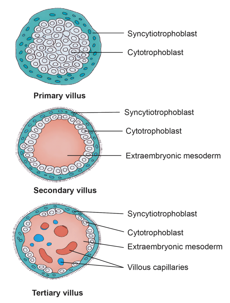
- During the second week of development, projections called primary chorionic villi arise from the trophoblast. Each primary villus consists of an inner layer of cytotrophoblast covered by syncytiotrophoblast.
- At the beginning of the third week, extraembryonic mesoderm grows into the core of the primary villi. These structures are then termed secondary villi.
- By the end of the third week, mesodermal cells within the villi differentiate into blood vessels and capillaries. Villi that contain fetal capillaries are called tertiary (definitive) chorionic villi. These villi establish the structural basis for placental circulation.
Types of Chorionic Villi
- Anchoring villi
Extend from the chorionic plate to the decidua basalis.
The cytotrophoblast cells from these villi proliferate and form a cytotrophoblastic shell, which secures the chorion to the maternal endometrium. - Free (floating) villi
Arise as branches of anchoring villi.
Project into the intervillous space and are surrounded by maternal blood.
These villi are the principal sites for maternal–fetal exchange.
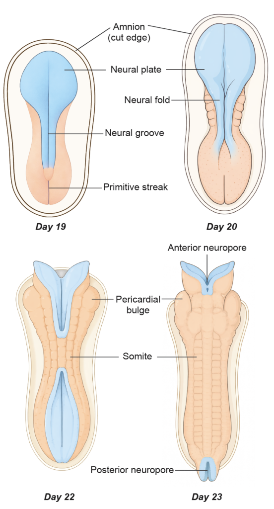
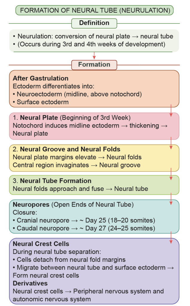
FORMATION OF NEURAL TUBE (NEURULATION)
Definition
- Neurulation is the process by which the neural plate forms, elevates into neural folds, and fuses to create the neural tube.
Process of Neurulation
After gastrulation, the ectoderm differentiates into:
- Neuroectoderm in the midline, located above the notochord
- Surrounding surface ectoderm
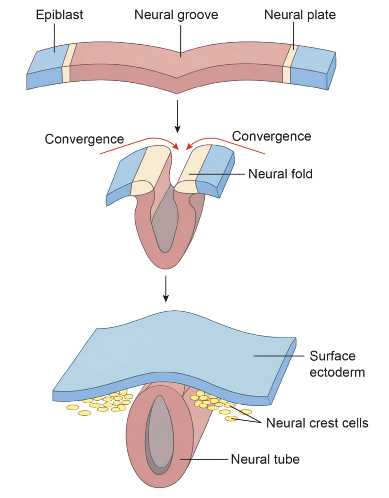
1. Formation of the Neural Plate
- At the beginning of the third week, the midline ectoderm between the primitive node and prechordal plate thickens under the inductive influence of the notochord. This thickened region forms the neural plate (medullary plate).
2. Formation of the Neural Groove and Neural Folds
- The lateral edges of the neural plate elevate to form neural folds, while the central region invaginates to form the neural groove.
3. Formation of the Neural Tube
- The neural folds approach each other and fuse, converting the neural plate into the neural tube.
- Fusion begins near the future cervical region, around the level of the fifth somite.
- Closure proceeds in both cranial and caudal directions.
- Current evidence supports initiation of closure at multiple sites, which then progress simultaneously.
Neuropores
The open ends of the neural tube are called:
- Cranial neuropore
- Caudal neuropore
Closure timeline:
- Cranial neuropore: around day 25 (approximately 18–20 somites)
- Caudal neuropore: around day 27 (approximately 24–25 somites)
Before complete closure, amniotic fluid can communicate with the neural canal.
Formation of Neural Crest Cells
During separation of the neural tube from the surface ectoderm:
- Cells from the lateral margins of the neural folds detach.
- These cells migrate between the neural tube and surface ectoderm.
- They are termed neural crest cells.
- They undergo epithelial-to-mesenchymal transition (EMT), enabling migration.
Derivatives
- Neural tube → Central nervous system
- Neural crest cells → Major components of the peripheral nervous system and autonomic nervous system, along with several other structures
This sequence ensures proper formation of the early central nervous system during the third and fourth weeks of development.
DEVELOPMENT OF CHORIONIC VILLI
Definition
- The neural crest is a group of specialized cells that arise from the lateral edges of the neural plate, at the junction between the developing neural tissue and the surface ectoderm. These cells appear during early neurulation and temporarily occupy a position between the neural tube and the surface ectoderm.
Process of Formation
- During formation of the neural tube, the neural plate folds and separates from the surface ectoderm. At this stage, cells located at the crest of the neural folds detach from the neural tube. These cells constitute the neural crest cells.
- After separation, neural crest cells migrate to different regions of the embryo. They proliferate and may organize into:
- Dorsal cell groups
- Ventral cell groups
- Neural crest cells differentiate into a wide range of tissues throughout the body. Because of their extensive migratory capacity and diverse derivatives, the neural crest is often referred to as the fourth germ layer, although it is derived from ectoderm.
Significance of neural crest
- The neural crest contributes to multiple structures, including components of the peripheral nervous system, certain craniofacial structures, and specific endocrine cells. Its proper development is essential for normal embryogenesis.
FURTHER DEVELOPMENT IN THE THIRD WEEK
- During the third week of intrauterine life, major structural changes occur in the embryo.
- Formation of the neural tube begins early in this week and is usually completed by the 27th day. This process establishes the primordium of the central nervous system.
- At the same time, the mesoderm differentiates into three distinct regions:
- Paraxial mesoderm
- Intermediate mesoderm
- Lateral plate mesoderm
- A curved, horseshoe-shaped cavity called the intraembryonic coelom develops within the lateral plate mesoderm. This cavity divides the lateral plate into two layers:
- Somatopleuric (parietal) layer
- Splanchnopleuric (visceral) layer
- Toward the end of the third week, embryonic folding begins. This folding converts the flat trilaminar disc into a more cylindrical body form and helps establish the basic body plan.
Important Questions
- Outline the major developmental events of the third week of gestation.
- Write short notes on the following early embryonic structures and processes: a. Gastrulation b. Prochordal plate c. Primitive streak.
- Describe the development and functions of the notochord.
- List the principal midline structures present in the embryonic germ disc during early development.
- Write a brief note on the allantois, including its origin and developmental significance.
- Describe chorionic villi and explain their role in placental development.
- Define neurulation and outline the sequential steps involved in this process.
- Write a short note on neural crest cells, including their origin and major derivatives.