
Competencies
- AN52.4: Describe the development of anterior abdominal wall.
- AN52.6: Describe the development and congenital anomalies of foregut, midgut and hindgut
Introduction
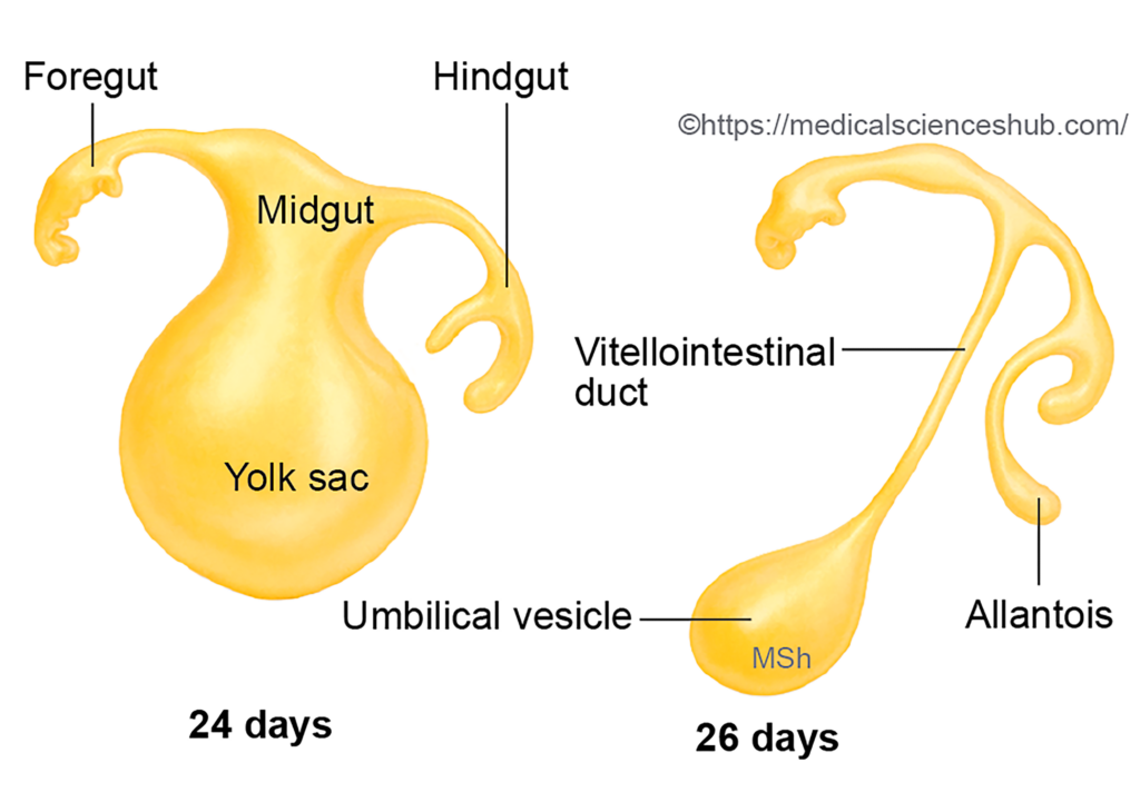
- The alimentary canal develops from the primitive gut, an endoderm-lined tube formed during early embryonic development.
- During formation of the head fold and tail fold, a portion of the definitive yolk sac becomes incorporated into the embryo. This incorporated segment forms the primitive gut.
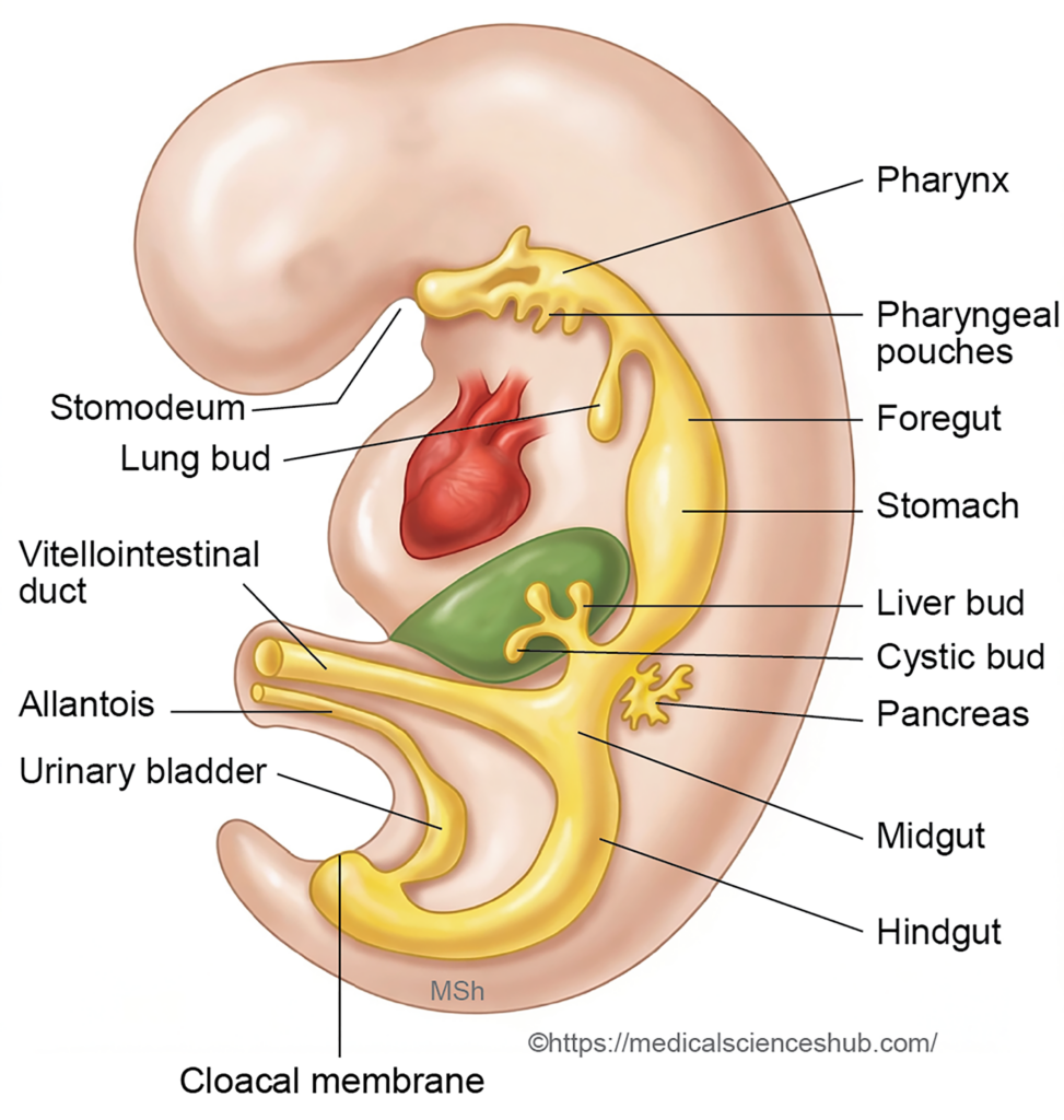
- The primitive gut extends from the buccopharyngeal membrane at the cranial end to the cloacal membrane at the caudal end. In both membranes, ectoderm and endoderm are in direct contact without an intervening mesodermal layer.
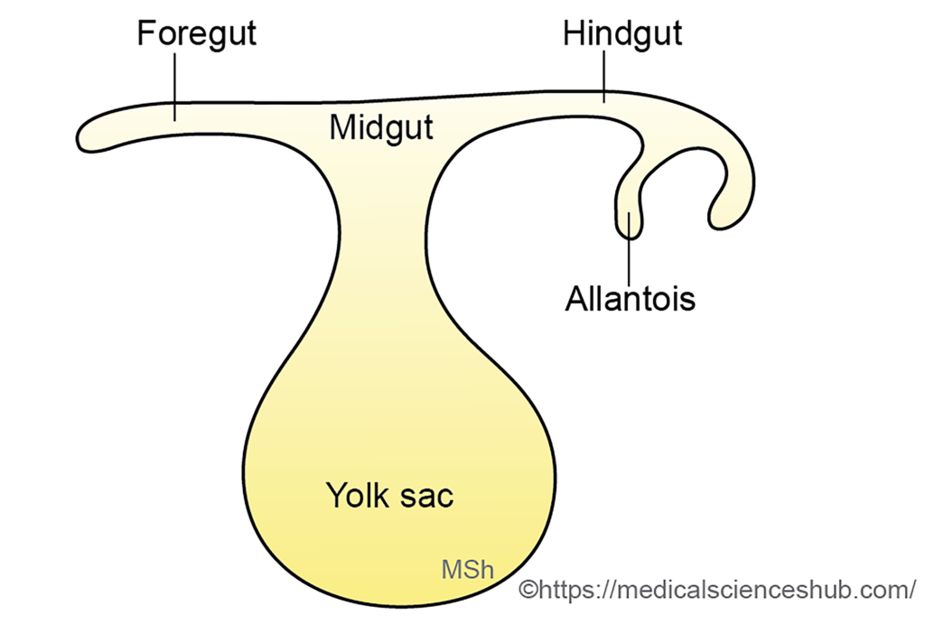
- The developing primitive gut communicates with:
- The yolk sac through the vitellointestinal (vitelline) duct.
- The allantoic diverticulum, which arises from the caudal part of the gut.
- Based on its location and connections, the primitive gut is divided into three regions:
- Foregut – the cranial segment located within the head fold.
- Midgut – the segment that remains temporarily connected to the yolk sac through the vitellointestinal duct.
- Hindgut – the caudal segment situated within the tail fold.
- The vitellointestinal duct normally degenerates and disappears by the fifth week of development.
- The anterior (cranial) intestinal portal connects the foregut with the midgut, while the posterior (caudal) intestinal portal connects the midgut with the hindgut.
- The buccopharyngeal membrane separates the stomodeum (primitive oral cavity) from the foregut, whereas the cloacal membrane separates the hindgut from the proctodeum.
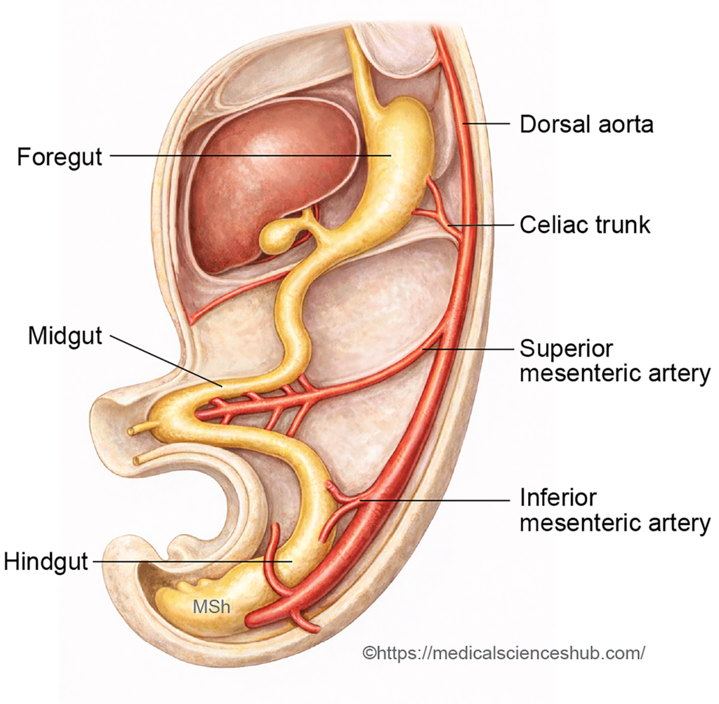
- The gut tube is suspended from the body wall by mesenteries, initially consisting of ventral and dorsal mesenteries.
- The arterial supply reflects the embryological divisions of the gut:
- Celiac trunk supplies the foregut.
- Superior mesenteric artery supplies the midgut.
- Inferior mesenteric artery supplies the hindgut.
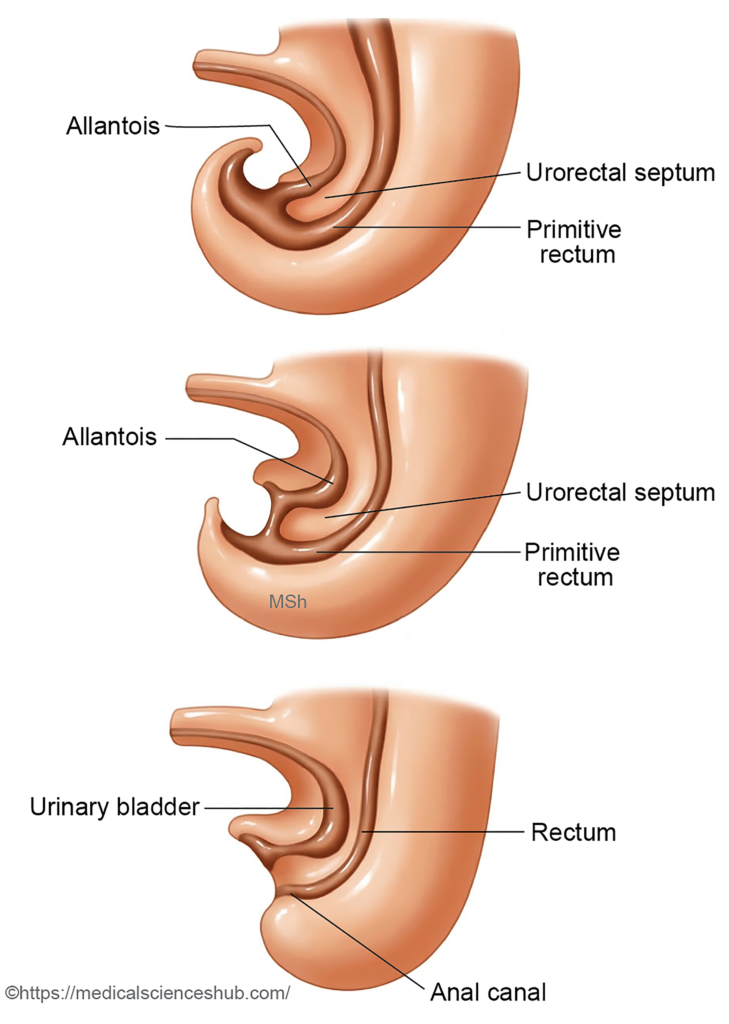
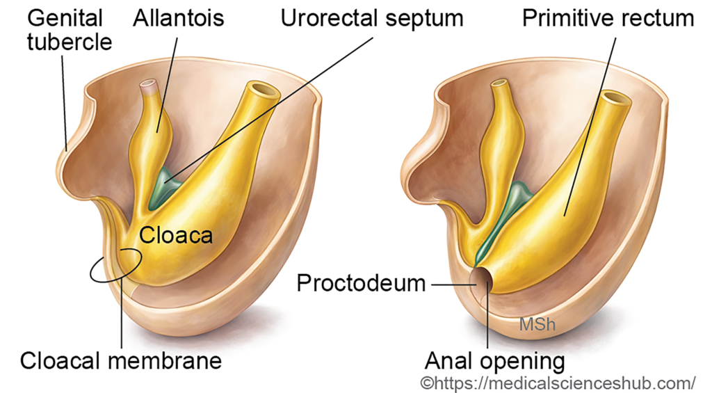
- In early development, the allantois opens into the caudal part of the hindgut called the cloaca. The urorectal septum subsequently divides the cloaca into a ventral primitive urogenital sinus and a dorsal primitive anorectal canal.
- Fusion of the urorectal septum with the cloacal membrane subdivides it into a ventral urogenital membrane and a dorsal anal membrane.
Development Of Oesophagus
The oesophagus develops from the segment of the foregut located between the pharynx and the stomach.
Derivation of Components
- Epithelium is derived from the endoderm of the foregut.
- Muscle layers and connective tissue originate from the surrounding splanchnopleuric mesoderm.
Stages of Development
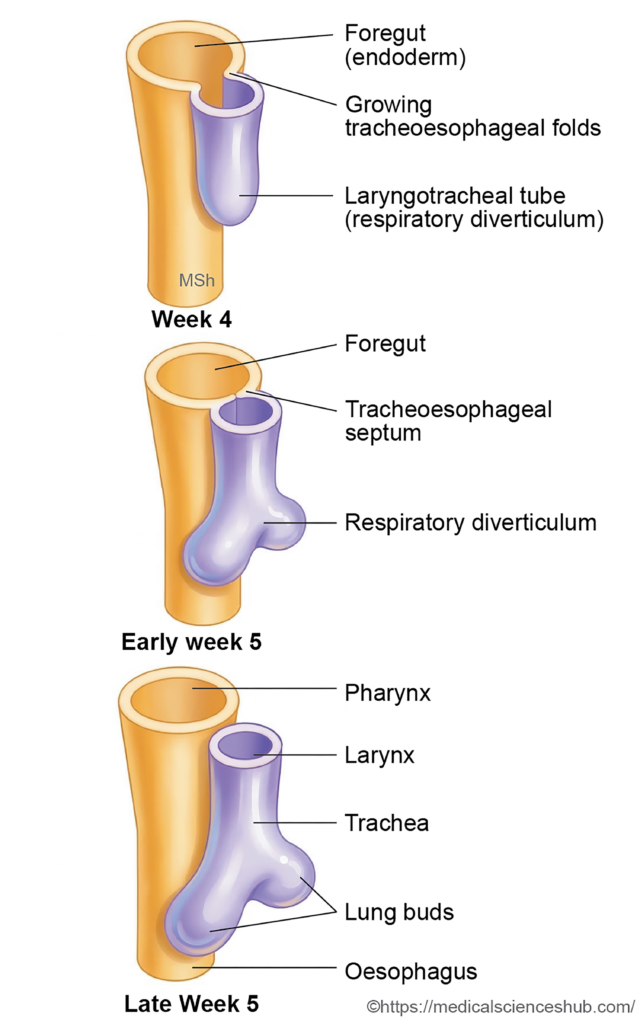
- In the lower region of the primitive pharynx, a laryngotracheal groove appears. This groove later forms the tracheobronchial (respiratory) diverticulum.
- A tracheoesophageal septum develops and divides the foregut segment below the diverticulum into two parts: a ventral trachea and a dorsal oesophagus.
- Initially the oesophagus is short, but it lengthens as the neck develops and the diaphragm, lungs, and heart descend during embryonic growth.
- The enteric nerve plexuses—Auerbach’s (myenteric) plexus and Meissner’s (submucosal) plexus—are derived from neural crest cells.
Muscular Arrangement
The muscular composition of the oesophagus varies along its length:
- The upper one-third contains striated muscle.
- The middle one-third contains a mixture of striated and smooth muscle.
- The lower one-third consists predominantly of smooth muscle.
CLINICAL EMBRYOLOGY
Congenital Anomalies of Oesophagus
- Oesophageal atresia
- This condition results from incomplete formation of a segment of the oesophagus, producing a discontinuous tube. The proximal segment usually ends as a blind pouch. A characteristic clinical feature in newborns is persistent excessive salivation and regurgitation during feeding.
- Oesophageal stenosis
- This anomaly involves narrowing of the oesophageal lumen, commonly due to incomplete recanalization during development. Severe obstruction may interfere with fetal swallowing of amniotic fluid, which can contribute to polyhydramnios.
- Tracheoesophageal fistula
- This defect occurs when the tracheoesophageal septum fails to develop properly, creating an abnormal communication between the trachea and oesophagus. The common pattern includes a blind-ending proximal oesophagus with the distal segment opening into the trachea.
- Achalasia cardia (cardiospasm)
- Degeneration or absence of ganglion cells in the Auerbach’s (myenteric) plexus prevents normal relaxation of the lower oesophageal sphincter. This leads to functional obstruction of the oesophagus. A barium swallow examination typically demonstrates a characteristic “bird-beak” narrowing at the lower end of the oesophagus.
Development Of Stomach
Stages of Development
- The stomach begins to develop during the 4th–5th week as a fusiform dilation of the foregut located distal to the oesophagus.
- Initially, the tubular foregut enlarges to form a spindle-shaped sac with distinct ventral and dorsal borders.
- The dorsal border grows more rapidly than the ventral border, producing the greater curvature of the stomach. In contrast, the slower-growing ventral border forms the lesser curvature.
- As development progresses, the stomach acquires its characteristic features, including anterior and posterior surfaces, the greater and lesser curvatures, and the fundus.
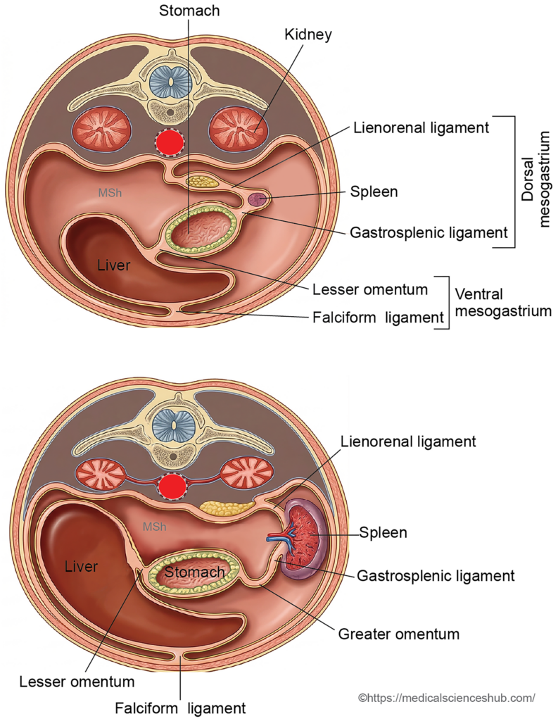
- The ventral border of the developing stomach is attached to the septum transversum through the ventral mesogastrium, while the dorsal border is connected to the posterior body wall by the dorsal mesogastrium.
- Several organs arise within these mesenteries. The spleen, celiac trunk, and dorsal pancreatic bud develop within the dorsal mesogastrium, whereas the liver, gallbladder, and ventral pancreatic bud develop within the ventral mesogastrium.
Rotation of the Stomach
The developing stomach undergoes two important rotations that determine its final anatomical position and relationships.
- First rotation (longitudinal axis):
The stomach rotates approximately 90° clockwise around its longitudinal axis. As a result, the left surface of the primitive foregut becomes the anterior surface of the adult stomach, while the right surface becomes the posterior surface. Consequently, the left vagus nerve supplies the anterior surface and the right vagus nerve supplies the posterior surface. - Second rotation (anteroposterior axis):
A further rotation occurs around the transverse (anteroposterior) axis. During this movement, the caudal end of the stomach shifts to the right and upward, forming the pyloric part, while the cranial end moves to the left, contributing to the final orientation of the stomach.
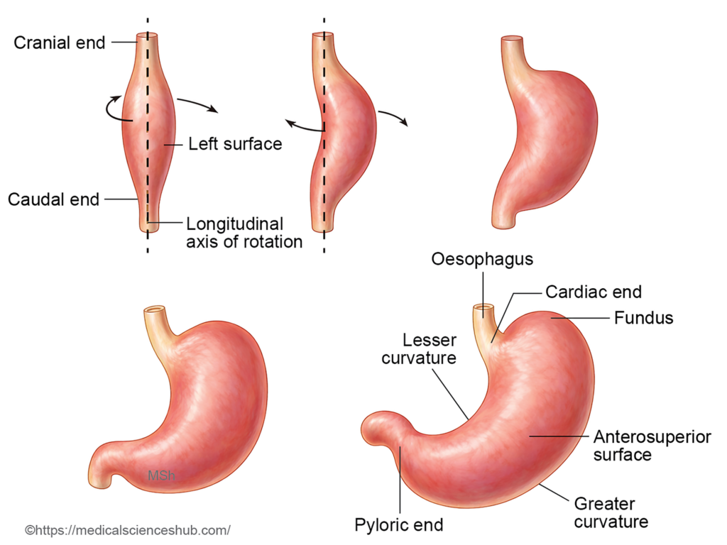
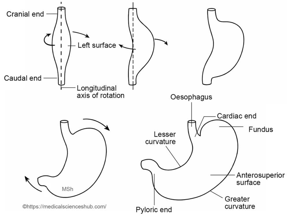

Changes in Mesenteries of Stomach
- The liver develops within the ventral mesogastrium during early embryonic life. As the liver enlarges, it divides the ventral mesogastrium into two main parts:
- Falciform ligament – extends between the liver and the anterior abdominal wall.
- Lesser omentum – connects the liver to the lesser curvature of the stomach and the proximal duodenum.
- The ventral mesogastrium also gives rise to the coronary ligament and triangular ligaments of the liver.
- The spleen develops within the dorsal mesogastrium. Its growth subdivides the dorsal mesogastrium into:
- Gastrosplenic ligament – located between the fundus and greater curvature of the stomach and the spleen; it contains the short gastric vessels.
- Lienorenal (splenorenal) ligament – extending between the spleen and the posterior abdominal wall, and containing the splenic vessels.
- The remaining part of the dorsal mesogastrium attached to the greater curvature elongates to form the greater omentum.
- A peritoneal recess called the lesser sac (omental bursa) develops behind the stomach, lesser omentum, and greater omentum.
Histogenesis of the Stomach
- The epithelial lining and gastric glands originate from the endoderm of the foregut.
- The muscular layers and connective tissue arise from the surrounding splanchnopleuric mesoderm.
CLINICAL EMBRYOLOGY
Congenital hypertrophic pyloric stenosis
- Congenital hypertrophic pyloric stenosis is a condition characterized by abnormal thickening (hypertrophy) of the circular muscle layer of the pylorus, leading to narrowing of the pyloric canal. It is one of the most common gastric outlet obstructions in infants.
- Incidence: The condition occurs in approximately 1 in 150 male infants and 1 in 750 female infants, demonstrating a clear male predominance.
- Clinical presentation: Affected infants usually appear normal at birth. Symptoms typically begin after feeding is established and include projectile, forceful vomiting occurring about 2–3 hours after feeding. The vomitus is non-bilious because the obstruction lies proximal to the duodenum.
- Treatment: Definitive management is surgical, most commonly pyloromyotomy, which relieves the obstruction by splitting the hypertrophied pyloric muscle.
- During stomach development, gastric glands begin to appear around the third month of intrauterine life, while specialized cells such as oxyntic (parietal) cells and zymogenic (chief) cells differentiate during the fourth month.
Development Of Duodenum
Stages of Development
The duodenum develops from two embryological regions of the primitive gut.
- The foregut forms the segment of the duodenum proximal to the opening of the bile duct.
- The midgut gives rise to the portion distal to the bile duct opening.
During development, rotation of the stomach shifts the duodenum toward the right side of the abdominal cavity. Initially, the duodenum is suspended from the posterior abdominal wall by a dorsal mesentery known as the mesoduodenum. As development proceeds, most of this mesentery undergoes zygosis (fusion with the posterior abdominal wall), causing the duodenum to become secondarily retroperitoneal. Only a small proximal segment near the pylorus remains intraperitoneal and mobile.
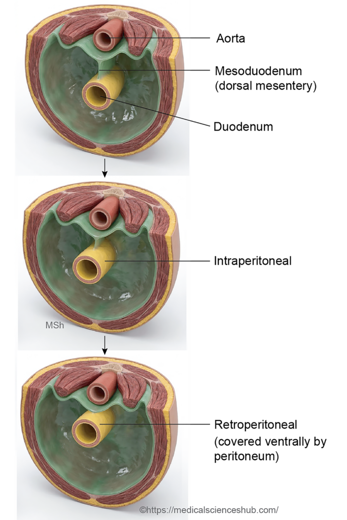
Development of the Lumen
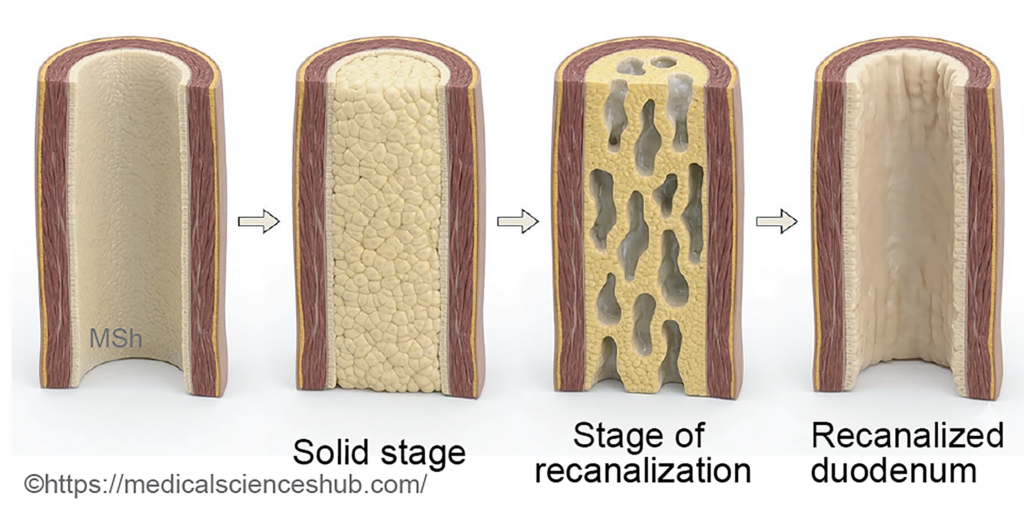
Rapid proliferation of endodermal epithelial cells temporarily obliterates the duodenal lumen around the 8th week of intrauterine life. The lumen subsequently reopens through recanalization, which is usually completed by the third month of fetal development.
Blood Supply
The dual embryological origin explains the arterial supply of the duodenum:
- The foregut-derived portion receives branches from the coeliac trunk.
- The midgut-derived portion is supplied by branches of the superior mesenteric artery.
CLINICAL EMBRYOLOGY
Anomalies of Duodenum
- Duodenal stenosis: It refers to partial narrowing of the duodenal lumen, usually caused by incomplete recanalization during embryonic development. Newborns commonly present with bilious vomiting because the obstruction typically lies distal to the entry of the bile duct.
- Duodenal atresia: It is the complete obstruction of the duodenal lumen, most often occurring distal to the hepatopancreatic ampulla. Affected infants develop persistent bilious vomiting soon after birth. This condition is the most common form of intestinal atresia and is frequently associated with polyhydramnios during pregnancy. On prenatal ultrasonography, dilation of the stomach and proximal duodenum produces the characteristic double-bubble sign.
- Duodenal diverticulum: It is an outpouching of the duodenal wall that most commonly arises from the second (descending) part of the duodenum. It results from localized weakness or developmental variation in the duodenal wall.
Development Of Midgut
The primitive midgut extends from the cranial intestinal portal to the caudal intestinal portal. It remains attached to the posterior abdominal wall by a dorsal mesentery. During early development, the midgut communicates with the yolk sac through the vitelline duct (also called the vitellointestinal duct or yolk stalk). Its arterial supply is provided by the superior mesenteric artery.
The superior mesenteric artery divides the midgut into two functional segments:
- Prearterial (proximal) segment
- Postarterial (distal) segment
Derivatives of the prearterial segment:
- Distal part of the duodenum
- Jejunum
- Most of the ileum
Derivatives of the postarterial segment:
- Terminal part of the ileum
- Caecum
- Appendix
- Ascending colon
- Proximal two-thirds of the transverse colon.
Rotation of Gut
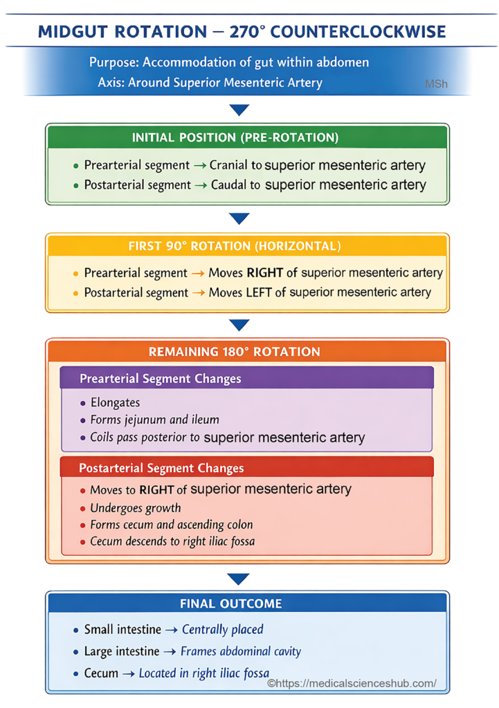
The midgut undergoes a total 270° counterclockwise rotation around the axis of the superior mesenteric artery during development. This rotation helps accommodate the rapidly growing midgut loop within the relatively small abdominal cavity.
Stages of Rotation
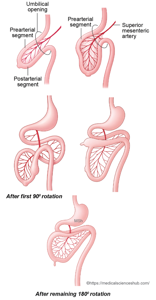
- The superior mesenteric artery divides the midgut into prearterial (proximal) and postarterial (distal) segments. Before rotation, the prearterial segment lies cranial to the artery in the midline, while the postarterial segment lies caudal to it. Rotation occurs in two stages: an initial 90° rotation followed by an additional 180° rotation.
1. First 90° Rotation
- During early development, the midgut rotates 90° counterclockwise around the superior mesenteric artery (viewed from the ventral side). This movement positions the prearterial segment on the right side and the postarterial segment on the left side. This stage is completed by the early eighth week and facilitates the gradual return of the herniated midgut loop into the abdominal cavity.
2. Remaining 180° Rotation
- After the midgut returns to the abdomen, it undergoes an additional 180° counterclockwise rotation. During this stage, the prearterial segment elongates to form the coils of the jejunum and ileum. These intestinal loops become positioned posterior to the superior mesenteric artery, while the artery lies anterior to the duodenum.
- Meanwhile, the postarterial segment, which gives rise to the caecum and appendix, shifts toward the right side of the abdomen. The caecal bud enlarges and descends to the right iliac fossa, reaching its final adult position. As a result of these rotational changes, the transverse colon passes anterior to the superior mesenteric artery.
Fixation of Gut
- Initially, all segments of the midgut are attached to the posterior abdominal wall by a dorsal mesentery. After the intestinal rotation is completed, certain portions of this mesentery undergo zygosis (fusion with the posterior abdominal wall). As a result, the duodenum, ascending colon, descending colon, and rectum become secondarily retroperitoneal, being covered by peritoneum only on their anterior surface.
- The remaining unfused mesentery persists and forms the mesentery of the small intestine, the transverse mesocolon, the sigmoid mesocolon, and the mesoappendix that supports the appendix.
CLINICAL EMBRYOLOGY
Congenital Anomalies of Midgut
- Umbilical fecal fistula (vitelline fistula):
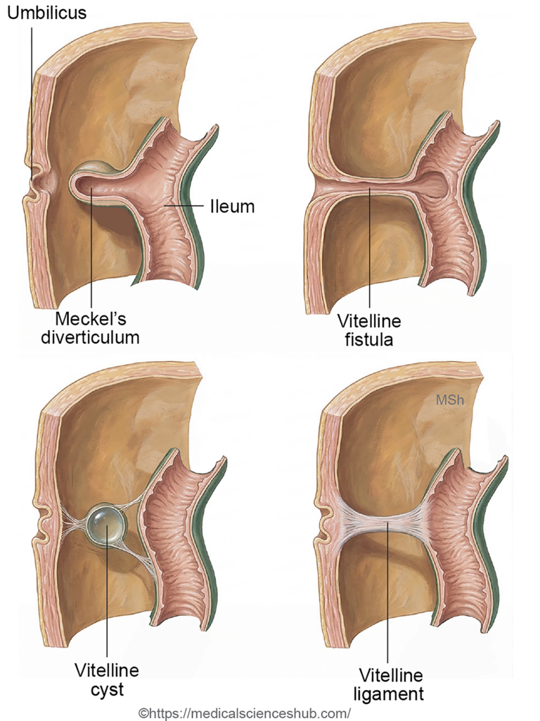
This condition results from a patent vitellointestinal duct, creating a direct communication between the ileum and the umbilicus. It leads to the discharge of intestinal contents or fecal material through the umbilicus. - Meckel’s diverticulum:
Meckel’s diverticulum arises from persistence of the proximal part of the vitellointestinal duct. It appears as a diverticular outpouching from the ileum and represents the most common congenital anomaly of the small intestine. - Vitelline cyst (enterocystoma):
A vitelline cyst develops when the central portion of the vitellointestinal duct persists while both ends close. This results in a cystic structure along the course of the duct. Persistence of different parts of the duct may produce Meckel’s diverticulum, vitelline cyst, umbilical fistula, or an umbilical sinus. - Raspberry tumor of the umbilicus:
Persistence of the distal segment of the vitellointestinal duct near the umbilicus may produce a small bright red granulomatous mass, often described as a raspberry-like tumor. - Exomphalos (omphalocele):
Normally, the physiological umbilical hernia returns to the abdominal cavity by the 12th week of intrauterine life. Failure of this return results in exomphalos, where abdominal viscera remain herniated at the umbilicus and are covered by a peritoneal sac and amnion. - Congenital umbilical hernia:
In this condition, intestinal loops protrude through the umbilical ring but are covered by skin, connective tissue, and peritoneum. The swelling becomes more prominent during crying or coughing. Most cases resolve spontaneously by 2–3 years of age, although surgical repair may be required if persistence occurs. - Gastroschisis:
Gastroschisis is a congenital defect of the anterior abdominal wall caused by failure of complete lateral body folding. Abdominal viscera protrude outside the abdominal cavity without a protective membranous sac. - Rotational anomalies of the gut:
Abnormal or incomplete midgut rotation may lead to altered positions of abdominal organs. The ligament of Treitz serves as an important landmark in diagnosing these conditions. Examples include nonrotation, producing a left-sided large intestine and right-sided small intestine, and mixed rotation, where the caecum may lie abnormally near the pylorus. In reverse rotation, the duodenum may lie anterior to the transverse colon, which passes posterior to the superior mesenteric artery. - Apple-peel atresia (type IIIb intestinal atresia):
This rare form of small-intestinal atresia accounts for about 10% of intestinal atresias. The duodenum or proximal jejunum ends blindly, while the distal small intestine coils around a narrow vascular pedicle, creating a spiral configuration resembling an apple peel.
CLINICAL EMBRYOLOGY
Meckel’s diverticulum (diverticulum ilei)
Meckel’s diverticulum was initially described by GF Hildanus in the sixteenth century and later detailed by Johann Friedrich Meckel in 1809. It is a true congenital diverticulum of the small intestine because it contains all layers of the intestinal wall. This anomaly represents persistence of the proximal part of the vitellointestinal duct, which normally disappears around the sixth week of intrauterine life. It is the most common congenital anomaly of the intestine.
Features
- Occurs in approximately 2% of the population.
- Male-to-female ratio: about 2:1.
- Clinical symptoms most commonly appear around 2 years of age.
- Average length is about 5 cm (2 inches).
- Located roughly 60 cm (2 feet) proximal to the ileocecal valve.
- Usually arises from the antimesenteric border of the ileum.
- The diameter is typically similar to that of the adjacent ileum.
- The apex may remain free or be connected to the umbilicus, mesentery, or other abdominal structures by a fibrous band.
- It may contain ectopic gastric or pancreatic tissue, which can lead to complications.
Clinical Significance
Meckel’s diverticulum may produce intestinal obstruction or bleeding due to ectopic gastric mucosa. Inflammation of the diverticulum (diverticulitis) can mimic the clinical features of acute appendicitis.
Treatment
Symptomatic or complicated cases are managed by surgical resection, commonly performed using laparoscopic techniques.
CLINICAL EMBRYOLOGY
Malrotation
Clinical Presentation
- Sudden onset of bilious vomiting in a newborn.
- In older children, recurrent abdominal pain accompanied by bilious vomiting due to intermittent intestinal obstruction or midgut volvulus.
Diagnosis
- Barium follow-through study may demonstrate an abnormally shaped (often S-shaped) duodenum.
- The ligament of Treitz is found in an abnormal position, which is an important diagnostic sign.
Management
Treatment is surgical and commonly performed through the Ladd’s procedure. This operation includes division of Ladd’s bands, widening of the mesenteric base to reduce the risk of volvulus, and appendectomy to prevent future diagnostic confusion.
Development Of Caecum And Appendix
- The caecum and vermiform appendix develop from a caecal bud, which appears as a dilation of the postarterial segment of the midgut loop. This bud becomes visible around the sixth week of intrauterine life.
- The proximal part of the caecal bud enlarges and differentiates to form the caecum, whereas the distal narrow portion persists as the vermiform appendix.
- During growth, the caecal bud forms two sacculations. The right saccule enlarges more rapidly than the left, causing the base of the appendix to shift toward the left side near the ileocaecal junction. This differential growth determines the final position of the appendiceal opening in the caecum.

Development Of Hindgut
The hindgut forms the terminal portion of the primitive gut and gives rise to several components of the large intestine. Its derivatives include:
- Distal one-third of the transverse colon
- Descending colon
- Sigmoid colon
- Rectum
- Upper part of the anal canal
- These structures develop from the caudal segment of the embryonic gut tube and contribute to the formation of the distal gastrointestinal tract.
- Development of Urorectal Septum
- The distal part of the hindgut that communicates with the allantois forms a common cavity called the cloaca. During development, a mesodermal partition known as the urorectal septum grows downward and divides the cloaca into two parts: a ventral primitive urogenital sinus and a dorsal primitive rectum.
- The urorectal septum also separates the cloacal membrane into an anterior urogenital membrane and a posterior anal membrane. The cloacal membrane normally ruptures around the seventh week of intrauterine life, establishing external openings. The anal membrane is located at the upper end of the proctodeum.
- The primitive urogenital sinus later forms the urinary bladder and urethra, while the primitive rectum develops into the rectum and the upper part of the anal canal.
- Development of the urorectal septum involves two main components:
- The Tourneux fold, a vertical midline fold that grows caudally between the rectum and the urogenital sinus.
- The Rathke folds, which arise from the lateral walls of the cloaca and contribute to formation of the septum.
Development Of Anal Canal
The anal canal develops from two embryological sources:
- The endodermal cloaca (distal part of the primitive rectum) forms the upper part of the anal canal, located above the pectinate line.
- The ectodermal anal pit, known as the proctodeum, forms the lower part of the anal canalbelow the pectinate line.
Stages of Development
- The proctodeum appears as a depression of the surface ectoderm. At its base lies the anal membrane, which separates the developing anal canal from the exterior.
- Around the ninth week of intrauterine life, the anal membrane perforates, establishing communication between the hindgut and the external environment. The former site of this membrane later corresponds to the pectinate line of the anal canal.
- Thus, the pectinate line represents the junction between the endoderm-derived upper anal canal and the ectoderm-derived lower anal canal. Because of this dual origin, the arterial supply, lymphatic drainage, and nerve supply differ above and below the pectinate line.
Table 13.1: Development of anal canal
| Feature | Above the Pectinate Line (Upper Anal Canal) | Below the Pectinate Line (Lower Anal Canal) |
|---|---|---|
| Embryonic origin | Endoderm | Ectoderm |
| Developmental source | Derived from the endodermal cloaca forming the primitive rectum | Derived from the proctodeum (anal pit) |
| Arterial supply | Superior rectal artery (branch of inferior mesenteric artery) | Inferior rectal artery (branch of internal pudendal artery) |
| Venous drainage | Superior rectal vein → portal system via inferior mesenteric vein | Inferior rectal vein → systemic circulation → inferior vena cava |
| Nerve supply | Autonomic (visceral); pain poorly localized | Somatic via inferior rectal nerve; pain well localized |
CLINICAL EMBRYOLOGY
Congenital Anomalies of the Large Intestine
1. Hirschsprung’s Disease (Congenital Megacolon)
Hirschsprung’s disease is a congenital disorder characterized by the absence of parasympathetic ganglion cells in the intestinal wall, specifically in the myenteric (Auerbach) plexus and submucosal (Meissner) plexus. This defect results from failure of neural crest cell migration during development.
The aganglionic segment remains persistently constricted due to unopposed sympathetic activity, while the proximal colon becomes markedly dilated because of accumulation of intestinal contents.
- Incidence: Approximately 1 in 5,000 newborns.
- Clinical sign: Delayed passage of meconium beyond the first 48 hours after birth.
- Treatment: Surgical removal of the aganglionic segment followed by reanastomosis.
2. Imperforate Anus
In imperforate anus, the distal gut fails to establish communication with the exterior. Possible causes include:
- Failure of anal membrane rupture
- Failure of development of the ectodermal proctodeum
- Rectal atresia due to defective rectal development
3. Ectopic Anus
Normally, growth of the perineal body divides the cloacal membrane into urogenital and anal membranes. Abnormal division can result in displacement of the anal opening, producing an ectopic anus.
- In females: Opening may occur within the vestibule.
- In males: It may appear near the base of the scrotum or within the bulbar urethra region.
4. Rectal Fistula
A fistula is an abnormal passage connecting two epithelial surfaces. In rectal anomalies, the rectum may communicate with nearby organs
- Rectovesical fistula (high type): Communication between the rectum and urinary bladder.
- Rectourethral fistula (low type): Connection between the rectum and urethra.
- Rectovaginal fistula (low type): Abnormal passage between the rectum and vagina.
Development Of Anterior Abdominal Wall
- The anterior abdominal wall consists of several layers, including the skin (epidermis and dermis), fascia, muscle layers, and the parietal peritoneum, which encloses the peritoneal cavity.
Stages of Development
- Initially, the embryo is trilaminar, consisting of three germ layers:
- Ectoderm
- Mesoderm—subdivided into paraxial, intermediate, and lateral plate mesoderm
- Endoderm
- The intraembryonic coelom divides the lateral plate mesoderm into two layers: the somatopleuric mesoderm and the splanchnopleuric mesoderm.
- During the fourth week, the embryo undergoes body folding, which includes the head fold, tail fold, and two lateral folds. These folds converge at the umbilical region on the ventral surface.
- As a result of this folding:
- A portion of the intraembryonic coelom becomes the peritoneal cavity.
- The somatopleuric layers from both sides fuse in the midline to form the connective tissue and muscular components of the anterior abdominal wall.
- The ectoderm develops into the epidermis covering the abdominal wall.
- A small unfused ventral region remains at the umbilicus, allowing passage of the umbilical vessels and forming the umbilical cord.
CLINICAL EMBRYOLOGY
Defects of Ventral Body Wall
Ventral body wall defects occur in approximately 1 in 2,000 live births and involve abnormal development of the anterior abdominal wall, allowing abdominal organs to protrude outside the abdominal cavity.
Gastroschisis: Gastroschisis is a congenital defect of the anterior abdominal wall, usually located near the umbilicus. Through this opening, intestinal loops and occasionally other abdominal organs protrude directly into the amniotic cavity. The herniated contents are not covered by a peritoneal sac, which exposes them to the surrounding fluid.
Omphalocele:Omphalocele is a congenital condition in which abdominal viscera remain outside the abdominal cavity due to failure of the midgut to return after physiological herniation. The protruding organs are enclosed within a peritoneal sac that is covered by amnion.
Derivatives Of Gut
The derivatives of the gut tube are listed in the Table 13.2.
Table 13.2: Derivatives of gut
| Embryonic Gut Region | Major Derivatives |
|---|---|
| Foregut | Forms the pharynx, oesophagus, stomach, and the proximal duodenum up to the major duodenal papilla. Gives rise to pharyngeal pouches and their derivatives. Also produces the liver, gallbladder, biliary apparatus, and pancreas. The respiratory tract develops from a ventral foregut diverticulum. Contributes to part of the floor of mouth and the tongue. |
| Midgut | Develops into the distal duodenum, jejunum, ileum, caecum, appendix, ascending colon, and the proximal two-thirds of the transverse colon. |
| Hindgut | Forms the distal one-third of the transverse colon, descending colon, sigmoid colon, rectum, and upper anal canal. Endoderm of this region also contributes to the epithelial lining of the urinary bladder and urethra through the cloaca. |
Important Questions
- Describe the development of the oesophagus.
- Describe the development of the stomach.
- Outline the embryological development of the duodenum.
- Enumerate the derivatives of the midgut.
- Describe the process of midgut rotation during development.
- Explain the embryological basis and clinical significance of Meckel’s diverticulum.
- Describe the development of the anal canal.
- Discuss the congenital anomalies associated with the anal canal.
- Explain the embryological basis and features of imperforate anus.
📝 Test Your Knowledge – Practice MCQs
Attempt the chapter MCQ quiz and assess your understanding of key concepts.
Click to see: Medical Disclaimer and Copyright Notice