
Competency
- AN62.3: Describe the white matter of cerebrum
Introduction
- The white matter of the cerebrum is composed of myelinated nerve fibers (axons) that lie deep to the cerebral cortex and form a large part of each cerebral hemisphere.
- These fibers connect the two cerebral hemispheres with each other and with other regions of the brain and spinal cord, enabling efficient communication across the nervous system.
- Functionally, the white matter integrates sensory and motor information, coordinates movement, and supports higher functions such as learning, memory, and cognition.
Types of Fibers
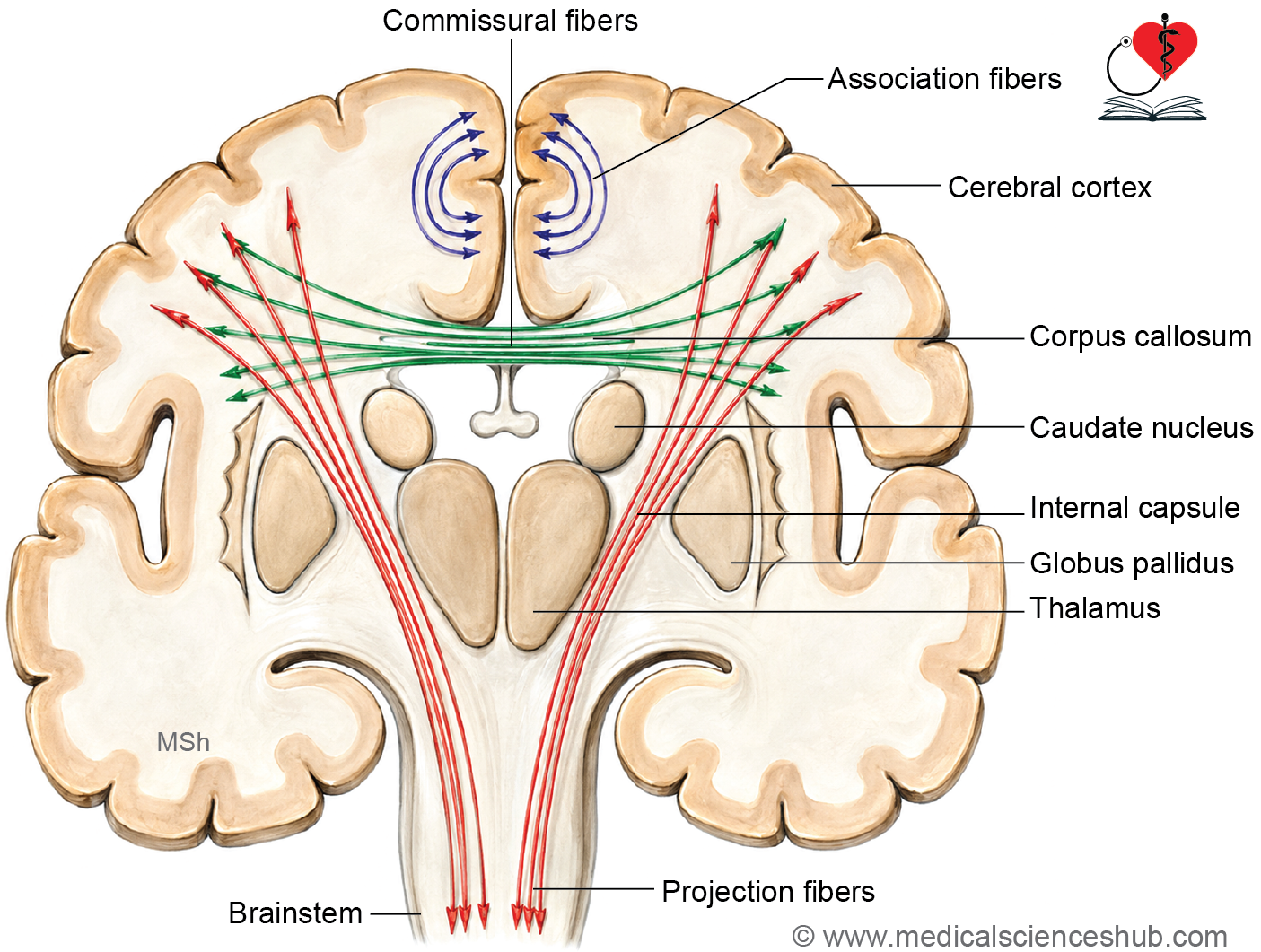
The white matter of the cerebrum contains three categories of nerve fibers, each serving a distinct role in neural communication:
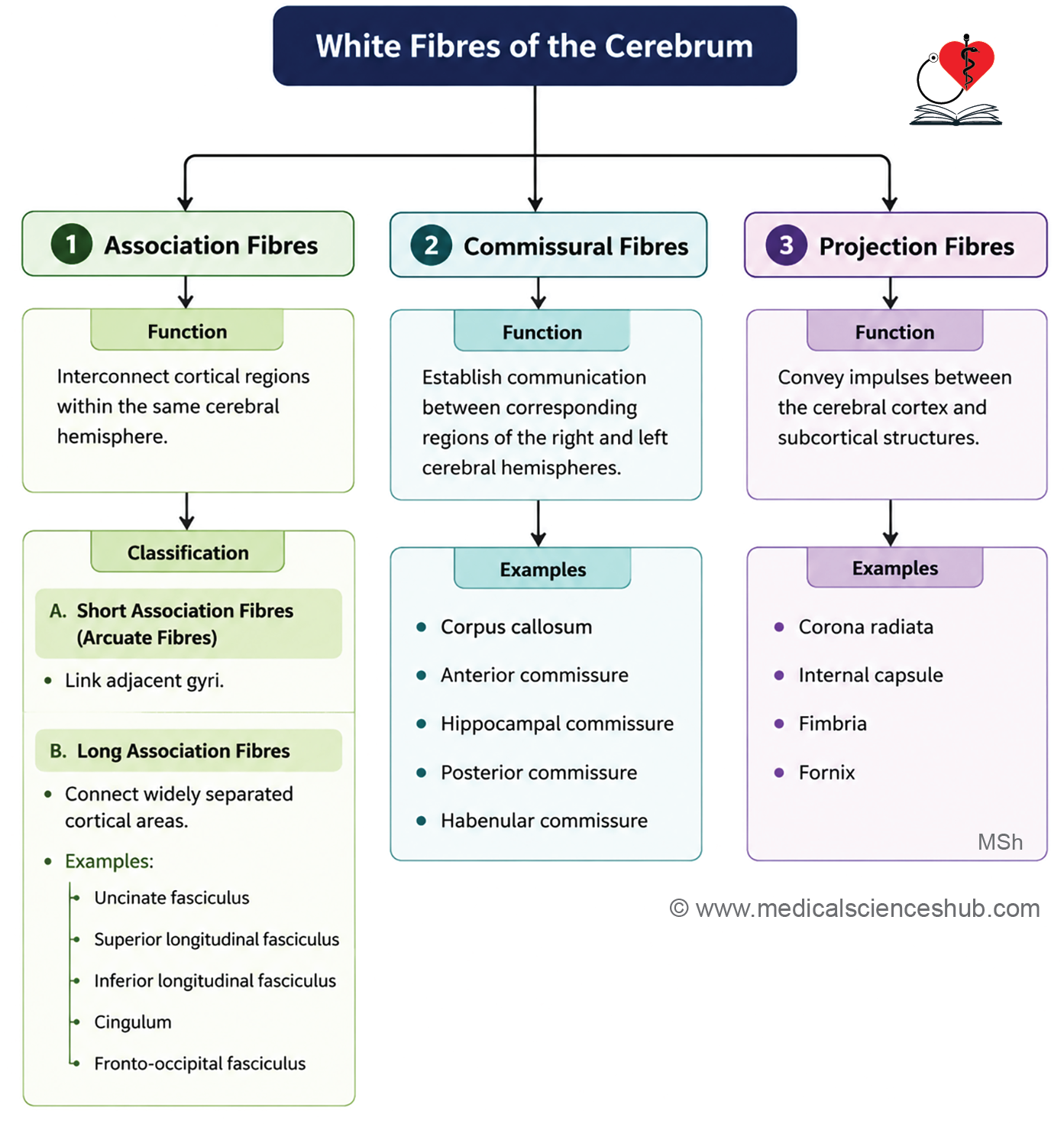
- Association fibers connect different regions within the same cerebral hemisphere.
- Commissural fibers connect corresponding areas across the two cerebral hemispheres.
- Projection fibers connect the cerebral cortex to subcortical structures in the brain and spinal cord.
Association Fibers
- Association fibers, also called intrahemispheric fibers, link different cortical regions within the same hemisphere and support complex functions such as language, memory, and higher-order cognition.
- They are divided into two types: short association fibers and long association fibers.
Short Association Fibers (Arcuate or U-fibers)
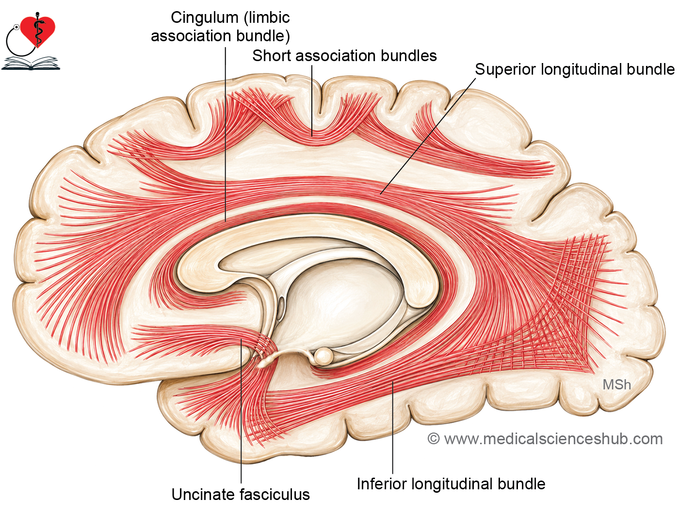
- These fibers connect adjacent gyri by looping around the intervening sulcus, giving them their characteristic U-shaped appearance.
Long Association Fibers
- These fibers connect widely separated cortical regions and include the following named bundles:
- Uncinate fasciculus — connects the motor speech area and orbital cortex of the frontal lobe with the cortex of the temporal pole, hooking around the stem of the lateral sulcus. It is involved in memory encoding and retrieval, processing of novel information, and emotional regulation. It also transmits visual word images to Broca’s area for speech production.
- Superior longitudinal fasciculus — the longest association bundle, connecting the frontal lobe with the occipital and temporal lobes, running lateral to the corona radiata above the insular region. It serves as a major link between the occipital and frontal eye fields.
- Inferior longitudinal fasciculus — connects the visual association cortex (areas 18 and 19) of the occipital lobe with the temporal lobe, running along the lateral wall of the posterior horn of the lateral ventricle. It contributes to face and object recognition as well as visual memory.
- Cingulum (cingulate fasciculus) — a thick fiber bundle running deep within the cingulate and parahippocampal gyri, extending from the anterior perforated substance to the uncus and temporal pole. It forms part of the Papez circuit and is involved in emotional processing, attention, decision-making, and behavioral flexibility.
- Fronto-occipital fasciculus — runs anteroposteriorly from the frontal pole to the occipitotemporal region, deep to the superior longitudinal fasciculus, separated from it by the corona radiata. It integrates visual information with motor planning to facilitate coordinated, visually guided movements.
Commissural Fibers
- Commissural fibers, also called interhemispheric fibers, connect corresponding areas of the two cerebral hemispheres by crossing the midline. A discrete bundle of such fibers is termed a commissure.
- They play an essential role in transferring information between hemispheres to coordinate bilateral responses and support learning.
- Examples include the corpus callosum, anterior commissure, posterior commissure, hippocampal commissure, and habenular commissure.
- It is worth noting that the primary visual area and the hand region of the somatosensory cortex do not receive commissural fibers.
Projection Fibers
- Projection fibers connect the cerebral cortex with subcortical structures in both directions.
- Corticofugal fibers carry signals away from the cortex toward subcortical centers, while corticopetal fibers carry signals from subcortical centers back to the cortex.
- Examples include the corona radiata, internal capsule, fimbria, and fornix.
Corpus Collosum
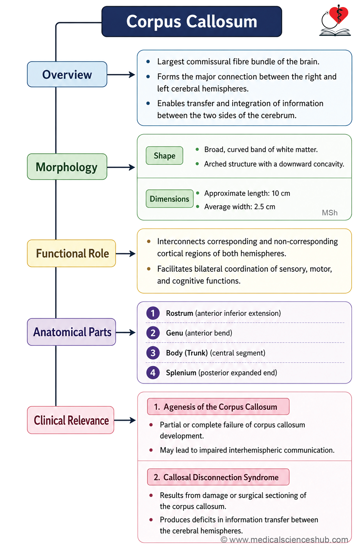
- The corpus callosum is the largest commissure of the brain, connecting the right and left cerebral hemispheres. It links all regions of the neocortex in both hemispheres, with the exception of the anterior and inferior parts of the temporal lobes, which are instead connected by the anterior commissure.
- It is a massive, arched structure with a downward concavity, appearing C-shaped in midsagittal section. It measures approximately 10 cm in length and 2.5 cm in breadth and contains roughly 150–200 million axons.
Parts
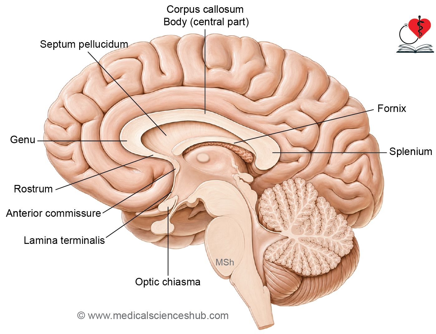
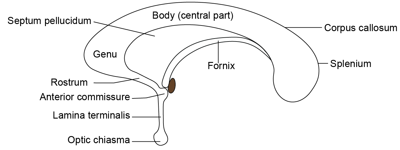
The corpus callosum is divided into four parts from front to back:
- Rostrum — the most anterior and inferior portion, extending from the upper end of the lamina terminalis to the genu. It forms the floor of the anterior horn of the lateral ventricle and carries fibers connecting the orbital surfaces of the two frontal lobes.
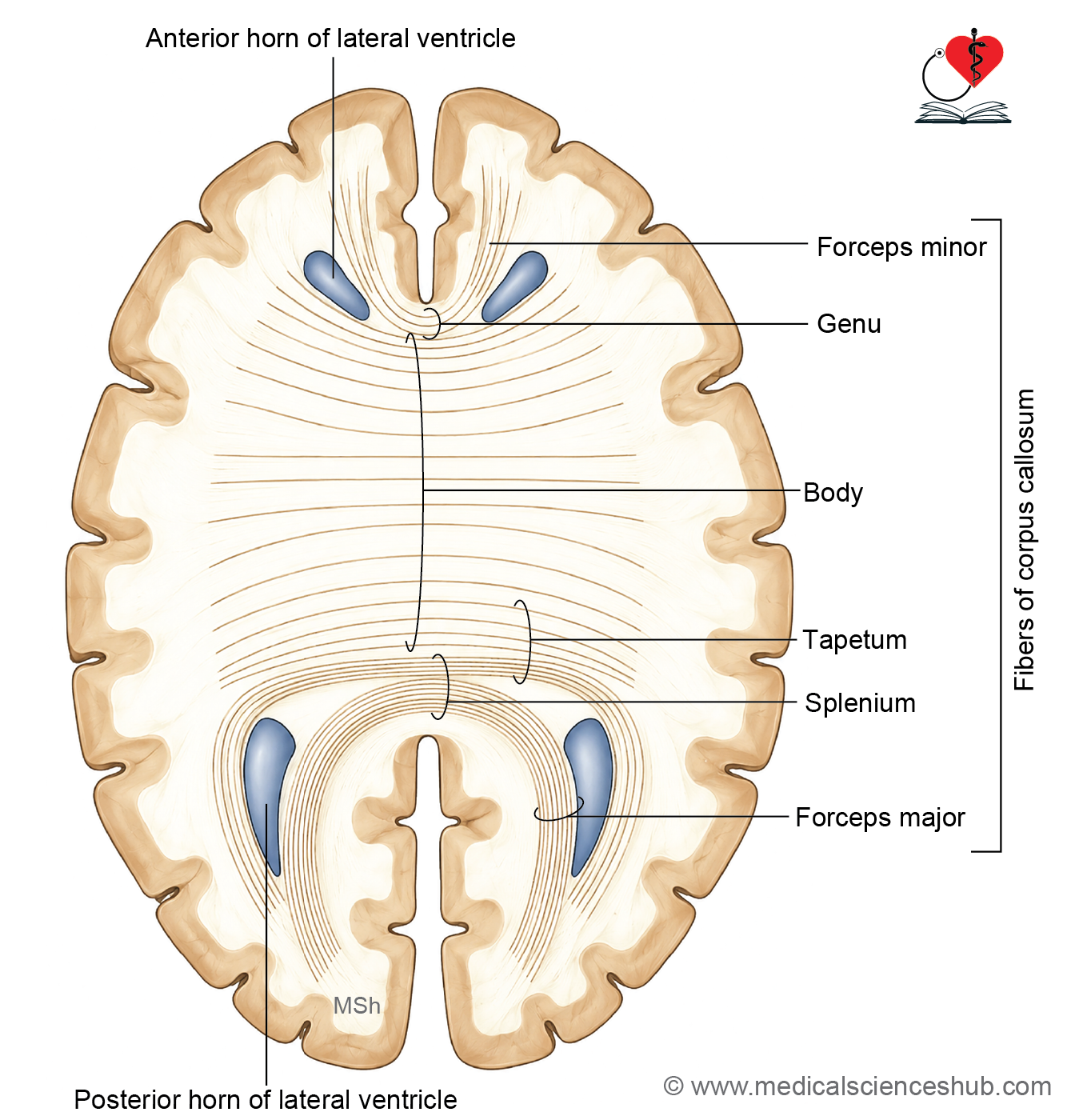
- Genu — the thick, curved anterior portion lying approximately 4 cm behind the frontal pole. It forms the anterior boundary of the anterior horn of the lateral ventricle. Its fibers form a fork-like bundle called the forceps minor, connecting the frontal poles of both hemispheres.
- Body (trunk) — the middle portion extending from the genu to the splenium. It connects the frontal and anterior parietal lobes of both hemispheres and forms the roof of the body of the lateral ventricle.
- Splenium — the thickest, most posterior part, located about 6 cm anterior to the occipital pole. It connects the posterior parietal, temporal, and occipital lobes of both hemispheres. Its fibers form a large fork-like bundle called the forceps major, connecting the occipital lobes. These fibers produce a bulge in the medial wall of the posterior horn of the lateral ventricle known as the bulb of the posterior horn. The splenium lies above the pulvinar of the thalamus, pineal gland, and tectum of the midbrain, and is related inferiorly to the great cerebral vein of Galen.
- Tapetum — fibers derived from the body and splenium that form the roof and lateral wall of the posterior horn and the lateral wall of the inferior horn of the lateral ventricle. These fibers are not intersected by the corona radiata.
Relations of Corpus Callosum
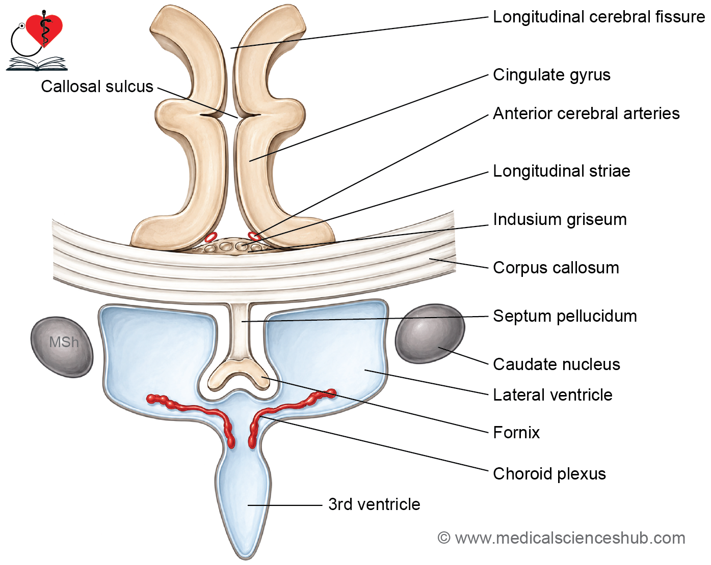
- The upper convex surface forms the floor of the median longitudinal fissure and is related to the inferior free margin of the falx cerebri and the inferior sagittal sinus. It is separated laterally from the cingulate gyrus by the callosal sulcus.
- The upper surface is covered by a thin layer of grey matter called the indusium griseum, which extends over the rostrum, genu, and body, and continues over the splenium as the gyrus fasciolaris.
- Embedded within the indusium griseum are two pairs of thin white fiber bundles called the medial and lateral longitudinal striae, which run from the hippocampus of the temporal lobe to the septal area. These striae and the indusium griseum represent remnants of the hippocampal formation.
- The lower concave surface has several important relations:
- The septum pellucidum connects the lower surface of the corpus callosum to the upper surface of the fornix.
- The fornix contacts the posterior part of the inferior surface.
- The cavity of the lateral ventricle, lined by ependyma, is related to the concave surface.
- Below the splenium, the tela choroidea of the third ventricle extends through the transverse fissure between the fornix and the roof of the third ventricle.
Functions
- It enables transfer of learned information between hemispheres — memory formed in one hemisphere using sensory and motor inputs is made available to the other hemisphere through the corpus callosum.
- It connects the speech and language areas of the dominant (usually left) hemisphere — including Wernicke’s area (area 22), the angular gyrus (area 39), the supramarginal gyrus (area 40), and Broca’s area (areas 44 and 45) — with the non-dominant hemisphere, enabling integrated language processing.
- It coordinates bilateral motor movements, ensuring that the two sides of the body work together smoothly.
- It supports problem-solving and creative thinking by facilitating communication between the two hemispheres.
CLINICAL NEUROANATOMY
Split-brain syndrome: · Congenital absence or surgical division of the corpus callosum results in split-brain syndrome, in which the two hemispheres function largely independently, as if the patient had two separate brains. Lesions of the anterior corpus callosum produce akinetic mutism (loss of spontaneous movement and speech) and tactile anomia (inability to name objects identified by touch). Lesions of the posterior corpus callosum produce alexia without agraphia — the patient loses the ability to read but retains the ability to write.
Fiber Bundles of the Corpus Callosum
- Forceps minor — fork-shaped fibers derived from the genu of the corpus callosum that connect the frontal lobes of both hemispheres.
- Tapetum — fibers from the body and splenium of the corpus callosum that form the roof and lateral wall of the posterior horn and the lateral wall of the inferior horn of the lateral ventricle. These fibers do not intersect with the fibers of the corona radiata.
- Forceps major — fork-shaped fibers derived from the splenium of the corpus callosum that connect the occipital lobes of both hemispheres.
- Bulb of the posterior horn — a distinct bulge on the medial wall of the posterior horn of the lateral ventricle, produced by the fibers of the forceps major passing through this region.
Other Commissures of the Brain
- Anterior commissure — a small bundle of fibers located in the upper part of the lamina terminalis, crossing the midline in front of the column of the fornix and the interventricular foramen. It consists of two parts:
- A small anterior part connects the olfactory regions of the two hemispheres.
- A larger posterior part connects the lower anterior portions of the temporal lobes of both hemispheres.
- Posterior commissure — located in the inferior lamina of the pineal stalk, it connects the superior colliculi, pretectal nuclei, and interstitial nuclei of both sides.
- Habenular commissure — located in the superior lamina of the pineal stalk, it connects the habenular nuclei of the two sides.
- Commissure of the fornix (hippocampal commissure) — connects the two crura of the fornix, linking the hippocampal formations of both hemispheres.
Internal Capsule
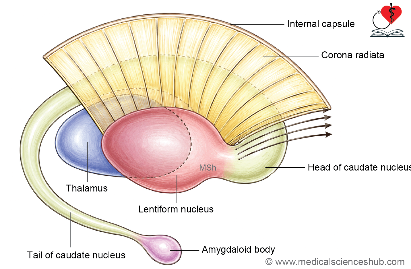
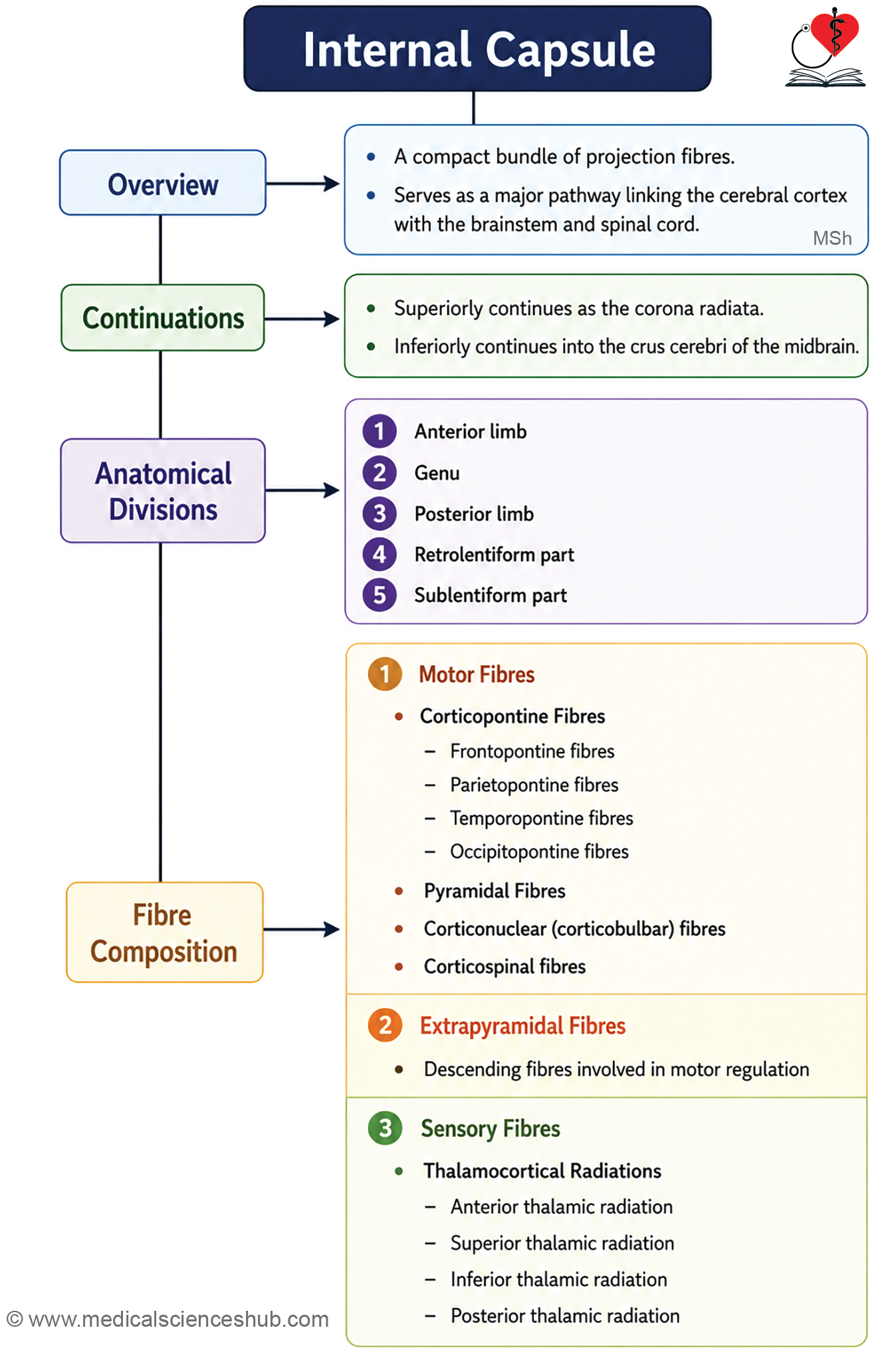
- The internal capsule is a compact bundle of projection fibers located between the thalamus and caudate nucleus medially, and the lentiform nucleus laterally.
- It carries motor signals from the cerebral cortex downward to the brainstem and spinal cord, and transmits sensory information from the body upward to the cortex, making it essential for motor control and sensory perception of the opposite side of the body.
- Because its fibers are densely packed into a small area, even a minor lesion can produce widespread paralysis and sensory loss affecting the entire contralateral half of the body.
Shape and Continuations of the Internal Capsule
- In a transverse section of the brain, the internal capsule appears V-shaped, with its concavity directed laterally and occupied by the lentiform nucleus.
- Superiorly, its fibers fan out to form the corona radiata, which spreads like radiating rays toward the cerebral cortex.
- Inferiorly, its fibers converge and continue as the crus cerebri of the midbrain.
Parts of the Internal Capsule
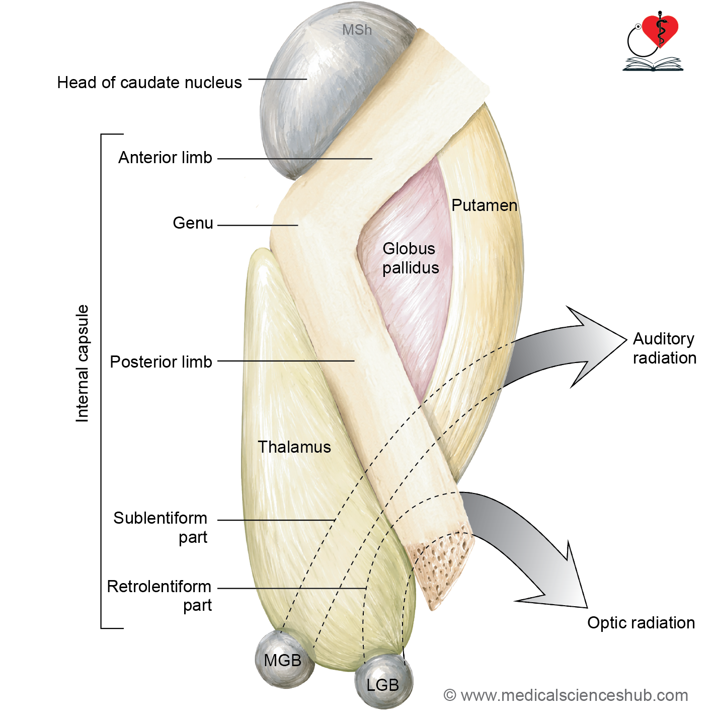
The internal capsule consists of five parts:
- Anterior limb — lies between the caudate nucleus and the anterior part of the lentiform nucleus.
- Genu — the bend between the anterior and posterior limbs.
- Posterior limb — lies between the thalamus and the posterior part of the lentiform nucleus.
- Retrolentiform part — lies posterior to the lentiform nucleus.
- Sublentiform part — lies inferior to the lentiform nucleus at the posterior end of the internal capsule.
Constituent Fibers of Internal Capsule
Because the internal capsule carries functionally distinct fiber groups in specific locations, a localized lesion produces predictable loss of motor or sensory function in a particular part of the body.
Motor Fibers
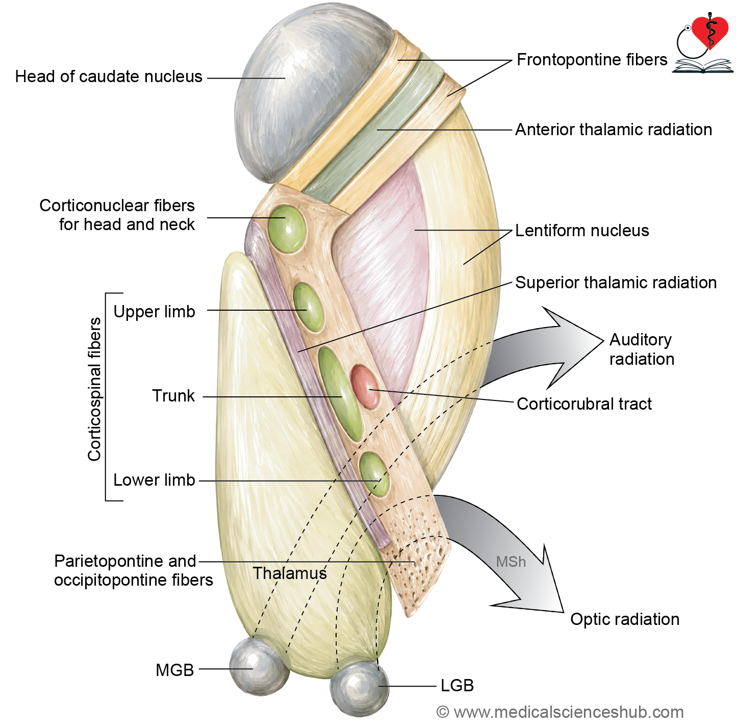
1. Corticopontine Fibers
- These fibers originate from the cortex of all four lobes of the cerebral hemisphere and account for approximately two-thirds of all fibers in the internal capsule.
- They form the cortico-ponto-cerebellar pathway, relaying information about voluntary movement and motor planning to the cerebellum, enabling it to fine-tune and coordinate smooth, precise movements.
- After synapsing in the pontine nuclei on the same side, the fibers cross the midline and enter the opposite cerebellar hemisphere via the middle cerebellar peduncle.
- They are named according to their lobe of origin and pass through the following regions:
- Frontopontine fibers — from the frontal lobe, pass through the anterior limb, genu, and posterior limb.
- Parietopontine fibers — from the parietal lobe, pass through the retrolentiform part.
- Occipitopontine fibers — from the occipital lobe, pass through the retrolentiform part.
- Temporopontine fibers — from the temporal lobe, pass through the sublentiform part.
2. Pyramidal Fibers
- These fibers arise in the cerebral cortex and relay to lower motor neurons on the contralateral side in the brainstem and spinal cord. They are divided into two groups:
- Corticonuclear fibers — pass through the genu of the internal capsule and relay in the contralateral motor nuclei of cranial nerves III through XII. They control voluntary movements of the face and head, including facial expression, speech, and swallowing.
- Corticospinal fibers — arise from the motor cortex, pass through the posterior limb of the internal capsule, and relay at the contralateral anterior horn cells of the spinal cord to control voluntary movements of the upper limb, trunk, and lower limb. Within the posterior limb, fibers are arranged in order: upper limb anteriorly, trunk in the middle, and lower limb posteriorly.
3. Extrapyramidal Fibers
- These fibers arise from the cerebral cortex and project to subcortical grey matter structures, including the corpus striatum (corticostriate fibers), red nucleus (corticorubral fibers), substantia nigra (corticonigral fibers), and reticular nuclei of the brainstem (corticoreticular fibers).
- They pass through the posterior limb and regulate involuntary motor functions including muscle tone, posture, balance, and automatic movements.
Sensory Fibers (Thalamocortical fibers)
- Most sensory information reaching the cerebral cortex is relayed from the thalamus via fibers called thalamocortical fibers or thalamic radiations, which carry third-order neurons of various sensory pathways and convey somatosensory information from the contralateral side of the body.
- These radiations are divided into four groups:
- Anterior thalamic radiation — passes through the anterior limb, connecting the anterior and dorsomedial nuclei of the thalamus with the cingulate gyrus as part of the Papez circuit. It is involved in emotion and recent memory; damage produces memory impairment and emotional disturbances.
- Superior thalamic radiation — passes through the genu and posterior limb, connecting the ventral group of thalamic nuclei with the primary somatosensory cortex (areas 3, 1, and 2) and adjacent frontal and parietal regions. It is responsible for transmitting somatosensory information to the cortex.
- Posterior thalamic radiation (optic radiation) — passes through the retrolentiform part, extending from the lateral geniculate body to the primary visual cortex (area 17) of the occipital lobe via the geniculocalcarine tract. It is essential for visual perception and acuity, and also carries a small number of corticofugal fibers from areas 18 and 19 to the superior colliculus and nuclei controlling extraocular muscles.
- Inferior thalamic radiation (auditory radiation) — passes through the sublentiform part, arising from the medial geniculate body and terminating in the anterior transverse temporal gyrus (areas 41 and 42). It carries auditory information essential for sound perception, localization, and discrimination.
Table 14.1: Constituent fibers of internal capsule
| Part | Major Fibre Tracts | Sensory Fibres | Principal Arterial Supply |
|---|---|---|---|
| Anterior limb | Frontopontine fibres | Anterior thalamic radiation | Direct branches from the anterior cerebral artery and recurrent artery of Heubner |
| Genu | Frontopontine fibres and corticonuclear (corticobulbar) fibres | Anterior portion of the superior thalamic radiation | Perforating branches from the internal carotid artery and the posterior communicating artery |
| Posterior limb | Corticospinal fibres, corticopontine fibres, and corticorubral fibres | Superior thalamic radiation | Lateral and medial striate branches of the middle cerebral artery together with the anterior choroidal artery |
| Retrolentiform part | Parietopontine and temporopontine fibres | Inferior thalamic radiation, including auditory pathways | Anterior choroidal artery, anterior cerebral artery, and posterior cerebral artery |
| Sublentiform part | Parietopontine and occipitopontine fibres | Posterior thalamic (optic) radiation | Primarily supplied by branches of the posterior cerebral artery |
Arterial Supply of Internal Capsule
Here’s the rewritten passage:
Arterial Supply of the Internal Capsule
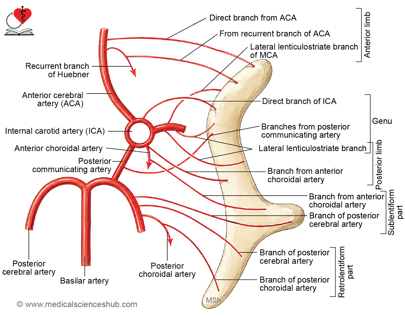
- The internal capsule receives its blood supply from multiple arterial sources. Because its fibers are densely packed, even a small vascular lesion can produce significant motor weakness and sensory loss on the opposite side of the body.
- The supplying arteries are as follows:
- Anterior cerebral artery — via the medial striate arteries and the recurrent artery of Heubner (a large striate branch that takes a recurrent course back toward the internal capsule).
- Internal carotid artery — via a few direct branches.
- Middle cerebral artery — via the medial striate artery, lateral striate arteries, and a branch from the anterior choroidal artery. The lateral striate branches are the most clinically significant, as they are the most prone to rupture.
- Posterior communicating artery — via its central branches.
- Posterior cerebral artery — via its posterolateral central branches.
- Charcot’s artery of cerebral hemorrhage is one of the larger lateral striate branches of the middle cerebral artery and is the vessel most frequently responsible for hypertensive intracerebral hemorrhage involving the internal capsule.
CLINICAL NEUROANATOMY
Lesion of internal capsule
- Because the internal capsule carries densely packed upper motor neuron fibers, any lesion typically produces contralateral upper motor neuron paralysis affecting the opposite half of the body and face.
- Vascular lesions of the internal capsule are common and result from either rupture of a blood vessel causing hemorrhage, or occlusion causing ischemic necrosis. The branches most frequently affected are the medial and lateral striate branches of the middle cerebral artery.
Specific Vascular Lesions
- Rupture of Charcot’s artery (artery of cerebral hemorrhage) — damages the posterior limb, resulting in paralysis of the opposite half of the body and face (hemiplegia). This vessel ruptures most commonly in patients with hypertension and represents one of the most frequent causes of stroke-related paralysis.
- Lesion of the recurrent artery of Heubner — damages the genu and anterior limb, producing paralysis of the face and upper limb on the opposite side.
- Thrombosis of the anterior choroidal artery — damages the posterior limb, sublentiform, and retrolentiform parts, resulting in motor hemiplegia combined with visual and auditory deficits.
Important Questions
- List the type of fibers in white matter of cerebrum with their examples.
- Write a short note on corpus callosum.
- Write a short note on arterial supply of internal capsule.
📝 Test Your Knowledge – Practice MCQs
Attempt the chapter MCQ quiz and assess your understanding of key concepts.
Click to see: Medical Disclaimer and Copyright Notice