
Competencies
- AN66.1 Describe and identify various types of Connective tissue with functional correlation.
- AN66.2 Describe the ultrastructure of Connective tissue.
Introduction
- A tissue is an organized group of cells, along with extracellular components, that collectively perform a specific function. The term derives from the Latin texo (‘to weave”).
- The human body is composed of four fundamental tissue types: epithelial, connective, muscle, and nervous tissue.
Epithelial tissue
- Epithelial tissue covers external body surfaces, lines internal body cavities, and forms the secretory units of glands.
Connective tissue
- Connective tissue supports and binds the other three tissue types, consisting of cells, connective tissue fibers, and extracellular matrix.
- Specialized forms include bone (mineralized matrix), cartilage (hydrated matrix), adipose tissue, lymphoid tissue, and blood (a fluid connective tissue).
Muscle tissue
- Muscle tissue is characterized by the property of contractility, enabling movement.
- It is subdivided into skeletal, cardiac, and smooth muscle.
Nervous tissue
- Nervous tissue is characterized by excitability and conductivity, enabling rapid communication throughout the body.
- It receives, integrates, and transmits information from both internal and external environments to regulate organ function.
- Nervous tissue consists of neurons (nerve cells) and supporting neuroglial cells.
General Connective Tissue
- Connective tissue comprises three components: cells, fibers, and extracellular matrix.
- It provides structural support, maintains organ shape, and performs diverse physiological functions throughout the body.
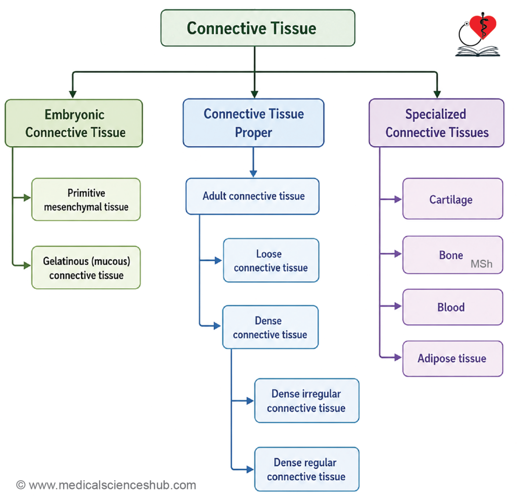
- onnective tissue is broadly classified into three categories.
- Embryonic connective tissue is mainly present during fetal development and includes mesenchymal and mucous (mucoid) connective tissue.
- Connective tissue proper is widely distributed throughout the body and is subdivided into loose and dense connective tissue.
- Specialized connective tissue possesses a highly modified extracellular matrix and includes cartilage, bone, blood, adipose tissue, and lymphoid tissue.
Components of Connective Tissue
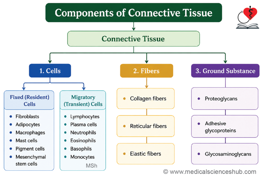
- Connective tissue consists of the following three basic components:
- Cells are categorized as:Resident cells (permanent inhabitants): fibroblasts, adipocytes, macrophages, mast cells, adult stem cells, and pigment cells.
- Wandering (transient) cells (recruited from blood): lymphocytes, plasma cells, neutrophils, eosinophils, basophils, and monocytes.
- Fibers include collagen, elastic, and reticular fibers.
- Ground substance is an amorphous, space-filling material composed of proteoglycans, glycoproteins, and glycosaminoglycans, providing structural and biochemical support between cells and fibers.
Fibers of Connective Tissue
- The extracellular matrix of connective tissue contains three principal fiber types: collagen, reticular, and elastic fibers.
Collagen Fibers
- Collagen fibers are the most abundant connective tissue fibers and occur in tendons, ligaments, bone, cartilage, skin, and most organs.
- They are strong, flexible, relatively inelastic, and possess high tensile strength, providing resistance to stretching forces.
- Under light microscopy, collagen fibers appear as wavy bundles that may branch and interconnect.
- Collagen molecules are composed of three α-chains rich in glycine, proline, and hydroxyproline.
- More than 28 genetically distinct collagen types have been identified; the most clinically important are Types I–IV.
Type I Collagen
- The most abundant collagen type, accounting for approximately 90% of body collagen.
- Found in tendons, ligaments, fascia, bone, skin, sclera, and organ capsules.
- Provides strength and resistance to tension.
- Defects are associated with osteogenesis imperfecta.
Type II Collagen
- Present in hyaline cartilage, elastic cartilage, vitreous body of the eye, nucleus pulposus, and remnants of the notochord.
- Provides resistance to compressive forces.
- Deficiency is associated with Kniest dysplasia.
Type III Collagen (Reticular Fibers)
- Forms delicate supporting networks in organs such as the liver, spleen, lymphoid tissues, blood vessels, and fetal skin.
- Contributes to the structural framework of soft tissues.
- Certain forms of Ehlers-Danlos syndrome are linked to abnormalities of Type III collagen.
Type IV Collagen
- Forms sheet-like networks rather than fibers.
- Located in the basal lamina, renal glomeruli, and lens capsule.
- Provides epithelial support and contributes to the renal filtration barrier.
- Defects may result in Alport syndrome, characterized by renal, auditory, and ocular abnormalities.
Reticular Fibers
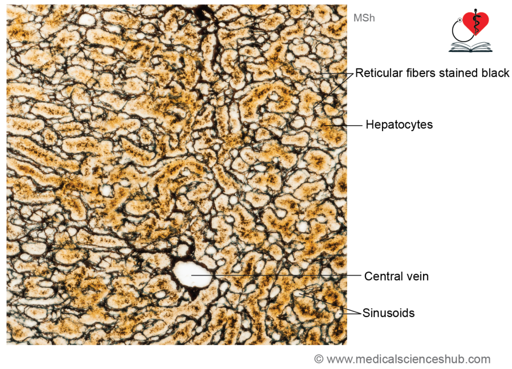
- Reticular fibers are thin fibers composed primarily of Type III collagen.
- They form a delicate supportive meshwork in the liver, lymphoid organs, bone marrow, glands, intestine, nerves, and muscles.
- Unlike collagen fibers, they do not form thick bundles.
- Reticular fibers are synthesized mainly by fibroblasts and specialized reticular cells.
- They are argyrophilic, appearing black with silver stains, and are PAS-positive because of their carbohydrate content.
- Their primary function is to provide structural support for cells and contribute to basement membrane organization.
Elastic Fibers
- Elastic fibers are thin, branching fibers that form networks rather than bundles.
- They are abundant in the skin, lungs, elastic cartilage, large arteries, ligamentum nuchae, ligamenta flava, periodontal ligament, and vocal folds.
- Ultrastructurally, they consist of a central core of elastin surrounded by fibrillin-rich microfibrils.
- Elastin contains unique amino acids, desmosine and isodesmosine, which enable elasticity and recoil.
- Elastic fibers are produced by fibroblasts, smooth muscle cells, endothelial cells, and chondrocytes.
- They are poorly visualized with routine H&E staining but can be demonstrated using orcein or Verhoeff’s stain.
- Degeneration and abnormal accumulation of elastic fibers may result in elastosis. In solar (actinic) elastosis, chronic ultraviolet exposure causes thickened, dry, and wrinkled skin.
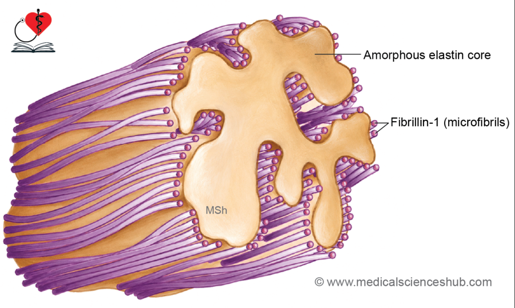
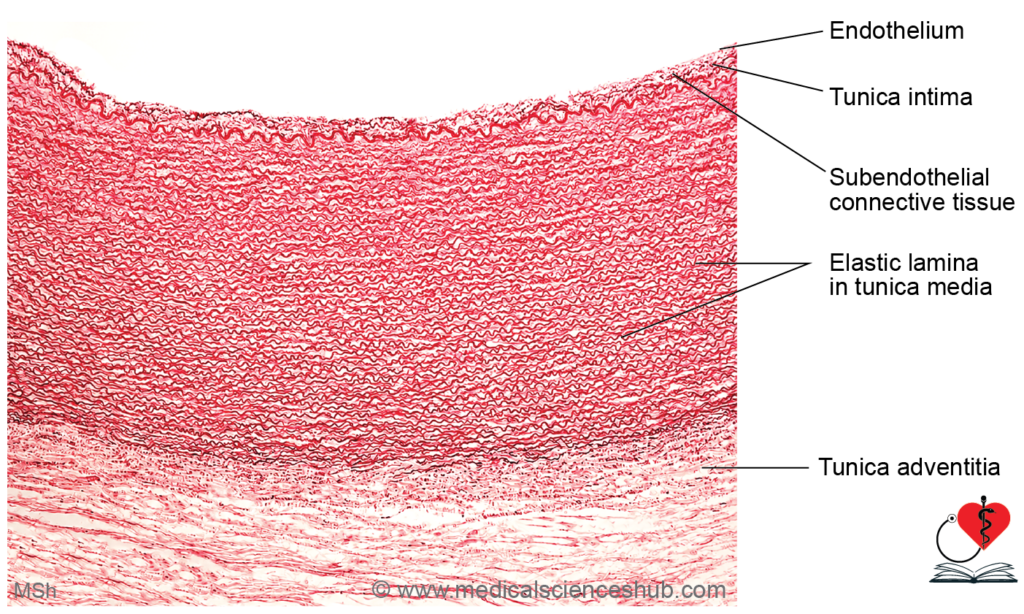
Table 4.1: Differences between collagen, elastic, and reticular fibers
| Property | Collagen Fibers | Elastic Fibers | Reticular Fibers |
|---|---|---|---|
| Location | Tendons, ligaments, organ capsules | Large vessels, lungs, ligamentum nuchae | Basal lamina, liver, spleen, lymph nodes |
| Arrangement | Parallel bundles with branching | Run individually with branching | Form a mesh-like network |
| TEM Striations | Present | Absent | Present |
| Physical Properties | High tensile strength; flexible but inelastic | Highly elastic; low tensile strength | Low elasticity and tensile strength |
| Special Stains | Van Gieson's stain; Masson's trichrome | Orcein; Verhoeff's method | Silver impregnation (argyrophilia) |
| Alternative Name | White fibers | Yellow fibers | Argyrophilic fibers |
CLINICAL INTEGRATION
Marfan syndrome
- Marfan syndrome is an inherited connective tissue disorder that primarily affects elastic fibers and microfibrils.
- It is caused by mutations in the FBN1 gene on chromosome 15, which encodes fibrillin-1, a key component of elastic fiber formation.
- The condition follows an autosomal dominant pattern of inheritance.
- Common features include a tall, slender body habitus, disproportionately long limbs and fingers (arachnodactyly), joint hypermobility, and ocular abnormalities such as lens dislocation.
- Cardiovascular complications, particularly aortic root dilatation and aneurysm formation, are major clinical concerns.
- Diagnosis is based on clinical findings, family history, and genetic testing.
- Although there is no cure, appropriate medical monitoring and treatment can reduce complications and improve outcomes.
Ground Substance/Extracellular Matrix
The extracellular matrix (ground substance) is a transparent, homogeneous material occupying the interfibrillar and intercellular spaces of connective tissue.
Staining Properties
- On routine H&E staining, ground substance is dissolved during tissue processing, leaving an empty appearance with only cells and fibers visible.
- Freeze-drying preserves the matrix, which stains metachromatically with toluidine blue.
- PAS staining yields a positive result due to the presence of proteoglycans.
Composition
- Ground substance is made up of three groups of substances as follows:
- Glycosaminoglycans (GAGs): dermatan sulfate, keratan sulfate, and hyaluronan.
- Proteoglycans: aggrecan and syndecan.
- Glycoproteins: fibronectin and laminin.
Functions of Ground substance
- Its semifluid consistency facilitates diffusion of oxygen, electrolytes, and water-soluble metabolites between blood and tissue cells.
- It acts as a selective barrier, restricting movement of large molecules and pathogens.
- Adhesive glycoproteins anchor connective tissue cells to fibers and to the basement membrane.
Cells of Connective Tissue
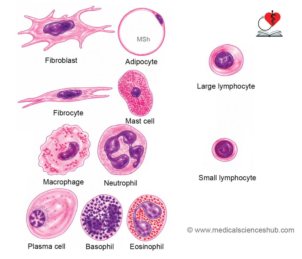
- Cells of connective tissue are classified as resident/ fixed cells and wandering cells.
Fibroblasts
- Fibroblasts are the most numerous cells of connective tissue, responsible for synthesizing collagen, elastic, and reticular fibers, as well as extracellular matrix components.
- Active fibroblasts have abundant cytoplasm, an oval to elongated nucleus, and branched cytoplasmic processes.
- Fibrocytes are quiescent, mature fibroblasts characterized by reduced cytoplasm and a flattened, elongated nucleus.
- On H&E staining, fibroblasts are difficult to distinguish from surrounding collagen fibers; nuclei are visible, and thin pale processes may be seen.
- Fibroblasts align parallel to adjacent collagen fibers and become most metabolically active during wound healing.
- The abundance of cytoplasmic organelles reflects the secretory activity of the cell.
Myofibroblast
- Myofibroblasts are elongated, spindle-shaped cells that share structural features of both fibroblasts and smooth muscle cells.
- They contain actin and myosin filaments, conferring contractile capability.
- Unlike smooth muscle cells, myofibroblasts lack a surrounding basal lamina.
- They cannot be distinguished from fibroblasts on routine H&E staining; immunohistochemical markers are required.
- They play a central role in wound healing and are present in the periodontal ligament, contributing to tooth eruption.
Pericytes
- Pericytes are cells that surround the endothelium of capillaries and venules, embedded within the endothelial basal lamina.
- They possess a large, oval, euchromatic nucleus with multiple cytoplasmic processes that wrap around the capillary wall.
- Pericytes share features of both fibroblasts and smooth muscle cells, contributing to vascular regulation and stability.
Adipocytes (Fat Cells)
Adipocytes are specialized connective tissue cells that synthesize and store lipids and are classified into two types: unilocular and multilocular.
Unilocular Adipocytes
- These large, spherical to polyhedral cells contain a single, massive lipid droplet that compresses the cytoplasm into a thin peripheral rim and displaces the nucleus to one side.
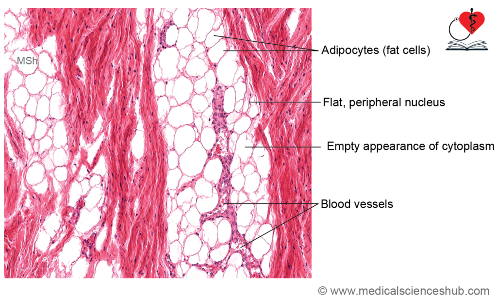
- On H&E staining, the lipid is dissolved during tissue processing, leaving an empty vacuole with a thin cytoplasmic rim and a flattened peripheral nucleus.
- Lipid content is best visualized in fresh frozen sections using special stains: Oil Red O, Sudan Black B, or Nile Blue Sulfate.
- Locations: Subcutaneous layer (panniculus adiposus), mammary fat pad, orbital fat, yellow bone marrow, retroperitoneal space, greater omentum, and cushioning around vital organs.
- Primary functions include thermal insulation, mechanical shock absorption, and energy storage.
Multilocular Adipocytes
- These cells are smaller than unilocular adipocytes and contain multiple small lipid droplets dispersed throughout the cytoplasm, along with an eccentric, round nucleus.
- On H&E staining, the cytoplasm appears foamy due to lipid dissolution, with the eccentric nucleus remaining visible.
- Locations: Abundant in newborns; in adults, found in the posterior neck, interscapular region, and posterior abdominal wall.
- Functions of brown adipose tissue:
- Brown adipose tissue is specialized for non-shivering thermogenesis — generating heat through lipid oxidation via uncoupling of mitochondrial respiration.
- This function is critically important in neonates for thermoregulation during the transition from the intrauterine environment to the cooler external environment.
Macrophages/Histocytes/Clasmatocytes
- Macrophages are derived from circulating monocytes and represent a key component of the innate immune system within connective tissue.
- They are difficult to identify on routine H&E staining without special markers.
- Two functional subtypes exist:
- Fixed macrophages are attached to connective tissue fibers, displaying an irregular shape, short branching cytoplasmic processes, and a kidney-shaped nucleus.
- Wandering macrophages are mobile and migrate to sites of infection or injury.
- Their cytoplasm contains numerous lysosomes essential for digesting phagocytosed material.
- When multiple macrophages fuse in response to large foreign bodies, they form Langhans giant cells — multinucleated cells containing up to 100 nuclei.
- Functions:
- Phagocytosis of bacteria, cellular debris, and foreign particles.
- Antigen presentation to lymphocytes, initiating the adaptive immune response.
Mast Cells
- Mast cells (histaminocytes) are large, ovoid connective tissue cells (~20–30 µm in diameter), typically located in the vicinity of blood vessels.
- They possess a small, centrally placed nucleus, numerous surface filopodia, and a cytoplasm densely packed with basophilic granules containing heparin and histamine.
- Staining:
- Granules stain positively with PAS (magenta), toluidine blue, and alcian blue.
- Mast cell granules exhibit metachromasia — appearing purple to red when stained with toluidine or alcian blue, despite the dye itself being blue.
- Glutaraldehyde fixation is required for optimal mast cell identification.
- Immunoglobulin E (IgE) receptors are expressed on the mast cell surface.
- Functions: Upon antigen–IgE binding, mast cells undergo degranulation, releasing:
- Histamine which causes bronchiolar smooth muscle contraction (asthma), vasodilation and increased vascular permeability (edema), and cutaneous itching.
- Heparin which acts as a local anticoagulant.
- Serine proteases which promote inflammation.
- Leukotriene C4 which induces bronchospasm by constricting respiratory smooth muscle.
Lymphocytes
- Lymphocytes are the primary cellular mediators of immune responses, identifiable by their deeply staining heterochromatic nucleus and thin cytoplasmic rim.
- They are classified based on surface cluster of differentiation (CD) markers:
- T-lymphocytes mediate cell-mediated immunity.
- B-lymphocytes mediate humoral (antibody-mediated) immunity.
- Natural killer (NK) cells destroy virus-infected and neoplastic cells.
Plasma Cells/Plasmatocytes
- Plasma cells are terminally differentiated B-lymphocytes specialized for antibody production.
- They are ovoid cells with deeply basophilic cytoplasm, reflecting abundant rough endoplasmic reticulum (rER).
- The nucleus is round and eccentrically placed, with a characteristic clock-face (cartwheel) chromatin pattern — a key identification feature.
- Russell bodies are large, homogeneous intracytoplasmic inclusions representing accumulated immunoglobulins within dilated rER cisternae; they are PAS-positive. Plasma cells containing Russell bodies are termed Mott cells.
Pigment Cells
- Melanocytes are star-shaped cells with long branching processes that synthesize melanin pigment.
- They are derived from neural crest cells and located in the skin, iris, and choroid.
- Their primary function is protecting deeper tissues from ultraviolet radiation damage.
Other Leucocytes
- Leukocytes originate in the bone marrow and migrate into connective tissue as needed.
- They are classified as:
- Granulocytes — contain prominent cytoplasmic granules; include neutrophils, eosinophils, and basophils.
- Agranulocytes — lack specific cytoplasmic granules; include lymphocytes and monocytes.
Table 4.2: Connective tissue cells
| Fixed (Resident) Cells | Wandering (Transient) Cells |
|---|---|
| Fibroblasts | Lymphocytes |
| Adipocytes | Plasma cells |
| Macrophages | Neutrophils |
| Mast cells | Eosinophils |
| Pericytes | Basophils |
| Monocytes |
Embryonic Connective Tissue
- Embryonic connective tissue is derived from mesoderm and comprises two subtypes: mesenchyme and mucous connective tissue.
Mesenchyme
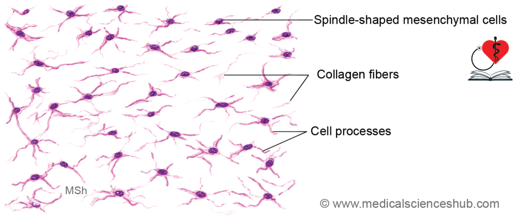
- Mesenchyme is the embryonic connective tissue composed of small, spindle-shaped mesenchymal cells with fine cytoplasmic processes interconnected by gap junctions.
- The intercellular spaces are occupied by ground substance, reticular fibers, and collagen fibers.
Mucous Connective Tissue
- Mucous connective tissue is primarily found in the umbilical cord.
- It surrounds and supports the umbilical blood vessels, including two umbilical arteries and one umbilical vein.
- This tissue is composed of two main components: cells and Wharton’s jelly.
- The cells are typically spindle-shaped with elongated cytoplasmic processes.
- These cells are embedded within a gelatinous extracellular matrix known as Wharton’s jelly.
- Wharton’s jelly is rich in hyaluronic acid, which contributes to its soft, mucoid consistency.
- During routine histological processing, much of the Wharton’s jelly is removed. As a result, tissue sections mainly show spindle-shaped cells with elongated nuclei and delicate cytoplasmic processes.
- A key identifying feature of this tissue is the presence of widely spaced spindle-shaped cells within a mucoid matrix.
- Mesenchymal stem cells isolated from Wharton’s jelly possess regenerative potential and are being investigated for applications in stem cell therapy and regenerative medicine.
Loose Connective/Areolar Tissue
- Loose (areolar) connective tissue is the most widely distributed connective tissue in the body.
- It consists of three major components:
- Fibers
- Cells
- Ground substance
- The fibers form a loosely arranged network and include collagen fibers, elastic fibers, and reticular fibers.
- The cellular component contains various connective tissue cells, including fibroblasts, macrophages, plasma cells, adipocytes (fat cells), and migrating leukocytes.
- Fibers and cells are embedded within an abundant, gel-like ground substance.
- During routine histological processing, much of the ground substance is removed, leaving clear spaces called areolae. This characteristic appearance gives the tissue its name, areolar tissue.
Locations
- It is present beneath most epithelia, particularly in the lamina propria.
- It forms a major component of the superficial fascia (subcutaneous tissue).
- It surrounds blood vessels, nerves, muscles, viscera, and glandular parenchyma.
- It is also found within the mesentery.
Functions
- It provides structural support and packing material for tissues and organs.
- It permits movement of adjacent structures, such as the skin over deeper tissues.
- It allows expansion of organs, blood vessels, and other soft tissues.
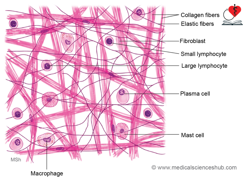
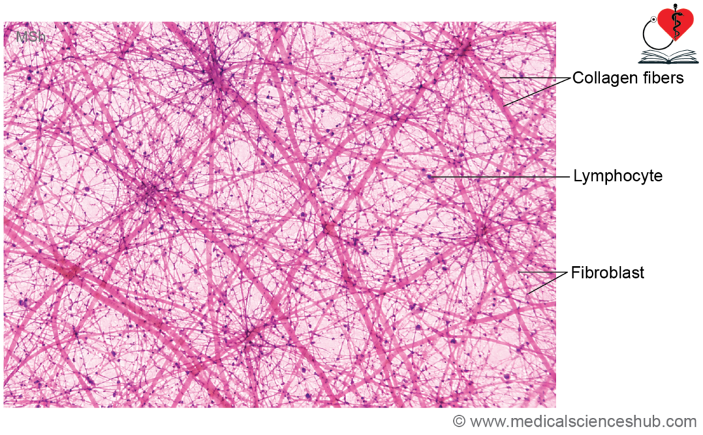
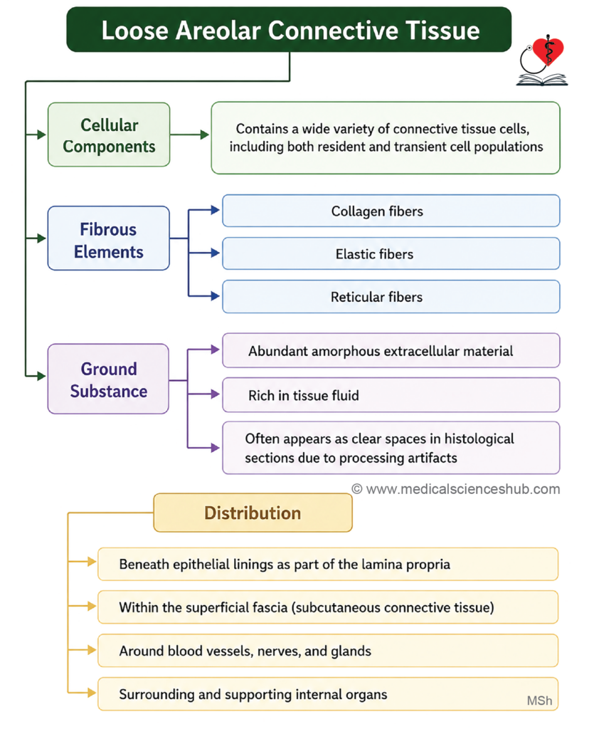
Table 4.3: White blood cells
| Cell Type | Morphology | Functions |
|---|---|---|
| Neutrophil (10–12 µm) | Nucleus: 3–5 lobes; cytoplasm: pale pink to reddish-purple granules | Phagocytosis of microorganisms; first-line defense against bacterial infection |
| Basophil (10–12 µm) | Nucleus: segmented; cytoplasm: large, dark basophilic granules containing histamine | Releases histamine and promotes inflammatory and allergic responses |
| Eosinophil (10–14 µm) | Nucleus: bilobed; cytoplasm: prominent eosinophilic granules | Phagocytosis of antigen–antibody complexes; mediates allergic reactions and asthma; defense against parasitic infections |
| Lymphocyte (6–14 µm) | Round, deeply staining nucleus; thin cytoplasmic rim | T cells: cell-mediated immunity B cells: humoral (antibody-mediated) immunity NK cells: destruction of neoplastic and virus-infected cells |
| Monocyte (12–20 µm) | Large kidney-shaped nucleus | Phagocytosis; differentiation into tissue macrophages and other mononuclear phagocytic cells |
Dense Connective Tissue
- Dense connective tissue contains abundant connective tissue fibers with relatively few cells and a limited amount of ground substance.
- Its primary function is to provide mechanical strength, support, and protection to tissues and organs.
- The tissue is characterized by densely packed fibers, predominantly collagen fibers, which enhance resistance to tensile forces.
- Based on the arrangement of its fibers, dense connective tissue is classified into:
- Dense irregular connective tissue, in which fibers are arranged in multiple directions.
- Dense regular connective tissue, in which fibers are arranged in parallel bundles.
- This structural organization enables the tissue to withstand stress applied from different directions or along a single axis, depending on the subtype.
Dense Irregular Connective Tissue
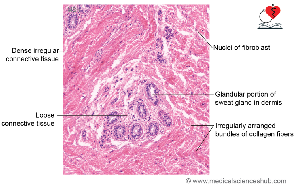
- Dense irregular connective tissue contains thick bundles of collagen fibers arranged in multiple directions, providing strength against stresses from various angles.
- It is commonly found in the dermis of the skin, sclera of the eye, fibrous capsules of organs and joints, and as sheet-like connective tissue in the periosteum and perichondrium.
- Its primary function is to provide structural support, protection, and resistance to multidirectional tensile forces.
Dense Regular Connective Tissue
- Dense regular connective tissue is composed predominantly of type I collagen fibers arranged in compact, parallel bundles with relatively few cells.
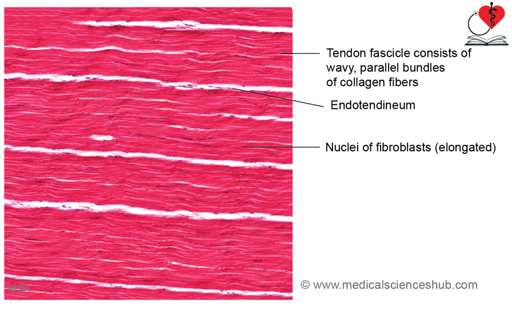
- The collagen bundles characteristically display a wavy appearance on histological sections.
- Also termed white fibrous tissue, it is found in tendons, ligaments, and the corneal stroma.
Tendon
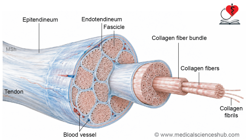
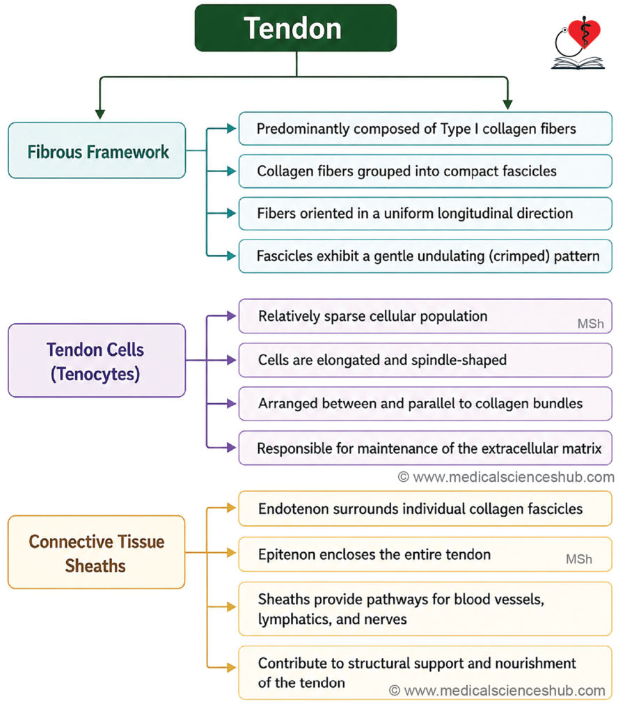
- Tendons consist of type I collagen fibers arranged in a tightly packed, parallel (cord-like) pattern.
- Individual bundles are separated by a thin vascular connective tissue layer called the endotendineum; the entire tendon is enclosed by the epitendineum.
- Elongated tendinocytes (tendon fibroblasts) are oriented parallel to the long axis of collagen bundles and are situated between fiber bundles.
Ligament
- Ligaments also consist primarily of type I collagen fibers, but these are arranged in a less regular, sheet-like pattern compared to tendons, with interspersed fibroblasts.
- Elastic ligaments (e.g., ligamentum nuchae and ligamentum flavum) contain a high proportion of elastic fibers, conferring greater extensibility.
- Special Arrangement — Aponeurosis and Corneal Stroma: In the aponeurosis and corneal stroma, collagen bundles are arranged in alternating layers oriented at 90° to one another (orthogonal array), providing multidirectional tensile strength and, in the cornea, optical transparency.
Function:
- The parallel, densely packed arrangement of type I collagen fibers provides exceptional tensile strength, enabling tendons and ligaments to withstand high unidirectional mechanical and pulling forces.
Adipose Tissue
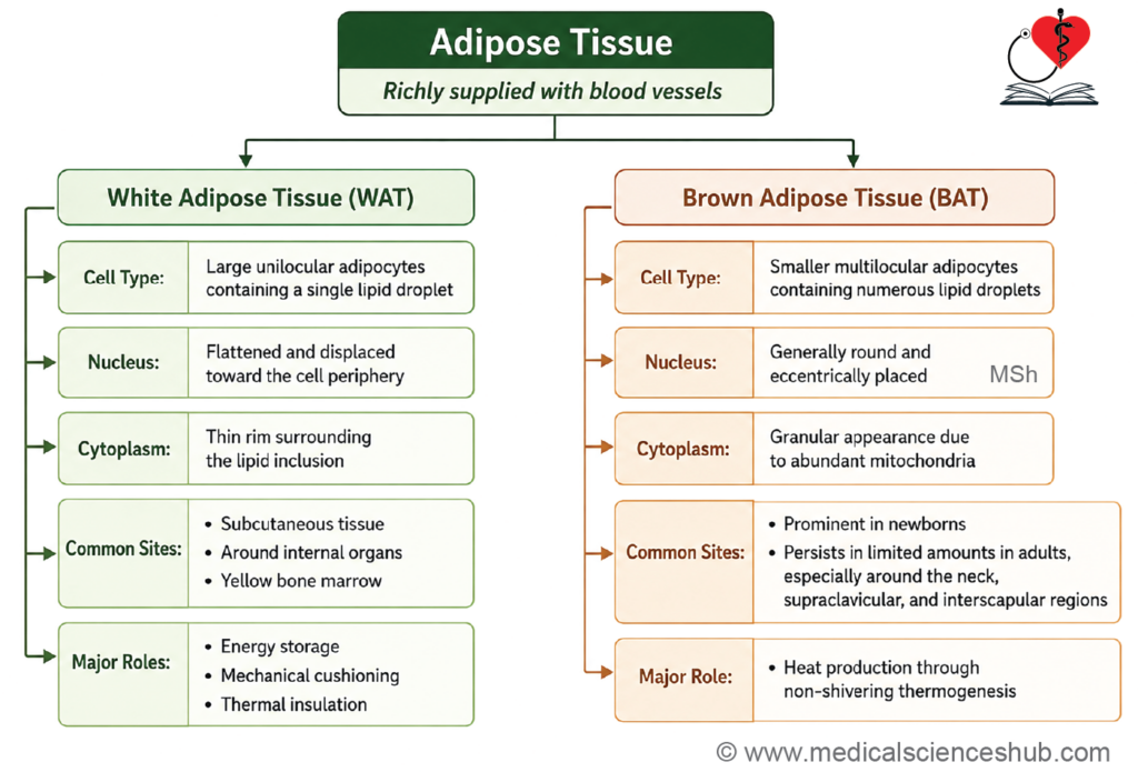
Adipose tissue is a specialized connective tissue composed of adipocytes, classified into two types: white (yellow) and brown adipose tissue.
Yellow Adipose Tissue
- Appears whitish to yellowish and is highly vascularized.
- Composed of unilocular adipocytes — large, oval cells containing a single massive lipid droplet that displaces the nucleus peripherally and compresses the cytoplasm into a thin rim, producing a characteristic signet ring appearance.
- On H&E staining, lipid is dissolved during processing, leaving cells with an empty vacuole, thin cytoplasmic rim, and flattened peripheral nucleus.
- Locations: Superficial fascia (breast, buttocks), yellow bone marrow, omentum, mesentery, and perivisceral fat depots.
- Functions: Energy storage, thermal insulation, and mechanical protection of visceral organs.
Brown Adipose Tissue
- Appears brownish due to a rich mitochondrial content and dense vascularization.
- Composed of multilocular adipocytes — smaller than unilocular adipocytes, with multiple small lipid droplets producing a foamy cytoplasmic appearance and a round, eccentrically placed nucleus.
- Locations: Predominant in neonates; in adults, found in the posterior neck, interscapular region, and posterior abdominal wall, with quantities declining progressively with age.
- Functions:
- Specialized for non-shivering thermogenesis — generating heat through mitochondrial uncoupling during lipid oxidation.
- Plays a critical role in thermoregulation in newborns, facilitating adaptation to the extrauterine thermal environment.
CLINICAL INTEGRATION
- Obesity results from excessive accumulation of adipose tissue, involving both hypertrophy (increased adipocyte size) and hyperplasia (increased adipocyte number).
- Lipoma is the most common benign tumor of adipose tissue, presenting as a soft, mobile, painless subcutaneous nodule that does not metastasize.
- Liposarcoma is a rare malignant tumor of adipose tissue with the capacity for local invasion and distant metastasis.
Important Questions
- List the components of connective tissue.
- List the cells of connective tissue and their functional correlation.
- Write a short note on macrophages.
- Write a short note on Wharton’s jelly.
- Write a short note on histology of tendon or ligament.
- Write a short note on adipose tissue.
- List the differences between yellow and brown adipose tissue.
📝 Test Your Knowledge – Practice MCQs
Attempt the chapter MCQ quiz and assess your understanding of key concepts.
Click to see: Medical Disclaimer and Copyright Notice