
Introduction
- Cells form the basic structural and functional units of all tissues. Each cell contains a membrane, cytoplasm, and nucleus. The selectively permeable cell membrane regulates exchange, while specialized junctions connect neighbouring cells, enabling adhesion, communication, mechanical stability, and coordinated activity within tissues.
Basic Structural Components of a Cell
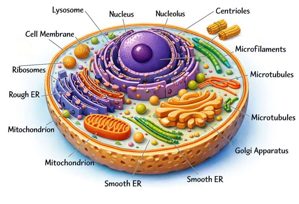
- A typical cell contains three principal components: the cell membrane, cytoplasm, and nucleus, which together maintain cellular structure and regulate vital functions.
- The cell membrane forms the outer boundary of the cell. It encloses the cytoplasm and regulates the movement of substances between the intracellular and extracellular environments through selective permeability.
- The cytoplasm is the internal fluid matrix that contains numerous membrane-bound organelles. Common organelles include mitochondria, ribosomes, lysosomes, peroxisomes, centrioles, endoplasmic reticulum, and the Golgi apparatus, each performing specialized metabolic or synthetic functions.
- The cytoplasm also contains a cytoskeleton composed of microfilaments, intermediate filaments, and microtubules, which maintain cell shape and support intracellular transport.
- The nucleus usually lies near the center of the cell and contains genetic material that regulates cellular activities.
- Contractile proteins such as actin and myosin contribute to cell movement, structural stability, and interactions with neighbouring cells.
Cell Membrane
- The cell membrane forms the outer protective boundary of the cell. It separates the intracellular contents from the external environment and maintains the integrity of the cell.
- The membrane regulates the movement of ions, nutrients, and waste products between the cytoplasm and the surrounding extracellular fluid. It also participates in cell communication, cell recognition, and adhesion between neighbouring cells.
- Membrane proteins and surface molecules help anchor cells to surrounding structures and to adjacent cells.
Structure of Cell Membrane
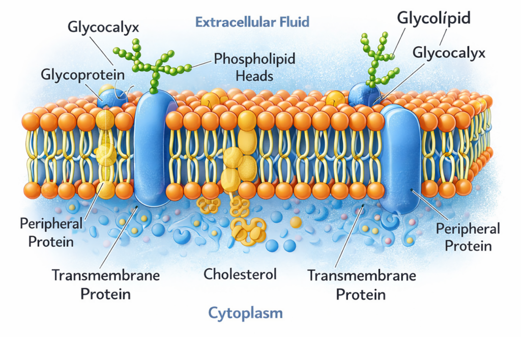
- The membrane is approximately 7–10 nanometres thick and mainly consists of a phospholipid bilayer.
- Lipids form about forty five percent of the membrane dry weight, proteins contribute about fifty percent, and carbohydrates constitute a small proportion.
- Phospholipid molecules arrange themselves in two layers with hydrophilic heads facing outward and hydrophobic tails directed inward.
Fluid-Mosaic Model of the Membrane
- The membrane follows the fluid mosaic model, which explains its flexible and dynamic structure (described by Singer and Nicolson in 1972).
- Proteins are embedded within or attached to the phospholipid bilayer, forming a mosaic pattern.
- Both phospholipids and proteins can move laterally within the membrane, a process called translational diffusion. This lateral mobility maintains membrane flexibility while preserving structural stability.
- The degree of membrane fluidity depends largely on the lipid composition of the phospholipid bilayer.
Effect of Temperature on Membrane Fluidity:
- The phospholipid bilayer contains hydrophobic fatty acid chains that remain tightly arranged at lower temperatures, producing a relatively rigid membrane structure.
- When temperature rises, these lipid chains shift from an ordered crystalline state to a more disordered and fluid arrangement.
- The temperature at which this structural change occurs is called the transition temperature.
- Phospholipids with longer and more saturated fatty acid chains interact strongly and therefore require higher temperatures to increase membrane fluidity.
- Greater unsaturation of fatty acids increases membrane flexibility and reduces the transition temperature.
Lipid Bilayer of the Cell Membrane
- The lipid bilayer forms the basic structural framework of the cell membrane and mainly consists of phospholipids, glycolipids, and cholesterol.
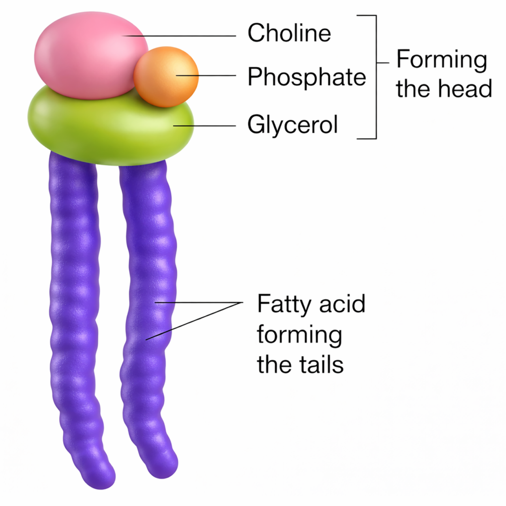
- Common phospholipids in the membrane include phosphatidylcholine, sphingomyelin, phosphatidylserine, and phosphatidylethanolamine. These molecules form the principal structural matrix of the membrane.
- Glycolipids are usually located in the outer layer of the membrane and contribute to cell recognition and surface interactions.
- Membrane lipids are amphipathic molecules, meaning they possess both hydrophilic and hydrophobic regions.
- The polar hydrophilic head contains phosphate or hydroxyl groups that interact with water, whereas the non-polar hydrophobic tails consist of fatty acid chains that avoid water.
- In the bilayer, hydrophobic tails face inward while hydrophilic heads face the aqueous environments inside and outside the cell.
- Cholesterol is embedded within the hydrophobic region and stabilizes the membrane by regulating permeability and fluidity.
Functions of the Lipid Bilayer
- The lipid bilayer forms the principal permeability barrier of the cell membrane. It separates the cytoplasm from the surrounding interstitial fluid and maintains the internal environment of the cell.
- The membrane allows substances to pass based on their lipid solubility.
- Lipid-soluble molecules, such as oxygen and certain small non-polar substances, diffuse easily through the bilayer.
- Water-soluble molecules, including glucose and urea, cannot pass freely and usually require specific transport proteins. Because of this selective movement, the membrane acts as a semipermeable barrier.
Membrane Proteins
- The proportion of membrane proteins varies depending on the function of the membrane.
- On average, proteins constitute about half of the membrane mass.
- Membrane proteins are broadly classified into integral proteins and peripheral proteins, each performing specialized structural and functional roles.
Integral Proteins
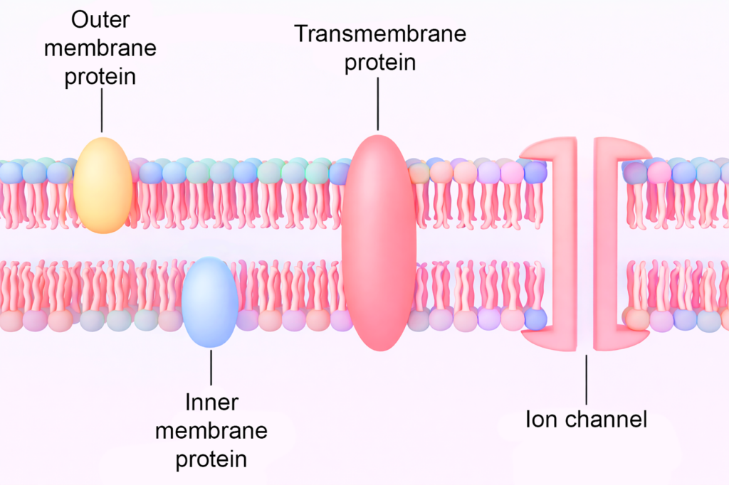
- Integral proteinsare membrane proteins embedded within the lipid bilayer. Many extend across the entire membrane and are therefore called transmembrane proteins.
- Some integral proteins penetrate only part of the membrane and are exposed either to the extracellular surface or to the cytoplasmic side.
- Several membrane proteins can move laterally within the lipid bilayer, which supports dynamic membrane activities such as endocytosis and signal transmission.
- Functions of Integral Proteins
- They function as channel proteins that form pores allowing diffusion of water-soluble substances such as ions and small molecules.
- They act as carrier proteins that transport specific molecules across the membrane by facilitated diffusion.
- Some proteins operate as ion pumps, which actively transport ions against their concentration gradients using cellular energy.
- Certain integral proteins serve as receptors, enzymes, or antigenic molecules involved in cell signalling and immune recognition.
Peripheral Proteins
- Peripheral proteins are loosely attached to the surface of the cell membrane rather than embedded within the lipid bilayer.
- These proteins are located either on the inner cytoplasmic surface or on the outer extracellular surface of the membrane.
- They are attached through weak interactions with membrane lipids or with integral membrane proteins.
- Types of Peripheral Proteins
- Intrinsic peripheral proteins are present on the inner surface of the membrane. They commonly function as enzymes or as anchoring sites for cytoskeletal elements that help maintain cell shape and structural stability.
- Extrinsic peripheral proteins are located on the outer surface of the membrane. They participate in cell adhesion, enabling cells to attach to neighbouring cells or to components of the basal lamina.
- These proteins can be removed from the membrane without disrupting the lipid bilayer.
Membrane Carbohydrates
- The outer surface of the cell membrane is covered by a carbohydrate-rich layer called the glycocalyx, also known as the cell coat.
- These carbohydrates are mainly short oligosaccharide chains attached to membrane proteins and lipids.
- Carbohydrates linked to proteins form glycoproteins, whereas those attached to lipids form glycolipids.
- Some membrane components also contain carbohydrate chains associated with proteoglycans.
- Together, these molecules create a carbohydrate layer on the external surface of the lipid bilayer.
- Functions of the Glycocalyx
- The glycocalyx provides protection to the cell surface and contributes to the stability of the membrane.
- Negatively charged carbohydrate groups help reduce unwanted interactions between adjacent cells and circulating molecules.
- Certain glycoproteins participate in cell adhesion, allowing temporary or stable attachment between neighbouring cells.
- Some membrane carbohydrates function as receptors involved in cell recognition and signalling.
Functions of Cell Membrane
- The cell membrane maintains a stable intracellular environment that supports normal cellular metabolism and organelle function.
- It regulates the composition of intracellular fluid, which typically contains lower concentrations of sodium and chloride ions and higher levels of potassium, magnesium, and organic phosphates.
- The membrane controls cell volume by regulating ion transport through specialized channels and active transport mechanisms, particularly by removing sodium ions from the cell.
- In neurons and muscle cells, the membrane maintains an electrical potential difference between the intracellular and extracellular surfaces, which is essential for excitability and signal transmission.
- Membrane surface molecules participate in cell recognition, allowing the immune system to identify foreign cells and initiate appropriate defense responses.
Special Features of RBC Membrane
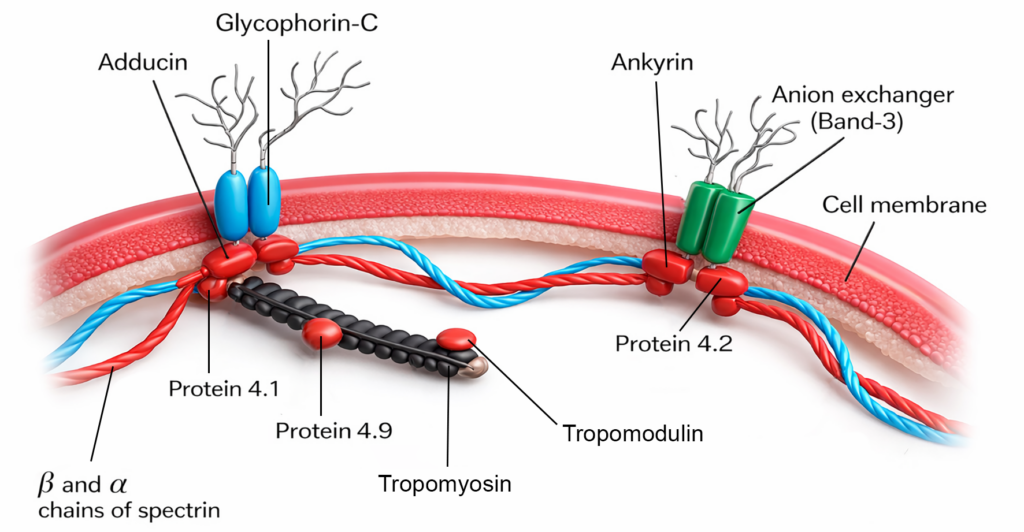
- The red blood cell membrane is extensively studied because of its simple structure and clinical importance. In addition to the usual membrane components, it contains specialized integral and peripheral proteins that maintain cell structure and function.
- The specialized membrane proteins—ankyrin, spectrin, and adducin—that form a flexible cytoskeletal network, giving the red cell membrane its characteristic deformability, along with integral components such as the anion exchanger (Band-3), glycophorin-C, and other submembrane proteins.
Integral Proteins
- Two important integral proteins in the red blood cell membrane are glycophorins and band-3 proteins.
- Glycophorins are glycoproteins that contain a high proportion of carbohydrate chains attached to a protein core.
- The carbohydrate components project on the outer surface of the membrane and contribute to blood group antigen expression, including determinants of the MN blood group system.
- Band-3 protein is a major transmembrane transport protein that crosses the membrane multiple times. It functions as an anion exchanger, allowing the exchange of bicarbonate and chloride ions during gas transport in blood.
Peripheral Proteins
- The inner surface of the membrane contains cytoskeletal proteins that stabilize the membrane and maintain the characteristic biconcave shape of red blood cells.
- Important peripheral proteins include spectrin and ankyrin.
- Spectrin forms a fibrous network beneath the membrane that provides mechanical strength.
- Ankyrin connects spectrin to integral membrane proteins such as band-3, thereby linking the membrane to the cytoskeleton and preserving membrane integrity.
Clinical Integration
- Hereditary spherocytosis occurs due to defects in red blood cell membrane proteins such as spectrin or ankyrin, leading to spherical and fragile cells. These cells are easily destroyed in the spleen, resulting in hemolytic anemia and jaundice.
- Hereditary elliptocytosis results from abnormalities in cytoskeletal proteins, producing elliptical red blood cells with reduced mechanical stability and increased susceptibility to hemolysis.
Cell Organelles
- Animal cells contain several organelles, including mitochondria, endoplasmic reticulum, Golgi apparatus, ribosomes, lysosomes, peroxisomes, and centrioles, each performing specialized cellular functions.
- Mature red blood cells lack a nucleus and most organelles, including mitochondria and ribosomes, which allows greater space for haemoglobin and efficient oxygen transport.
Mitochondria
- Mitochondria are membrane-bound organelles responsible for most energy production in the cell.
- They generate adenosine triphosphate through oxidative metabolism and therefore support cellular activities requiring energy. Their number and size vary among cells and are greater in tissues with high metabolic demand, such as liver and cardiac muscle.
Structure
- Each mitochondrion is surrounded by two membranes: an outer membrane and an inner membrane.
Outer Mitochondrial Membrane
- The outer membrane forms the external boundary of the organelle.
- It contains channel-forming proteins called porins that permit the diffusion of small molecules and ions.
Inner Mitochondrial Membrane
- The inner membrane is rich in proteins and is relatively impermeable to most polar molecules and ions.
- It forms numerous inward folds known as cristae, which increase the surface area for metabolic reactions.
- The inner membrane contains enzymes and electron carriers involved in the electron transport chain and oxidative phosphorylation.
Mitochondrial Matrix
- The internal space enclosed by the inner membrane is called the matrix.
- The matrix contains enzymes required for the citric acid cycle and fatty acid oxidation.
- It also contains mitochondrial DNA, ribosomes, and enzymes necessary for the synthesis of certain mitochondrial proteins.
Functions
- Mitochondria play a central role in cellular respiration and energy production.
- Acetyl coenzyme A enters the citric acid cycle within the matrix to generate reducing equivalents for the electron transport chain.
- The final products of these reactions include carbon dioxide, water, and adenosine triphosphate.
- Mitochondria possess their own DNA and can undergo self-replication to maintain their population within cells.
- Mitochondrial enzymes and their main roles are listed in Table 4.1.
Clinical Integration
- Mitochondrial diseases occur when defects impair mitochondrial energy production, leading to reduced adenosine triphosphate generation in cells with high metabolic demand.
- Mutations in mitochondrial DNA can cause mitochondrial cytopathies, which commonly present with muscle weakness, neurological degeneration, and elevated lactic acid levels in blood.
- Mitochondrial damage caused by free radicals contributes to cellular injury and is associated with several age-related degenerative disorders.
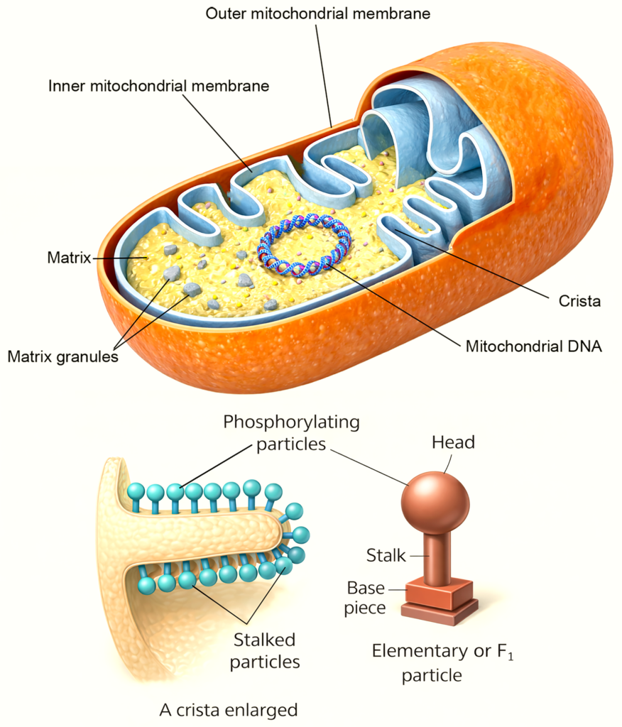
Table 1.1: Mitochondrial enzymes
| Mitochondrial Region | Major Enzymes | Primary Functional Role |
|---|---|---|
| Outer Mitochondrial Membrane | Cytochrome b5 and cytochrome b5 reductase | Participate in electron transfer reactions and fatty acid metabolism. |
| Fatty acyl-CoA synthetase | Activates fatty acids by converting them to fatty acyl-coenzyme A for metabolic processing. | |
| Phospholipase A | Involved in phospholipid metabolism and membrane lipid remodeling. | |
| Nucleoside diphosphate kinase | Maintains cellular nucleotide balance by interconverting nucleoside diphosphates and triphosphates. | |
| Inner Mitochondrial Membrane | Cytochromes b, c1, c, a, and a3 | Components of the electron transport chain responsible for oxidative phosphorylation. |
| NADH dehydrogenase | Transfers electrons from reduced nicotinamide adenine dinucleotide to the electron transport chain. | |
| Succinate dehydrogenase | Functions in both the citric acid cycle and electron transport chain. | |
| Electron-transferring flavoproteins | Carry electrons from fatty acid oxidation to the respiratory chain. | |
| β-Hydroxybutyrate dehydrogenase | Catalyzes reactions involved in ketone body metabolism. | |
| Carnitine palmitoyltransferase | Facilitates transport of long-chain fatty acids into mitochondria for β-oxidation. | |
| Membrane translocases | Transport metabolites and ions across the inner mitochondrial membrane. | |
| Intermembrane Space | Adenylate kinase | Converts adenine nucleotides and supports cellular energy balance. |
| Nucleoside diphosphate kinase | Regulates nucleotide interconversion for metabolic processes. | |
| Sulfite oxidase | Catalyzes oxidation of sulfite during sulfur amino acid metabolism. | |
| Mitochondrial Matrix | Pyruvate dehydrogenase complex | Converts pyruvate into acetyl-coenzyme A, linking glycolysis with the citric acid cycle. |
| Citrate synthase, aconitase, isocitrate dehydrogenase, α-oxoglutarate dehydrogenase, malate dehydrogenase | Catalyze sequential reactions of the citric acid cycle for energy production. | |
| Fatty acid oxidation enzymes | Carry out β-oxidation to generate acetyl-coenzyme A and reducing equivalents. | |
| Ornithine transcarbamylase | Participates in the urea cycle, contributing to ammonia detoxification. |
Endoplasmic Reticulum
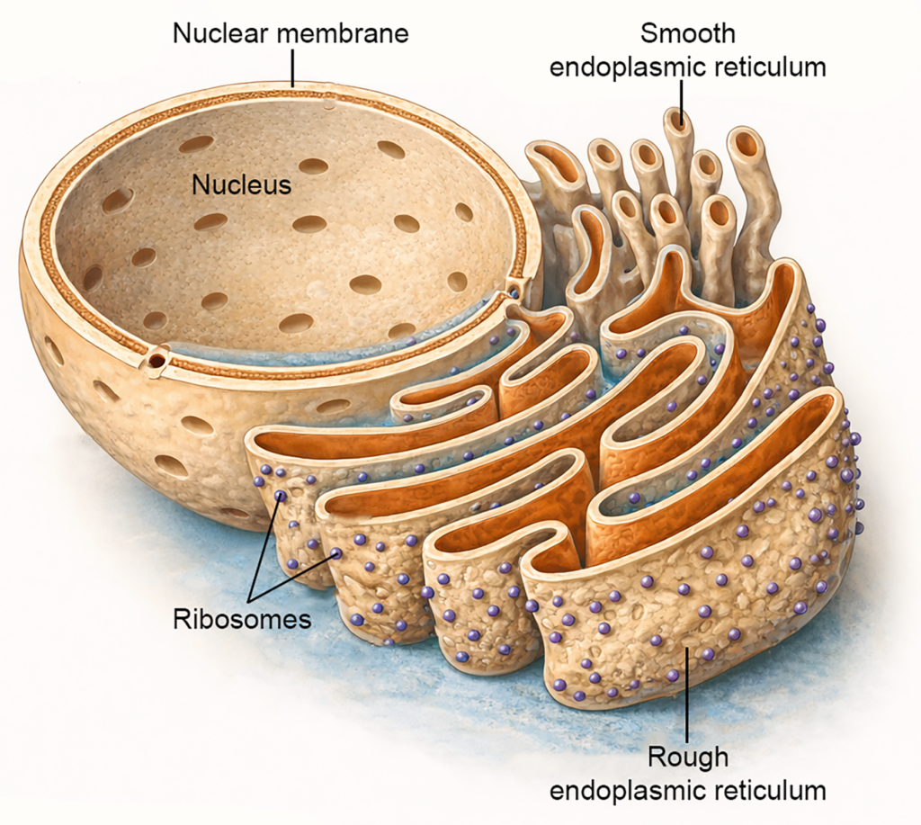
- The endoplasmic reticulum is an extensive intracellular membrane system composed of interconnected tubules, vesicles, and flattened sacs known as cisternae.
- Its membrane is continuous with the outer membrane of the nucleus and maintains functional connections with the Golgi apparatus.
- This organelle participates in the synthesis, processing, and intracellular transport of molecules required for cell structure and function.
- The endoplasmic reticulum is structurally and functionally divided into two forms: rough endoplasmic reticulum and smooth endoplasmic reticulum.
Rough Endoplasmic Reticulum
- The rough endoplasmic reticulum contains numerous ribosomes attached to its cytoplasmic surface, giving it a granular appearance under the microscope.
- It is highly developed in cells that actively produce proteins for secretion or membrane incorporation.
- Examples include pancreatic acinar cells, plasma cells, and many types of secretory epithelial cells.
- In neurons, aggregates of rough endoplasmic reticulum appear as Nissl bodies, which are involved in the synthesis of neuronal proteins.
- Functions
- The rough endoplasmic reticulum is the primary site of protein synthesis for secretory proteins, membrane proteins, and proteins destined for intracellular organelles.
- Newly synthesized proteins enter the lumen of the rough endoplasmic reticulum for folding and initial modification.
- It contributes to the early stages of glycoprotein formation by adding carbohydrate groups to specific proteins before they are transported to the Golgi apparatus for further processing.
Smooth Endoplasmic Reticulum
- The smooth endoplasmic reticulum lacks ribosomes on its surface and therefore appears smooth when viewed microscopically.
- It is abundant in cells that specialize in lipid metabolism and chemical processing.
- Functions
- The smooth endoplasmic reticulum is the principal site of lipid synthesis, including phospholipids, cholesterol, and steroid hormones.
- In muscle cells, it forms the sarcoplasmic reticulum, which stores and releases calcium ions during muscle contraction.
- It participates in intracellular transport by forming a continuous membrane system with the rough endoplasmic reticulum and the Golgi apparatus.
- It also plays an important role in detoxification, where enzymes modify drugs, metabolic products, and toxic substances to facilitate their removal from the cell.
Golgi Apparatus
- The Golgi apparatus is a membrane-bound organelle present in most eukaryotic cells.
- It is usually located near the nucleus and shows a close structural relationship with the endoplasmic reticulum.
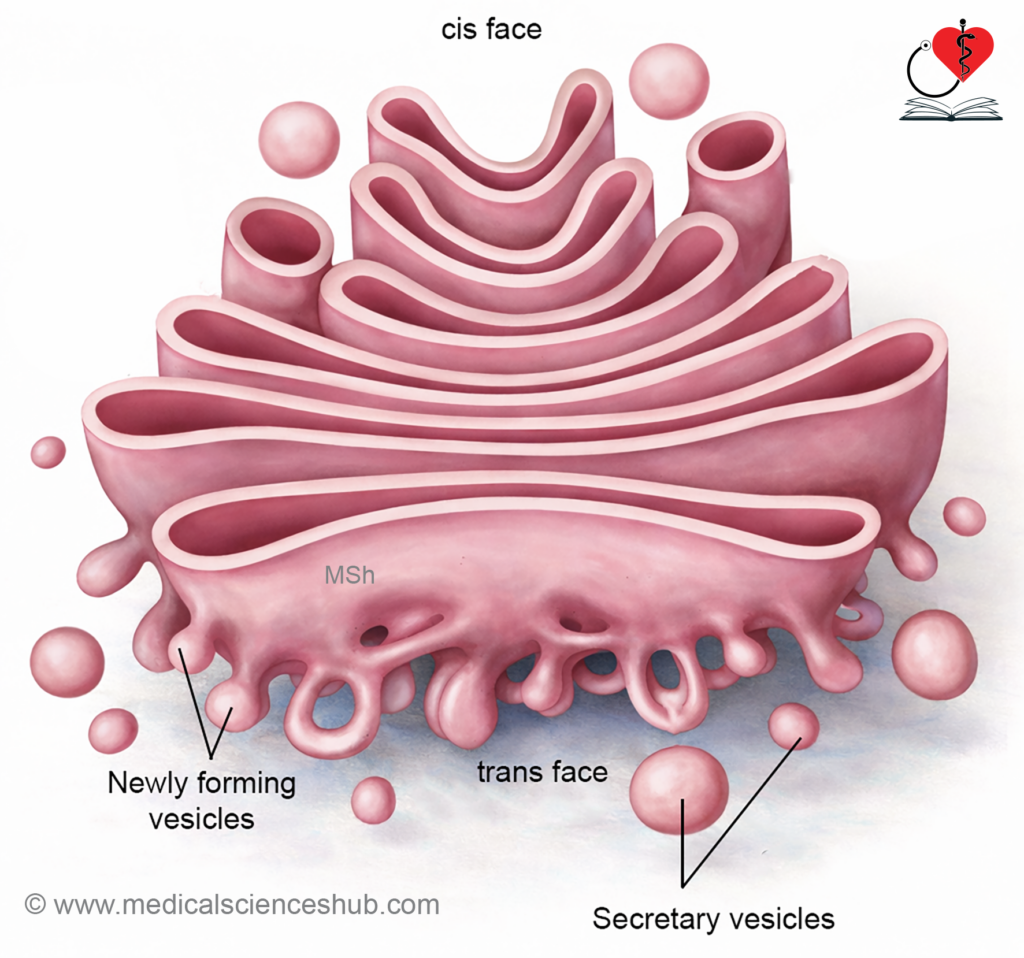
- The organelle consists of flattened membrane sacs called cisternae, arranged in stacked layers with small vesicles at their margins.
- The Golgi apparatus has two functional regions. The cis face receives transport vesicles from the endoplasmic reticulum, while the trans face directs processed materials toward their final destinations.
Major Functions of GA
- The Golgi apparatus modifies and packages proteins that arrive from the rough endoplasmic reticulum into membrane-bound secretory vesicles.
- It adds carbohydrate groups to certain proteins to produce glycoproteins required for secretion or membrane formation.
- The organelle participates in the formation of lysosomes by packaging digestive enzymes.
- It also sorts and transports proteins and lipids to different intracellular compartments or to the cell membrane for secretion.
Lysosomes
- Lysosomes are small membrane-bound organelles that contain numerous digestive enzymes responsible for intracellular degradation.
- These enzymes belong mainly to the group of hydrolytic enzymes, which function optimally in an acidic internal environment.
- Lysosomes occur in most animal cells but are absent in mature erythrocytes.
- They are especially numerous in cells that perform active phagocytosis, such as neutrophils, macrophages, and monocytes.
- In granulocytes, lysosomes appear as cytoplasmic granules containing digestive enzymes.
- Lysosomal enzymes are synthesized in the rough endoplasmic reticulum and packaged by the Golgi apparatus before being enclosed within lysosomal membranes.
Types of Lysosomes
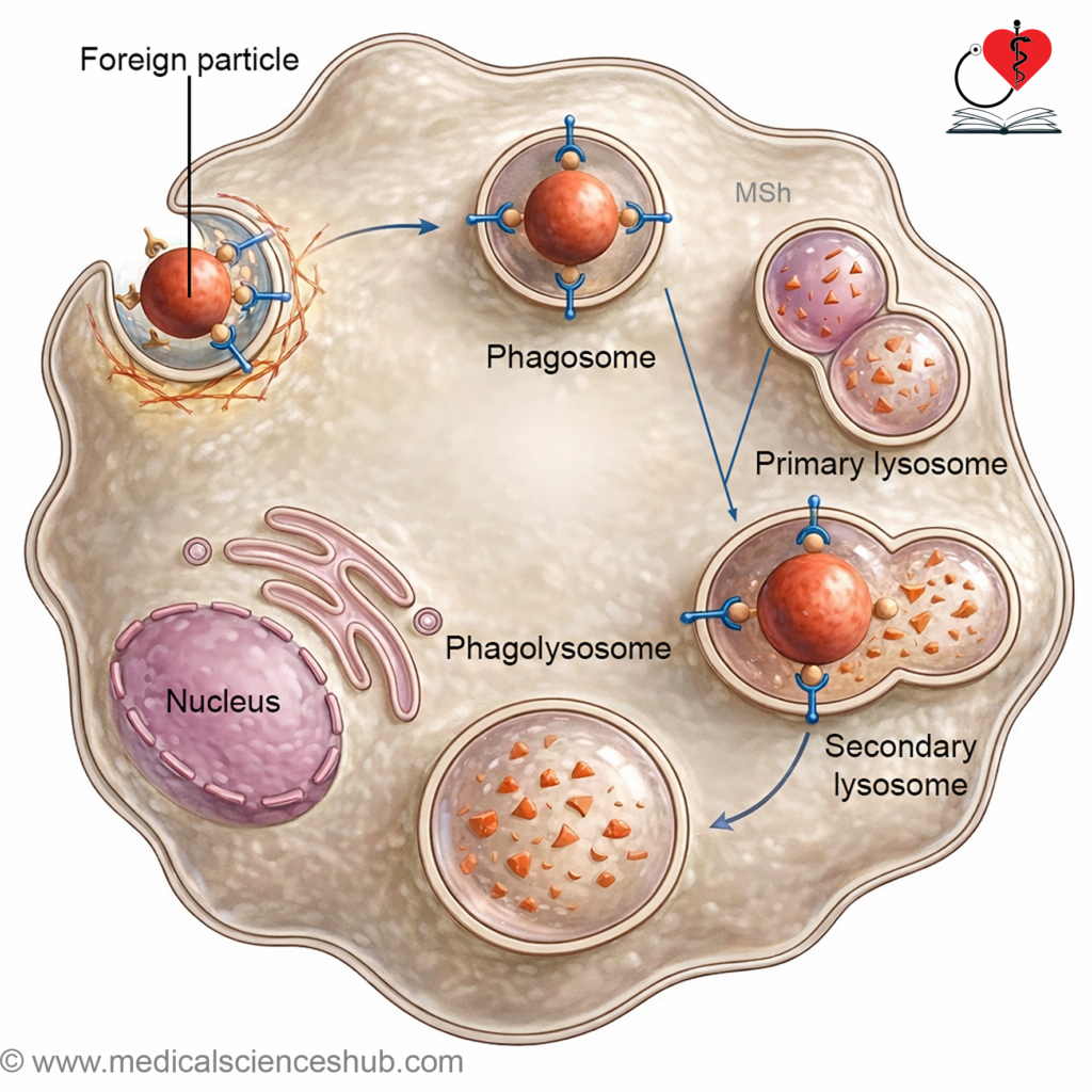
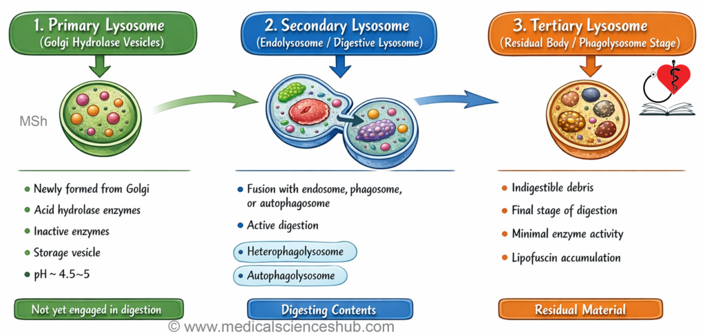
- Primary lysosomes are newly formed vesicles that contain digestive enzymes but have not yet participated in degradation.
- Secondary lysosomes form when a lysosome fuses with an endocytic vesicle or endosome, allowing digestion of engulfed material. Phagolysosomes develop when lysosomes fuse with phagocytic vacuoles that contain microorganisms or large particles.
- Tertiary lysosome or Autophagic vacuoles are formed when lysosomes digest worn-out organelles of the same cell.
Functions of Lysosome
- Lysosomes carry out intracellular digestion of macromolecules, cellular debris, and engulfed particles.
- They destroy bacteria and other foreign materials taken into the cell.
- The acrosome of sperm is a modified lysosome that contains enzymes needed for penetration of the ovum.
- Lysosomal enzymes can digest damaged cellular components, a process known as autophagy.
- Deficiency of specific lysosomal enzymes leads to lysosomal storage disorders due to accumulation of undigested substances.
Clinical Integration
- Lysosomes are absent in mature erythrocytes because these cells lack most cytoplasmic organelles. Neutrophils contain numerous lysosomes, which enable efficient destruction of microorganisms during phagocytosis.
- Acid phosphatase activity is commonly used as a biochemical marker to identify lysosomal function in cells and tissues.
- Residual bodies are undigested remnants that remain within lysosomes after intracellular digestion of phagocytosed material.
Table 1.2: Important lysosomal enzymes
| Enzyme Category | Representative Lysosomal Enzymes | Primary Function in the Cell |
|---|---|---|
| Proteolytic Enzymes | Cathepsins, Collagenases, Elastase | These enzymes degrade proteins and structural components of the extracellular matrix. They participate in the breakdown of cytoplasmic proteins, collagen, and elastic fibers during intracellular digestion and tissue remodeling. |
| Lipid-Degrading Enzymes | Lipases, Phospholipases, Fatty acyl esterases | These enzymes hydrolyze lipids into fatty acids and glycerol. They contribute to the digestion of cellular membranes and lipid-containing particles that enter the lysosomal compartment. |
| Carbohydrate-Degrading Enzymes | Alpha-glycosidase, Beta-galactosidase, Hyaluronidase, Arylsulfatase | These enzymes cleave complex carbohydrates and glycosaminoglycans. They help degrade glycoconjugates and polysaccharides during lysosomal processing of cellular and extracellular material. |
| Nucleic Acid–Degrading Enzymes | Ribonuclease, Deoxyribonuclease | These enzymes digest nucleic acids by breaking down ribonucleic acid and deoxyribonucleic acid into smaller nucleotide components during intracellular degradation. |
| Other Lysosomal Enzymes | Acid phosphatase, Catalase | Acid phosphatase removes phosphate groups from various molecules and is commonly used as a biochemical marker of lysosomal activity. Catalase helps in the breakdown of hydrogen peroxide and contributes to cellular protection from oxidative damage. |
| Intermembrane Space | Adenylate kinase | Converts adenine nucleotides and supports cellular energy balance. |
Clinical Integration
- Lysosomal storage disorders
- These are inherited conditions caused by deficiency or absence of specific lysosomal enzymes.
- The missing enzyme prevents normal degradation of certain cellular substances inside lysosomes.
- As a result, materials such as glycogen, gangliosides, cerebrosides, or sphingomyelin accumulate within cells.
- Progressive accumulation disrupts normal cellular structure and interferes with organ function.
- Partially digested substances remain within lysosomes and appear as intracellular inclusion bodies.
Table 1.3: Common lysosomal storage diseases
| Lysosomal Storage Disorder | Deficient Enzyme | Accumulated Substance | Major Clinical Features |
|---|---|---|---|
| Fabry disease | Alpha-galactosidase A | Glycosphingolipids, mainly globotriaosylceramide | Patients develop angiokeratomas, reduced sweating (hypohidrosis), neuropathic pain, and progressive involvement of kidneys and heart. |
| Gaucher disease | Acid beta-glucocerebrosidase | Glucocerebroside | Characterized by hepatosplenomegaly, bone pain, skeletal deformities, and accumulation of lipid-laden macrophages in various organs. |
| Niemann–Pick disease | Sphingomyelinase | Sphingomyelin | Leads to neurodegeneration, developmental delay, enlargement of liver and spleen, and progressive pulmonary involvement. |
| Tay–Sachs disease | Beta-hexosaminidase A | GM2 ganglioside | Infants show progressive neurological deterioration, exaggerated startle response, visual impairment, and macrocephaly due to neuronal storage of gangliosides. |
| Hurler syndrome | Alpha-L-iduronidase | Glycosaminoglycans such as dermatan sulfate and heparan sulfate | Causes coarse facial features, developmental delay, skeletal abnormalities, and cardiovascular involvement. |
| Pompe disease | Acid alpha-glucosidase | Glycogen within lysosomes | Produces severe cardiomyopathy, muscle weakness, and respiratory difficulty due to glycogen accumulation in muscle cells. |
Ribosomes
- Ribosomes are small cellular structures responsible for protein synthesis.
- They may attach to the rough endoplasmic reticulum or remain free in the cytoplasm.
- Ribosomes contain a large proportion of cellular ribonucleic acid, which participates in translation of genetic information.
- They may occur individually or form clusters called polyribosomes, which increase efficiency of protein production.
Peroxisomes
- Peroxisomes are small spherical organelles present in the cytoplasm of many cells.
- Their diameter is approximately 0.5 micrometres, and they are sometimes referred to as microbodies.
- These organelles are formed by growth and division of pre-existing peroxisomes and by contributions from the smooth endoplasmic reticulum.
- Peroxisomes contain several oxidative enzymes that participate in cellular metabolic reactions.
Structural and Functional Features
- Peroxisomes contain oxidases that catalyze oxidation reactions, particularly the beta oxidation of long-chain fatty acids.
- These reactions generate hydrogen peroxide, which is rapidly broken down by the enzyme catalase, thereby protecting the cell from oxidative injury.
- Specialized proteins called peroxins guide newly synthesized proteins into the peroxisomal matrix.
- Transport proteins in the peroxisomal membrane regulate movement of metabolites between the cytoplasm and the organelle.
- The peroxisomal matrix contains numerous enzymes involved in metabolism of lipids, amino acids, purines, cholesterol, and bile acids.
- Deficiency of peroxisomal enzymes can lead to metabolic disorders affecting multiple organs.
- Cellular signals mediated through peroxisome proliferator-activated receptors regulate the number and metabolic activity of peroxisomes.
Clinical Integration
- Clinical Significance of Peroxisomes
- Peroxisomes protect cells from oxidative stress generated during normal metabolic reactions.
- Many metabolic processes produce hydrogen peroxide, a highly reactive oxidizing molecule that can damage cellular components.
- The peroxisomal enzyme catalase converts hydrogen peroxide into water and oxygen, thereby preventing cellular injury.
- Increased activity of catalase may indicate elevated oxidative stress within tissues.
- Inherited Disorders of Peroxisomes
- Zellweger syndrome results from mutations affecting proteins required for peroxisome formation. It causes severe neurological impairment, accumulation of very long-chain fatty acids, abnormal bile acid synthesis, and reduced plasmalogen production. Most affected infants die within the first year of life.
- Infantile Refsum disease is a related but milder disorder caused by partial defects in peroxisomal proteins.
- Adrenoleukodystrophy occurs due to impaired peroxisomal oxidation of very long-chain fatty acids, leading to progressive degeneration of the brain and adrenal glands.
- Primary hyperoxaluria develops when defective peroxisomal metabolism causes excessive oxalate production, resulting in recurrent kidney stones and renal damage.
Centrosomes
- The centrosome is located near the nucleus in most eukaryotic animal cells.
- It consists of two centrioles arranged perpendicular to each other and embedded in the surrounding pericentriolar material.
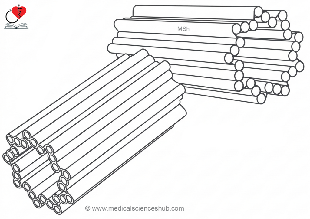
- Each centriole is a short cylindrical structure composed of microtubules organized into nine sets of triplets arranged around its wall.
- The microtubules contain γ-tubulin, which assists in the nucleation and organization of new microtubules.
- The centrosome functions as the primary microtubule-organizing center of the cell.
- During cell division, the centrosome duplicates and the two centrosomes migrate to opposite poles of the cell.
- They help form the mitotic spindle and regulate the accurate movement and separation of chromosomes.
Cytoskeleton
- The cytoskeleton is a network of protein filaments present within the cytoplasm of cells.
- It provides structural support and helps maintain the shape and mechanical stability of the cell.
- The cytoskeleton also enables controlled changes in cell shape required for cell movement and other physiological activities.
- It participates in intracellular transport, cell division, and organization of cellular components.
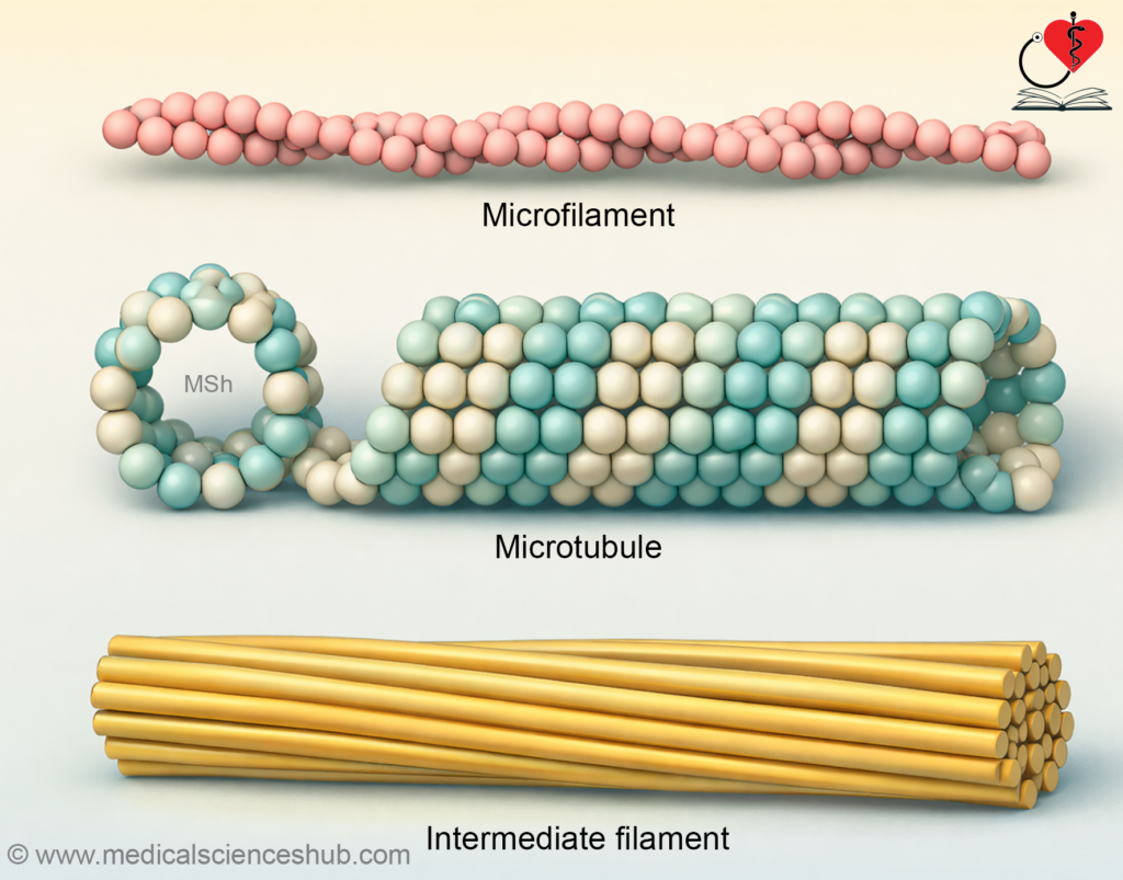
- The cytoskeleton is composed of three main filament systems: microfilaments, microtubules, and intermediate filaments.
- Each component is formed by specific structural proteins that assemble into filamentous networks to perform specialized cellular functions.
Microfilaments
Structure
- Microfilaments are thin protein filaments that form an important component of the cytoskeleton in eukaryotic cells.
- They are solid filaments with an approximate diameter of 7 nanometres.
- Microfilaments are primarily composed of the protein actin, which is one of the most abundant proteins in many cells.
- Actin exists as globular actin, a single unpolymerized subunit, which can assemble to form filamentous actin.
- These actin subunits polymerize to form long filaments and can also disassemble when required.
- In living cells, actin filaments continuously undergo polymerization at one end and depolymerization at the opposite end.
- Microfilaments are present in most cells but are highly organized in cells involved in contraction, secretion, and cell movement.
- They extend into cellular projections such as microvilli in epithelial cells.
Functions
- Microfilaments provide mechanical support and help maintain the shape of the cell.
- They function as the primary contractile elements responsible for cell movement and changes in cell form.
- They participate in cellular processes such as phagocytosis, intracellular transport, secretion, and contraction.
- Microfilaments assist in the movement of secretory vesicles and cytoplasmic granules during cellular release mechanisms.
- Actin filaments interact with membrane proteins to form focal adhesion complexes, which help cells attach to surfaces and generate traction for movement.
- In epithelial cells, microfilaments extend into microvilli and contribute to their stability and limited movement.
- They also play supportive roles in cell division and organization of the cytoplasm.
Microtubules
Structure
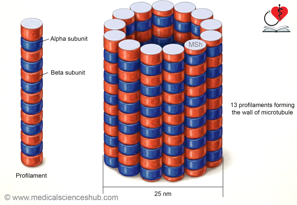
- Microtubules are long, hollow cylindrical structures that form an essential part of the cytoskeleton.
- They have an approximate external diameter of 25 nanometres and a central hollow lumen of about 15 nanometres.
- Microtubules are composed of globular protein subunits known as tubulin.
- Two main forms of tubulin, alpha-tubulin and beta-tubulin, combine to form heterodimers.
- These heterodimers assemble in a helical arrangement to create linear strands called protofilaments.
- Thirteen protofilaments typically align side by side to form the wall of a microtubule.
- Microtubules are polar structures with a growing positive end and a relatively stable negative end.
- Tubulin molecules can continuously assemble and disassemble, giving microtubules a highly dynamic organization within the cell.
- Assembly of microtubules requires guanosine triphosphate, and their stability is influenced by temperature and cellular conditions.
Functions
- Microtubules maintain cell shape and contribute to the mechanical stability of the cytoplasm.
- They act as intracellular tracks for the movement of vesicles and organelles using motor proteins such as kinesin and dynein.
- Microtubules form the structural framework of cilia and flagella, enabling cellular and fluid movement.
- They participate in the formation of the mitotic spindle, which ensures accurate separation of chromosomes during cell division.
- Microtubules also assist in intracellular transport and overall cell motility.
Intermediate Filaments
- Intermediate filaments are cytoskeletal protein fibers with a diameter of approximately 8 to 14 nanometres.
- They are composed of different structural proteins that vary according to the type of cell.
- These filaments form a stable internal framework that supports the structural integrity of the cell.
- Intermediate filaments help anchor the nuclear membrane, plasma membrane, and certain organelles within the cytoplasm.
- They provide mechanical strength and protect cells from damage caused by external stress.
- Abnormalities in these filaments can weaken tissues and may contribute to skin blistering disorders.
- Specific intermediate filament proteins are also used as cellular markers in diagnostic pathology.
Table 1.4: Protein subunit and diameter of cytoskeletal elements of cell.
| Feature | Microfilaments (Actin Filaments) | Intermediate Filaments | Microtubules |
|---|---|---|---|
| Structural organization | Two intertwined strands forming a helical filament composed of actin polymers. | Rope-like, flexible protein fibers that form stable cytoskeletal cables. | Long, hollow cylindrical tubes built from polymerized tubulin subunits. |
| Typical diameter | Approximately 7 nanometres. | Usually 8–14 nanometres (commonly about 10 nanometres). | Approximately 25 nanometres. |
| Principal protein components | Actin proteins arranged as filamentous actin polymers. | Diverse proteins depending on cell type, such as keratin, vimentin, desmin, and lamins. | Tubulin heterodimers consisting of alpha-tubulin and beta-tubulin. |
| Common cellular distribution | Concentrated beneath the plasma membrane, present in the core of microvilli, and abundant in muscle contractile structures. | Form networks across the cytoplasm, connect to desmosomes and hemidesmosomes, create the nuclear lamina, and are prominent in epithelial cells as keratin filaments. | Present in the mitotic spindle, extend through the cytoplasm as intracellular tracks, and form the structural core of cilia and flagella. |
| Major biological roles | Enable cell contraction, cell shape changes, and movement of the cell membrane and cytoplasm. | Provide mechanical strength, stabilize cellular architecture, and maintain tissue integrity under mechanical stress. | Facilitate intracellular transport of vesicles and organelles, support chromosome separation during cell division, and enable movement of cilia and flagella. |
Cilia
- Cilia are minute, hair-like projections present on the surface of certain epithelial cells.
- They beat in coordinated, rhythmic movements that generate a directed flow of fluid or particles across the cell surface.
- Each cilium arises from a basal body located just beneath the plasma membrane.
- The internal core of a cilium is the axoneme, which contains microtubules arranged in a nine plus two pattern.
- Nine outer microtubule doublets surround a central pair of microtubules.
- Dynein motor proteins attached to microtubules generate sliding movements that produce ciliary beating.
- The basal body contains nine microtubule triplets and structurally resembles a centriole.
Clinical Integration
- Several anticancer drugs act by altering the function of microtubules, which are essential for cell division. Drugs such as vincristine and vinblastine promote disassembly of microtubules and disrupt the mitotic spindle. Paclitaxel stabilizes microtubules and prevents their normal dynamic remodeling during the cell cycle. These effects block chromosome separation, inhibit mitosis, and lead to death of rapidly dividing cancer cells.
- Colchicine also interferes with microtubule assembly and inhibits spindle formation.
- Intermediate filament proteins serve as important diagnostic markers in pathology. For example, cytokeratin indicates epithelial origin, whereas vimentin is typically expressed in fibroblasts and mesenchymal cells.
The Nucleus
Structure of Nucleus
- The nucleus is a membrane-bound organelle that contains the genetic material of the cell.
- Nearly all human cells possess a nucleus, except mature red blood cells, which lose it during development.
- Platelets do not contain a nucleus because they are fragments derived from larger precursor cells.
- Most cells contain a single centrally placed nucleus, although its size, shape, and position may vary in different cell types.
- Certain cells, such as skeletal muscle fibers, may contain multiple nuclei.
Nuclear Envelope
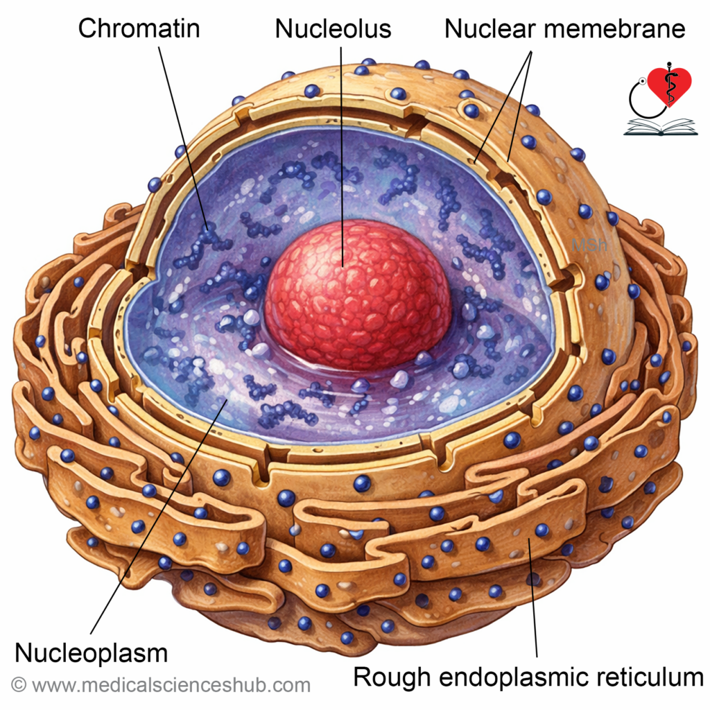
- The nucleus is surrounded by a double-layered membrane known as the nuclear envelope.
- The narrow space between the two membranes is called the perinuclear space.
- In many cells, the outer membrane of the nuclear envelope is continuous with the endoplasmic reticulum.
- Numerous nuclear pores are present in the envelope to permit selective exchange of molecules between the nucleus and cytoplasm.
- Transport of proteins and ribonucleic acid molecules through these pores is regulated by specialized transport proteins.
Nucleoplasm and Chromatin
- The interior of the nucleus contains a gel-like matrix called nucleoplasm.
- Genetic material is present as deoxyribonucleic acid associated with histone proteins, forming chromatin.
- During cell division, chromatin condenses and becomes visible as chromosomes.
Nucleolus
- The nucleolus is a dense region within the nucleus rich in ribonucleic acid.
- One or more nucleoli may be present in active cells.
- The nucleolus is responsible for ribosome production.
Functions
- The nucleus stores and protects genetic information.
- It regulates gene expression, protein synthesis, and overall cellular activities.
- The nucleus also controls cell division and transmission of hereditary information.
Ihintercellular Junctions
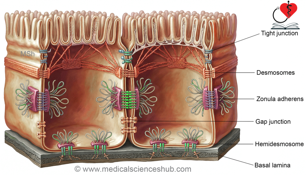
- Intercellular junctions are specialized structural connections that link adjacent cells within tissues.
- These junctions maintain tissue integrity, support communication between cells, and regulate movement of substances across cellular layers.
- Cells in tissues are often embedded within an extracellular matrix, which provides structural support and spatial organization.
- In connective tissues such as cartilage and bone, the extracellular matrix is abundant and cells are widely separated within this matrix.
- In muscle and some connective tissues, cells are closely associated and are connected mainly by cell-to-cell adhesive structures.
- In epithelial tissues that cover body surfaces or line cavities, cells are tightly packed and connected by specialized junctional complexes.
- The space between adjacent cells, called the intercellular space, normally measures approximately 20–25 nanometers.
- In epithelial tissues, the extracellular matrix is reduced to a thin supporting layer known as the basal lamina, which lies beneath the epithelial cells.
Functional Classification of Intercellular Junctions
- Intercellular junctions are broadly classified according to their primary function.
- Some junctions provide mechanical attachment and hold cells firmly together.
- Other junctions allow direct communication and transfer of ions or small molecules between neighboring cells.
Tight Junctions
- Tight junctions are specialized sealing junctions commonly present in epithelial tissues.
- They are abundant in the epithelium of the gastrointestinal tract, renal tubules, urinary tract, hepatobiliary system, and choroid plexus.
- They form a belt-like structure near the apical region of epithelial cells.
Structural Features
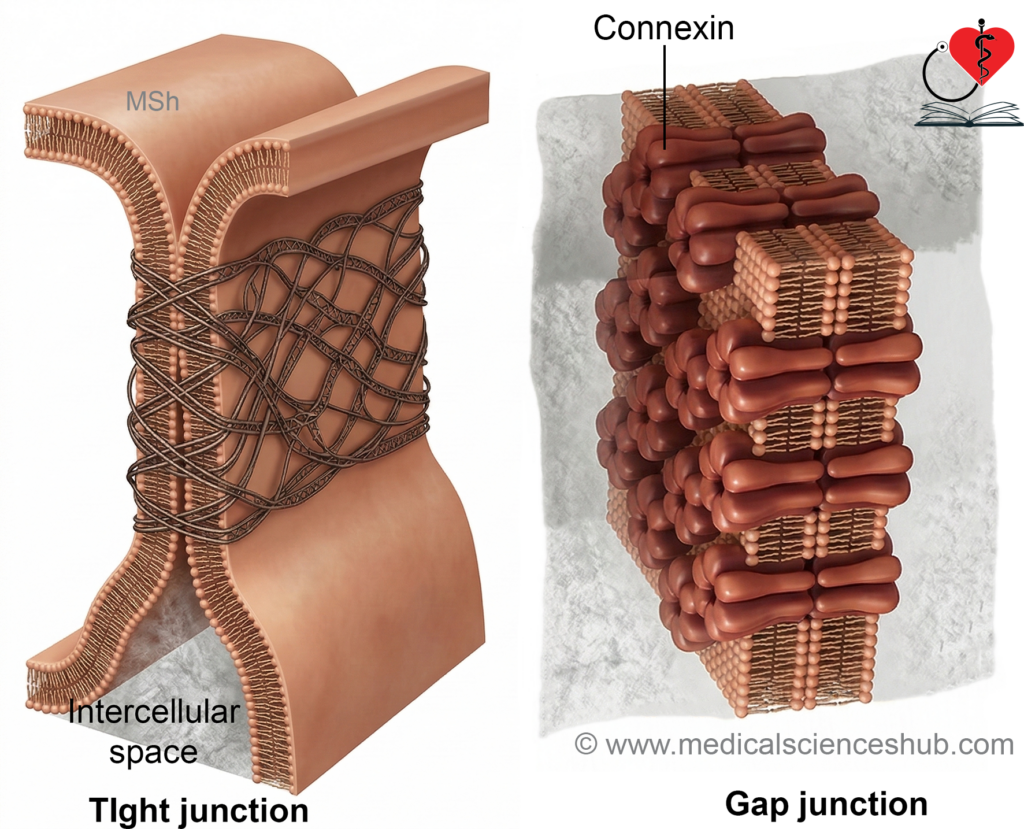
- At tight junctions, the outer layers of the plasma membranes of adjacent cells closely approach and fuse at several points.
- This arrangement greatly reduces or eliminates the intercellular space near the apical surface.
- The junction consists of interconnected protein strands contributed by both neighboring cells.
- These strands create a barrier that restricts the passage of substances between cells.
- Important structural proteins include claudins, occludin, and junctional adhesion molecules.
- Cytoplasmic proteins attach these membrane components to the intracellular cytoskeleton, providing additional stability.
Functions of Tight Junctions
- Tight junctions form a selective permeability barrier that restricts movement of large molecules through the intercellular space.
- Large molecules from luminal fluids usually cross epithelial cells through vesicular transport rather than passing between cells.
- Small ions and water-soluble molecules may pass through certain tight junctions depending on epithelial type.
- This movement between cells is known as paracellular transport.
- The permeability of tight junctions varies among tissues.
- For example, sodium ions can move relatively easily across intestinal epithelium but are strongly restricted in the urinary bladder.
- Tight junctions between endothelial cells of cerebral blood vessels contribute to the blood–brain barrier.
- Similar junctions in the ciliary epithelium participate in forming the blood–aqueous barrier of the eye.
Anchoring Junctions
- Anchoring junctions provide strong mechanical attachments that stabilize tissues exposed to mechanical stress.
- These junctions connect cells either to neighboring cells or to the basal lamina.
Types of Anchoring Junctions
- Anchoring junctions are divided into two main groups:
- Cell-to-cell anchoring junctions.
- Cell-to-basal lamina anchoring junctions.
Cell-to-Cell Anchoring Junctions
These junctions directly connect adjacent cells and maintain tissue cohesion.
Desmosomes
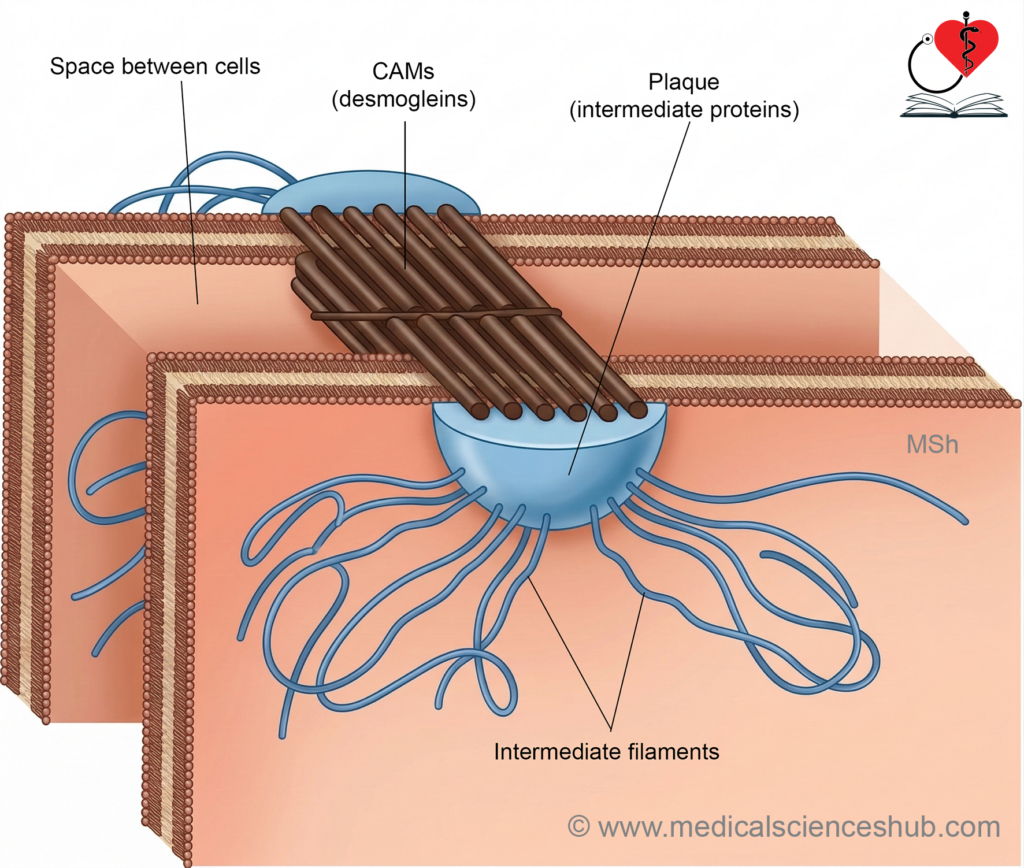
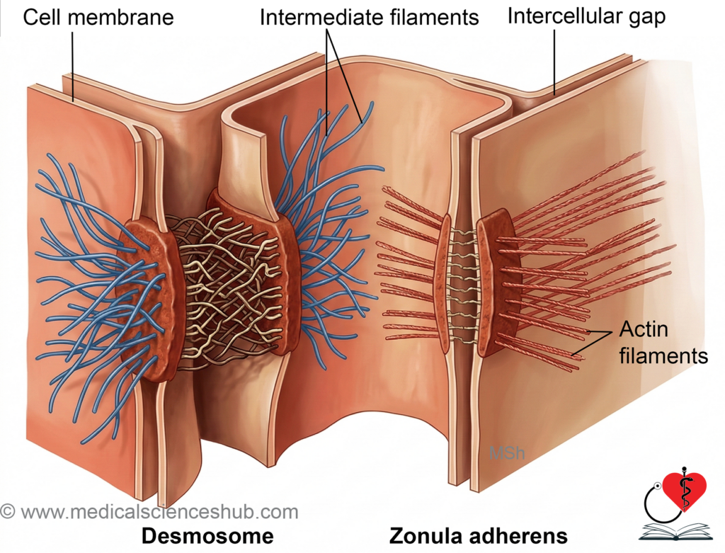
- Desmosomes are strong adhesive junctions that link neighboring cells.
- They appear as localized thickened regions on the cytoplasmic surface of adjacent plasma membranes.
- The opposing membranes are separated by a narrow intercellular gap of about 25 nanometers.
- Dense protein plaques are present on the cytoplasmic side of each membrane.
- Intermediate filaments from the cytoskeleton attach to these plaques, providing mechanical strength.
- The intercellular space contains adhesive proteins such as cadherins, including desmogleins and desmocollins.
- These proteins bind the two cells together and help tissues resist mechanical stress.
Zonula Adherens
- Zonula adherens forms a continuous belt-like junction located just below tight junctions in epithelial cells.
- This junction is a major site of attachment for intracellular actin microfilaments.
- Adhesive proteins called cadherins extend across the intercellular space and connect neighboring cells.
- These structures help maintain cell shape and coordinate mechanical forces across epithelial sheets.
Cell-to-Basal Lamina Anchoring Junctions
These junctions attach cells to the underlying extracellular matrix.
Hemidesmosomes
- Hemidesmosomes resemble half of a desmosome in structure.
- They anchor epithelial cells firmly to the basal lamina.
- The junction contains transmembrane proteins known as integrins rather than cadherins.
- Intracellularly, hemidesmosomes connect to intermediate filaments of the cytoskeleton.
- This arrangement stabilizes epithelial tissues and prevents separation from the basement membrane.
Focal Adhesions
- Focal adhesions are dynamic attachment sites between cells and the basal lamina.
- These junctions connect the cell membrane to extracellular matrix proteins through integrins.
- Internally, they are linked to actin filaments of the cytoskeleton.
- Focal adhesions participate in cell movement, migration, and changes in cell shape.
Gap Junctions
- Gap junctions are specialized communication junctions that allow direct exchange of small molecules between neighboring cells.
- At these sites, the intercellular space narrows from approximately 25 nanometers to about 3 nanometers.
Structural Organization
- Gap junctions are formed by transmembrane protein complexes called connexons.
- Each connexon consists of six protein subunits known as connexins arranged around a central aqueous channel.
- Connexons from adjacent cells align with each other to form a continuous channel between the two cytoplasms.
- The diameter of this channel is approximately 2 nanometers.
Functional Significance
- These channels permit direct transfer of water, ions, and small molecules between cells.
- Substances such as amino acids, sugars, and small signaling molecules can diffuse through gap junction channels.
- Gap junctions allow rapid transmission of electrical signals between cells.
- For this reason, they function as electrical synapses in tissues such as cardiac muscle and certain neurons.
- Cells connected by numerous gap junctions often behave as a coordinated functional unit called a physiological syncytium.
Regulation of Gap Junction Activity
- The permeability of gap junction channels is regulated by intracellular conditions.
- Increased intracellular calcium concentration can close the channels and reduce communication between cells.
- Changes in cytoplasmic acidity also influence channel opening and closing.
- These regulatory mechanisms protect tissues during cellular injury.
Cell Adhesion Molecules
- Cell adhesion molecules are specialized proteins that mediate attachment between cells and between cells and the extracellular matrix.
- These molecules are abundant at intercellular junctions and play a crucial role in maintaining tissue organization.
Structural Characteristics
- Cell adhesion molecules possess extracellular domains that bind to specific ligands or receptors.
- Many of these interactions involve extracellular matrix proteins such as laminin.
- Laminin is a large cross-shaped glycoprotein present in the basal lamina and contains multiple binding sites for cell adhesion molecules.
- Intracellular portions of these molecules attach to the cytoskeleton, strengthening cellular attachment.
Types of Binding
Cell adhesion molecules display two main binding patterns.
- Homophilic binding occurs when a molecule binds to an identical molecule on another cell.
- Heterophilic binding occurs when a molecule binds to a different type of molecule on a neighboring cell or extracellular matrix component.
Major Classes of Cell Adhesion Molecules
- Integrins are heterodimeric proteins that connect cells to extracellular matrix components and participate in signaling.
- Immunoglobulin superfamily molecules resemble immunoglobulin proteins and contribute to cell recognition and adhesion.
- Cadherins are calcium-dependent adhesion proteins that primarily mediate homophilic cell-to-cell binding.
- Selectins possess carbohydrate-binding domains and are important in transient cell interactions.
Functions of Cell Adhesion Molecules
- They promote strong attachment between neighboring cells and stabilize tissues.
- Their interaction with the cytoskeleton contributes to cellular movement and structural organization.
- They participate in intracellular signaling by transmitting signals from the extracellular environment into the cell.
- These molecules play important roles in inflammation, immune cell migration, and wound repair.
- Proper cell adhesion helps maintain cell survival, whereas loss of attachment to the extracellular matrix can trigger programmed cell death.
Table 1.5: Types of intercellular connections
| Functional Category | Type of Junction | Key Structural Features and Functions |
|---|---|---|
| Junctions that hold cells together | Tight junctions (Zonula occludens) | Adjacent plasma membranes fuse near the apical region of epithelial cells. Junctions are formed by proteins such as claudins, occludin, and junctional adhesion molecules. They create a selective barrier that limits movement of substances through the intercellular space and helps maintain epithelial polarity. |
| Junctions that hold cells together | Anchoring junctions | These junctions provide strong mechanical attachment between cells or between cells and the basal lamina. They link cytoskeletal elements to neighboring cells or to extracellular matrix components, thereby maintaining tissue stability. |
| Cell-to-cell anchoring junctions | Desmosomes (Macula adherens) | Localized adhesive junctions that connect adjacent cells through cadherin family proteins. Cytoplasmic plaques anchor intermediate filaments, providing strong resistance to mechanical stress in tissues such as skin and cardiac muscle. |
| Cell-to-cell anchoring junctions | Zonula adherens | Belt-like junction located below tight junctions in epithelial cells. Cadherins link neighboring cells, while intracellular plaques attach to actin filaments. These junctions maintain cell shape and transmit mechanical forces across epithelial layers. |
| Cell-to-basal lamina anchoring junctions | Hemidesmosomes | Junctions that attach epithelial cells to the basement membrane. Transmembrane integrins connect extracellular matrix proteins with intracellular intermediate filaments, providing strong anchorage to underlying tissues. |
| Cell-to-basal lamina anchoring junctions | Focal adhesions | Dynamic attachment sites between cells and the extracellular matrix. Integrins connect extracellular matrix components with intracellular actin filaments. These junctions play an important role in cell movement, migration, and mechanical signaling. |
| Junctions that allow transfer of ions and small molecules | Gap junctions (Nexus) | Specialized communication channels formed by connexons, each composed of six connexins. These channels link the cytoplasm of adjacent cells and permit direct passage of ions, metabolites, and small signaling molecules. They enable electrical and metabolic coupling between cells. |
Clinical Integration
- Mutations in genes that encode connexin proteins can disrupt the structure and function of gap junctions, which are essential for direct cell-to-cell communication. Humans possess multiple connexin genes, and mutations may produce tissue-specific disorders because different tissues express different connexin types.
- Certain mutations in connexin genes are associated with Charcot–Marie–Tooth disease, a hereditary neurological disorder. In this condition, defective gap junction communication affects peripheral nerves, leading to peripheral neuropathy characterized by muscle weakness, sensory loss, and reduced reflexes.
- Gap junctions are abundant in cardiac muscle, where they allow rapid spread of electrical impulses between adjacent cells. Because of this electrical coupling, cardiac muscle fibers function as a functional syncytium, allowing coordinated contraction of the heart. The atrial and ventricular muscles act as two separate functional syncytia because fibrous septa electrically isolate them. Gap junctions in visceral smooth muscle also permit electrical signal transmission, producing coordinated rhythmic contractions.
Important Questions
- Describe the structure and list the functions of cell membrane.
- Describe the details of the structure, functions and dysfunctions of each of the following cell organelles: a. Mitochondrion b. Lysosome c. Nucleus d. Microfilaments.
- Write a short note on intercellular junctions.
📝 Test Your Knowledge – Practice MCQs
Attempt the chapter MCQ quiz and assess your understanding of key concepts.
Click to see: Medical Disclaimer and Copyright Notice