
Competencies
- AN52.1 Describe and identify the microanatomical features of gastrointestinal system: Oesophagus, fundus of stomach, pylorus of stomach.
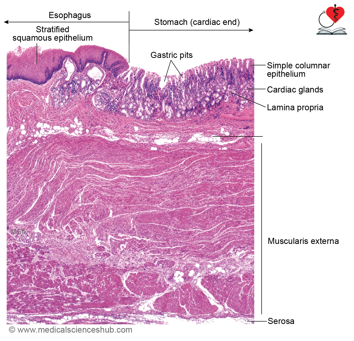
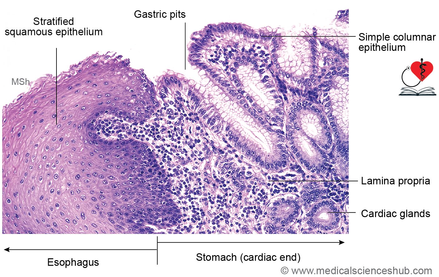
- AN52.3 Describe and identify the microanatomical features of cardiooesophageal junction.
Introduction
- The gastrointestinal tract (GIT) is a continuous muscular tube extending from the esophagus to the anal canal.
- It includes the esophagus, stomach, small intestine (duodenum, jejunum, ileum), large intestine (colon), vermiform appendix, and anal canal.
- Despite regional specialization, all parts of the GIT share common structural features adapted for digestion, absorption, secretion, propulsion, and transport of food and waste materials.
General Structure of Git
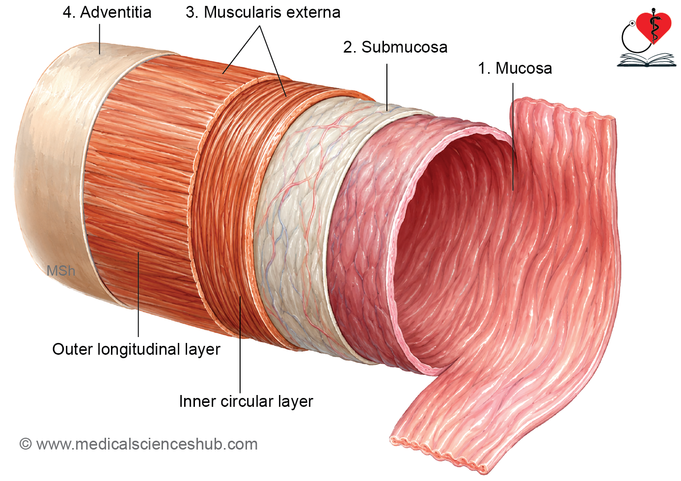
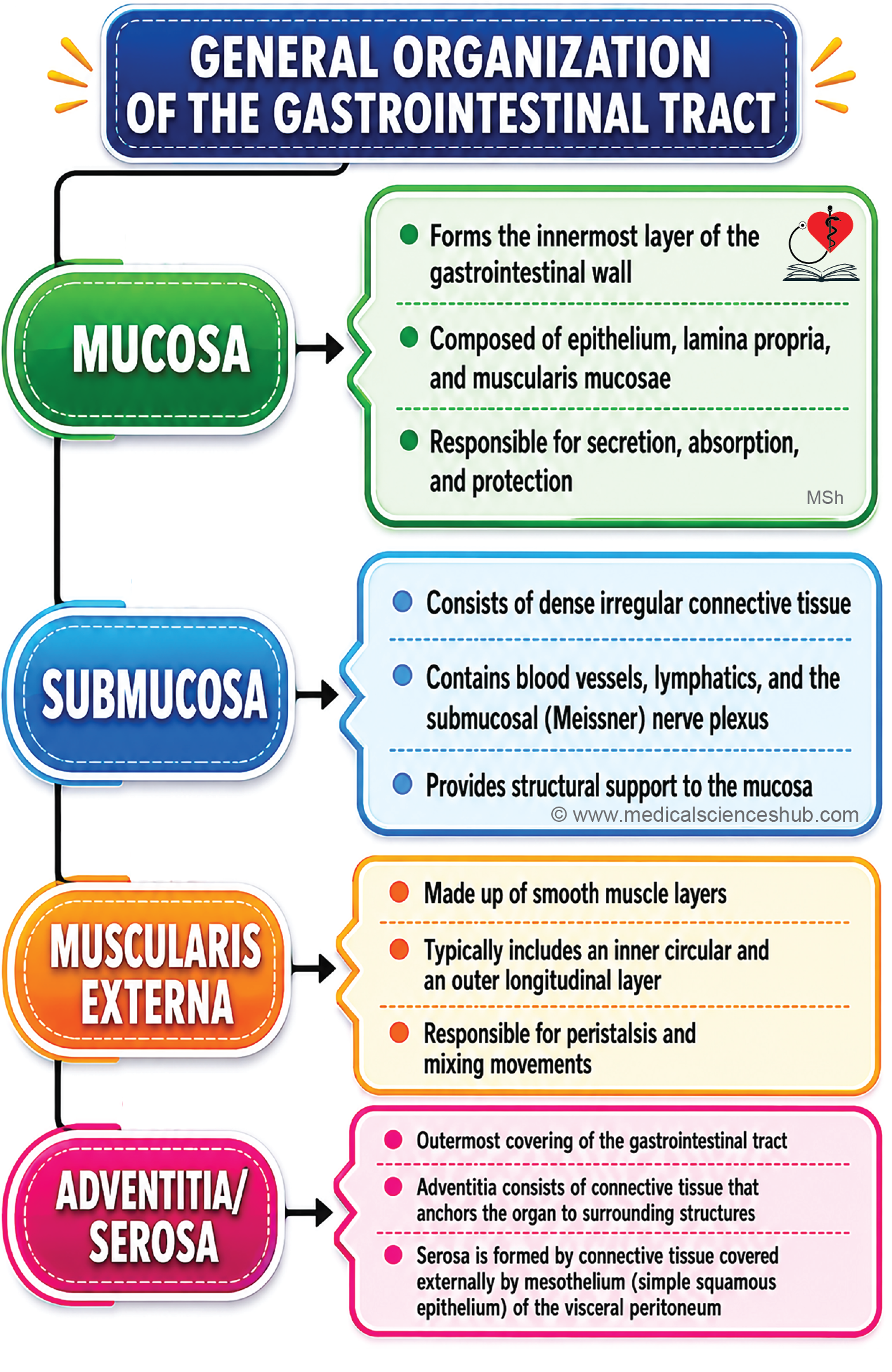
- The wall of the gastrointestinal tract (GIT) is organized into four principal layers from the lumen outward.
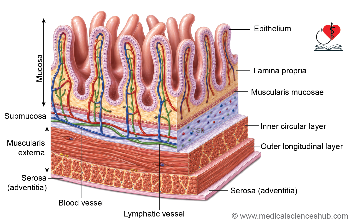
- Mucosa consists of the lining epithelium, lamina propria, and muscularis mucosae.
- Submucosa is composed of dense irregular connective tissue containing blood vessels, lymphatics, and nerves.
- Muscularis externa typically includes an inner circular and an outer longitudinal layer of smooth muscle, responsible for peristalsis.
- Serosa is the outer connective tissue layer covered by mesothelium (visceral peritoneum).
- In regions not covered by peritoneum, the outer layer is called adventitia, which consists only of connective tissue.
Mucosa
The mucosa is the innermost layer of the gastrointestinal tract and consists of:
- Epithelium
- Lamina propria
- Muscularis mucosae
Epithelium
- Most of the gastrointestinal tract is lined by simple columnar epithelium.
- Exceptions include:
- Esophagus: lined by nonkeratinized stratified squamous epithelium.
- Terminal part of the anal canal: lined by stratified squamous epithelium.
- Major functions include protection, absorption, and secretion.
Adaptations for absorption (especially in the small intestine):
- Plicae circulares: permanent circular folds involving mucosa and submucosa.
- Villi: finger-like projections of the mucosa.
- Microvilli: microscopic projections on absorptive cells forming the brush border.
- Intestinal length and crypts of Lieberkühn further enhance absorptive capacity.
Secretory components of the gut:
- Unicellular secretory cells within the epithelium.
- Intestinal glands (crypts) extending into the lamina propria.
- Submucosal glands in the esophagus and duodenum.
- Extramural glands such as the liver and pancreas, which discharge secretions into the gut lumen through ducts.
Lamina Propria
- Consists of loose connective tissue supporting the epithelium.
- Contains blood vessels and lymphatics involved in nutrient transport.
- Houses gut-associated lymphoid tissue (GALT), including diffuse lymphoid tissue and lymphoid nodules.
- In the ileum, aggregated lymphoid nodules form Peyer’s patches.
- Provides support for mucosal glands such as gastric glands and intestinal crypts.
Muscularis Mucosae
- Thin layer of smooth muscle separating mucosa from submucosa.
- Usually arranged into inner circular and outer longitudinal layers.
- Extends into mucosal folds but does not enter villi.
- Its contractions alter mucosal shape and facilitate absorption and secretion.
Submucosa
- Composed of dense irregular connective tissue.
- Contains blood vessels, lymphatics, nerves, and occasional glands.
- Houses the submucosal (Meissner’s) plexus, which regulates glandular secretion and local blood flow.
- Allows mobility of the mucosa over the deeper muscular layers.
Mucularis Externa
- Usually consists of:
- Inner circular smooth muscle layer
- Outer longitudinal smooth muscle layer
Important exceptions:
- Upper third of the esophagus contains skeletal muscle; the middle third contains both skeletal and smooth muscle.
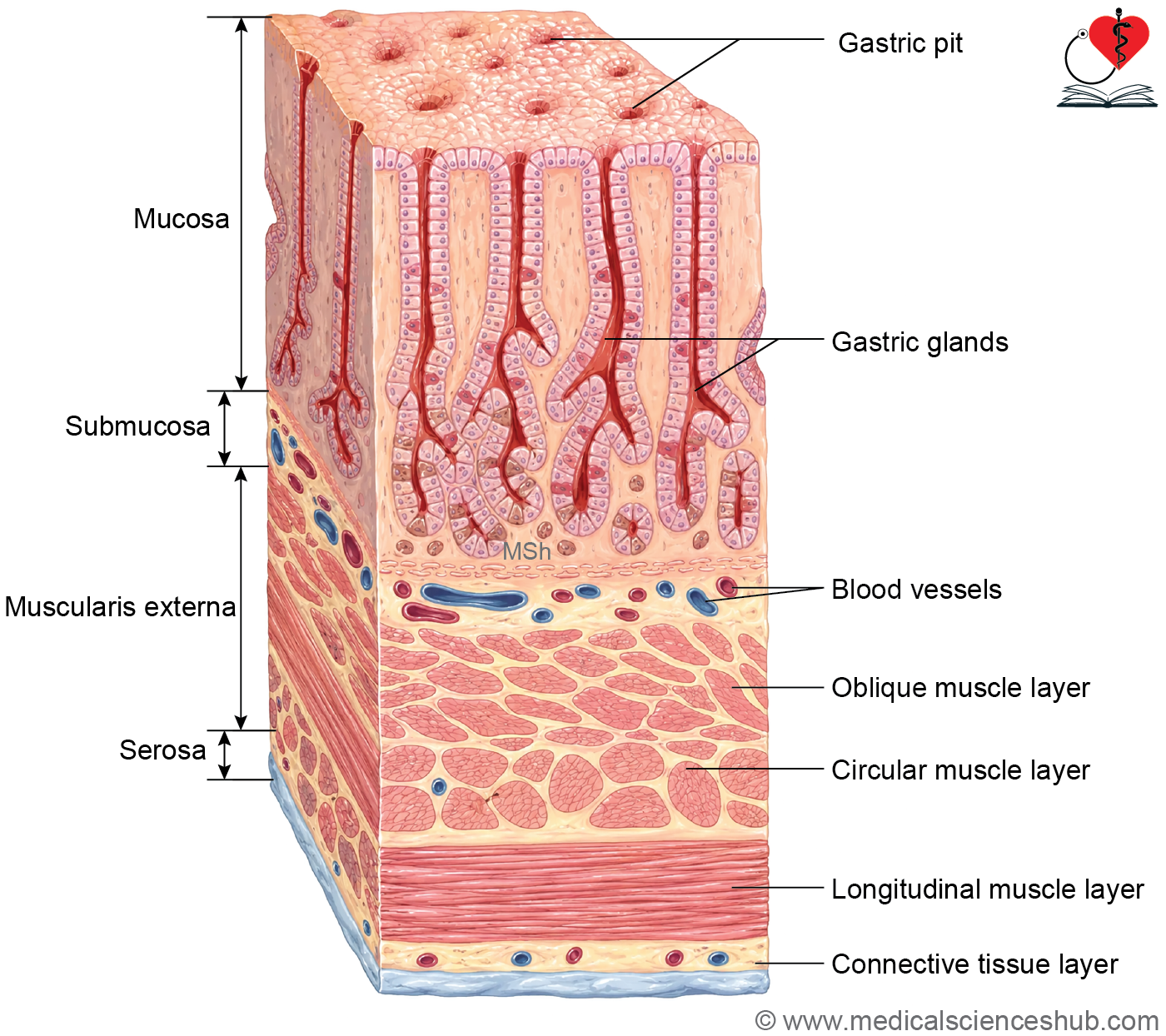
- The stomach has three layers: inner oblique, middle circular, and outer longitudinal.
- In the colon, the longitudinal layer forms taeniae coli.
- Localized thickening of the circular layer forms sphincters such as:
- Lower esophageal sphincter
- Pyloric sphincter
- Ileocecal valve region
- Internal anal sphincter
- The myenteric (Auerbach’s) plexus lies between the two muscle layers.
- Coordinated contractions of this layer produce peristalsis, which propels luminal contents through the gastrointestinal tract.
Serosa/Adventitia
- The outermost covering of the gastrointestinal tract is either serosa or adventitia.
- Serosa consists of connective tissue covered by mesothelium (simple squamous epithelium) and is present where the organ is covered by peritoneum.
- Adventitia consists only of connective tissue and occurs in regions lacking a peritoneal covering.
- This outer layer contains larger blood vessels, lymphatics, and nerves supplying the gut wall.
Esophagus
- The esophagus is a muscular tube that transports food and liquids from the pharynx to the stomach.
- It extends through the neck and thorax and is surrounded by connective tissue that anchors it to adjacent structures.
- The adult esophagus measures approximately 25 cm in length.
- Its lumen is usually collapsed and opens transiently during swallowing.
- The mucosa forms longitudinal folds, allowing expansion during passage of food.
- Four normal constrictions are present:
- At the pharyngoesophageal junction (cricopharyngeal sphincter).
- Where crossed by the arch of the aorta.
- Where crossed by the left main bronchus.
- At the esophageal hiatus of the diaphragm.
- These constrictions are clinically important sites where foreign bodies may lodge.
Histology of Esophagus
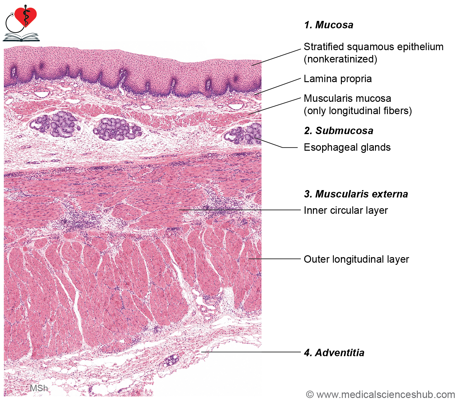
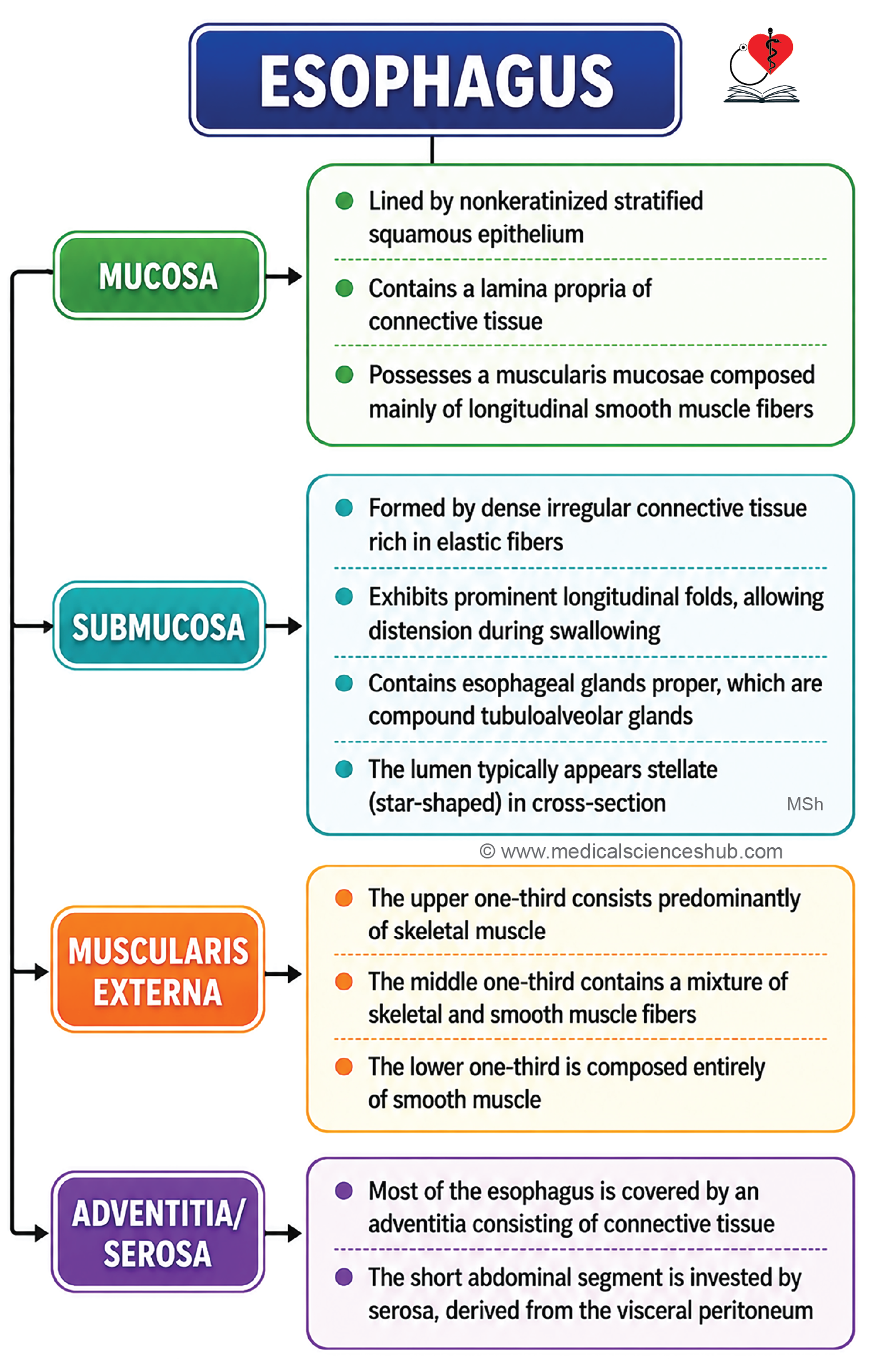
The wall of the esophagus consists of four layers: mucosa, submucosa, muscularis externa, and adventitia.
Mucosa
- The mucosa is composed of:
- Epithelium
- Lamina propria
- Muscularis mucosae
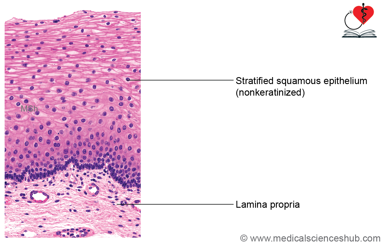
- The epithelium is nonkeratinized stratified squamous epithelium, providing protection against abrasion during swallowing.
- The lamina propria contains connective tissue papillae that project into the epithelium, helping anchor it to underlying tissues.
- The muscularis mucosae is unusually prominent and consists mainly of longitudinal smooth muscle fibers.
- The mucosa forms longitudinal folds, which flatten when the esophagus distends during food passage.
Submucosa
- The submucosa is composed of dense irregular connective tissue rich in collagen and elastic fibers.
- It contains large blood vessels, lymphatics, and nerves.
- Abundant elastic fibers contribute to the folded appearance of the lumen, which may appear irregular or star-shaped in cross-section.
- Esophageal glands proper, which are compound tubuloalveolar mucous glands, are present within the submucosa.
- Their ducts pass through the mucosa and open into the esophageal lumen, providing lubrication and protection.
- These glands are especially numerous in the lower part of the esophagus.
Muscularis Externa
- The muscularis externa is arranged into inner circular and outer longitudinal muscle layers.
- Muscle composition varies along the length of the esophagus:
- Upper third: skeletal muscle
- Middle third: mixture of skeletal and smooth muscle
- Lower third: smooth muscle only
- The myenteric (Auerbach’s) plexus lies between the two muscle layers and coordinates peristaltic activity.
- This is the strongest structural layer of the esophageal wall.
Adventitia/Serosa
- Most of the esophagus is surrounded by adventitia, a fibrous connective tissue layer that anchors it to surrounding structures.
- A short abdominal segment near the stomach is covered by serosa (visceral peritoneum).
Functions of Esophagus
- Transports food and liquids from the pharynx to the stomach by peristalsis.
- Produces protective mucus through esophageal glands.
- Does not participate significantly in digestion or nutrient absorption.
CLINICAL CORRELATION
- Achalasia cardia is a motility disorder in which the lower esophageal sphincter (LES) fails to relax adequately during swallowing.
- This leads to progressive dilatation of the esophagus (megaesophagus) and symptoms such as dysphagia, chest discomfort, and regurgitation of undigested food.
- Gastroesophageal reflux disease (GERD) occurs when the LES is incompetent, allowing acidic gastric contents to reflux into the esophagus.
- Chronic reflux causes heartburn, irritation, and inflammation of the esophageal mucosa (esophagitis).
- Barrett’s esophagus is a complication of long-standing GERD in which the normal stratified squamous epithelium is replaced by intestinal-type columnar epithelium with goblet cells.
- This metaplastic change is considered a premalignant condition because it increases the risk of esophageal adenocarcinoma.
Stomach
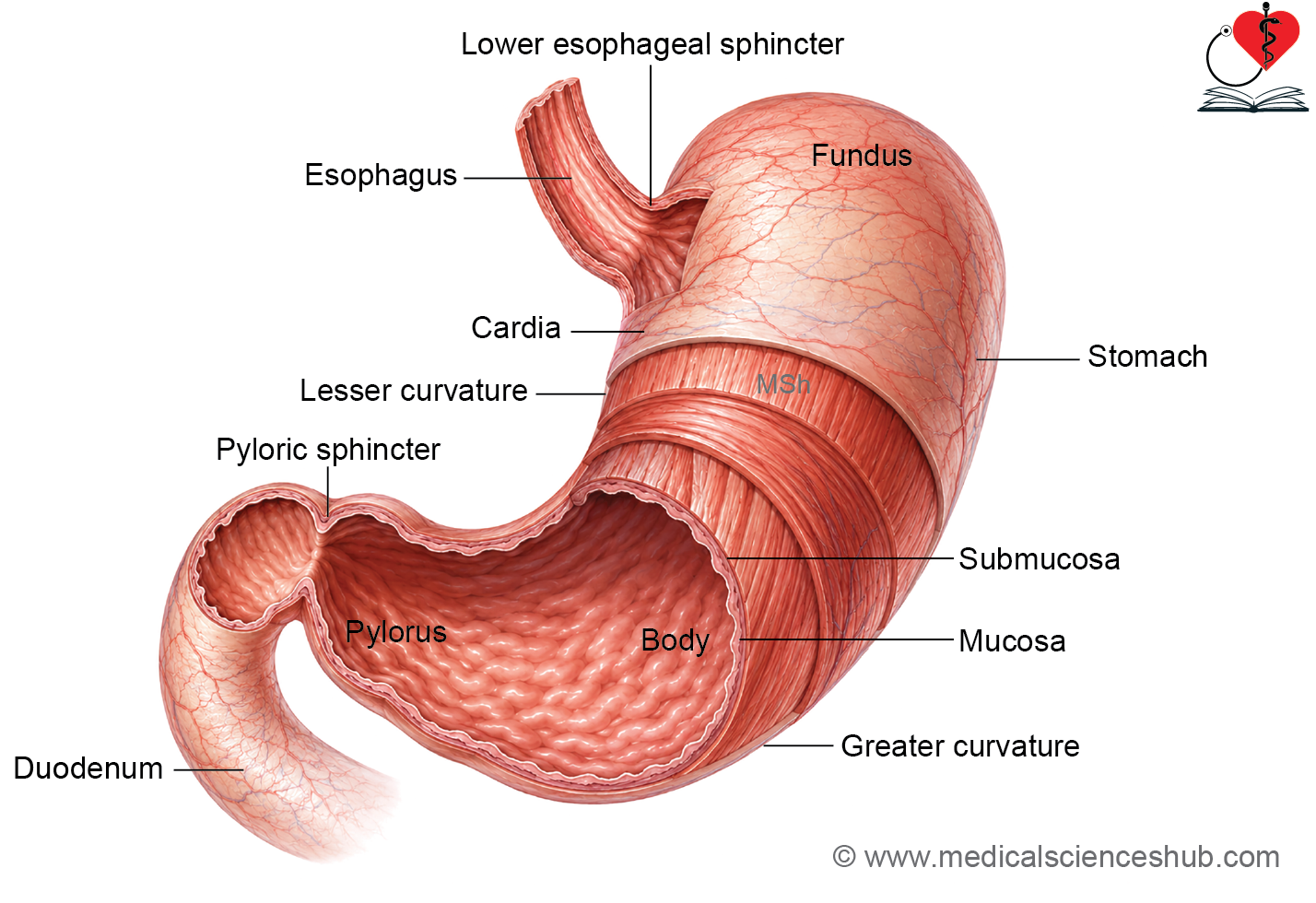
- The stomach is a hollow muscular organ located between the esophagus and the duodenum, serving as a temporary reservoir and site of digestion.
- Anatomically, it is divided into the fundus, body, and pylorus, with cardiac and pyloric ends.
- Histologically, the stomach is organized into three regions:
- Cardiac region: a narrow zone near the gastroesophageal junction.
- Fundic (body) region: includes the fundus and body, which share similar microscopic features.
- Pyloric region: the distal part that communicates with the duodenum.
- Like other parts of the gastrointestinal tract, the stomach wall consists of mucosa, submucosa, muscularis externa, and serosa.
- The gastric mucosa contains specialized gastric glands that produce secretions essential for digestion and mucosal protection.
Internal Structure of Stomach
- The inner surface of the stomach contains longitudinal mucosal folds called rugae, which allow expansion when the stomach fills with food. These folds flatten during distension.
- The mucosa contains numerous gastric pits, which are epithelial invaginations that open into the underlying gastric glands.
- Along the lesser curvature, the mucosal folds form longitudinal channels known as gastric canals, which may facilitate the passage of liquids toward the pylorus.
- Surface mucous cells secrete a thick mucus layer that coats the gastric lining.
- This mucus, together with bicarbonate, forms a protective barrier that shields the stomach mucosa from gastric acid and digestive enzymes.
Cardiac Region (Cardia) of Stomach
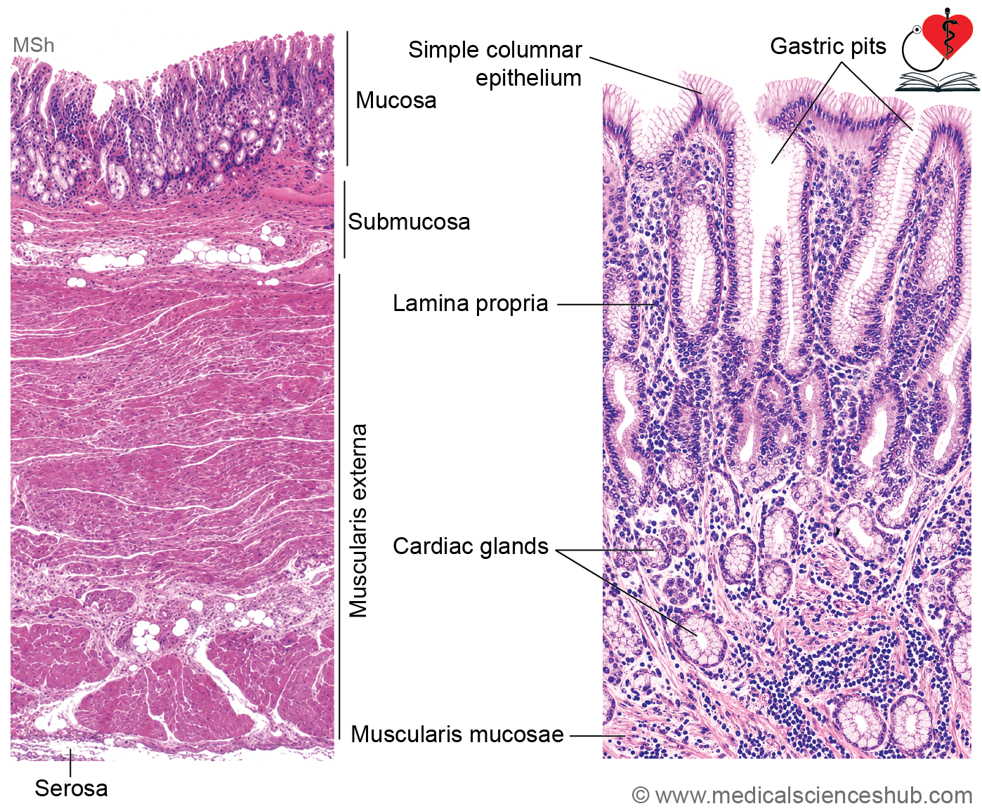
The cardiac region is the area of the stomach adjacent to the gastroesophageal junction and exhibits the typical four layers of the gastrointestinal tract: mucosa, submucosa, muscularis externa, and serosa.
Mucosa
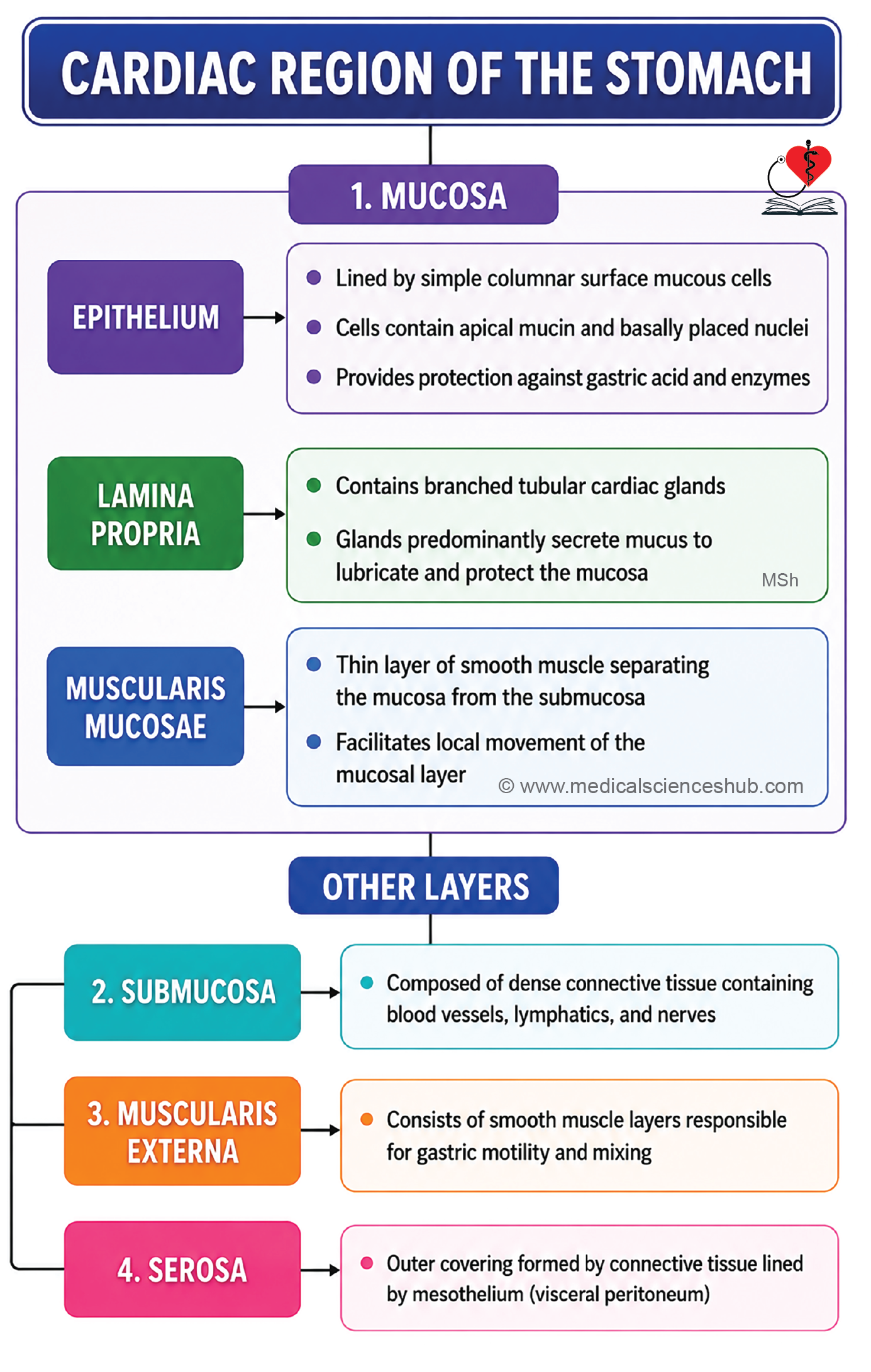
- The mucosa consists of epithelium, lamina propria, and muscularis mucosae.
- The surface is lined by simple columnar mucous cells with basally placed nuclei.
- These cells secrete protective mucus and often appear vacuolated in histological sections because mucin is removed during tissue processing.
- Unlike the small intestine, the gastric epithelium lacks goblet cells and a well-developed absorptive brush border.
- The lamina propria contains branched cardiac glands, which predominantly secrete mucus.
- These glands open into gastric pits and contribute to protection of the mucosa from gastric acid.
- The muscularis mucosae is composed of smooth muscle arranged in inner circular and outer longitudinal layers.
Submucosa
- The submucosa consists of dense irregular connective tissue containing blood vessels, lymphatics, and nerves.
- It also contains the submucosal (Meissner’s) plexus, which helps regulate glandular secretion and local blood flow.
Muscularis Externa
- The muscularis externa is formed by smooth muscle responsible for gastric motility.
- Functionally, the stomach contains oblique, circular, and longitudinal muscle fibers; however, these layers may not always be distinctly separated in routine histological sections.
- The myenteric (Auerbach’s) plexus lies within this layer and coordinates gastric contractions.
Serosa
- The outermost layer is serosa, consisting of connective tissue covered by mesothelium (visceral peritoneum).
- Along the greater and lesser curvatures, the serosa continues into the greater omentum, lesser omentum, and associated peritoneal ligaments.
Fundic or Body Region of Stomach
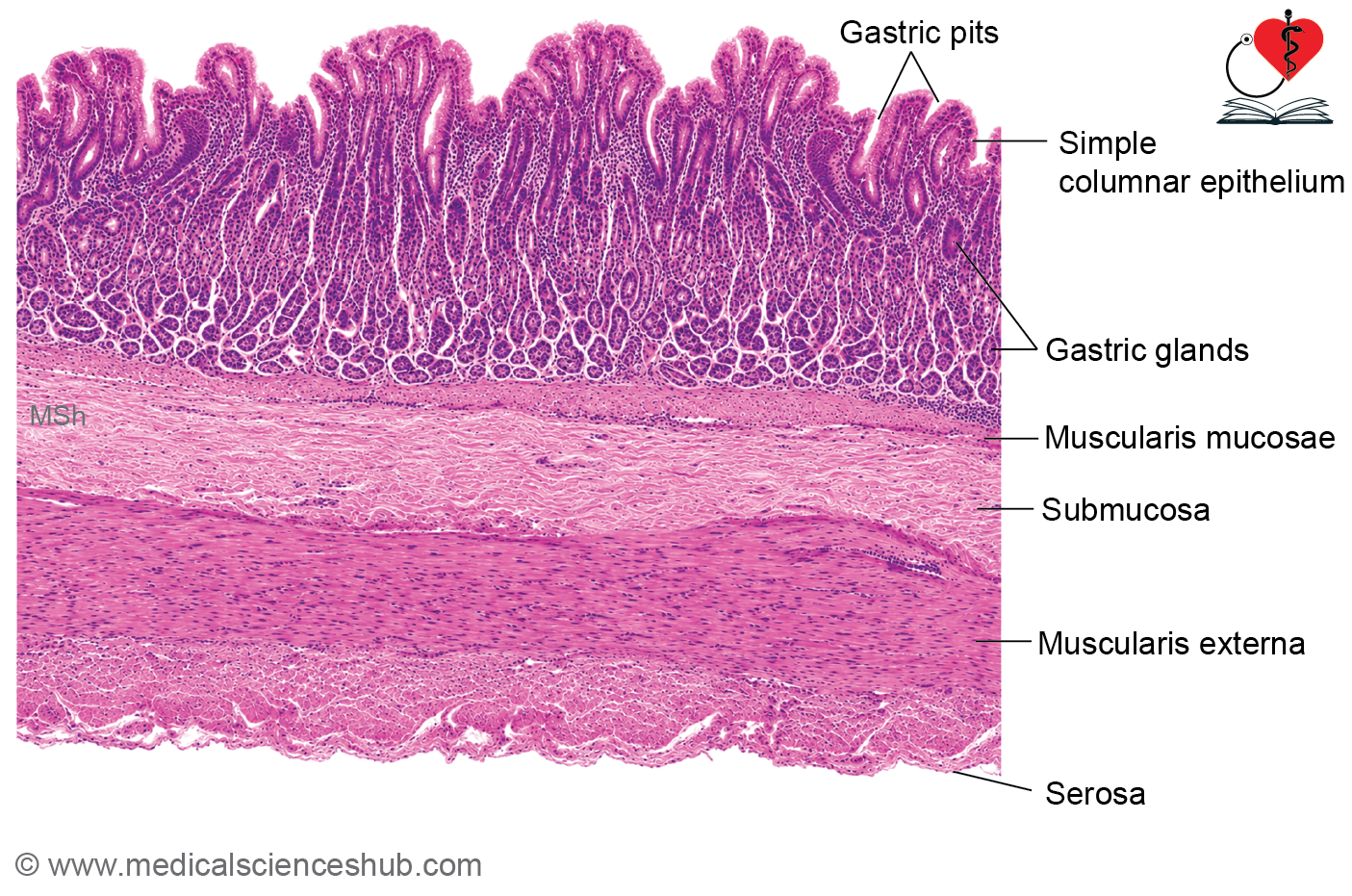
The fundic (body) region constitutes the major secretory portion of the stomach and exhibits the typical four layers: mucosa, submucosa, muscularis externa, and serosa.
Mucosa
- The mucosa consists of epithelium, lamina propria, and muscularis mucosae.
- The surface is lined by simple columnar mucous cells that secrete protective mucus.
- These cells contain apical mucin granules and basally placed oval nuclei.
- In routine histological sections, the apical cytoplasm often appears vacuolated because mucus is removed during processing.
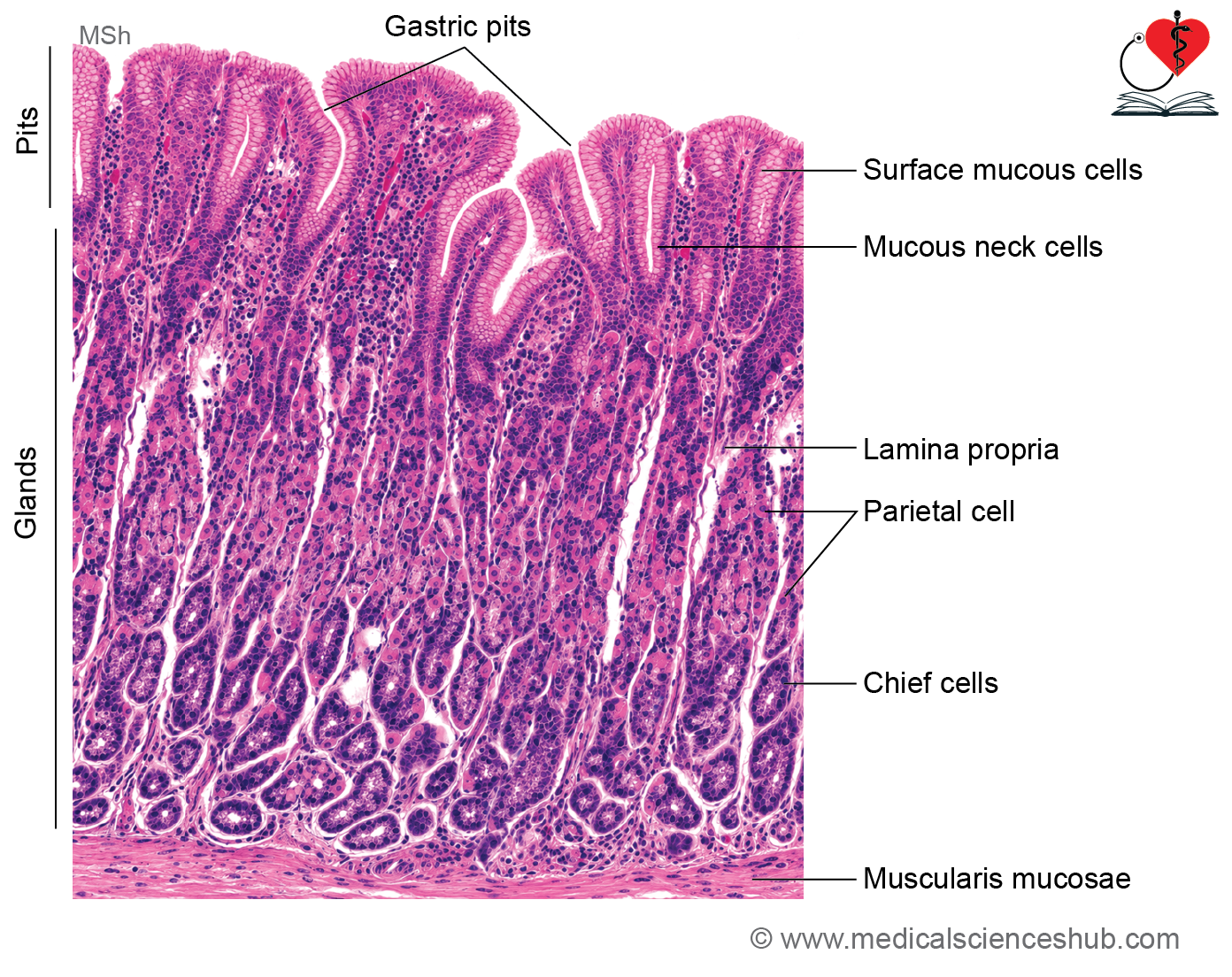
- The lamina propria contains numerous fundic (gastric) glands, which are simple branched tubular glands extending from the base of the gastric pits to the muscularis mucosae.
- Each gland communicates with a gastric pit through a narrow region called the isthmus.
- The isthmus contains stem cells that continuously renew both surface epithelial cells and glandular cells.
Regions of Gastric Glands
- Neck region: A narrow segment located just below the isthmus.
- Base (fundic region): A wider, deeper portion that may branch into two or more terminal segments.
Cell Types in Fundic Glands
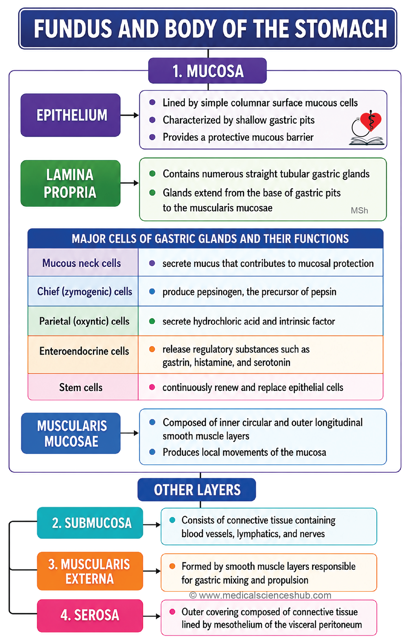
- Mucous neck cells produce mucus that contributes to mucosal protection.
- Parietal (oxyntic) cells secrete hydrochloric acid and intrinsic factor, which is essential for vitamin B₁₂ absorption.
- Chief cells synthesize pepsinogen, the precursor of pepsin.
- Enteroendocrine cells release hormones and regulatory peptides that influence gastric activity.
- Undifferentiated stem cells maintain epithelial renewal and glandular regeneration.
Muscularis Mucosae, Submucosa, Muscle Layer, Serosa
The muscularis mucosae, submucosa, muscularis externa, and serosa have structural features similar to those found in the cardiac region of the stomach.
Cells of Fundic Glands of Stomach
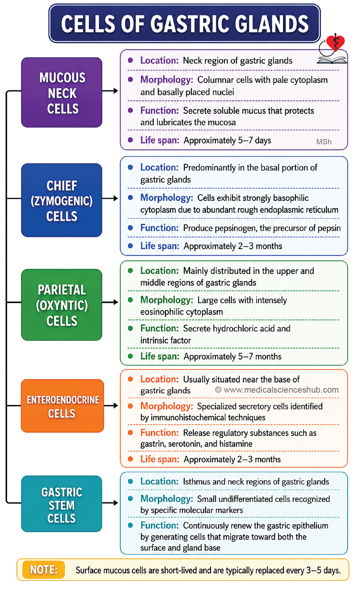
The fundus and body of the stomach contain branched tubular fundic glands composed of mucous neck cells, chief cells, parietal cells, enteroendocrine cells, and stem cells.
Mucous Neck Cells
- These cells are located in the neck region of the gastric glands, just below the gastric pits.
- They are columnar cells with basally placed oval nuclei.
- Their apical cytoplasm contains mucin granules and often appears foamy in routine histological sections.
- They produce soluble mucus, which differs from the thicker mucus secreted by surface mucous cells.
- Some cells in this region contribute to the stem cell population responsible for epithelial renewal.
- Gastric surface mucous cells are replaced approximately every 5–7 days.
Chief or Zymogenic Cells
- Chief cells are concentrated in the deeper portions of fundic glands.
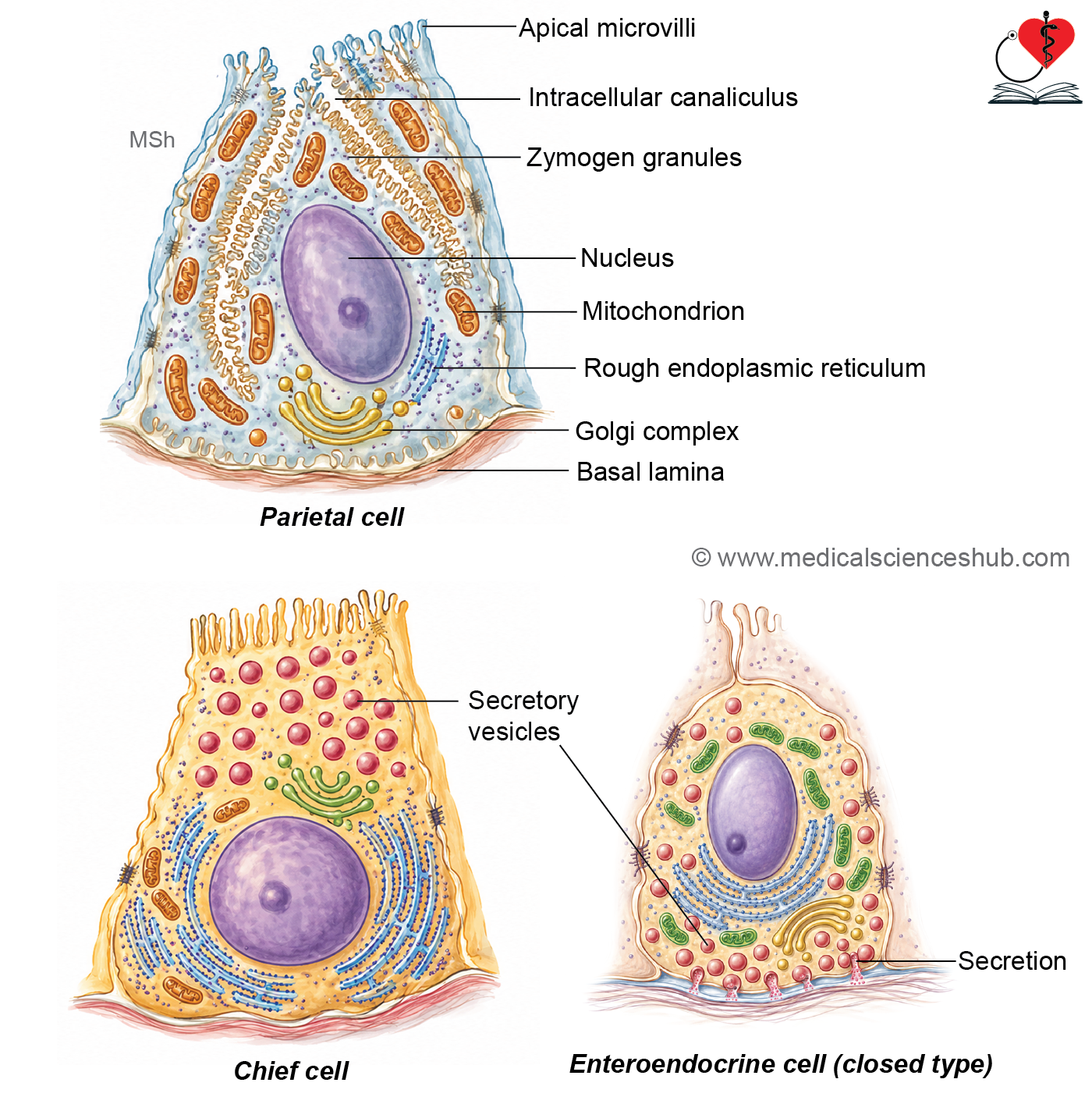
- They are strongly basophilic because of abundant rough endoplasmic reticulum, reflecting active protein synthesis.
- The apical cytoplasm contains zymogen granules that store digestive enzyme precursors.
- Ultrastructurally, these cells possess extensive rough endoplasmic reticulum, a prominent Golgi apparatus, and short apical microvilli.
- Their primary function is secretion of pepsinogen, which is converted into pepsin in the acidic gastric environment.
Parietal (Oxyntic) Cells
- Parietal cells are most abundant in the upper and middle regions of fundic glands.
- They are large, pyramidal or rounded cells with centrally located nuclei and intensely eosinophilic cytoplasm.
- Their cytoplasm contains numerous mitochondria, reflecting the high energy requirement for acid secretion.
- Specialized intracellular canaliculi with microvilli greatly increase the secretory surface area.
- The cell membrane contains proton pumps (H⁺/K⁺-ATPase) responsible for hydrochloric acid secretion.
Functions of Parietal Cells
- Secretion of hydrochloric acid (HCl).
- Production of intrinsic factor, which is essential for vitamin B₁₂ absorption in the ileum.
- Deficiency of intrinsic factor can lead to vitamin B₁₂ deficiency and pernicious anemia.
- These cells respond to gastrin, histamine (H₂ receptors), and acetylcholine.
Enteroendocrine Cells
- These cells are mainly located in the basal portions of gastric glands.
- They may be:
- Open type, with apical processes reaching the gland lumen.
- Closed type, which do not contact the lumen.
- They are difficult to identify on routine H&E staining but can be demonstrated using special staining techniques.
- They release regulatory substances such as gastrin, serotonin, and other bioactive mediators that influence gastric secretion and motility.
Stem Cells
- Stem cells are located primarily in the isthmus and neck regions of gastric glands.
- They continuously divide and migrate upward or downward to replace surface epithelial cells and glandular cells.
- These cells are essential for the rapid renewal and maintenance of the gastric mucosa.
Pyloric Region Of Stomach
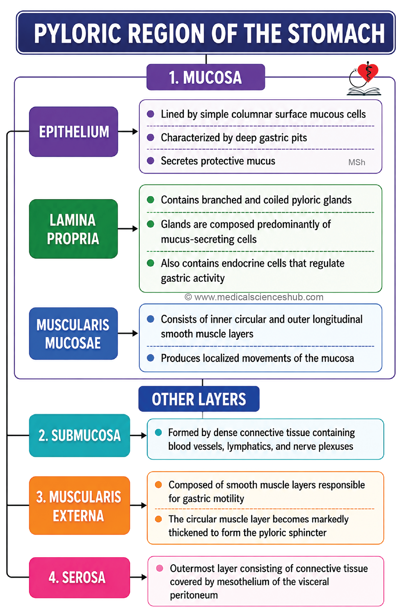
The pyloric region forms the distal part of the stomach and, like other regions, consists of mucosa, submucosa, muscularis externa, and serosa.
Mucosa
- The mucosa is composed of epithelium, lamina propria, and muscularis mucosae.
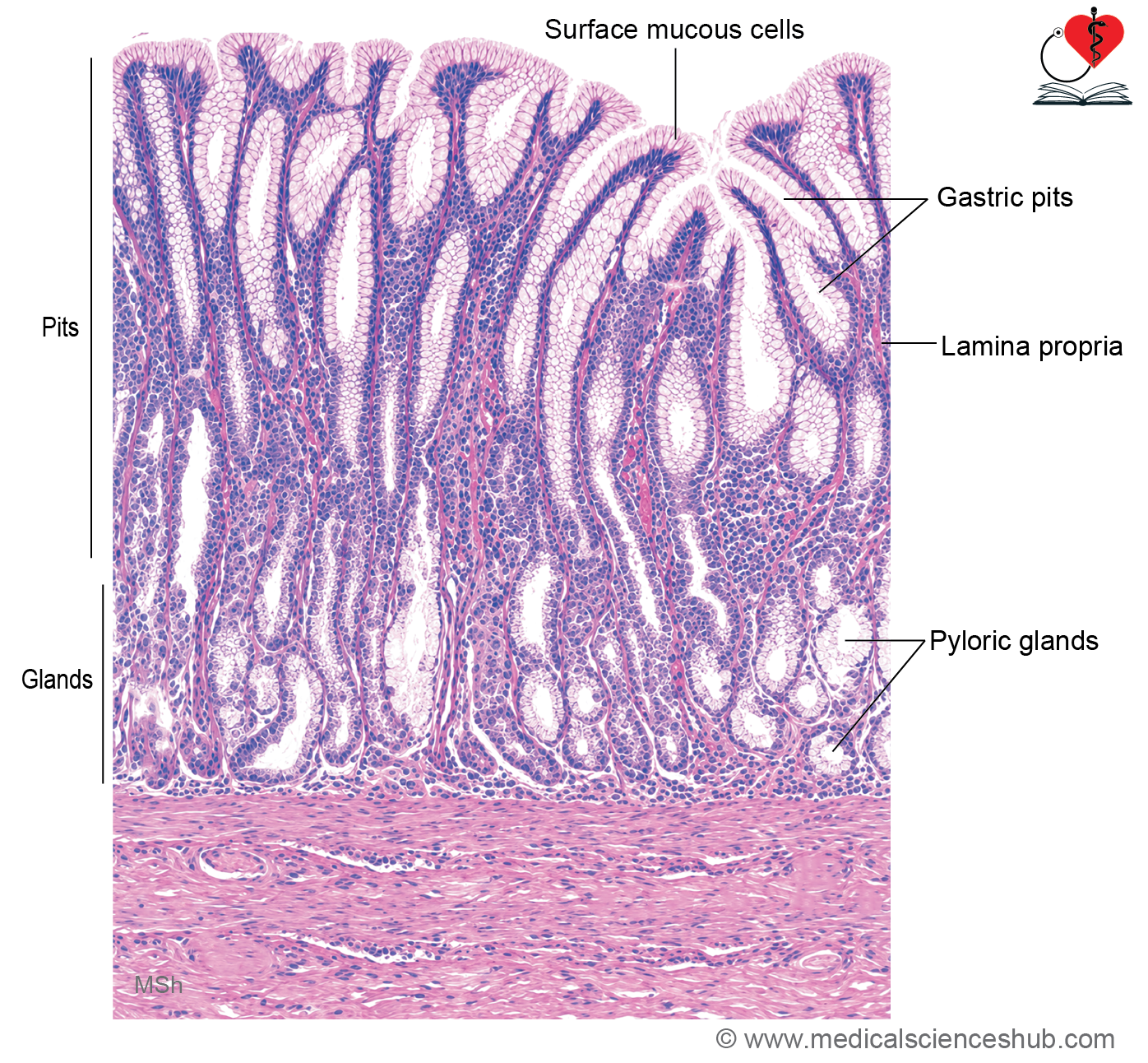
- The surface is lined by tall simple columnar mucous cells that secrete protective mucus.
- The lamina propria contains numerous pyloric glands.
- Gastric pits are characteristically deep, extending through nearly two-thirds of the mucosal thickness.
- Pyloric glands are shorter, coiled, branched tubular glands with relatively wide lumina.
- These glands are composed predominantly of mucus-secreting cells resembling mucous neck cells.
- Scattered enteroendocrine cells are also present.
- G cells, located mainly in the deeper portions of pyloric glands and proximal duodenum, secrete gastrin, which stimulates gastric acid secretion and gastric motility.
Muscularis Mucosae, Lamina Propria, and Submucosa
- The lamina propria, muscularis mucosae, and submucosa have structural features similar to those of the cardiac region of the stomach.
- The submucosa contains connective tissue, blood vessels, lymphatics, and the submucosal (Meissner’s) plexus.
Muscularis Externa
- The circular layer of the muscularis externa becomes markedly thickened in this region.
- This thickened circular muscle forms the pyloric sphincter, which regulates the passage of gastric contents into the duodenum.
Serosa
- The outer surface is covered by serosa, consisting of connective tissue lined by mesothelium (visceral peritoneum).
CLINICAL CORRELATION
- Gastritis is inflammation of the gastric mucosa and commonly presents with upper abdominal pain, heartburn, nausea, and vomiting.
- A gastric ulcer is a localized defect in the stomach lining that may cause epigastric pain. Major causes include Helicobacter pylori infection, prolonged NSAID use, smoking, and other factors that impair mucosal protection.
- Pernicious anemia results from deficiency of intrinsic factor due to dysfunction or loss of parietal cells.
- Because intrinsic factor is essential for vitamin B₁₂ absorption in the ileum, its deficiency leads to megaloblastic anemia.
- Treatment typically involves vitamin B₁₂ (cobalamin) supplementation, often administered by injection.
Table 14.1: Differences between histology of fundic (body) and pyloric region of stomach
| Feature | Fundic (Body) Region of Stomach | Pyloric Region of Stomach |
|---|---|---|
| Gastric pits | Short and shallow; extend through approximately the upper one-third of the mucosal thickness | Long and deep; extend through approximately the upper two-thirds of the mucosal thickness |
| Extent of gastric glands | Occupy the lower two-thirds of the mucosa | Occupy the lower one-third of the mucosa |
| Configuration of glands | Relatively straight with minimal coiling | More tortuous and highly coiled |
| Glandular lumen | Narrow lumen | Wider lumen |
| Predominant cell types | Contain numerous parietal cells, chief cells, and mucous neck cells | Predominantly composed of mucus-secreting cells; chief cells are generally absent and parietal cells are sparse |
| Primary secretion | Hydrochloric acid, intrinsic factor, pepsinogen, and mucus | Primary secretion Hydrochloric acid, intrinsic factor, pepsinogen, and mucus Mainly mucus, with secretion contributing to protection of the gastric mucosa and neutralization of acidic chyme |
Important Questions
- Write a short note on histology of cardiac region or cardia of stomach.
- Write a short note on the histology of fundic or body region of the stomach.
- List the cells of fundic glands of stomach.
- Write a short note on histology of pyloric region of stomach.
- List the differences between histology of fundic (body) and pyloric part of stomach.
📝 Test Your Knowledge – Practice MCQs
Attempt the chapter MCQ quiz and assess your understanding of key concepts.
Click to see: Medical Disclaimer and Copyright Notice