
Introduction
- The autonomic nervous system (ANS) is the part of the nervous system that regulates the functions of internal organs without conscious control.
- It plays a vital role in maintaining homeostasis by continuously adjusting the activity of various body systems according to the body’s needs.
- The ANS controls the function of smooth muscle, cardiac muscle, and glands.
- It regulates essential involuntary activities such as heart rate, blood pressure, respiratory rate, digestion, glandular secretion, body temperature, and pupillary diameter.
- Through its actions, the ANS helps maintain a stable internal environment despite changes in external or internal conditions.
- Because it primarily governs the activity of the thoracic, abdominal, and pelvic viscera, it is also known as the visceral nervous system.
- The autonomic nervous system is divided into sympathetic and parasympathetic divisions, which usually exert complementary effects to maintain physiological balance.
- Proper autonomic function is essential for normal cardiovascular, respiratory, gastrointestinal, urinary, and endocrine regulation.
Divisions of Autonomic Nervous System
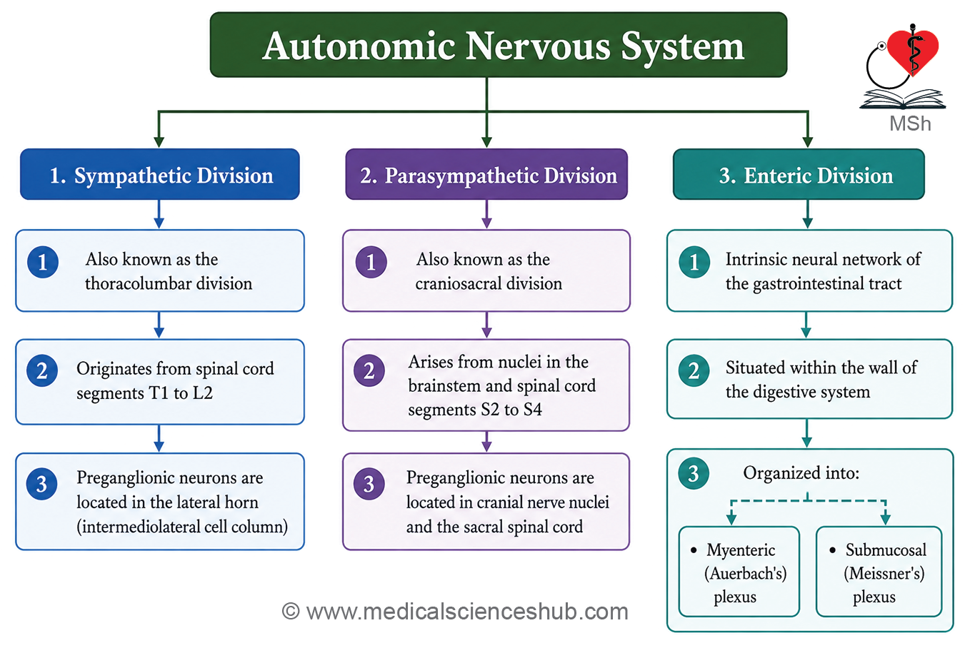
The autonomic nervous system (ANS) is divided into three functional components:
- Sympathetic nervous system
- Parasympathetic nervous system
- Enteric nervous system
Sympathetic Nervous System
- The sympathetic nervous system prepares the body to respond to physical or emotional stress and is often described as the thoracolumbar outflow.
- It consists of sympathetic ganglia, preganglionic neurons, and postganglionic neurons.
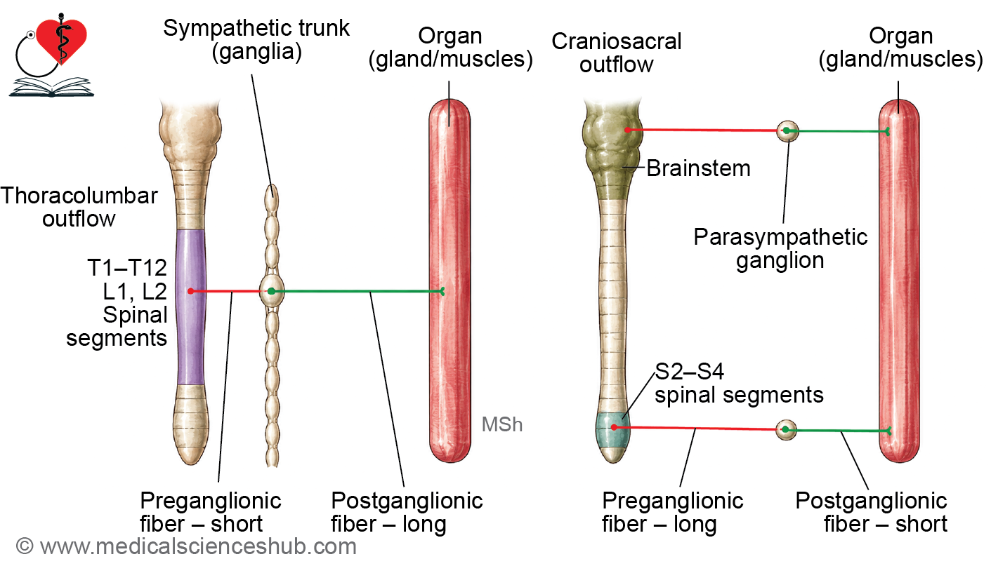
- Preganglionic neuron cell bodies are located in the lateral horn of the spinal cord segments T1 to L2.
- Their fibers leave the spinal cord through the anterior roots and enter the sympathetic trunk through white rami communicantes.
- Sympathetic ganglia are arranged as interconnected chains on either side of the vertebral column, forming the sympathetic trunks.
- Postganglionic fibers travel to various organs and regulate functions such as heart rate, blood pressure, airway diameter, sweating, and vascular tone.
- The sympathetic division generally increases energy expenditure and enhances the body’s ability to respond to stress.
Parasympathetic Nervous System
- The parasympathetic nervous system promotes maintenance, recovery, and conservation of energy and is known as the craniosacral outflow.
- It is composed of preganglionic neurons, parasympathetic ganglia, and postganglionic neurons.
- Preganglionic fibers arise from:
- Brainstem nuclei associated with the oculomotor (III), facial (VII), glossopharyngeal (IX), and vagus (X) nerves
- Sacral spinal cord segments S2-S4
- Most parasympathetic fibers supplying thoracic and abdominal organs travel through the vagus nerve.
- Parasympathetic ganglia are located within or very close to the target organs.
- Because the ganglia lie near the effector tissues, postganglionic fibers are typically short.
- This division slows the heart rate, stimulates gastrointestinal activity, promotes glandular secretion, and supports normal visceral function.
Enteric Nervous System
- The enteric nervous system is an extensive network of neurons located within the wall of the gastrointestinal tract.
- It functions as the intrinsic nervous system of the digestive tract and can perform many activities independently.
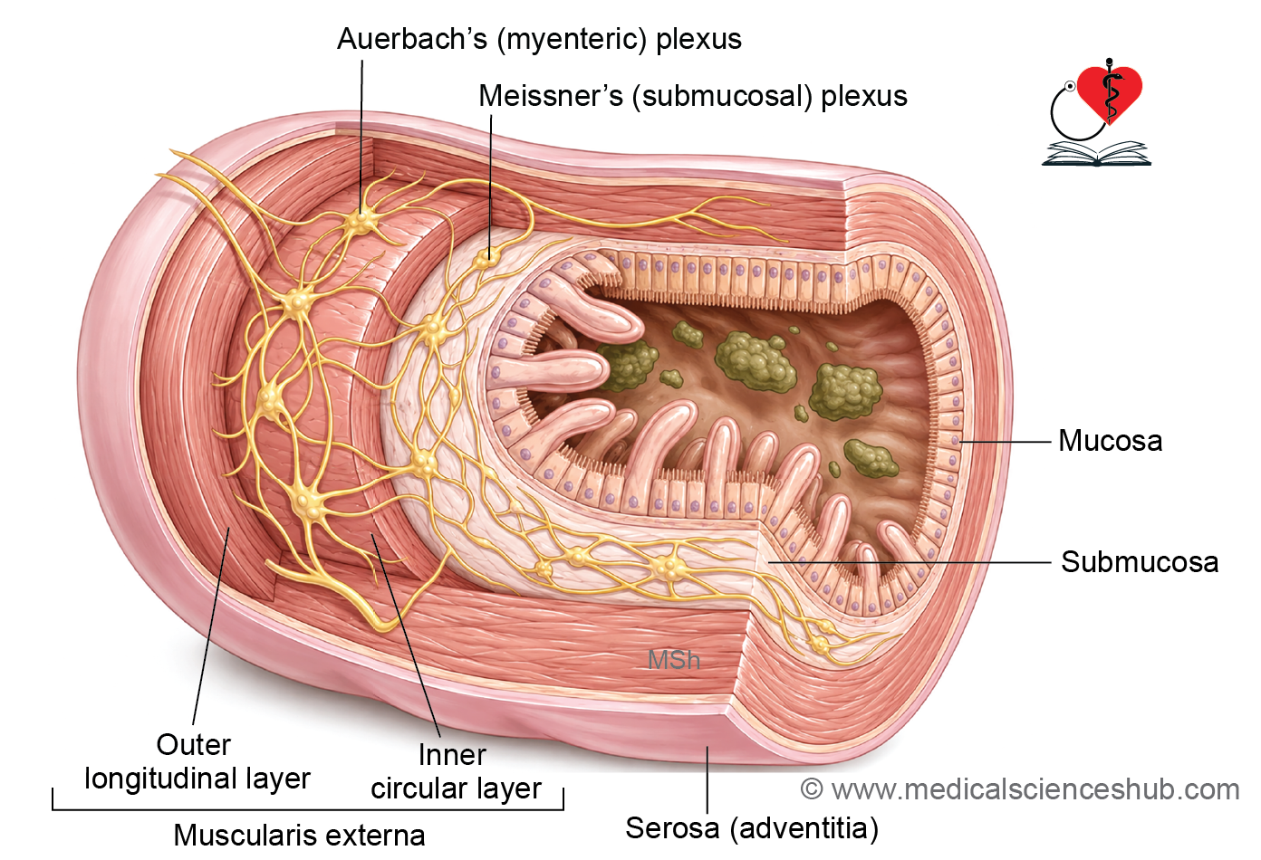
- The enteric nervous system is organized into two major nerve plexuses:
- Myenteric (Auerbach) plexus, located between the longitudinal and circular muscle layers
- Submucosal (Meissner) plexus, located within the submucosa
- It regulates gastrointestinal motility, secretion, absorption, and local blood flow.
- The enteric nervous system receives modulatory input from both sympathetic and parasympathetic divisions but is capable of coordinating many digestive functions on its own.
- It plays a critical role in the normal movement and processing of food throughout the gastrointestinal tract.
Sympathetic Nervous System
- The sympathetic nervous system is the thoracolumbar division of the autonomic nervous system.
- Preganglionic neuron cell bodies are located in the lateral horn of spinal cord segments T1-L2.
- Their myelinated fibers leave the spinal cord through anterior roots and enter the sympathetic trunk via white rami communicantes.
- After entering the sympathetic trunk, these fibers may:
- Synapse in a ganglion at the same level.
- Ascend or descend before synapsing.
- Pass through the trunk without synapsing and continue as splanchnic nerves to prevertebral ganglia.
- Postganglionic neuron cell bodies are located in sympathetic ganglia.
- Postganglionic fibers reach target organs directly or re-enter spinal nerves through gray rami communicantes.
- The sympathetic system regulates cardiovascular, respiratory, glandular, and smooth muscle activity and helps maintain homeostasis during physiological stress.
Sympathetic Trunk (Chain)
- The sympathetic trunks are paired chains of interconnected sympathetic ganglia located on either side of the vertebral column.
- They extend from the base of the skull to the coccyx and are connected inferiorly by the ganglion impar, an unpaired terminal ganglion.
- Each trunk typically contains 22 ganglia, although the number may vary.
- The ganglia are arranged as:
- Cervical ganglia: 3
- Thoracic ganglia: 10-12
- Lumbar ganglia: 4
- Sacral ganglia: 4
- The sympathetic trunk serves as a pathway for ascending, descending, and synapsing preganglionic sympathetic fibers.
- It allows sympathetic outflow from spinal cord segments T1-L2 to be distributed throughout the body.
- Through its extensive connections, the sympathetic trunk coordinates autonomic responses in the head, neck, thorax, abdomen, pelvis, and limbs.
Cervical Sympathetic Chain
- The cervical part of the sympathetic trunk lies on either side of the cervical vertebral column, posterior to the carotid sheath and anterior to the prevertebral muscles.
- Unlike the thoracic region, the cervical sympathetic trunk usually has no white rami communicantes because preganglionic sympathetic fibers originate only from spinal cord segments T1-L2.
- Preganglionic fibers ascend from the upper thoracic sympathetic trunk to synapse in cervical sympathetic ganglia.
- Postganglionic fibers from these ganglia are distributed to structures of the head, neck, heart, and upper thorax.
- The cervical sympathetic trunk typically contains three ganglia:
- Superior cervical ganglion
- Middle cervical ganglion
- Inferior cervical ganglion (often fused with the first thoracic ganglion to form the stellate ganglion)
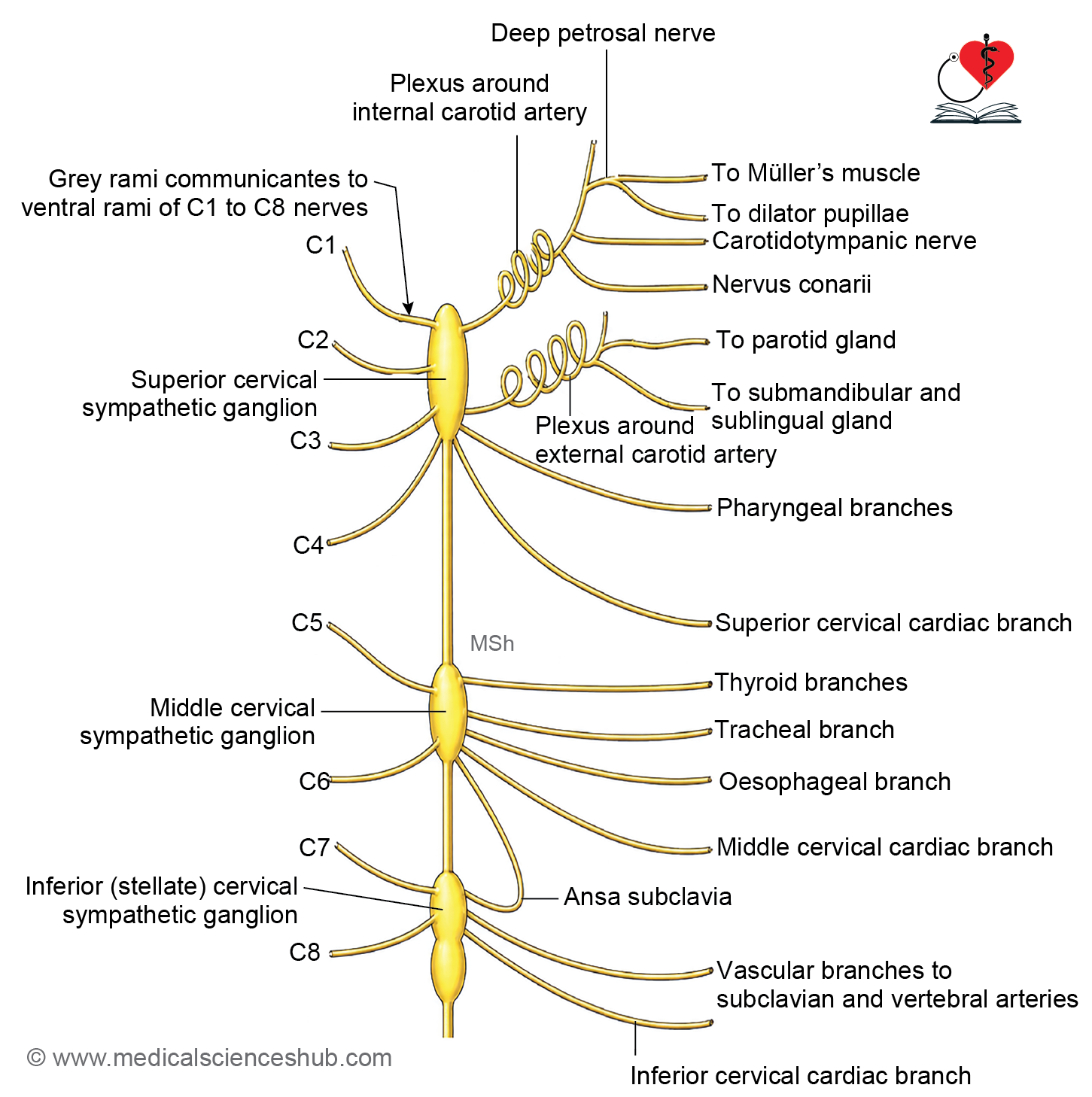
Superior Cervical Ganglion
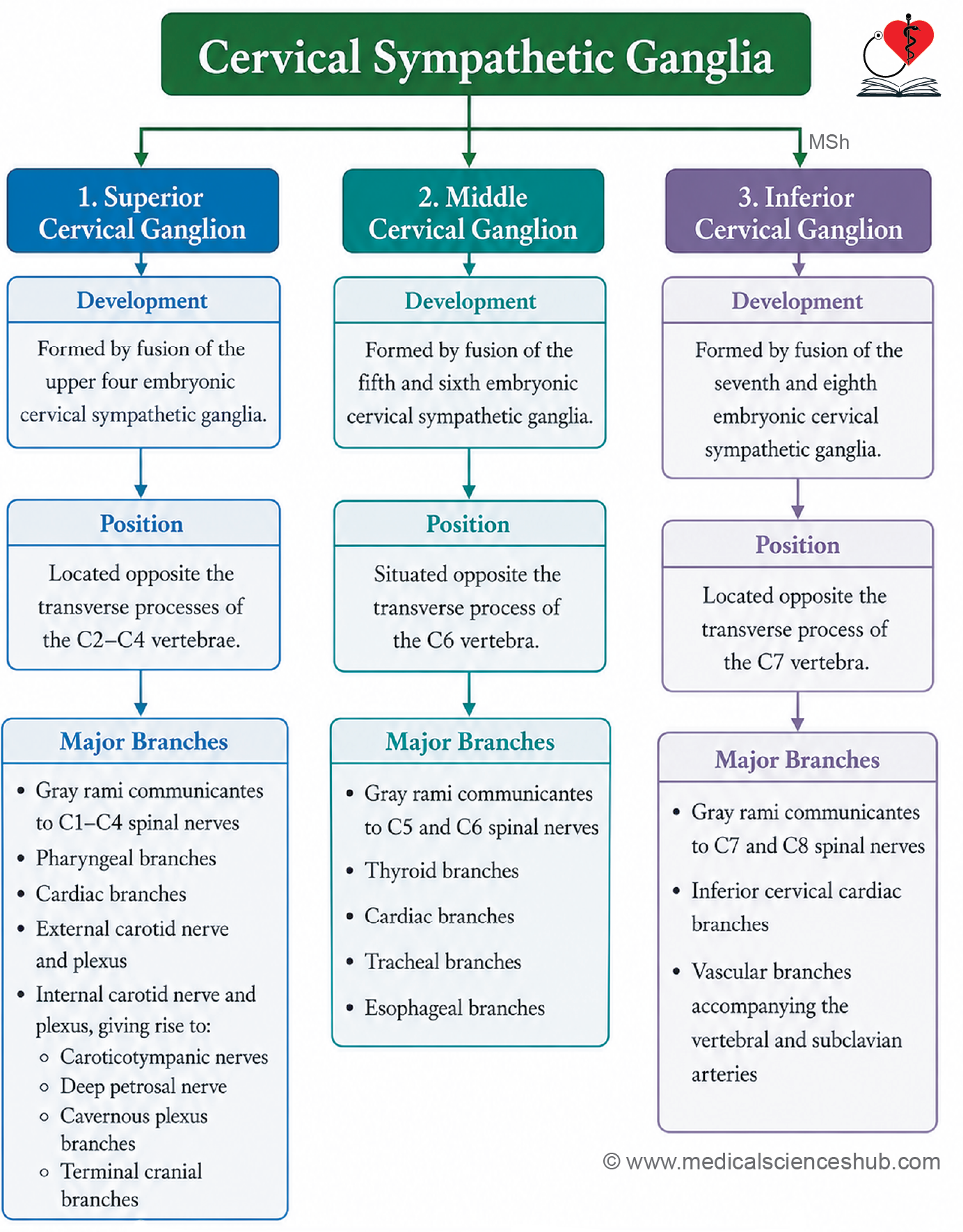
- The superior cervical ganglion is the largest cervical sympathetic ganglion.
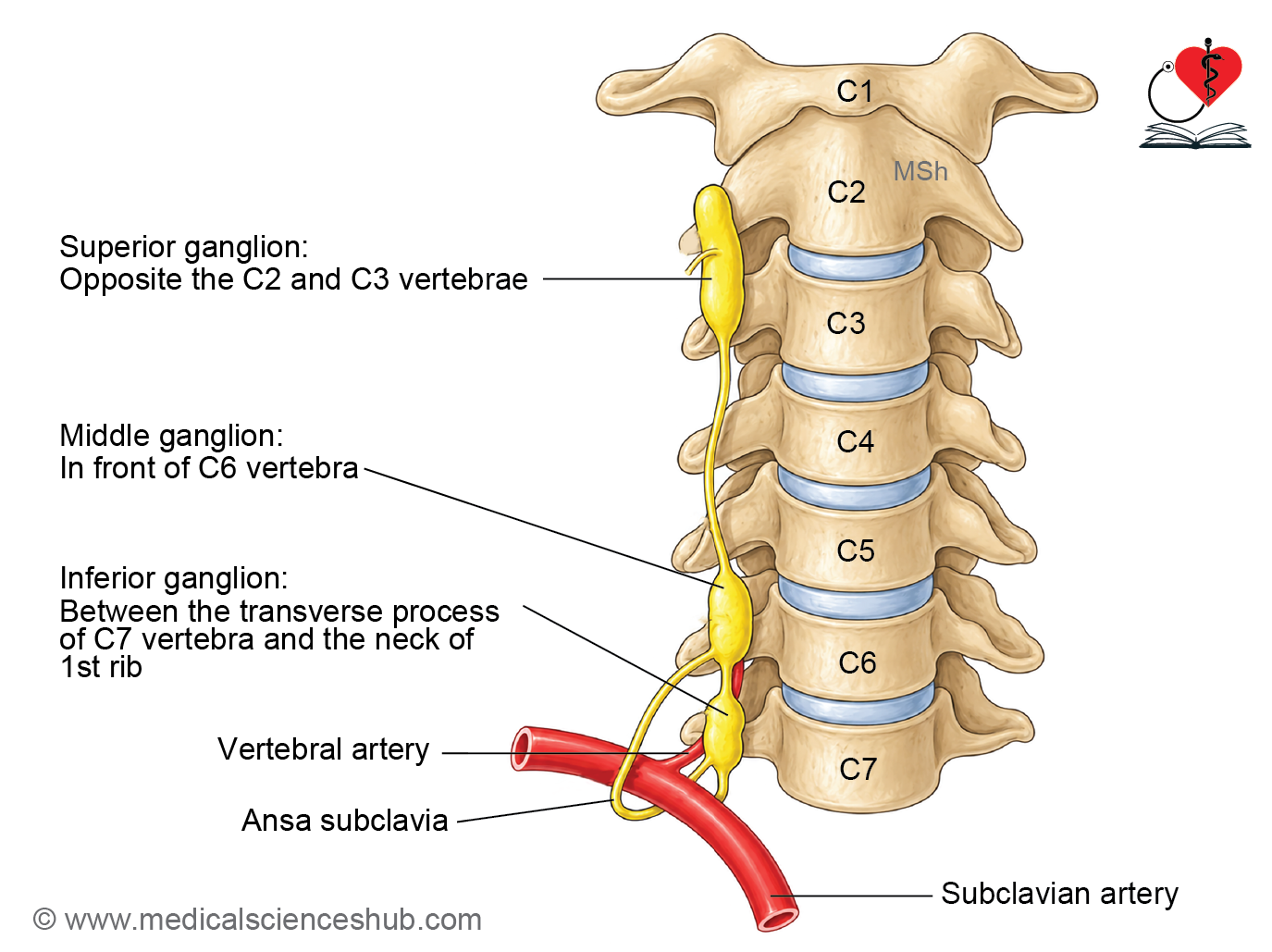
- It is usually located opposite the C2-C3 vertebrae, posterior to the internal carotid artery and medial to the vagus nerve.
- It is formed by fusion of the upper cervical sympathetic ganglia during development.
- This ganglion serves as the principal source of sympathetic innervation to the head and neck.
Major Branches and Distribution
- Gray rami communicantes join the upper cervical spinal nerves.
- Internal carotid nerves form the internal carotid plexus, which distributes sympathetic fibers to intracranial structures and the orbit.
- External carotid nerves form plexuses around branches of the external carotid artery and supply blood vessels and glands of the face and scalp.
- Jugular branches communicate with the glossopharyngeal, vagus, and hypoglossal nerves.
- Pharyngeal branches contribute to the pharyngeal plexus and supply the pharynx.
- Superior cardiac nerves descend into the thorax and participate in the cardiac plexus.
- Sympathetic fibers passing through the carotid plexuses supply the dilator pupillae muscle, superior tarsal muscle, sweat glands, and blood vessels of the head and neck.
CLINICAL NEUROANATOMY
- Interruption of cervical sympathetic pathways may produce Horner syndrome, characterized by ptosis, miosis, anhidrosis, and apparent enophthalmos.
- Knowledge of cervical sympathetic anatomy is important during surgical procedures involving the carotid arteries, thyroid gland, cervical spine, and neck tumors.
Middle Cervical Ganglion
- The middle cervical ganglion is the smallest and most variable of the cervical sympathetic ganglia and may occasionally be absent.
- It is usually located at the level of the C6 vertebra, near the inferior thyroid artery and anterior to the transverse process.
- The ganglion is formed by the fusion of adjacent cervical sympathetic ganglia during development.
- It communicates with the superior and inferior cervical ganglia through interganglionic sympathetic fibers.
Connections
- It is linked to the cervical sympathetic trunk by ascending and descending fibers.
- Small communicating branches connect it with nearby cervical spinal nerves.
- Sympathetic fibers from this ganglion contribute to autonomic plexuses in the neck.
Branches
- Gray rami communicantes join the C5 and C6 spinal nerves.
- Thyroid branches accompany the inferior thyroid artery and supply the thyroid and parathyroid glands as well as adjacent blood vessels.
- The middle cervical cardiac nerve arises from this ganglion and descends into the thorax to contribute to the cardiac plexus.
- Vascular branches form plexuses around nearby arteries and distribute sympathetic fibers to cervical structures.
- Additional branches may communicate with the recurrent laryngeal nerve and neighboring autonomic plexuses.
CLINICAL NEUROANATOMY
- The middle cervical ganglion is an important landmark during surgical procedures involving the thyroid gland, carotid sheath, and lower cervical region.
- Injury to its sympathetic fibers may alter vasomotor, sudomotor, and cardiac functions.
- Its variable size and position should be considered during neck dissections and imaging studies.
Inferior Cervical Ganglion
- The inferior cervical ganglion is formed by fusion of the lower cervical sympathetic ganglia during development.
- It is commonly fused with the first thoracic ganglion to form the cervicothoracic (stellate) ganglion.
- The ganglion is usually located near the neck of the first rib, between the transverse process of C7 and the first rib, close to the vertebral artery.
- It serves as an important relay station for sympathetic fibers supplying the neck, upper limb, thoracic viscera, and blood vessels.
Branches
- Gray rami communicantes join the C7 and C8 spinal nerves, distributing postganglionic sympathetic fibers to the upper limb and adjacent regions.
- Inferior cervical cardiac nerves descend into the thorax and contribute to the cardiac plexus, influencing cardiac activity.
- Vascular branches form plexuses around the vertebral and subclavian arteries and distribute sympathetic fibers along these vessels.
- Additional branches may contribute to sympathetic innervation of the upper thoracic structures and nearby autonomic plexuses.
CLINICAL NEUROANATOMY
- The stellate ganglion is a clinically important target for stellate ganglion block, a procedure used to treat certain pain syndromes, vascular disorders, and sympathetic overactivity.
- Injury to the inferior cervical ganglion or its fibers may produce features of Horner syndrome.
- Because of its close relationship to major vessels, the pleura, and the apex of the lung, detailed anatomical knowledge is essential during surgical and interventional procedures in the lower neck and thoracic inlet.
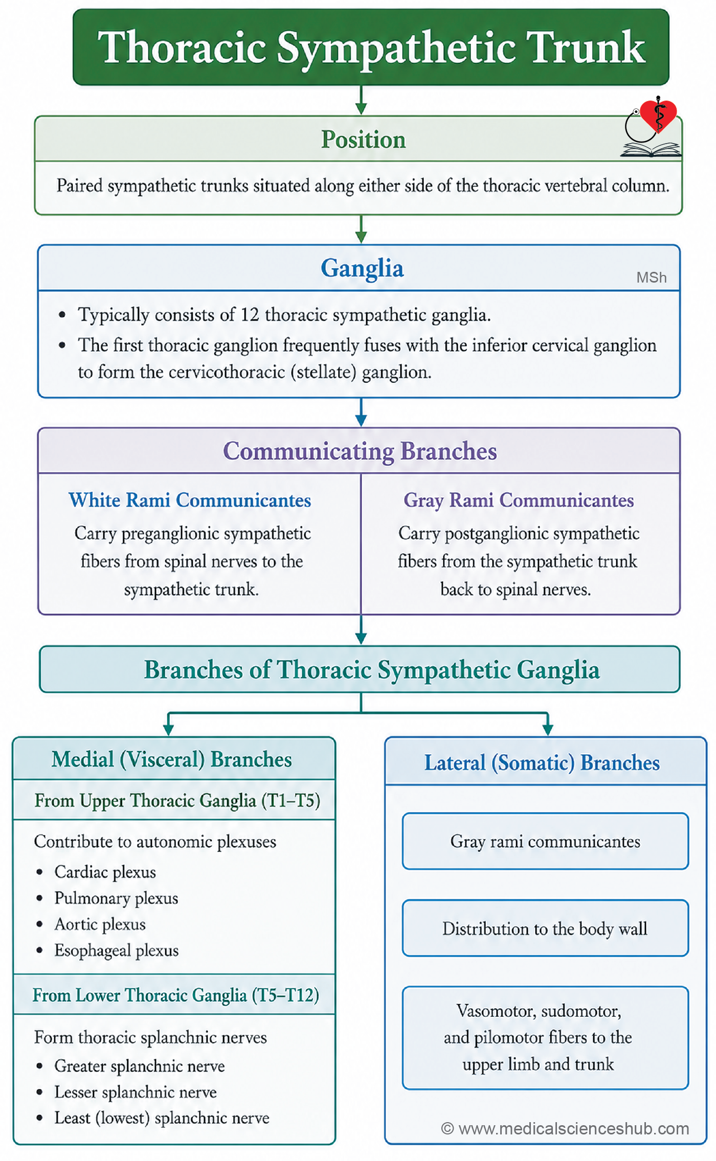
Thoracic Part of Sympathetic Chain
- The thoracic sympathetic trunk usually consists of 10-12 thoracic ganglia located along either side of the thoracic vertebral column.
- The first thoracic ganglion is frequently fused with the inferior cervical ganglion, forming the stellate (cervicothoracic) ganglion.
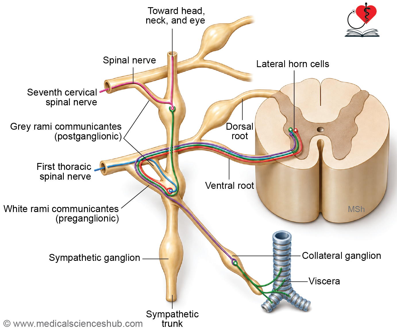
- Each thoracic ganglion is connected to the corresponding thoracic spinal nerve by communicating branches.
Connections with Spinal Nerves
- White rami communicantes carry myelinated preganglionic sympathetic fibers from spinal nerves T1-L2 to the sympathetic trunk.
- Gray rami communicantes carry unmyelinated postganglionic fibers from the sympathetic trunk back to the spinal nerves.
- These connections distribute sympathetic fibers to blood vessels, sweat glands, and arrector pili muscles of the body wall and limbs.
Visceral Branches
- Thoracic ganglia give rise to visceral branches that supply thoracic, abdominal, and pelvic organs through autonomic plexuses.
Cardiopulmonary Branches
- Upper thoracic ganglia send postganglionic fibers to:
- Cardiac plexus
- Pulmonary plexus
- Esophageal plexus
- Thoracic aortic plexus
- These fibers regulate the activity of the heart, lungs, blood vessels, and esophagus.
Thoracic Splanchnic Nerves
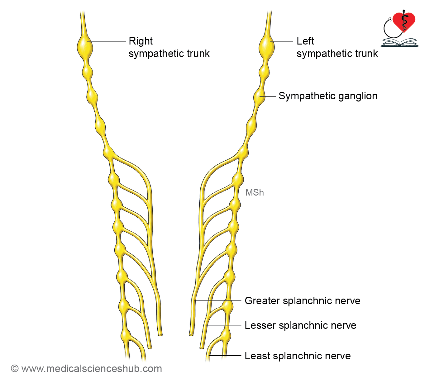
- Lower thoracic ganglia give rise to thoracic splanchnic nerves, which contain preganglionic sympathetic fibers.
- These nerves pass through the diaphragm and terminate in prevertebral ganglia within the abdomen.
- The major thoracic splanchnic nerves are:
- Greater splanchnic nerve (T5-T9)
- Lesser splanchnic nerve (T10-T11)
- Least splanchnic nerve (T12)
- They provide sympathetic innervation to abdominal and pelvic viscera.
CLINICAL NEUROANATOMY
- The thoracic sympathetic trunk plays a major role in regulating cardiovascular, respiratory, and visceral functions.
- Surgical interruption of thoracic sympathetic pathways (sympathectomy) may be performed for conditions such as hyperhidrosis and certain vascular disorders.
- Knowledge of its anatomy is important during thoracic surgery and procedures involving the mediastinum and vertebral column.
Prevertebral Ganglia
- Prevertebral ganglia are sympathetic ganglia located anterior to the vertebral column, mainly around the origins of major abdominal arteries.
- They serve as relay stations for preganglionic sympathetic fibers destined for abdominal and pelvic viscera.
- The principal prevertebral ganglia include:
- Celiac ganglia, situated near the origin of the celiac trunk.
- Superior mesenteric ganglia, located around the superior mesenteric artery.
- Aorticorenal ganglia, associated with the renal arteries.
- Inferior mesenteric ganglia, located near the origin of the inferior mesenteric artery.
- Postganglionic fibers from these ganglia form periarterial plexuses that accompany blood vessels to target organs.
Lumbar Sympathetic Chain
- he lumbar sympathetic trunk extends along either side of the lumbar vertebral bodies.
- It usually consists of four interconnected lumbar ganglia.
- On the right side, it lies posterior to the inferior vena cava; on the left side, it lies close to the abdominal aorta.
- The trunk continues superiorly with the thoracic sympathetic chain and inferiorly with the sacral sympathetic trunk.
Branches
- White rami communicantes connect the upper lumbar spinal nerves (L1 and L2) to the sympathetic trunk and carry preganglionic sympathetic fibers.
- Gray rami communicantes carry postganglionic sympathetic fibers from the lumbar ganglia to all lumbar spinal nerves.
- Lumbar splanchnic nerves arise from the lumbar ganglia and contain preganglionic sympathetic fibers.
- These nerves terminate in prevertebral ganglia and contribute to the intermesenteric, aortic, superior hypogastric, and inferior mesenteric plexuses.
Functions and Clinical Importance
- The lumbar sympathetic trunk provides sympathetic innervation to abdominal viscera, pelvic organs, blood vessels, sweat glands, and arrector pili muscles of the lower trunk and lower limbs.
- It plays an important role in regulating vasomotor activity and visceral function.
- Lumbar sympathectomy may be performed in selected vascular and pain disorders involving the lower limb.
- Knowledge of lumbar sympathetic anatomy is important during retroperitoneal and vascular surgical procedures.
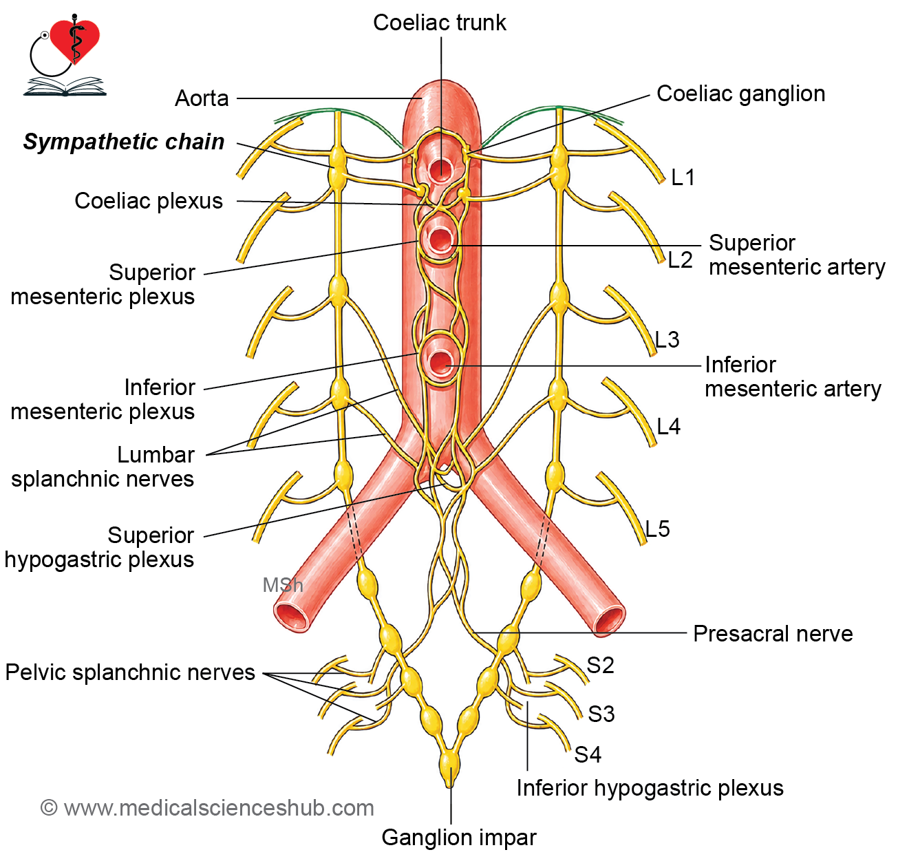
Autonomic Plexuses of the Posterior Abdominal Wall
- Several autonomic nerve plexuses are present on the posterior abdominal wall and distribute autonomic fibers to abdominal and pelvic organs.
- The major plexuses are:
- Celiac plexus
- Superior mesenteric plexus
- Inferior mesenteric plexus
Celiac Plexus
- The celiac plexus is the largest autonomic plexus in the abdomen and is often referred to as the solar plexus.
- It surrounds the origin of the celiac trunk and lies anterior to the abdominal aorta near the upper lumbar vertebrae.
- The plexus contains both sympathetic and parasympathetic fibers, as well as sensory fibers.
Sources of Fibers
- Preganglionic sympathetic fibers reach the plexus mainly through the greater and lesser thoracic splanchnic nerves.
- Postganglionic sympathetic fibers arise from neurons within the celiac ganglia.
- Preganglionic parasympathetic fibers are supplied by the vagus nerves.
- Visceral afferent fibers carrying sensory information from abdominal organs also pass through the plexus.
Celiac Ganglia
- The celiac plexus contains a pair of celiac ganglia, one on each side of the celiac trunk.
- These are the largest prevertebral sympathetic ganglia in the body.
- They serve as important relay stations for sympathetic innervation of abdominal viscera.
- The upper part of each ganglion primarily receives fibers from the greater splanchnic nerve, whereas the lower part receives fibers from the lesser splanchnic nerve and contributes to the renal plexus.
Branches of the Celiac Plexus
The celiac plexus gives rise to several secondary plexuses that accompany major abdominal arteries:
- Phrenic plexus
- Hepatic plexus
- Left gastric plexus
- Splenic plexus
- Suprarenal plexus
- Renal plexus
- Testicular or ovarian plexus
- Superior mesenteric plexus
- Intermesenteric (abdominal aortic) plexus
- Inferior mesenteric plexus
Clinical Importance
- The celiac plexus plays a central role in autonomic control of the foregut and many upper abdominal organs.
- A celiac plexus block may be used to relieve severe abdominal pain, particularly in pancreatic and upper abdominal malignancies.
- Knowledge of these plexuses is important during abdominal surgery, interventional procedures, and imaging interpretation.
Superior Hypogastric Plexus (presacral nerve)
- Located anterior to the bifurcation of the abdominal aorta, at the level of the body of L5 vertebra.
Formation
- Receives sympathetic fibers from the aortic plexus and the third and fourth lumbar splanchnic nerves.
- Receives parasympathetic fibers from the pelvic splanchnic nerves via the inferior hypogastric plexus.
Branches
- The right and left hypogastric nerves descend into the pelvis to form the two inferior hypogastric plexuses.
- Additional branches supply the ureteric, gonadal, and common iliac plexuses.
Inferior Hypogastric Plexuses
- There are two inferior hypogastric plexuses (right and left), each containing numerous small ganglia.
- They are located in the extraperitoneal connective tissue of the pelvis, on either side of the rectum.
Formation Each plexus receives three types of input:
- Sympathetic fibers from the superior hypogastric plexus and hypogastric nerves.
- Parasympathetic fibers from the pelvic splanchnic nerves (S2, S3, S4).
- Sensory (afferent) fibers from the pelvic viscera.
Branches
- Rectal plexus: runs along the middle rectal artery, supplying the rectum.
- Vesical plexus: runs along the vesical arteries, supplying the urinary bladder, adjacent ureters, ejaculatory ducts, and seminal vesicles.
- Prostatic plexus: surrounds the prostate, supplying the prostate, seminal vesicles, and ejaculatory ducts.
- Uterine and vaginal plexuses: run along the uterine and vaginal arteries, supplying the uterus and vagina.
Autonomic Nerves of True Pelvis
The autonomic innervation of the true pelvis is provided by four components:
- Pelvic part of the sympathetic trunk
- Presacral nerve (superior hypogastric plexus)
- Inferior hypogastric plexus
- Pelvic splanchnic nerves (nervi erigentes)
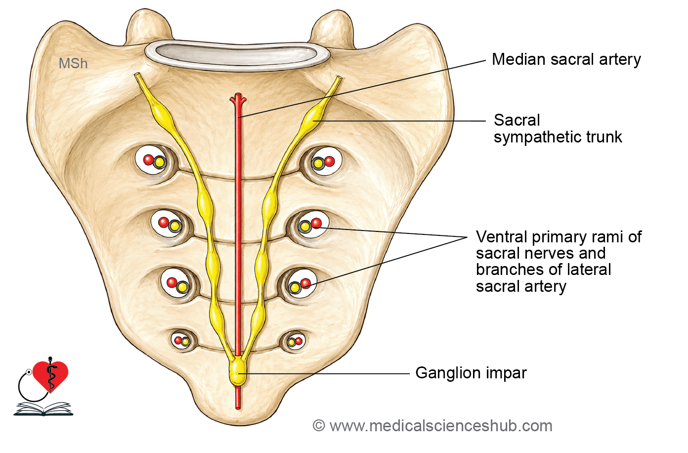
Sacral Sympathetic Chain
- The right and left sympathetic trunks descend through the pelvis between the sacral vertebral bodies and the pelvic sacral foramina.
- Inferiorly, both trunks unite anterior to the coccyx to form the unpaired ganglion impar.
- The sacral portion of the sympathetic trunk contains four pairs of sacral ganglia, connected to the sacral and coccygeal spinal nerves via grey rami communicantes carrying postganglionic sympathetic fibers.
Branches
- Grey rami communicantes to the ventral primary rami of the sacral and coccygeal nerves.
- Sacral splanchnic nerves to the inferior hypogastric plexus and rectum.
- Branches to the median sacral artery.
- Branches from the ganglion impar to the coccygeal body.
Presacral Nerve or Superior Hypogastric Plexus
- Lies anterior to the bifurcation of the abdominal aorta at the level of L5 vertebra.
- Detailed description covered in the preceding section.
Inferior Hypogastric Plexus
- Two plexuses (right and left) located in the extraperitoneal connective tissue of the pelvis, on either side of the rectum.
- Detailed description covered in the preceding section.
Coccygeal Body or Glomus Coccygeum
- A small, oval structure located anterior to the coccyx, near the ganglion impar.
- Contains chromaffin cells derived from neural crest cells.
- Also known as Luschka’s gland.
Functions of Sympathetic Nervous System
- Pupillary dilation (mydriasis): dilates the pupils to increase light entry, improving vision under low-light or stress conditions.
- Widening of the palpebral fissure: elevates the upper eyelid, broadening the visual field and producing an alert appearance.
- Stimulation of sweat glands: increases perspiration to regulate body temperature during physical activity or stress.
- Activation of arrector pili muscles: causes piloerection in response to cold or emotional stimuli.
- Vasoconstriction: constricts most peripheral blood vessels to raise blood pressure and redirect blood to the heart, brain, and skeletal muscles; notably, vessels supplying skeletal muscles dilate.
- Cardiac stimulation: increases both heart rate (chronotropy) and force of contraction (inotropy) to meet increased circulatory demands.
- Bronchodilation: widens the airways and reduces bronchial secretions, maximizing pulmonary airflow.
- Suppression of gastrointestinal activity: reduces gut motility and secretions, diverting energy away from digestion during stress.
- Visceral pain transmission: conveys afferent pain signals from internal organs to the central nervous system.
- Contraction of sphincters: maintains closure of the bladder and bowel sphincters, preventing involuntary voiding during stress responses.
- Control of ejaculation: mediates ejaculation via sympathetic motor pathways in the reproductive system.
CLINICAL NEUROANATOMY
Cardiac Pain
- Cardiac pain is transmitted via the middle and inferior cervical cardiac branches (not the superior cervical cardiac branch), with preganglionic fibers originating from T1–T5 spinal cord segments.
- Pain is referred to the corresponding T1–T5 dermatomes, including the medial aspect of the left upper limb extending to the little finger, the anterior left pectoral region, and the left shoulder.
Horner’s Syndrome
- Results from a lesion of preganglionic sympathetic fibers originating from T1, typically at the level of the inferior cervical ganglion.
- Features:
- Ptosis — drooping of the upper eyelid due to paralysis of the superior tarsal muscle.
- Miosis — pupillary constriction due to paralysis of the dilator pupillae.
- Anhidrosis — loss of sweating on the ipsilateral face due to disrupted sudomotor and vasomotor innervation.
- Enophthalmos — posterior displacement of the eyeball within the orbit.
- Loss of the ciliospinal reflex — pinching the skin of the back of the neck fails to produce pupillary dilation.
- Causes include lesions of the hypothalamospinal tract (e.g., spinal cord transection), compression of the sympathetic chain, or injury to postganglionic fibers surrounding the internal carotid artery.
Thoracoabdominal Sympathectomy
- Performed in cases of severe hypertension, involving surgical removal of T5–T12 ganglia and excision of the splanchnic nerves.
Raynaud Syndrome
Characterized by episodic arterial spasm causing reduced blood flow to the fingers, toes, lips, nose, and ears, producing pallor followed by a burning sensation upon reperfusion.
Upper limb sympathectomy (excision of the ganglion chain above the stellate ganglion) may provide relief.
Lumbar Sympathectomy
- Involves surgical excision of the 2nd, 3rd, and 4th lumbar sympathetic ganglia to produce vasodilation in the lower limb.
- Used in the treatment of Buerger’s disease (vaso-occlusive disease of the lower limb).
- The 1st lumbar ganglion is preserved, as it mediates ejaculation by contracting the sphincter vesicae and preventing retrograde entry of semen into the bladder; its excision results in dry coitus (retrograde ejaculation).
Presacral Neurectomy
- Surgical excision of the presacral branch of the superior hypogastric plexus, performed to treat intractable dysmenorrhea, as uterine pain is transmitted through the presacral nerve.
- In males, this procedure results in loss of ejaculation, while erection is preserved as it is mediated by the pelvic splanchnic nerves (nervi erigentes).
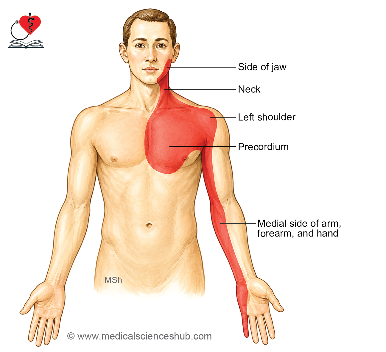
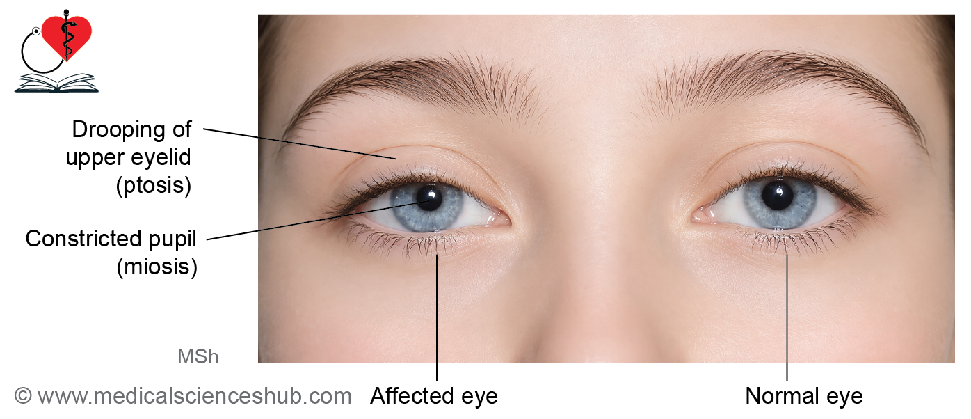
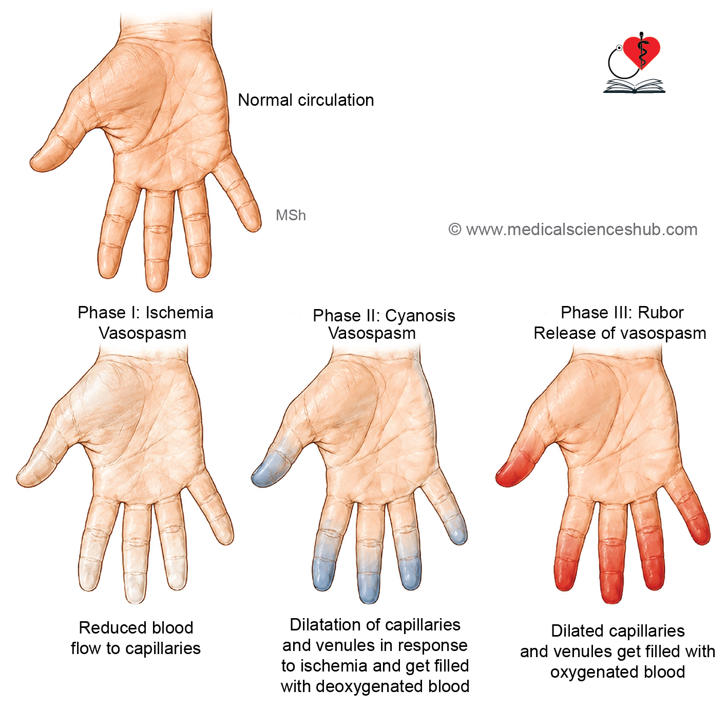
Table 22.1: Major differences between sympathetic and parasympathetic nervous systems
| Feature | Sympathetic Division | Parasympathetic Division |
|---|---|---|
| Alternative Name | Thoracolumbar division | Craniosacral division |
| Origin of Preganglionic Neurons | Lateral horn of spinal cord segments T1-L2 | Brainstem nuclei of cranial nerves III, VII, IX, X and sacral spinal cord segments S2-S4 |
| Preganglionic Fibers | Short | Long |
| Neurotransmitter Released by Preganglionic Fibers | Acetylcholine | Acetylcholine |
| Location of Ganglia | Near the spinal cord (paravertebral and prevertebral ganglia) | Close to or within the target organ |
| Postganglionic Fibers | Long | Short |
| Myelination of Postganglionic Fibers | Unmyelinated | Unmyelinated |
| Neurotransmitter Released by Postganglionic Fibers | Mainly norepinephrine (noradrenaline)* | Acetylcholine |
| Pattern of Response | Diffuse and widespread | Localized and specific |
| Primary Function | Prepares the body for stress, emergency, and increased activity ("fight or flight") | Promotes maintenance, recovery, and energy conservation ("rest and digest") |
Table 22.2: Functional differences in sympathetic and parasympathetic nervous systems
| Organ/System | Sympathetic Effect | Parasympathetic Effect |
|---|---|---|
| Heart | Increases heart rate, force of contraction, and cardiac output | Decreases heart rate, force of contraction, and cardiac output |
| Blood Pressure | Increases blood pressure | Decreases blood pressure |
| Blood Vessels | Vasoconstriction in skin and gastrointestinal tract; vasodilation in skeletal muscle vessels | Little or no direct effect on most blood vessels |
| Respiration | Increases respiratory rate | Decreases respiratory rate |
| Bronchial Smooth Muscle | Bronchodilation | Bronchoconstriction |
| Gastrointestinal Tract | Reduces peristalsis and secretions; contracts sphincters | Enhances peristalsis and secretions; relaxes sphincters |
| Urinary Bladder | Relaxes bladder wall and contracts internal sphincter, promoting urine retention | Contracts bladder wall and relaxes sphincter, facilitating micturition |
| Eye (Pupil) | Pupil dilation (mydriasis) | Pupil constriction (miosis) |
| Lacrimal and Salivary Glands | Produces scanty, thick secretions | Produces abundant, watery secretions |
| Sweat Glands | Stimulates sweating | No significant effect |
| Arrector Pili Muscles | Causes contraction (goosebumps) | No significant effect |
Table 22.3: Secondary autonomic plexuses arising from coeliac plexus
| Plexus | Associated Artery / Location | Distribution and Major Supply |
|---|---|---|
| Phrenic Plexus | Inferior phrenic artery | Supplies the diaphragm and contributes fibers to the suprarenal gland. |
| Hepatic Plexus | Hepatic artery | Distributed to the liver, gallbladder, bile ducts, and adjacent vascular structures. |
| Left Gastric Plexus | Left gastric artery | Supplies the stomach, particularly along the lesser curvature. |
| Splenic Plexus | Splenic artery | Accompanies the splenic artery to supply the spleen and contributes fibers to the pancreas. |
| Suprarenal Plexus | Suprarenal arteries | Innervates the adrenal gland; contains a large proportion of preganglionic sympathetic fibers. |
| Renal Plexus | Renal artery | Supplies the kidneys, upper ureter, and renal blood vessels; receives fibers from aorticorenal, celiac, and thoracolumbar splanchnic nerves. |
| Testicular Plexus | Testicular artery | Distributed to the testis, epididymis, and vas deferens. |
| Ovarian Plexus | Ovarian artery | Supplies the ovary and uterine tube. |
| Superior Mesenteric Plexus | Superior mesenteric artery | Innervates the midgut derivatives and accompanies branches of the superior mesenteric artery. |
| Intermesenteric (Abdominal Aortic) Plexus | Along the abdominal aorta between the superior and inferior mesenteric arteries | Connects the celiac, superior mesenteric, inferior mesenteric, and other periarterial plexuses. |
| Inferior Mesenteric Plexus | Inferior mesenteric artery | Supplies hindgut derivatives through branches accompanying the inferior mesenteric artery. |
Parasympathetic Nervous System
The parasympathetic nervous system is a division of the autonomic nervous system that primarily regulates involuntary activities associated with conservation of energy, glandular secretion, digestion, and maintenance of normal body functions.
Components of the Parasympathetic Nervous System
1. Preganglionic Neurons
- Preganglionic parasympathetic neurons arise from specific nuclei in the brainstem and from the sacral spinal cord.
- The Edinger-Westphal nucleus in the midbrain provides parasympathetic fibers to the sphincter pupillae and ciliary muscle of the eye through the oculomotor nerve.
- The superior salivatory nucleus in the pons gives rise to fibers that supply the lacrimal gland, glands of the nasal cavity, palate, and submandibular and sublingual salivary glands.
- The inferior salivatory nucleus in the medulla sends fibers to the parotid gland through the glossopharyngeal nerve.
- The dorsal motor nucleus of the vagus nerve and the nucleus ambiguus in the medulla provide parasympathetic fibers to the thoracic and abdominal viscera.
- Preganglionic neurons located in the lateral gray matter of the S2-S4 spinal cord segments form the pelvic splanchnic nerves, which supply the distal large intestine, urinary bladder, and reproductive organs.
2. Parasympathetic Ganglia
- Parasympathetic ganglia are situated close to their target organs or within the walls of the organs they supply.
- Because the ganglia are located near the effector organs, preganglionic fibers are relatively long, whereas postganglionic fibers are short.
- The major parasympathetic ganglia in the head include:
- Ciliary ganglion
- Pterygopalatine ganglion
- Submandibular ganglion
- Otic ganglion
- In the thoracic, abdominal, and pelvic cavities, parasympathetic fibers synapse in numerous terminal (intramural) ganglia located within or near the walls of the viscera.
3. Postganglionic Neurons
- Postganglionic neurons arise from parasympathetic ganglia and extend short fibers to the target tissues.
- These fibers innervate smooth muscle, cardiac muscle, and glands, producing responses such as pupillary constriction, glandular secretion, increased gastrointestinal activity, and contraction of the urinary bladder.
Craniosacral Outflow
- The parasympathetic nervous system is also known as the craniosacral outflow because its preganglionic neurons originate from:
- Brainstem nuclei associated with cranial nerves III, VII, IX, and X.
- Sacral spinal cord segments S2-S4.
Ciliary Ganglion
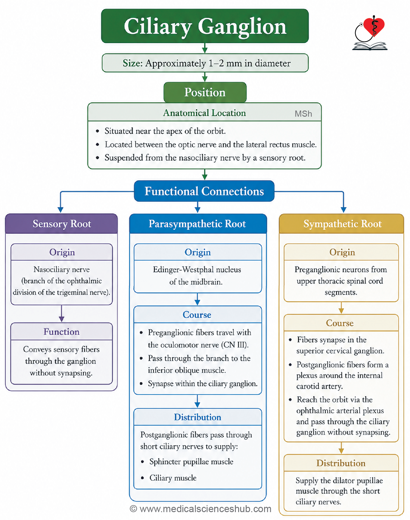
- The ciliary ganglion is a small parasympathetic ganglion located within the orbit.
- It measures approximately 1–2 mm in diameter.
- The ganglion is associated functionally with the oculomotor nerve (cranial nerve III), although it is anatomically connected to the nasociliary nerve, a branch of the ophthalmic division of the trigeminal nerve (V1).
Location and Relations
- The ciliary ganglion is situated in the posterior part of the orbit.
- It lies between the optic nerve and the lateral rectus muscle.
- The ganglion is positioned near the apex of the orbit.
- It is connected to the nasociliary nerve by a short sensory branch.
- Medially, it is related to the optic nerve.
- Laterally, it is related to the lateral rectus muscle.
Roots of the Ciliary Ganglion
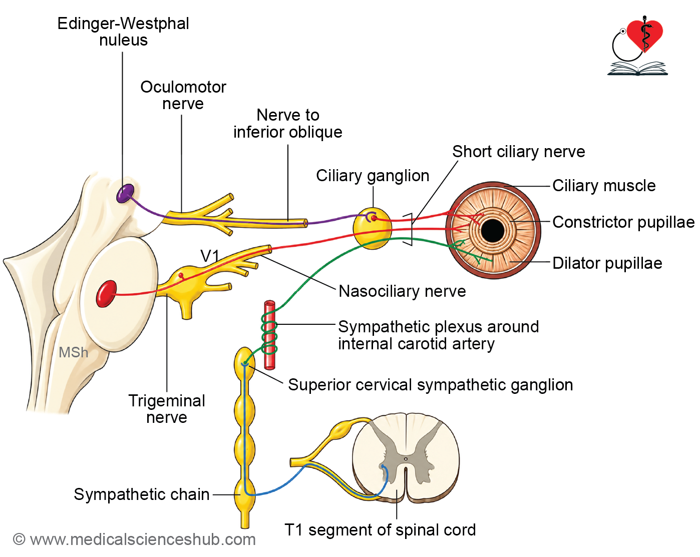
The ciliary ganglion receives three roots: parasympathetic, sympathetic, and sensory.
1. Parasympathetic (Motor) Root
- This root carries preganglionic parasympathetic fibers from the Edinger-Westphal nucleus in the midbrain.
- The fibers travel through the oculomotor nerve and enter its inferior division within the orbit.
- They reach the ciliary ganglion through the nerve to the inferior oblique muscle.
- These fibers synapse within the ciliary ganglion.
- Postganglionic fibers leave the ganglion through the short ciliary nerves.
- They supply the sphincter pupillae muscle, producing pupillary constriction.
- They also innervate the ciliary muscle, which is responsible for accommodation of the lens.
2. Sympathetic Root
- The sympathetic fibers originate from neurons in the upper thoracic spinal cord segments.
- They synapse in the superior cervical ganglion.
- Postganglionic fibers form a plexus around the internal carotid artery and reach the orbit through the ophthalmic artery.
- These fibers pass through the ciliary ganglion without synapsing.
- They enter the short ciliary nerves and supply blood vessels of the eyeball.
- Some sympathetic fibers also contribute to the innervation of the dilator pupillae muscle.
3. Sensory Root
- The sensory root is derived from the nasociliary nerve.
- Sensory fibers pass through the ciliary ganglion without synapsing.
- These fibers convey sensory information from the eyeball to the trigeminal nerve.
Branches
- The ciliary ganglion gives rise to 8–10 short ciliary nerves.
- These nerves pierce the sclera around the optic nerve and enter the eyeball.
- The short ciliary nerves carry:
- Postganglionic parasympathetic fibers to the sphincter pupillae and ciliary muscles.
- Postganglionic sympathetic fibers to ocular blood vessels and the dilator pupillae muscle.
- Sensory fibers from the eyeball.
Functions
- The ciliary ganglion participates in the parasympathetic control of pupillary constriction.
- It is involved in accommodation of the lens for near vision.
- It provides a pathway for sympathetic and sensory fibers entering and leaving the eyeball.
CLINICAL NEUROANATOMY
- A ciliary nerve block may be used during ophthalmic procedures to reduce pain and ocular reflexes.
- Interruption of parasympathetic fibers passing through the ganglion can result in pupillary dilation and loss of accommodation.
Otic Ganglion
- The otic ganglion is a small parasympathetic ganglion located in the infratemporal fossa.
- It is oval in shape and measures approximately 2–3 mm in diameter.
- Functionally, it is associated with the glossopharyngeal nerve (cranial nerve IX).
- Anatomically, it is closely related to the mandibular division of the trigeminal nerve (V3).
Location and Relations
- The otic ganglion lies in the infratemporal fossa, just below the foramen ovale.
- It is situated on the medial surface of the mandibular nerve.
- The ganglion is located close to the origin of the nerve to the medial pterygoid muscle.
- It lies medial to the mandibular nerve and lateral to the tensor veli palatini muscle.
Roots of the Otic Ganglion
The otic ganglion receives parasympathetic, sympathetic, sensory, and motor roots.
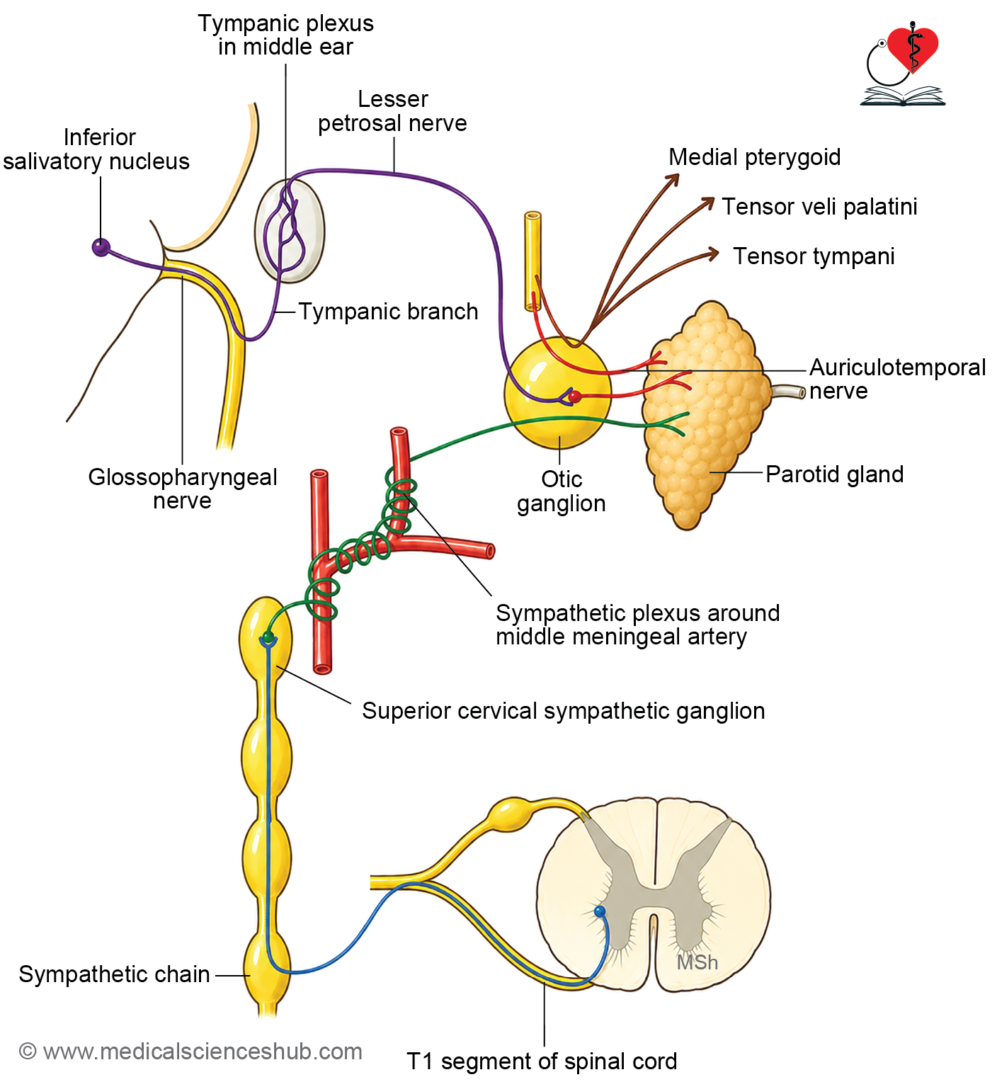
1. Parasympathetic (Secretomotor) Root
- Preganglionic parasympathetic fibers arise from the inferior salivatory nucleus in the medulla.
- These fibers travel through the glossopharyngeal nerve and its tympanic branch.
- The tympanic branch contributes to the tympanic plexus in the middle ear.
- Fibers then continue as the lesser petrosal nerve and reach the otic ganglion.
- Preganglionic fibers synapse within the otic ganglion.
- Postganglionic fibers leave the ganglion and pass through the auriculotemporal nerve.
- These fibers provide secretomotor innervation to the parotid gland and stimulate salivary secretion.
2. Sympathetic Root
- Sympathetic fibers originate from neurons in the upper thoracic spinal cord.
- After synapsing in the superior cervical ganglion, postganglionic fibers form a plexus around the middle meningeal artery.
- These fibers reach the otic ganglion but pass through it without synapsing.
- Sympathetic fibers accompany branches of the external carotid artery to the parotid gland.
- Their primary function is vasomotor control of blood vessels.
3. Sensory Root
- Sensory fibers are derived from the auriculotemporal nerve.
- These fibers pass through the ganglion without relay.
- They carry general sensory information from structures supplied by the auriculotemporal nerve.
4. Motor Root
- Motor fibers from the nerve to the medial pterygoid pass through the ganglion without synapsing.
- These fibers supply the tensor veli palatini and tensor tympani muscles.
Branches and Distribution
- Fibers emerging from the otic ganglion reach the parotid gland through the auriculotemporal nerve.
- The ganglion serves as a relay station for parasympathetic fibers destined for the parotid gland.
Functional Components
- Parasympathetic fibers: Secretomotor fibers to the parotid gland.
- Sympathetic fibers: Vasomotor fibers to blood vessels.
- Sensory fibers: General sensory fibers passing through the ganglion.
- Motor fibers: Fibers to the tensor veli palatini and tensor tympani muscles.
CLINICAL NEUROANATOMY
Frey Syndrome (Gustatory Sweating)
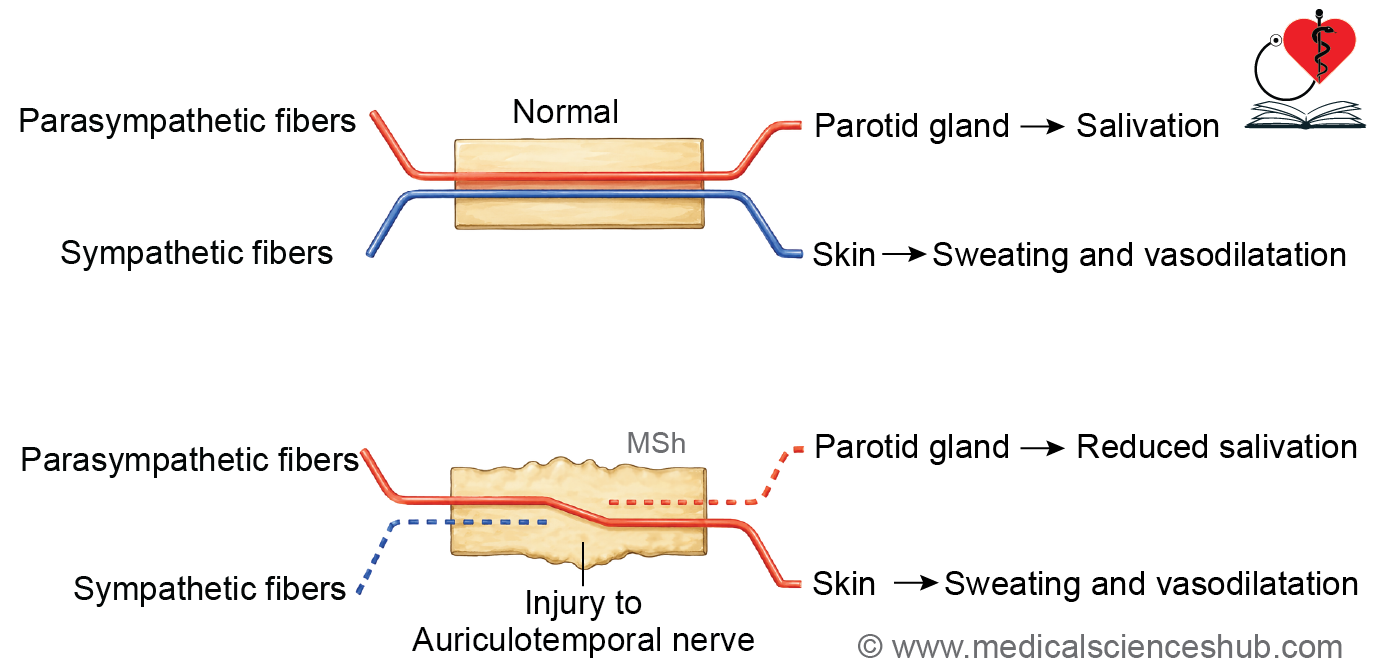
- Frey syndrome is characterized by sweating and flushing over the parotid region during eating or even while thinking about food.
- It commonly occurs after injury, inflammation, or surgery involving the parotid gland.
- Damage to the auriculotemporal nerve may lead to abnormal regeneration of parasympathetic fibers.
- During healing, some secretomotor fibers intended for the parotid gland may become connected to sweat glands and cutaneous blood vessels of the overlying skin.
- As a result, stimulation that normally produces salivation causes sweating and flushing in the affected area.
- This condition is also known as gustatory sweating syndrome or auriculotemporal nerve syndrome.
Submandibular Ganglion
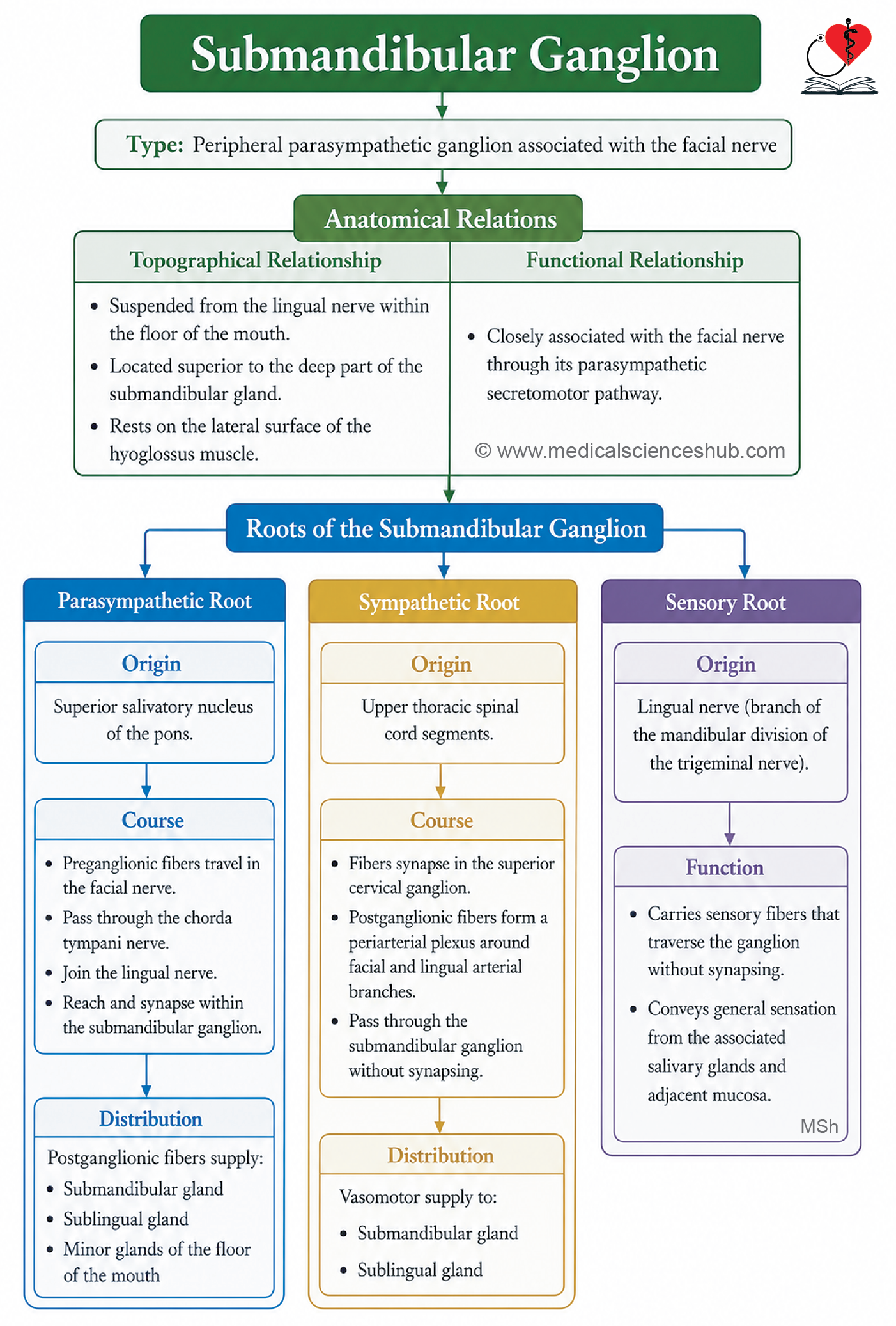
- The submandibular ganglion is a peripheral parasympathetic ganglion located in the floor of the oral cavity.
- It is one of the major parasympathetic ganglia of the head and neck.
- Functionally, it is associated with the facial nerve (cranial nerve VII).
- Anatomically, it is suspended from the lingual nerve, a branch of the mandibular division of the trigeminal nerve (V3).
Location and Relations
- The submandibular ganglion lies above the deep part of the submandibular gland.
- It is situated on the lateral surface of the hyoglossus muscle.
- The ganglion is connected to the lingual nerve by short ganglionic branches.
- It occupies a position between the lingual nerve superiorly and the deep part of the submandibular gland inferiorly.
Roots of the Submandibular Ganglion
The submandibular ganglion receives parasympathetic, sympathetic, and sensory roots.
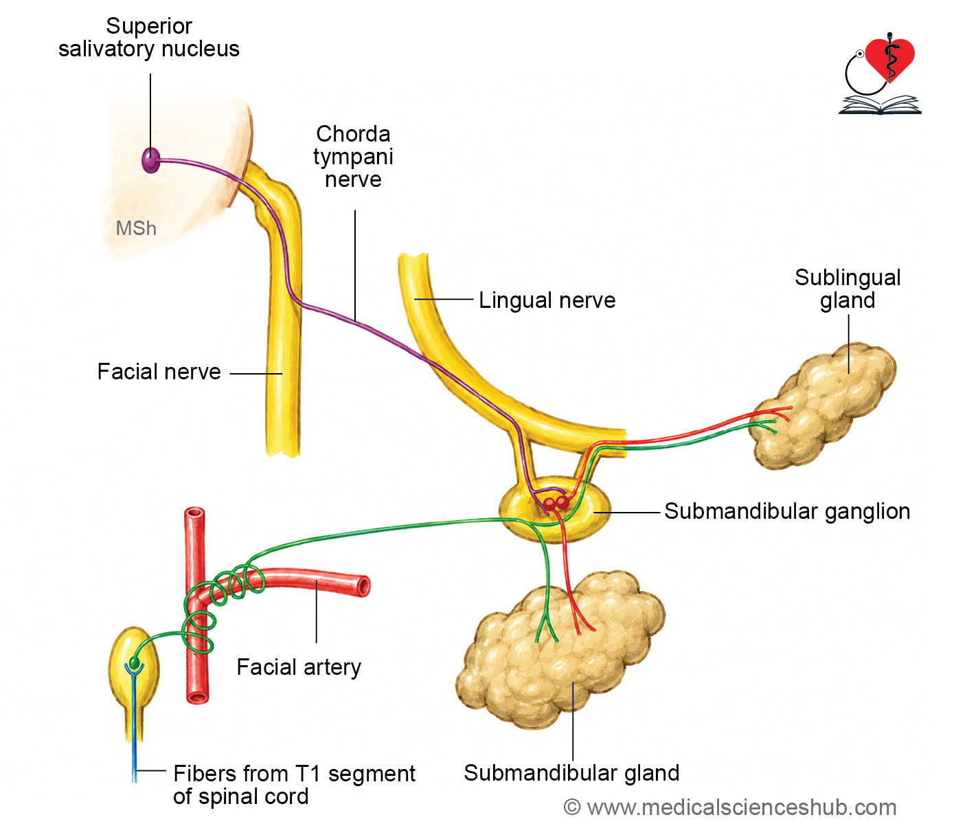
1. Parasympathetic (Secretomotor) Root
- Preganglionic parasympathetic fibers arise from the superior salivatory nucleus in the pons.
- These fibers travel through the facial nerve and enter its branch, the chorda tympani.
- The chorda tympani joins the lingual nerve in the infratemporal fossa.
- Preganglionic fibers are carried by the lingual nerve to the submandibular ganglion.
- These fibers synapse within the ganglion.
- Postganglionic fibers pass directly to the submandibular gland and stimulate salivary secretion.
- Some postganglionic fibers re-enter the lingual nerve and reach the sublingual gland and minor glands of the floor of the mouth.
2. Sympathetic Root
- Sympathetic fibers originate from neurons in the upper thoracic spinal cord.
- After synapsing in the superior cervical ganglion, postganglionic fibers form plexuses around branches of the external carotid artery, particularly the facial artery.
- These fibers reach the submandibular ganglion but pass through it without synapsing.
- Sympathetic fibers supply blood vessels of the submandibular and sublingual glands and regulate vascular tone.
3. Sensory Root
- Sensory fibers are derived from the lingual nerve.
- These fibers pass through the ganglion without relay.
- They carry general sensory information from the glands and adjacent mucosal structures.
Branches and Distribution
- Branches arising from the ganglion supply the submandibular gland directly.
- Additional fibers pass through the lingual nerve to reach the sublingual gland.
- The ganglion also contributes secretomotor fibers to minor salivary glands located in the floor of the mouth.
Functional Components
- Parasympathetic fibers: Stimulate secretion from the submandibular and sublingual salivary glands.
- Sympathetic fibers: Provide vasomotor innervation to glandular blood vessels.
- Sensory fibers: Convey general sensory information through the lingual nerve.
Functions
- The submandibular ganglion serves as the relay station for parasympathetic fibers destined for the submandibular and sublingual glands.
- It plays an important role in regulating salivary secretion.
- Parasympathetic stimulation produces abundant watery saliva, whereas sympathetic stimulation primarily influences vascular supply and glandular blood flow.
CLINICAL NEUROANATOMY
- Injury to the chorda tympani or lingual nerve may reduce salivary secretion from the submandibular and sublingual glands.
- Surgical procedures involving the submandibular region may damage ganglionic connections, leading to alterations in salivary gland function.
- Disorders affecting parasympathetic innervation may result in xerostomia (dry mouth) and associated difficulties in speech, mastication, and swallowing.
Pterygopalatine Ganglion
- The pterygopalatine ganglion is the largest parasympathetic ganglion in the head.
- It is also known as the sphenopalatine ganglion, nasal ganglion, or Meckel’s ganglion.
- Functionally, it is associated with the facial nerve (cranial nerve VII).
- It provides parasympathetic innervation to the lacrimal gland and to the glands of the nasal cavity, palate, and nasopharynx.
- Anatomically, it is suspended from the maxillary nerve (V2), a division of the trigeminal nerve.
Location and Relations
- The ganglion is situated within the pterygopalatine fossa.
- It lies inferior to the maxillary nerve.
- It is located anterior to the pterygoid canal and lateral to the sphenopalatine foramen.
- The ganglion is connected to the maxillary nerve by short ganglionic branches.
Roots of the Pterygopalatine Ganglion
The ganglion receives parasympathetic, sympathetic, and sensory roots.
1. Parasympathetic (Secretomotor) Root
- Preganglionic parasympathetic fibers arise from the superior salivatory nucleus in the pons.
- These fibers travel through the facial nerve and enter the greater petrosal nerve.
- The greater petrosal nerve joins the deep petrosal nerve to form the nerve of the pterygoid canal (Vidian nerve).
- Preganglionic parasympathetic fibers synapse within the pterygopalatine ganglion.
- Postganglionic fibers are distributed to:
- The lacrimal gland.
- Glands of the nasal cavity.
- Glands of the palate.
- Glands of the nasopharynx.
2. Sympathetic Root
- Sympathetic fibers originate from neurons in the upper thoracic spinal cord.
- After synapsing in the superior cervical ganglion, postganglionic fibers form the internal carotid plexus.
- These fibers continue as the deep petrosal nerve and reach the ganglion through the nerve of the pterygoid canal.
- Sympathetic fibers pass through the ganglion without synapsing.
- They supply blood vessels of the nasal cavity, palate, nasopharynx, and lacrimal gland, producing vasomotor effects.
3. Sensory Root
- Sensory fibers are derived from the maxillary nerve (V2).
- These fibers pass through the ganglion without relay.
- They convey general sensory information from areas supplied by branches associated with the ganglion.
Branches and Distribution
The pterygopalatine ganglion gives rise to several branches that distribute parasympathetic, sympathetic, and sensory fibers to surrounding structures.
Orbital Branches
- These branches enter the orbit through the inferior orbital fissure.
- They supply the periosteum of the orbit and contribute to innervation of structures within the posterior orbit.
Nasal Branches
- Posterior superior lateral nasal branches supply the lateral wall of the nasal cavity.
- Posterior superior medial nasal branches supply the nasal septum.
- These branches provide secretomotor fibers to the mucous glands of the nasal cavity.
Nasopalatine Nerve
- The nasopalatine nerve passes through the sphenopalatine foramen and crosses the roof of the nasal cavity.
- It descends through the incisive canal to reach the anterior part of the hard palate.
- It supplies the mucosa of the anterior hard palate and adjacent gingiva.
Palatine Branches
- The greater palatine nerve descends through the greater palatine canal and supplies the mucosa and glands of the hard palate.
- The lesser palatine nerves pass through the lesser palatine foramina and supply the soft palate, uvula, and palatine tonsillar region.
Pharyngeal Branch
- The pharyngeal branch passes through the palatovaginal canal.
- It supplies the mucosa and glands of the nasopharynx, particularly the region near the opening of the auditory tube.
Lacrimal Pathway
- Postganglionic parasympathetic fibers destined for the lacrimal gland travel through the zygomatic nerve (branch of V2).
- These fibers then communicate with the lacrimal nerve (branch of the ophthalmic nerve, V1) to reach the lacrimal gland.
- They stimulate tear secretion.
Functions
- The pterygopalatine ganglion serves as the relay station for parasympathetic fibers supplying the lacrimal gland.
- It provides secretomotor innervation to glands of the nasal cavity, palate, and nasopharynx.
- It transmits sympathetic vasomotor fibers to blood vessels in these regions.
- It acts as a distribution center for sensory fibers associated with the maxillary nerve.
CLINICAL NEUROANATOMY
- Irritation of the pterygopalatine ganglion may produce facial pain, lacrimation, and nasal secretion.
- A pterygopalatine ganglion block may be used in the management of certain headache disorders and facial pain syndromes.
- Disorders affecting the ganglion can alter tear production and glandular secretion within the nasal cavity and palate.
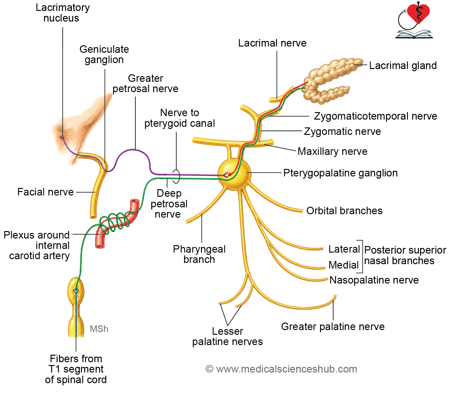
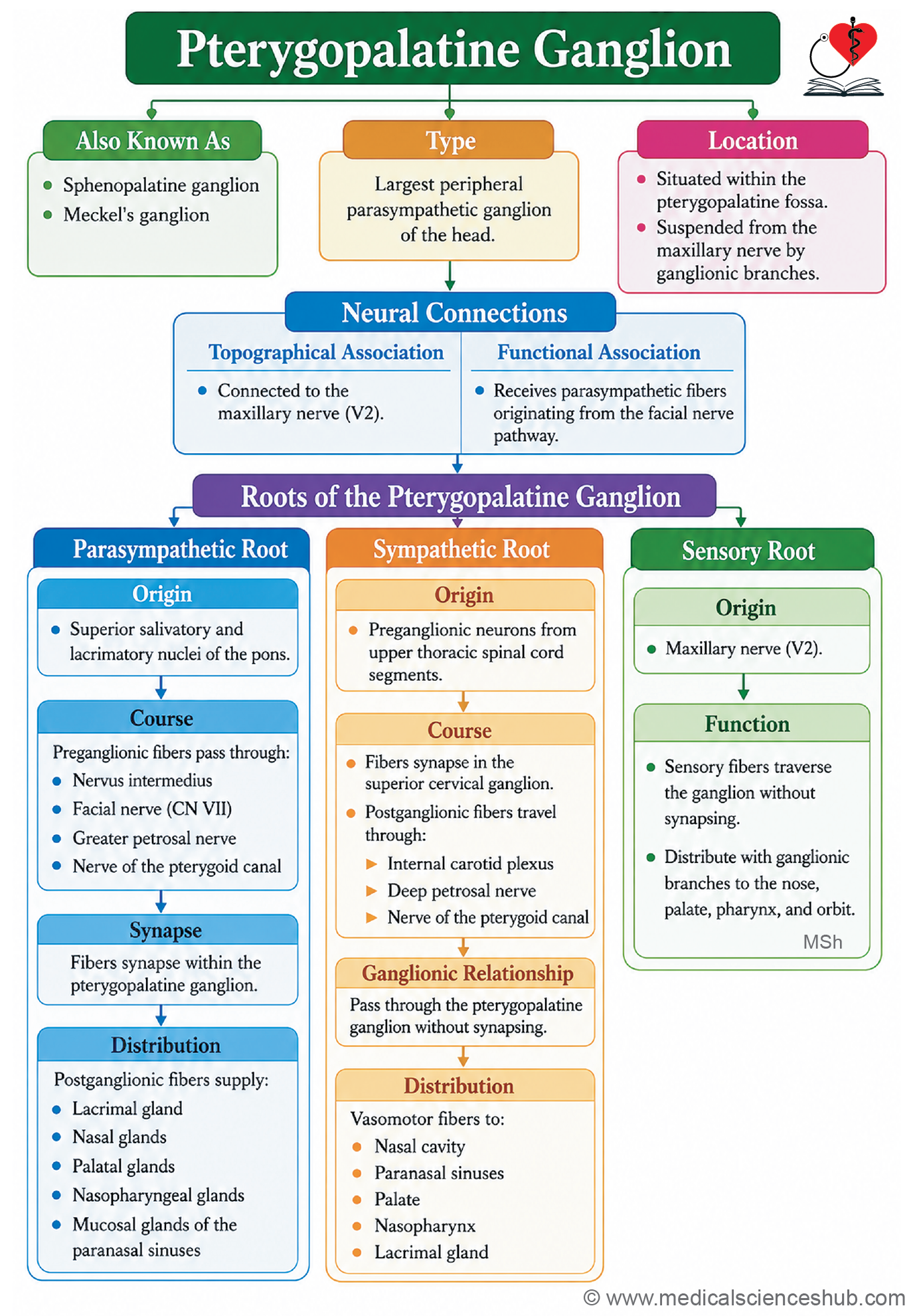
Table 22.4: Branches of pterygopalatine ganglion
| Branch | Distribution / Structures Supplied |
|---|---|
| Orbital Branches | Periosteum of the orbit, orbitalis muscle, and sphenoidal and ethmoidal air sinuses |
| Palatine Branches | Hard palate, soft palate, and palatine glands |
| Nasal Branches | Mucosa of the nasal cavity and paranasal sinuses |
| Posterior Superior Lateral Nasal Branches | Lateral wall of the nasal cavity, including superior and middle nasal conchae |
| Posterior Superior Medial Nasal Branches | Nasal septum; one large branch continues as the nasopalatine nerve |
| Nasopalatine Nerve | Anterior part of the hard palate and adjacent gingiva |
| Pharyngeal Branch | Mucosa of the nasopharynx, especially behind the auditory tube |
| Lacrimal Branch (Communicating Fibers) | Secretomotor fibers to the lacrimal gland via the zygomatic and lacrimal nerves |
Visceral Sensation
General Visceral Afferent Fibers
- General visceral afferent (GVA) fibers are sensory fibers that carry signals from internal organs to the central nervous system, traveling alongside autonomic nerve fibers.
- The cell bodies of these neurons reside in the sensory ganglia of cranial nerves or in the dorsal root ganglia of spinal nerves.
Types of Sensations Carried by GVA Fibers
- GVA fibers transmit a range of physiological and pathological signals, including stretch and distension of hollow organs, fluctuations in blood pressure and blood chemistry, gastric and intestinal fullness, urinary bladder and rectal distension, and sensations such as hunger, thirst, and nausea.
Characteristics of Visceral Sensation
- Visceral sensations are poorly localized compared to somatic sensations, largely because visceral afferent innervation is sparse and widely distributed.
- The majority of visceral sensory activity does not reach conscious awareness; pain is the most frequently perceived visceral sensation, and even this tends to be diffuse and difficult to pinpoint.
H4: Visceral Pain
Causes
- Visceral pain arises from distension or spasm of hollow organs, ischemia of visceral tissue, stretching of organ capsules or serous membranes, and inflammatory processes involving internal organs.
Characteristics
- The pain is typically described as dull, aching, or cramping, and is poorly localized.
- It is frequently accompanied by autonomic responses such as diaphoresis (sweating), nausea, vomiting, and alterations in heart rate, reflecting simultaneous activation of autonomic pathways.
Referred Pain
Mechanism
- Referred pain is pain that is consciously perceived at a body surface location that is anatomically distant from the organ actually generating the painful stimulus.
- This occurs because visceral afferent fibers and somatic afferent fibers from the same spinal segment converge onto the same second-order neurons in the dorsal horn of the spinal cord.
- The cerebral cortex, being more experienced in processing somatic signals, misinterprets the origin of the signal as coming from the dermatome corresponding to that spinal segment rather than from the internal organ.
Common Referred Pain Patterns
- Cardiac pain is referred to the left side of the chest, the left shoulder, and the medial aspect of the left upper limb.
- Gallbladder pain is referred to the right shoulder and the right scapular region.
- Splenic pain is referred to the left shoulder, a clinical sign known as Kehr’s sign.
- Diaphragmatic irritation produces shoulder pain on the corresponding side, because the phrenic nerve, which supplies the diaphragm, originates from cervical segments C3–C5, which also supply the shoulder region.
- In the early stage of appendicitis, pain is referred to the periumbilical region, as the appendix shares its nerve supply with the T10 spinal segment; as inflammation progresses and involves the parietal peritoneum, pain localizes to the right iliac fossa.
- Testicular and ovarian pain is referred to the periumbilical and lower abdominal regions, reflecting their shared segmental innervation from T10–T11.
- Ureteric pain is referred to the loin, groin, scrotum, or labium majus, depending on the location of the obstruction along the ureter.
- Gastric pain is referred to the epigastric region.
CLINICAL NEUROANATOMY
- Understanding referred pain patterns is essential in clinical practice, as the site of perceived pain on the body surface can provide reliable diagnostic clues about the involved viscus.
- Recognizing these patterns is a fundamental component of clinical examination and helps guide differential diagnosis.
Innervations of Bladder
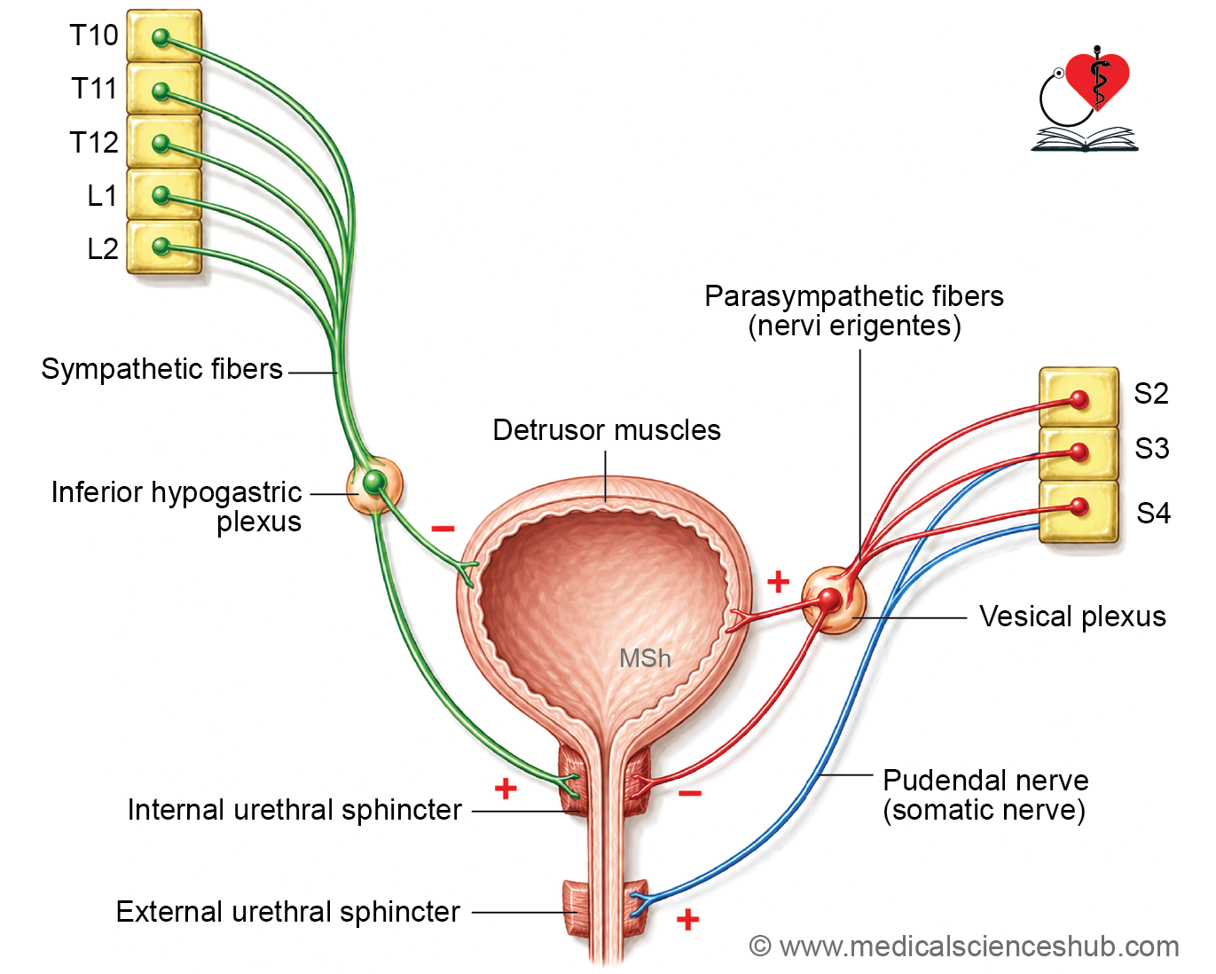
- The urinary bladder receives its nerve supply through the vesical nerve plexus, which lies on the lateral aspects of the rectum and the base of the bladder, and carries both motor and sensory fibers.
Efferent (Motor) Innervation
- The motor supply to the bladder consists of three components: parasympathetic, sympathetic, and somatic fibers.
- Parasympathetic fibers originate from the lateral horn cells of spinal segments S2, S3, and S4, and reach the vesical plexus via the pelvic splanchnic nerves (nervi erigentes). These fibers are excitatory to the detrusor muscle, stimulating bladder contraction during micturition. They do not supply the preprostatic sphincter.
- Sympathetic fibers arise from spinal segments T10–T12 and L1–L2. They exert an inhibitory effect on the detrusor muscle, thereby promoting urine storage, and are simultaneously excitatory to the preprostatic (internal urethral) sphincter, keeping it contracted during bladder filling.
- Somatic fibers are derived from the pudendal nerve (S2, S3, S4) and supply the external urethral sphincter (sphincter urethrae), which is the principal mechanism of voluntary control over micturition.
Afferent (Sensory) Innervation
- Sensory fibers from the bladder wall transmit two distinct types of signals: sensations of bladder distension and smooth muscle spasm are carried predominantly by parasympathetic fibers and partly by sympathetic fibers.
- Pain signals from the bladder travel in the anterolateral white column of the spinal cord, whereas signals conveying the sensation of bladder filling travel in the posterior white column (via the tract of Goll).
- Because these two types of signals travel in anatomically separate pathways, bilateral anterolateral cordotomy (sectioning of the anterolateral white columns) can be performed to abolish bladder pain while preserving the normal sensation of filling and the ability to micturate.
Neuronal Control of Micturition
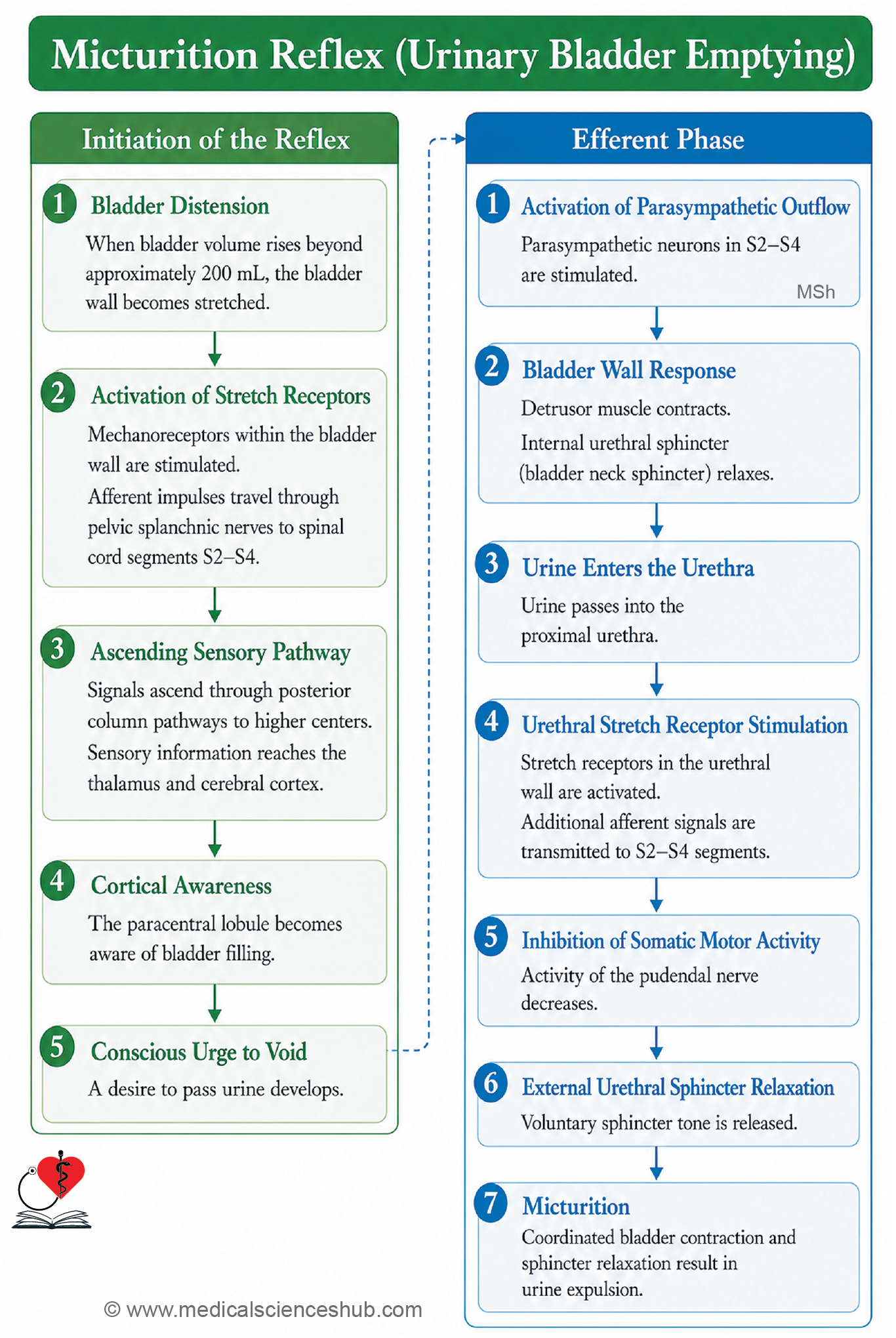
- Sensation of filling: When bladder volume exceeds approximately 220 ml, the bladder wall stretches, activating mechanoreceptors that send afferent signals mainly through the parasympathetic fibers of S2, S3, and S4.
- Conscious perception of fullness: These afferent signals ascend in the ipsilateral tract of Goll (fasciculus gracilis) to the nucleus gracilis, then continue via the internal arcuate fibers, medial lemniscus, and thalamus, ultimately reaching the paracentral lobule of the cerebral cortex, where fullness is consciously perceived and the desire to void is generated.
- Voluntary retention of urine: The detrusor center within the paracentral lobule suppresses the sacral parasympathetic neurons (lateral horn cells of S2–S4), maintaining continence until an appropriate time and place for micturition is available.
- Voiding (micturition): When voluntary inhibition from the paracentral lobule is withdrawn, parasympathetic fibers (S2–S4) are activated, producing detrusor contraction and simultaneous relaxation of the preprostatic sphincter. As urine enters the proximal urethra, urethral stretch receptors are activated and send additional parasympathetic signals that reflexively inhibit the pudendal nerve, resulting in relaxation of the external urethral sphincter and completion of micturition.
CLINICAL NEUROANATOMY
- Automatic (reflex) bladder results from a spinal cord lesion above the S2 segment, which disconnects the sacral micturition centers from higher cortical control. Voluntary initiation and control of micturition are lost. However, when the bladder fills sufficiently, stretch receptors trigger a spinal reflex arc that contracts the detrusor and produces involuntary voiding. This reflex pattern is the normal mode of bladder emptying in early infancy.
- Atonic bladder results from damage to the sensory (afferent) fibers supplying the bladder, most commonly in conditions such as tabes dorsalis. Because afferent signals from stretch receptors cannot reach the spinal cord, the reflex arc is broken; the bladder becomes progressively overdistended, thin-walled, and hypotonic, leading to overflow incontinence.
- Autonomous bladder occurs when the bladder loses all neural connections, as may follow damage to spinal segments S1–S3 or extensive pelvic surgery disrupting the pelvic plexus. Both voluntary and reflex control are abolished; the bladder becomes flaccid with increased capacity, and urine is continuously dribbled. Bladder emptying can only occur passively through overfilling or manual compression to raise intra-abdominal pressure, and long-term catheterization is typically required.
- Uninhibited bladder results from upper motor neuron lesions involving the brain, such as stroke or intracranial tumors, which impair the cortical inhibitory influence over the detrusor. The result is sudden, involuntary, and uncontrollable bladder contraction, a condition clinically referred to as urge incontinence.
Important Questions
- Write a short note on the ciliary ganglion.
- Write a short note on otic ganglion.
- Write a short note on submandibular ganglion.
- Write a short note on nerve supply of urinary bladder.
- Write a short note on micturition.
📝 Test Your Knowledge – Practice MCQs
Attempt the chapter MCQ quiz and assess your understanding of key concepts.
Click to see: Medical Disclaimer and Copyright Notice