
Competencies
- AN7.5 Describe principles of sensory and motor innervation of muscles.
Introduction
- Explore the vital roles of receptors and effectors in the nervous system. Receptors detect changes in the internal and external environment, while effectors respond to signals, enabling the body to sense, process, and act effectively.
Receptors
- Sensory receptors are specialized neuronal structures that detect changes in the internal or external environment.
- They transform stimuli into electrical signals (nerve impulses) that can be interpreted by the nervous system.
Classification of Receptors
Receptors can be grouped using different criteria:
1. Functional Classification
- Exteroceptors respond to stimuli from the external environment.
- Proprioceptors provide information about body position and movement.
- Interoceptors detect changes within internal organs.
2. Based on Type of Stimulus
- Mechanoreceptors respond to mechanical forces such as touch or pressure.
- Chemoreceptors detect chemical changes in the body.
- Other types exist depending on the nature of the activating stimulus.
3. Anatomical Classification
- Encapsulated receptors are surrounded by a connective tissue capsule.
- Nonencapsulated receptors lack a capsule and are relatively simple in structure.
4. Based on Location
- Cutaneous receptors are located in the skin.
- Muscle receptors are present within muscles.
- Tendon receptors are found in tendons.
- Joint receptors are located in joints.
Anatomical Types of Receptors
A. Encapsulated Receptors
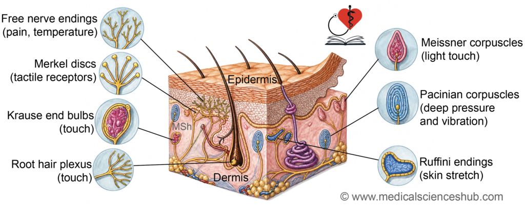
- Meissner’s corpuscles
- Pacinian corpuscles
- End bulbs of Krause
- Ruffini’s corpuscles
B. Nonencapsulated Receptors
- Free nerve endings
- Root hair plexus (peritrichial endings)
- Merkel discs
Table 3.1: Receptors and their activating stimulus
| Receptor Type | Primary Stimulus Detected |
|---|---|
| Meissner’s corpuscles | Detect gentle touch and low-frequency vibrations |
| Pacinian corpuscles | Respond to deep pressure and high-frequency vibrations |
| End bulbs of Krause | Sense cold sensations |
| Ruffini’s corpuscles | Detect skin stretch, continuous pressure, and rotational (torque) forces |
| Free nerve endings | Perceive pain, temperature (both hot and cold), and crude touch |
| Merkel discs (tactile discs) | Responsible for fine touch, sustained pressure, and texture recognition |
| Root hair plexus (peritrichial endings) | Detect movement of hair and light touch |
Major Receptors
Encapsulated Receptors
- Meissner’s corpuscles are located in the dermal papillae of hairless skin.
- They consist of nerve endings surrounded by vertically arranged supporting cells.
- In routine staining, they appear like a loosely coiled structure.
- They are specialized for light touch perception.
- Pacinian corpuscles are large, oval receptors found in the deep dermis and subcutaneous tissue.
- They show a characteristic onion-like lamellar pattern in cross-section.
- Concentric layers of flattened cells surround a central nerve ending.
- They detect deep pressure and vibration, especially high-frequency stimuli.
- End bulbs of Krause are small, rounded receptors acting as cold-sensitive thermoreceptors.
- They are present in areas such as the conjunctiva, mucosa of lips and tongue, genital regions, and joint linings.
- Ruffini’s corpuscles are elongated receptors with branching nerve endings enclosed in a capsule.
- They respond to skin stretch and sustained mechanical forces, including torque.
Nonencapsulated Receptors
- Free nerve endings extend into the superficial layers of the epidermis and around hair follicles.
- They detect pain, temperature, crude touch, and hair movement.
- These endings lack a well-defined capsule and are minimally supported.
- Merkel discs are located in the basal layer of the epidermis and near hair follicles.
- They function as mechanoreceptors for fine touch and pressure discrimination.
- Root hair plexus (peritrichial plexus) surrounds the outer root sheath of hair follicles.
- It is activated by movement of hair, contributing to light touch sensation.
Additional Functional Points
- Nonmyelinated autonomic fibers supply glands, cardiac muscle, and smooth muscle, regulating involuntary functions.
- Nonencapsulated mechanoreceptors (e.g., free nerve endings and Merkel discs) are generally slowly adapting, allowing sustained response to stimuli.
- Nociceptors respond to harmful or painful stimuli and typically show rapid adaptation.
Types of Sensory Input
- Exteroceptors detect stimuli from the external environment and are located near the body surface.
- Proprioceptors provide information about body position and movement.
- Interoceptors monitor internal conditions within the body.
Notes
- Pacinian corpuscles are the largest mechanoreceptors in the body.
- Muscle spindle fibers transmit sensory input to pseudounipolar neurons of dorsal root ganglia, contributing to proprioception.
Motor Innervation of Skeletal Muscle
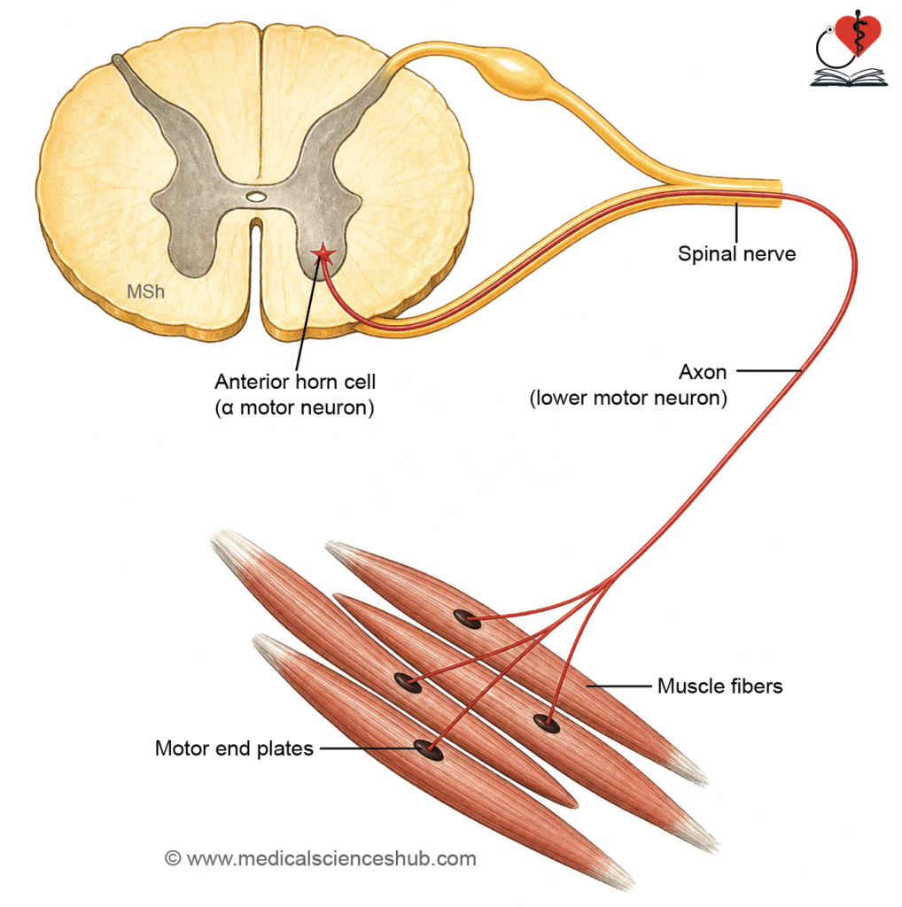
- Motor innervation allows skeletal muscles to contract by transmitting impulses from spinal motor neurons to muscle fibers.This system enables both fine control and powerful movements through organized neural connections.
Motor Nerve Fibers and Motor Unit
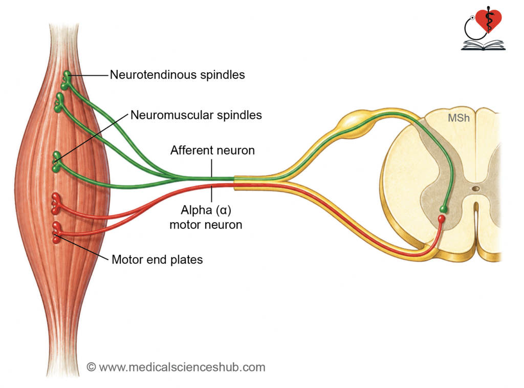
- Skeletal muscle fibers are supplied by alpha efferent fibers arising from multipolar neurons in the anterior horn of the spinal cord.
- These fibers are large and myelinated, ensuring rapid impulse conduction.
- A motor unit consists of a single motor neuron and all the muscle fibers it innervates.
- Small motor units are found in muscles requiring precise movements (e.g., eye muscles).
- Large motor units are present in muscles responsible for posture and gross movements.
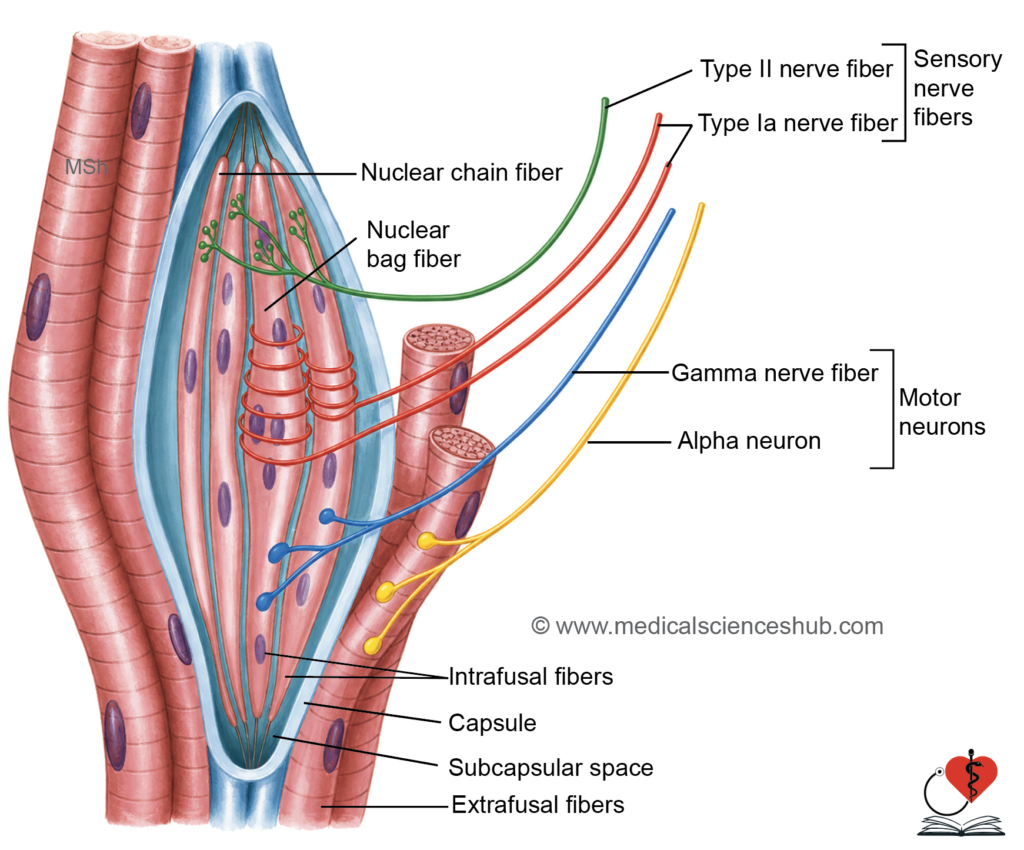
- Muscle spindles are specialized sensory receptors located within skeletal muscles.
- They detect changes in muscle length and contribute to proprioception.
- They receive gamma efferent fibers from gamma motor neurons of the spinal cord.
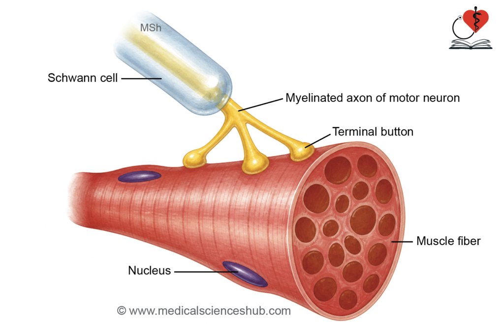
- The neuromuscular junction is the site where a motor neuron communicates with a muscle fiber.
- At this junction, the axon loses its myelin sheath and divides into multiple terminal branches.
- Each branch ends in an expanded terminal bouton, forming contact with the muscle membrane (sarcolemma).
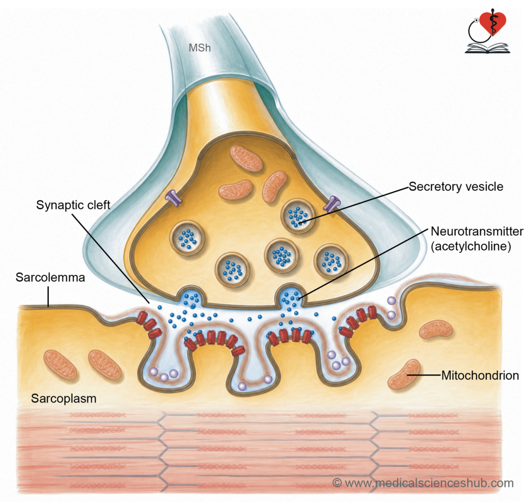
- The muscle membrane at this site forms a specialized region called the motor end plate, which has multiple folds to increase surface area.
- These folds contain numerous acetylcholine receptors.
- A narrow synaptic cleft separates the nerve terminal and muscle fiber.
- The neurotransmitter acetylcholine is released from vesicles in the nerve terminal and diffuses across this gap.
- The enzyme acetylcholinesterase rapidly breaks down acetylcholine, ensuring controlled and brief muscle stimulation.
Muscle Spindle
- Skeletal muscle fibers are supplied by alpha efferent fibers arising from multipolar neurons in the anterior horn of the spinal cord.
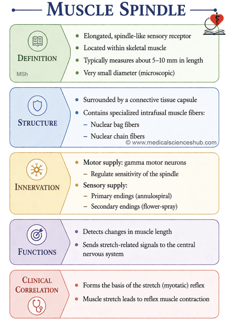
- The muscle spindle is a specialized spindle-shaped sensory receptor present within skeletal muscles.
- It measures approximately 5–10 mm in length and about 100 μm in diameter.
Structure
- The spindle is enclosed by a connective tissue capsule.
- It contains intrafusal muscle fibers, sensory nerve endings, and gamma motor fibers.
Types of Intrafusal Fibers
- Nuclear bag fibers:
- Possess multiple nuclei clustered centrally.
- They are longer and may extend beyond the capsule.
- Nuclear chain fibers:
- Have nuclei arranged in a linear sequence.
Nerve Supply
- The spindle receives motor innervation from gamma motor neurons.
- Sensory input is carried by afferent fibers:
- Type Ia fibers form annulospiral endings around the central region of intrafusal fibers.
- Type II fibers form flower-like endings near the peripheral regions.
- Extrafusal muscle fibers (main contractile fibers) are supplied by alpha motor neurons.
Functions
- The muscle spindle detects changes in muscle length (stretch).
- It provides continuous feedback to the CNS for regulation of muscle tone and coordination.
- It plays a key role in the stretch reflex, which prevents excessive muscle stretching.
- Activation of sensory fibers during stretch helps adjust motor neuron activity for smooth and controlled movement.
Golgi Tendon Organ
Location and Structure
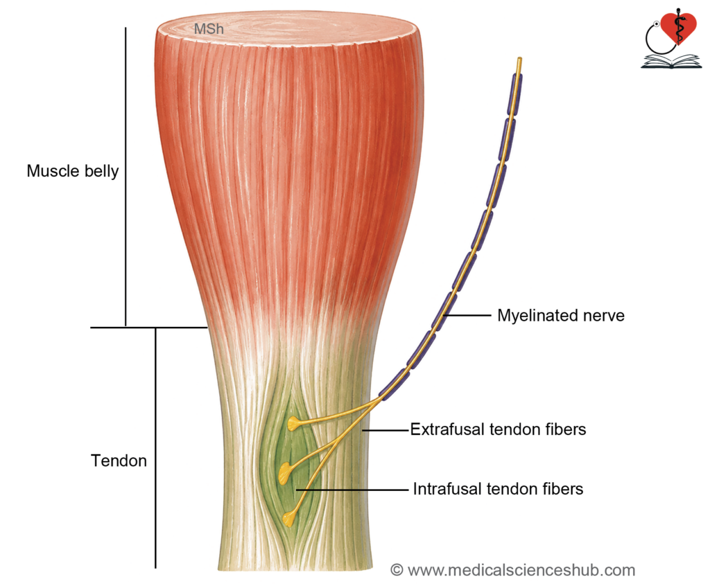
- The Golgi tendon organ is a fusiform receptor situated at the junction of muscle fibers and tendon.
- It contains interwoven collagen fibers enclosed within a connective tissue capsule.
- It is supplied by type A sensory nerve fibers, which enter the capsule and branch among the collagen bundles.
Function
- The GTO detects tension generated during muscle contraction.
- It sends signals that inhibit alpha motor neuron activity via interneurons.
- This mechanism prevents excessive force, protecting muscles and tendons while ensuring smooth and controlled movements.
Segmental Innervation
Segmental Innervation of Skin
Dermatome: Definition
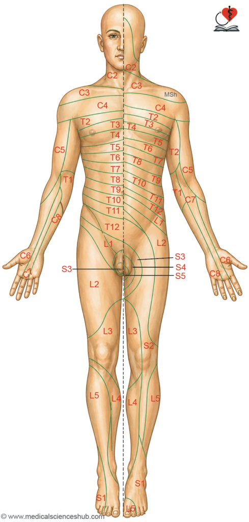
- A dermatome is a specific area of skin supplied by sensory fibers from a single spinal nerve.
- Each dermatome corresponds to one spinal segment.
- All spinal nerves contribute to dermatomes except C1, which usually lacks cutaneous supply.
Dermatomes of the Trunk
- Dermatomes over the trunk are arranged in a regular, segmental pattern.
- Thoracic and upper lumbar nerves supply the trunk in an orderly sequence.
- Each dermatome forms a horizontal band extending from the back (posterior midline) to the front (anterior midline).
- This simple arrangement makes trunk dermatomes clinically easy to interpret.
Overlap of Dermatomes
- Adjacent dermatomes show considerable overlap.
- Damage to a single spinal nerve rarely causes complete sensory loss.
- Complete anesthesia usually requires involvement of at least three consecutive spinal nerves.
- Pain and temperature sensations overlap more than touch, so their loss is less localized.
Dermatomes of the Limbs
- Dermatomal patterns in limbs are complex due to:
- Embryological limb rotation
- Formation of nerve plexuses
Embryological Basis
- Limbs develop as paddle-shaped buds with preaxial (cranial) and postaxial (caudal) borders.
- The thumb and great toe arise from the preaxial border.
- During development:
- Upper limb rotates laterally
- Lower limb rotates medially
Resulting Pattern
- Upper limb: Dermatomes descend along the lateral side and ascend along the medial side.
- Lower limb: Dermatomes descend medially and ascend along lateral and posterior aspects.
Role of Nerve Plexuses
- Limb innervation involves brachial and lumbosacral plexuses.
- Peripheral nerves contain fibers from multiple spinal segments.
- Therefore, the cutaneous area supplied by a named nerve differs from a single dermatome.
Dermatomes of Head and Neck
- The Trigeminal nerve supplies most of the face with well-defined areas.
- Upper cervical nerves supply the posterior scalp.
- The C2 dermatome meets trigeminal territory near the ear.
- On the trunk, C4 lies adjacent to T2 due to developmental changes.
Clinical Importance
- Dermatomes are essential for localizing spinal nerve lesions.
- They assist in neurological examination and pain mapping.
- Understanding dermatomes helps correlate symptoms with specific spinal segments.
Homunculus and its Significance
- The homunculus is a distorted representation of the human body mapped onto the cerebral cortex.
- The size of each body part reflects its functional importance, not its actual anatomical size.
- Regions requiring fine motor control or high sensory input appear disproportionately large.
Significance of Motor and Sensory Homunculus
- The homunculus illustrates how different body regions are organized within the cortex.
- Body parts such as the hands, face, and lips occupy larger cortical areas due to precise movements and heightened sensory perception.
- It demonstrates that greater cortical space is required for complex motor activities and detailed sensory processing.
- It is clinically useful in localizing brain lesions, as specific deficits in movement or sensation correspond to defined cortical regions.
Segmental Innervation of Muscles
- Skeletal muscles receive motor supply in a segmental pattern from the spinal cord.
- Each spinal segment contributes fibers to specific muscles.
- Most muscles are supplied by more than one spinal nerve.
Multiple Segment Supply
- Muscles commonly receive input from two or more spinal segments.
- Therefore, damage to a single nerve rarely causes complete paralysis.
- Total loss of function usually requires injury to multiple nerves or segments.
- This arrangement provides a protective mechanism, preserving partial muscle activity.
Pattern of Innervation
- Most skeletal muscles are multisegmental, except certain muscles like the intrinsic hand muscles, which may have single-segment dominance.
- Muscles performing the same action are supplied by the same spinal segments.
- Muscles with opposite actions are innervated by adjacent lower segments.
- For example:
- Elbow flexors are mainly supplied by C5–C6 segments.
- Elbow extensors are mainly supplied by C7–C8 segments.
- This organized pattern helps in identifying the level of neurological injury.
Correlation with Joints and Skin
- There is a close relationship between muscles, joints, and skin innervation.
- A spinal nerve supplying a muscle also supplies:
- The associated joint
- The skin over the muscle insertion area
- This explains why joint pain may be referred to specific skin regions.
CLINICAL EMBRYOLOGY
Clinical Importance of Myotomes
- It is not essential to memorize the segmental supply of every muscle.
- Understanding key muscles associated with reflexes is more important.
- These muscles are evaluated during examination using stretch reflexes (tendon jerks).
- Reflex testing helps determine the functional integrity of specific spinal segments.
Localization of Spinal Cord Lesions
- Motor examination of joint movements helps identify the level of spinal cord injury.
- Each movement corresponds to a specific myotome, aiding accurate localization.
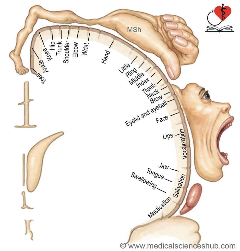
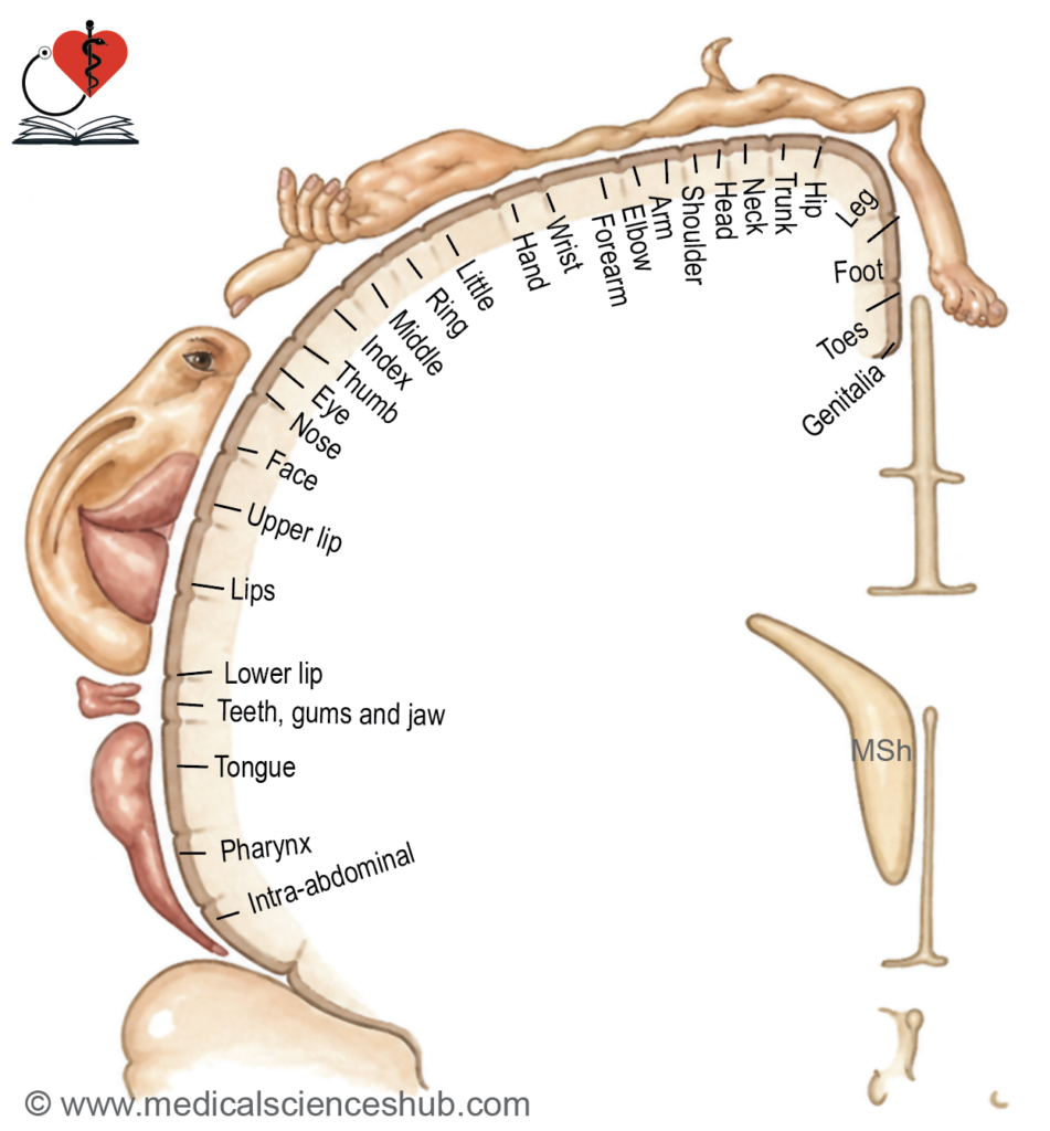
Table 3.2: Spinal level and corresponding myotome functions
| Spinal Level | Principal Myotome Function |
|---|---|
| C2–C3 | Neck flexion and lateral bending |
| C4 | Shoulder elevation (trapezius) |
| C5 | Shoulder abduction (deltoid) |
| C5–C6 | Elbow flexion |
| C6 | Wrist extension |
| C7 | Elbow extension |
| C7–C8 | Wrist flexion |
| C8 | Finger flexion |
| T1 | Finger abduction and adduction (interossei) |
| L1–L2 | Hip flexion |
| L3 | Knee extension |
| L4 | Ankle dorsiflexion |
| L5 | Great toe extension |
| L5–S1 | Foot eversion |
| S1 | Ankle plantarflexion |
| S1–S2 | Knee flexion |
| S2–S3 | Toe flexion |
| S3–S4 | Anal sphincter contraction |
| S2–S4 | Bladder and bowel sphincter control |
CLINICAL NEUROANATOMY
Stretch (Myotatic) Reflex
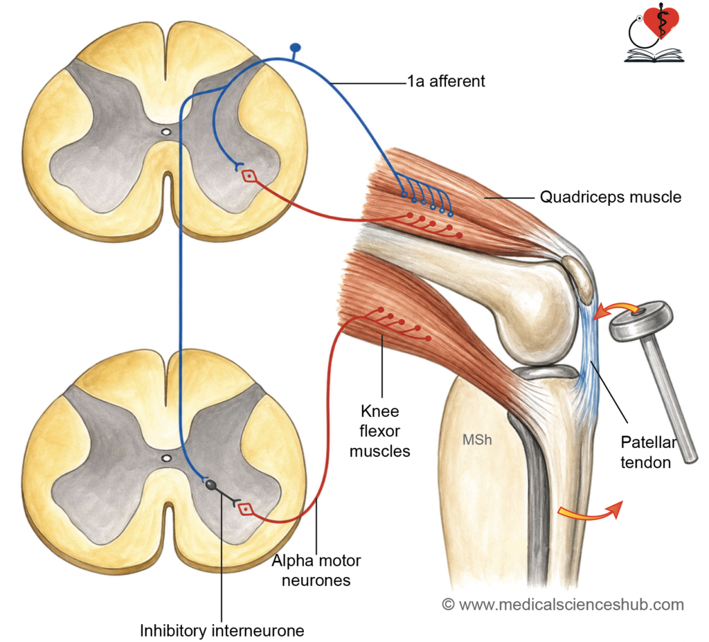
- The stretch reflex is a rapid contraction of a muscle in response to its sudden stretching.
- It begins when muscle spindles detect elongation of intrafusal fibers.
- Sequence:
- Muscle stretch → activation of type Ia and II sensory fibers → direct stimulation of alpha motor neurons → muscle contraction.
- This reflex is monosynaptic, as it involves a direct synapse without interneurons.
Knee Jerk (Patellar Reflex)
- The knee jerk is a classic example of the stretch reflex used in clinical examination.
Method
- Tapping the patellar ligament stretches the quadriceps muscle.
Reflex Pathway
- Receptors: Muscle spindles in quadriceps detect stretch.
- Afferent limb: Sensory impulses travel to spinal segments L2–L4.
- Central connection: Sensory neurons directly activate alpha motor neurons.
- Efferent limb: Motor impulses return to quadriceps muscle.
Response
- The quadriceps contracts, producing extension at the knee joint.
- A visible leg movement indicates a normal reflex.
Clinical Significance
- Loss of the reflex suggests damage to the reflex arc (sensory or motor pathway).
- The affected muscle becomes flaccid and hypotonic.
- Long-standing nerve injury leads to muscle wasting.
- Reflex testing helps evaluate spinal cord segments and neural pathways.
Tendon Reflexes
- Tendon reflexes are stretch reflexes used to assess specific spinal segments.
- Each reflex is produced by sudden stretching of a muscle via tendon tapping.
- Example: The brachioradialis (supinator) reflex.
- These reflexes are useful in assessing peripheral nerve and spinal cord integrity.
Protective Reflexes
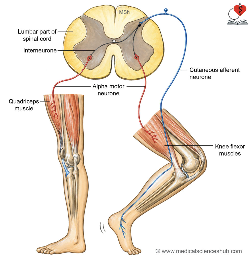
- The flexor reflex causes withdrawal of a limb from a painful stimulus.
- The crossed extensor reflex extends the opposite limb to maintain balance and posture.
Muscle Tone
- Muscle tone is the slight, continuous contraction present in a muscle at rest.
- It helps maintain posture and allows rapid response to movement.
- Clinically, tone is assessed by evaluating resistance during passive stretching.
- Since individual fibers contract fully or relax completely, tone is maintained by alternating activity of different fiber groups, preventing fatigue.
Neural Basis
- Muscle tone depends on an intact monosynaptic reflex arc.
- Sensory impulses from muscle receptors reach the spinal cord and directly activate alpha motor neurons.
Regulation
- Muscle spindles detect changes in muscle length.
- Tendon receptors monitor tension.
- Gamma motor neurons adjust spindle sensitivity, allowing fine control of tone.
Muscle Power
- Muscle power is the force generated during muscle contraction.
- It reflects the ability of muscles to perform work and movement.
Variation
- Muscle strength varies with size and function.
- Larger muscles, such as those in the thigh and back, generate greater force than smaller muscles responsible for precise movements.
Assessment
- Evaluation of muscle power is essential in neurological examination.
- It helps detect weakness due to nerve, muscle, or spinal cord disorders.
- The Medical Research Council (MRC) grading system is commonly used to standardize assessment and monitor progression or recovery.
Muscle Wasting
- Muscle wasting is a reduction in muscle bulk due to loss of nerve supply, disuse, or chronic disease.
Causes and Features
- Peripheral nerve injury or anterior horn cell disorders (e.g., Poliomyelitis) lead to weakness and possible paralysis.
- Wasting typically begins within 2–3 weeks after denervation.
- Fasciculations may appear as fine, involuntary muscle twitches in chronic conditions.
- Contractures can develop in opposing muscles, causing permanent shortening.
Types
- Localized wasting occurs in lower motor neuron lesions and disuse.
- Generalized wasting is seen in chronic illnesses such as cancer and prolonged systemic disease.
Table 3.3: MRC grading of muscle power
| Grade | Muscle Strength | Description |
|---|---|---|
| 0 | No activity | No visible or palpable muscle contraction is detected. |
| 1 | Trace contraction | A minimal contraction is present, but it does not produce any joint movement. |
| 2 | Movement without gravity | The muscle can move the joint only when the effect of gravity is eliminated. |
| 3 | Movement against gravity | The muscle can move the joint through full range against gravity, but not against added resistance. |
| 4 | Movement against resistance | The muscle can act against gravity and moderate resistance, but strength is reduced compared to normal. |
| 5 | Normal strength | The muscle performs full movement against gravity and strong resistance without weakness. |
📝 Test Your Knowledge – Practice MCQs
Attempt the chapter MCQ quiz and assess your understanding of key concepts.
Click to see: Medical Disclaimer and Copyright Notice