
Competencies
- AN56.1: Describe and identify various layers of meninges with their extent and modifications.
- AN56.2: Describe circulation of CSF with its applied anatomy.
- AN63.2: Describe anatomical basis of congenital hydrocephalus.
Introducti0n
- The meninges are three protective connective tissue layers—dura mater, arachnoid mater, and pia mater—that surround the brain and spinal cord. They provide mechanical protection, support blood vessels, enclose cerebrospinal fluid, and help maintain a stable environment for the central nervous system.
Meninges
- The meninges are three concentric protective layers surrounding the brain and spinal cord.
- From outermost to innermost, they are:
- Dura mater: the thick, tough outermost layer; also called pachymeninx (Greek: pachys = thick).
- Arachnoid mater: a delicate, avascular middle layer, with numerous thread-like trabeculae extending from it to the pia mater (Greek: arachno = cobweb-like).
- Pia mater: a thin, highly vascular innermost layer that is closely adherent to the surface of the brain and spinal cord.
- The arachnoid mater and pia mater are collectively referred to as the leptomeninges (Greek: leptos = thin), owing to their delicate structure.
- Meninges covering the brain are termed cranial meninges, while those surrounding the spinal cord are called spinal meninges.
- Correspondingly, the cerebral dura mater covers the brain, and the spinal dura mater covers the spinal cord.
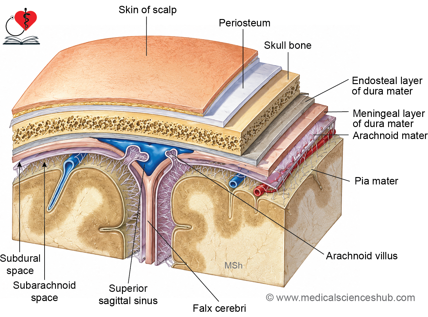
Cranial Meninges
- The cranial meninges cover and protect the brain, consisting of three layers arranged from outside inward.
- The cerebral dura mater is thick and firmly adherent to the base of the skull.
Layers of the Cranial Meninges
- Cranial dura mater — the outermost layer, consisting of two sublayers:
- Endosteal layer: the outer sublayer, which is adherent to the inner surface of the skull, functioning as its internal periosteum.
- Meningeal layer: the inner sublayer, which forms the actual protective covering of the brain and gives rise to the dural folds (reflections).
- Arachnoid mater — the delicate, avascular middle layer lying deep to the dura mater.
- Pia mater — the thin, vascular innermost layer, closely adherent to the surface of the brain.
Dura Mater
- The dura mater is the outermost and thickest meningeal layer, consisting of two sublayers: the endosteal layer and the meningeal layer.
Endosteal Layer
- This layer represents the internal periosteum of the skull bones (endocranium) and is firmly adherent to them.
- At the cranial foramina, it becomes continuous with the external periosteum of the skull.
Note: Fractures of the skull base can tear the dura mater and underlying dural venous sinuses, resulting in bleeding that may present as hemorrhage from the nose, ear, or pharynx.
Meningeal Layer
- This is a strong membranous layer that, at the foramen magnum, becomes continuous with the spinal dura mater.
- At the points where cranial nerves exit through skull foramina, it forms tubular sheaths around them, which subsequently fuse with the epineurium of each nerve.
- The sheath around the optic nerve is unique in that it fuses with the sclera of the eyeball.
Relationship Between the Two Layers
- The endosteal and meningeal layers are tightly fused throughout, except at two locations:
- Where they split apart to enclose the dural venous sinuses.
- Where the meningeal layer folds inward to form the dural folds (reflections).
Folds of Dura Mater
The meningeal layer of the dura mater folds inward to form four dural septa, which divide the cranial cavity into compartments and provide structural support to the brain.
- There are four main dural folds:
- Falx cerebri
- Falx cerebelli
- Tentorium cerebelli
- Diaphragma Sellae
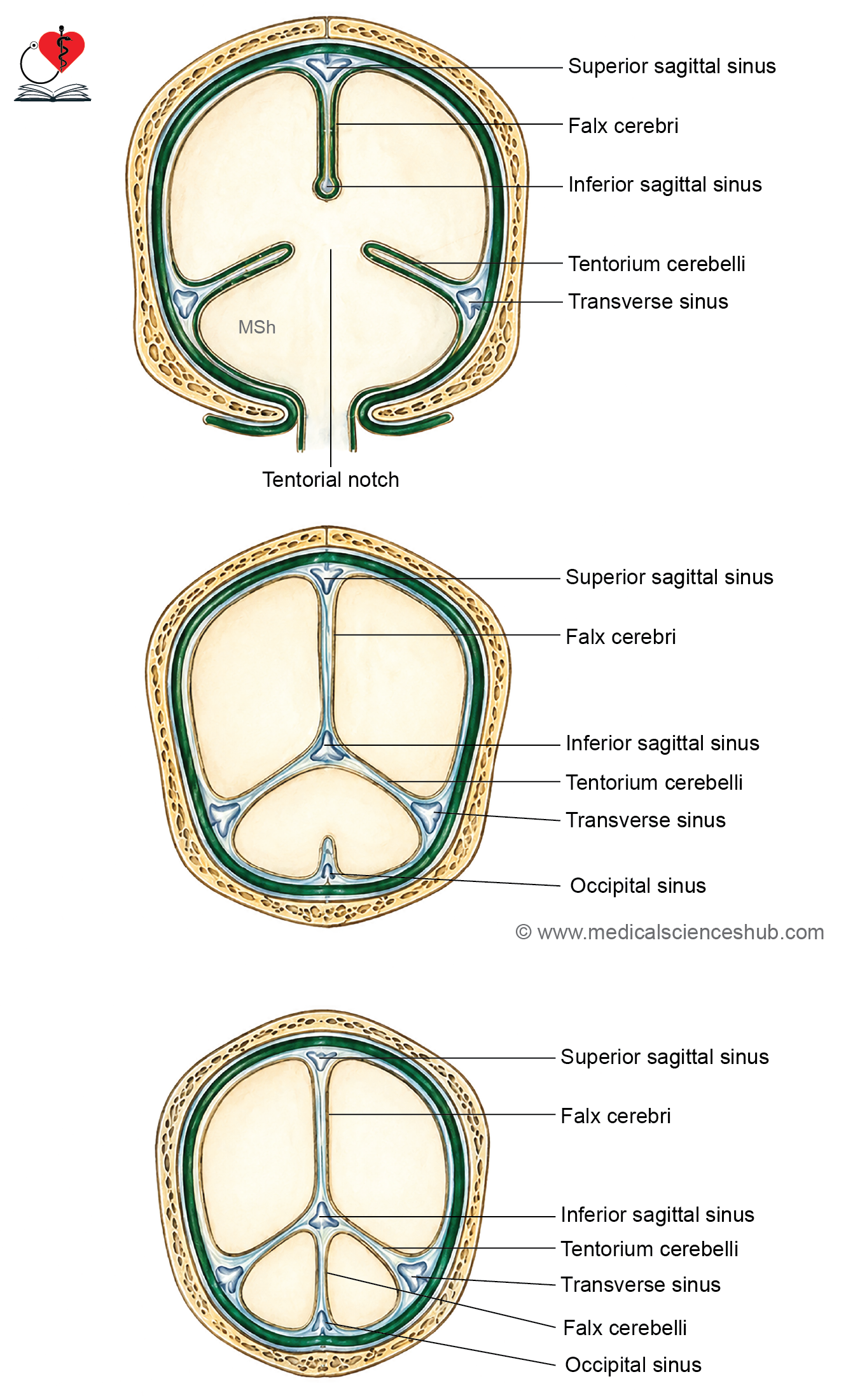
Falx Cerebri
- A sickle-shaped midline fold that projects into the longitudinal fissure, separating the two cerebral hemispheres.
- Attachments:
- Anteriorly to the crista galli
- Posteriorly to the tentorium cerebelli
- Its convex upper border to the vault of the skull along the sagittal suture
- Its lower concave border is free
- Enclosed sinuses: the superior sagittal sinus (upper border), inferior sagittal sinus (lower free border), and straight sinus (junction with tentorium cerebelli).
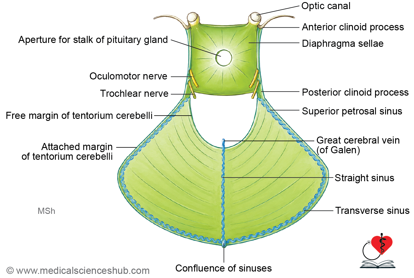
Tentorium Cerebelli
- A tent-shaped, horizontally oriented fold forming the roof of the posterior cranial fossa, separating the cerebrum from the cerebellum.
- Attachments:
- Outer convex border to the posterior clinoid processes, superior border of the petrous temporal bone, and lips of the transverse sulcus of the occipital bone.
- Inner concave (U-shaped) border is free, with its anterior ends attached to the anterior clinoid processes.
- The free inner margin forms the tentorial notch, which serves as a communication passage between the supratentorial and infratentorial compartments.
- Enclosed sinuses: the superior petrosal sinus (anterior attached margin) and transverse sinus (posterior attached margin).
Falx Cerebelli
- A small sickle-shaped fold lying in the midline of the posterior cranial fossa, between the two cerebellar hemispheres.
- Attachments:
- Base to the inferior surface of the tentorium cerebelli
- Apex toward the posterior margin of the foramen magnum
- Posterior border to the internal occipital crest
- Enclosed sinus: the occipital sinus along its attached margin.
Diaphragma Sellae
- Also called the tentorium hypophysis, it is a small circular fold forming the roof of the pituitary fossa (sella turcica).
- Attachments: anteriorly to the tuberculum sellae and posteriorly to the dorsum sellae.
- It contains a central aperture that transmits the stalk (infundibulum) of the pituitary gland.
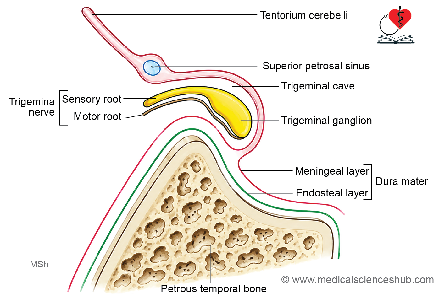
Trigeminal or Meckel’s cave
- The trigeminal cave, also known as Meckel’s cave, is a dural recess situated along the attached border of the tentorium cerebelli. It is formed by an outward extension of the meningeal layer of the dura mater over the trigeminal impression on the petrous part of the temporal bone. This cavity is located inferior to the superior petrosal sinus and encloses the trigeminal ganglion within a cerebrospinal fluid-filled space.
Arterial Supply of Dura
The dura mater receives its arterial supply from branches of three main arteries:
- External carotid artery: via the middle meningeal artery (branch of the maxillary artery), meningeal branches of the ascending pharyngeal artery, and the occipital artery.
- Internal carotid artery
- Vertebral artery
Nerve Supply of Dura
- The dura mater is innervated by:
- All three divisions of the trigeminal nerve (CN V)
- The upper three cervical spinal nerves (C1–C3)
- The cervical sympathetic trunk
- Unlike the dura, the brain, pia mater, and arachnoid mater are insensitive to pain, as they lack nociceptors.
Nerve Supply of Supra- and Infra-tentorial Dura
- The supratentorial dura is supplied by meningeal branches of the trigeminal nerve: the anterior and posterior ethmoidal nerves in the anterior cranial fossa, and meningeal branches of the maxillary and mandibular nerves in the middle cranial fossa.
- The infratentorial dura is supplied by ascending meningeal branches of the upper three cervical nerves (C1–C3).
CLINICAL NEUROANATOMY
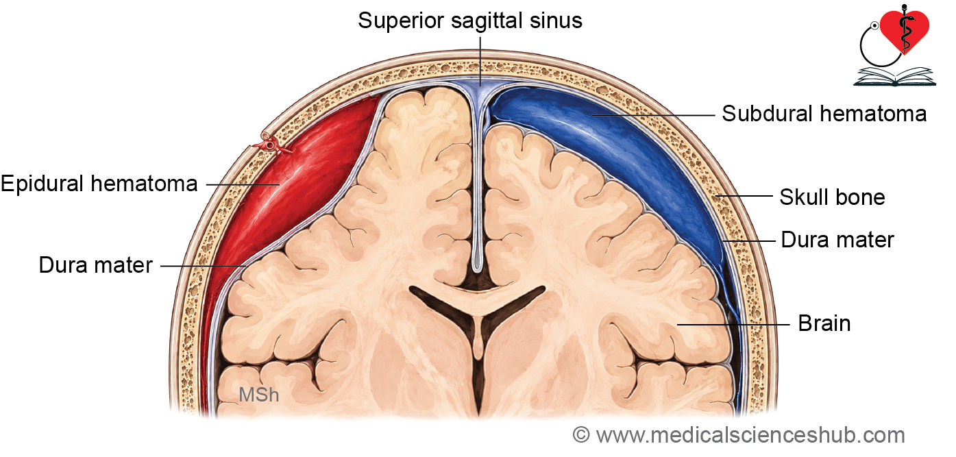
- The middle meningeal artery is frequently damaged in head injuries, leading to extradural hemorrhage, where blood accumulates between the skull and the dura mater.
- Stretching or pressure on the dura causes referred pain — pain originating from the supratentorial dura is referred to the forehead, while pain from the infratentorial dura is referred to the occipital region and back of the neck. This is a common mechanism underlying headache.
Table 18.1: Differences between extradural and subdural hemorrhages
| Feature | Extradural (epidural) hemorrhage | Subdural hemorrhage |
|---|---|---|
| Location | Between the skull and endosteal layer of dura mater | Between the dura mater and arachnoid mater |
| Involved vessels | Arteries (middle meningeal artery) | Veins |
| Symptoms | Rapid onset of symptoms (sudden onset of paralysis) | Gradually increasing symptoms |
| Blood in CSF | Absent | Present |
| CT scan appearance of hematoma | Biconvex lens | Crescent-shaped |
Arachnoid Mater
- The arachnoid mater is a thin, delicate, transparent avascular membrane lying between the dura mater and pia mater.
- It is separated from the dura mater by a narrow subdural space, which contains a thin film of fluid and is traversed by cerebral veins.
Arachnoid Villi and Granulations
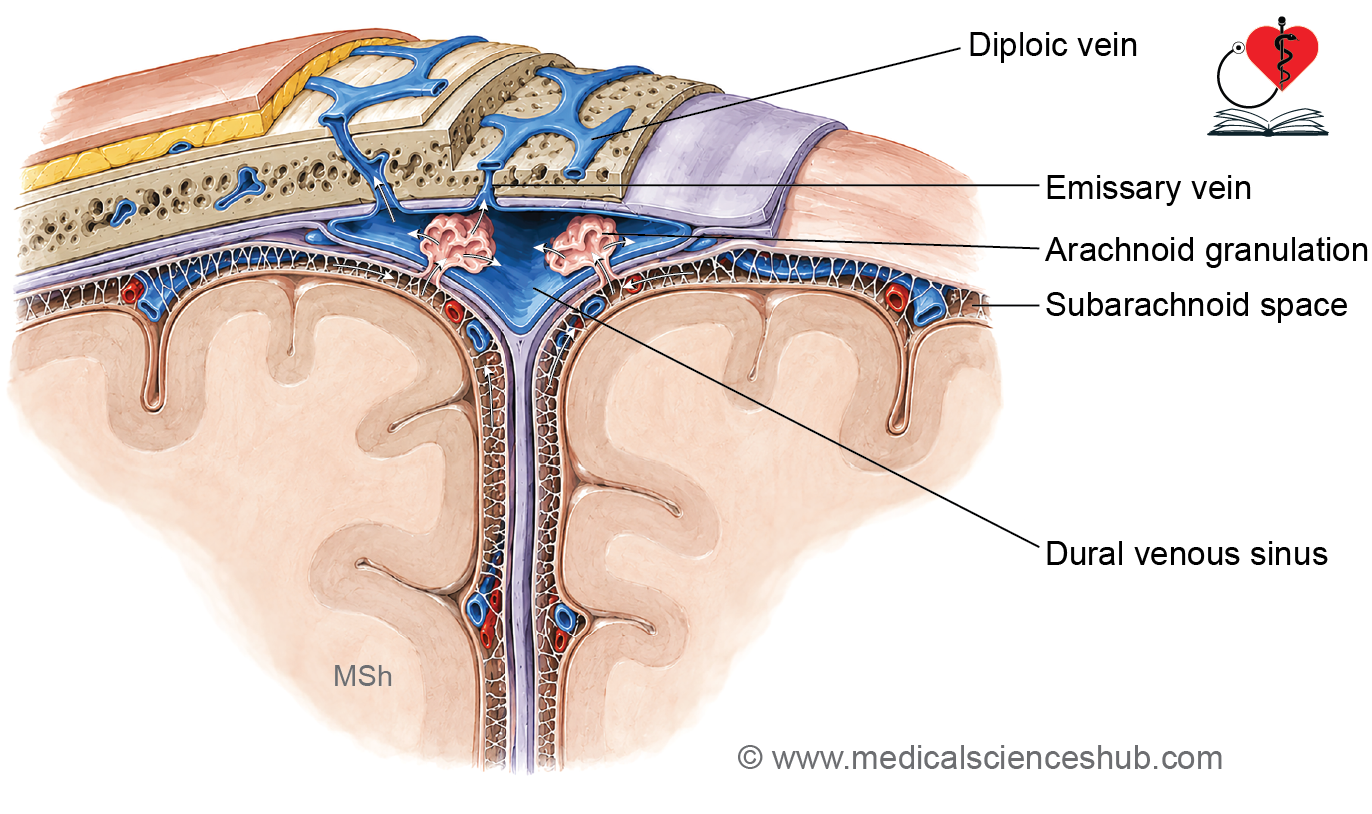
- Arachnoid villi are fine, finger-like projections of the arachnoid mater that perforate the dura mater and project into the dural venous sinuses.
- With advancing age, these villi enlarge and become globular, forming arachnoid granulations (also called Pacchionian bodies).
- Arachnoid granulations produce visible parasagittal depressions on the inner surface of the cranial vault.
Function
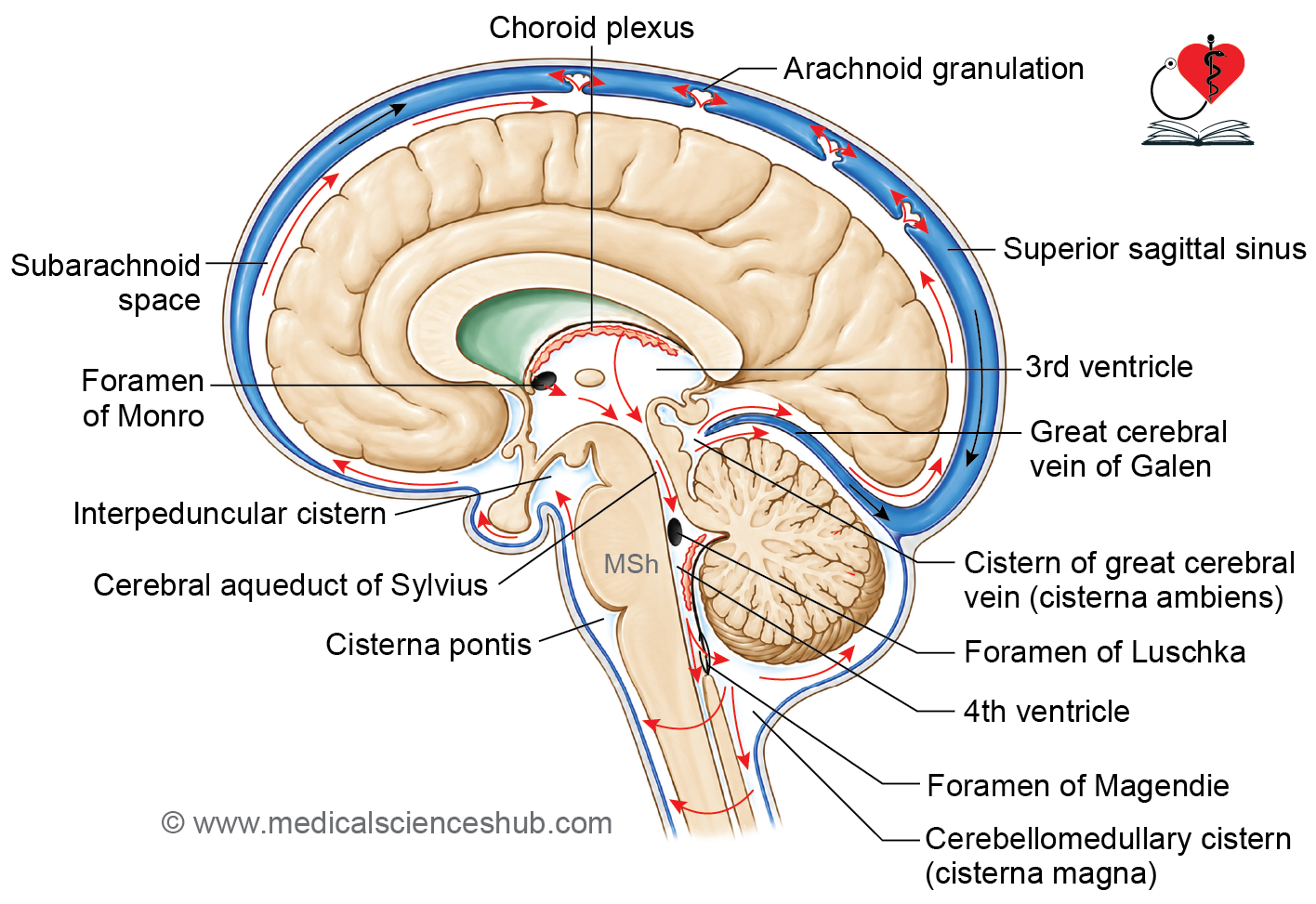
- Both arachnoid villi and granulations are responsible for CSF reabsorption, conveying CSF from the subarachnoid space into the dural venous sinuses and ultimately into the bloodstream via the following pathway: Subarachnoid space → Arachnoid villi and granulations → Dural venous sinuses → Bloodstream
Pia Mater
- The pia mater is a thin, delicate, highly vascular membrane that adheres closely to the surface of the brain, following all its contours including fissures and sulci.
- Blood vessels pierce the pia mater to enter the substance of the brain.
Processes of Pia
- Perivascular sheaths: The pia mater and arachnoid mater together form a sleeve around blood vessels entering the brain. Within this sheath, the pia mater lies outer (in contact with brain tissue) and the arachnoid mater lies inner (in contact with the vessel wall). The space between the two layers is called the perivascular space (Virchow–Robin space).
- Tela choroidea: At certain regions, the brain wall is represented only by ependyma, with no underlying grey or white matter. At these locations, the ependyma fuses with the pia mater to form the tela choroidea of the third and fourth ventricles. The tela choroidea, together with a tuft of capillaries, forms the choroid plexus, which is responsible for CSF production.
- Sheaths and folds: The pia mater forms sheaths around cranial nerves and dips into the sulci of the brain to form supporting septa.
Histology of Choroid Plexus
- The choroid plexus consists of three components:
- An ependymal lining of simple cuboidal epithelium resting on a basement membrane, with the ventricular surface bearing numerous microvilli and cells connected by tight junctions.
- A connective tissue core derived from the pia mater.
- A capillary plexus of fenestrated endothelium, enabling fluid exchange.
- The choroid plexus displays numerous villous projections, giving it a total surface area of approximately 150–300 cm².
Blood-CSF Barrier
The blood–CSF barrier acts as a selective filter, preventing large proteins, toxins, and harmful substances from entering the CSF while regulating the passage of essential molecules.
Structural Components (from blood to CSF):
- Fenestrated endothelial cells of choroidal capillaries
- Basement membrane of endothelial cells
- Thin layer of pia mater with pale cells
- Basement membrane of ependymal (choroidal) cells
- Ependymal (choroidal) cells connected by tight junctions
Functions
- Protects the brain from circulating toxins and harmful substances.
- Participates in CSF production.
- Acts as a selective barrier:
- Allows free passage of water, gases, and lipid-soluble substances.
- Mediates active transport of glucose, amino acids, Na⁺, and Ca²⁺.
Brain–CSF Barrier
- This barrier separates brain tissue from the CSF within the ventricles and consists of:
- Extrachoroidal ependymal cells lining the ventricles
- Basement membrane of these ependymal cells
- Subependymal glial membrane
- Unlike the blood–CSF barrier, the brain–CSF barrier is comparatively weak, as the extrachoroidal ependymal cells are connected by gap junctions rather than tight junctions, allowing relatively free exchange of substances between the CSF and brain tissue.
Subarachnoid Space
- The subarachnoid space is located between the arachnoid mater and the pia mater, and is traversed by numerous spider-like trabeculae extending between the two layers.
Contents
- Cerebrospinal fluid (CSF)
- Arteries — whose pulsations assist in CSF circulation
- Veins
Communications
- With the ventricular system via the openings of the fourth ventricle (foramina of Magendie and Luschka)
- With the spinal subarachnoid space inferiorly
Extensions
- The subarachnoid space extends along cranial nerves, and notably continues around the optic nerve all the way to the eyeball.
- It also extends around blood vessels entering the substance of the nervous tissue; however, these vessels are covered by leptomeninges and therefore do not directly contact the CSF.
Note: The perivascular spaces (Virchow–Robin spaces) lie between the meningeal sheaths and the blood vessel walls. These spaces are separated from the subarachnoid space and CSF by the leptomeninges and should not be confused with the subarachnoid space itself.
Subarachnoid Cisterns
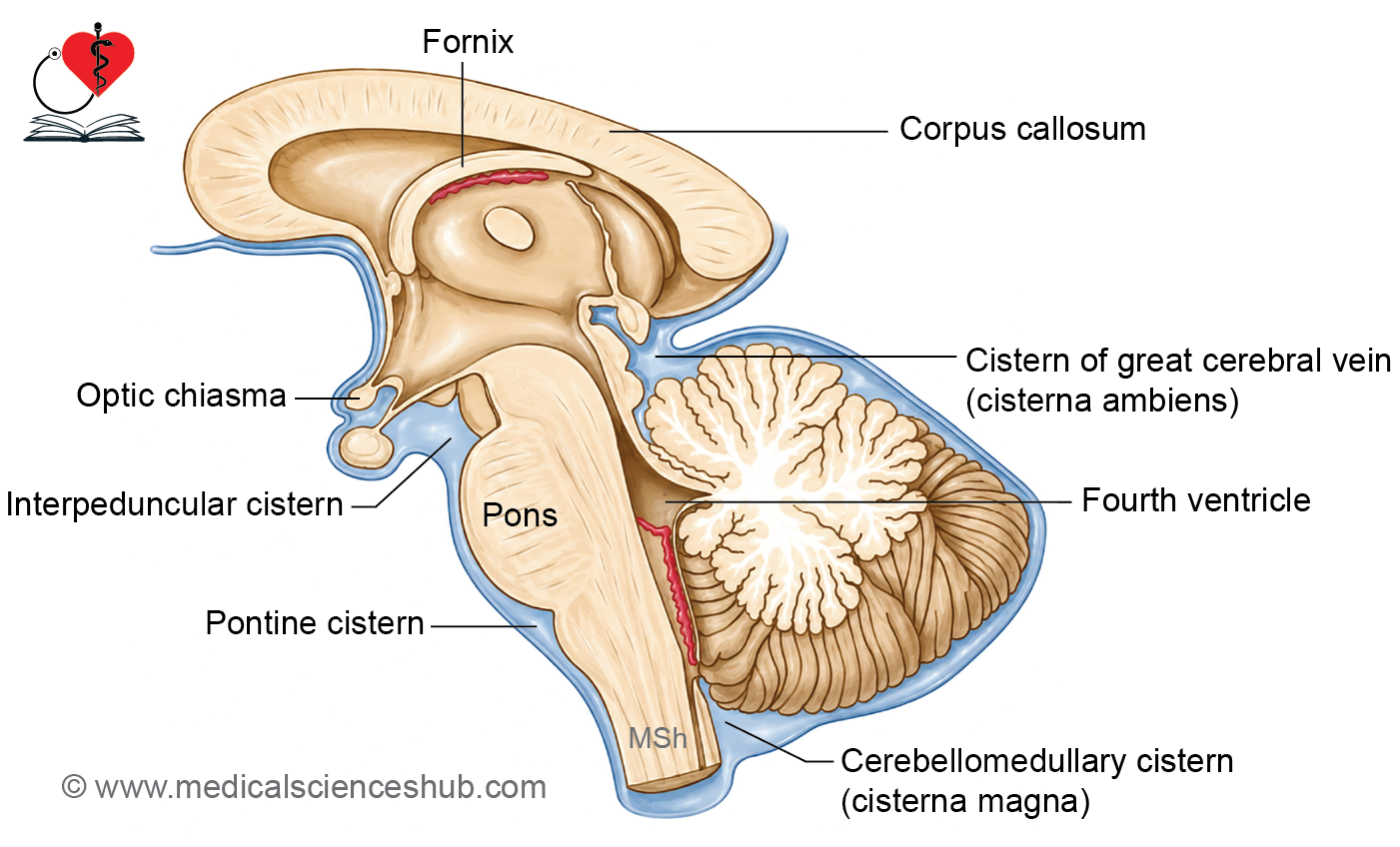
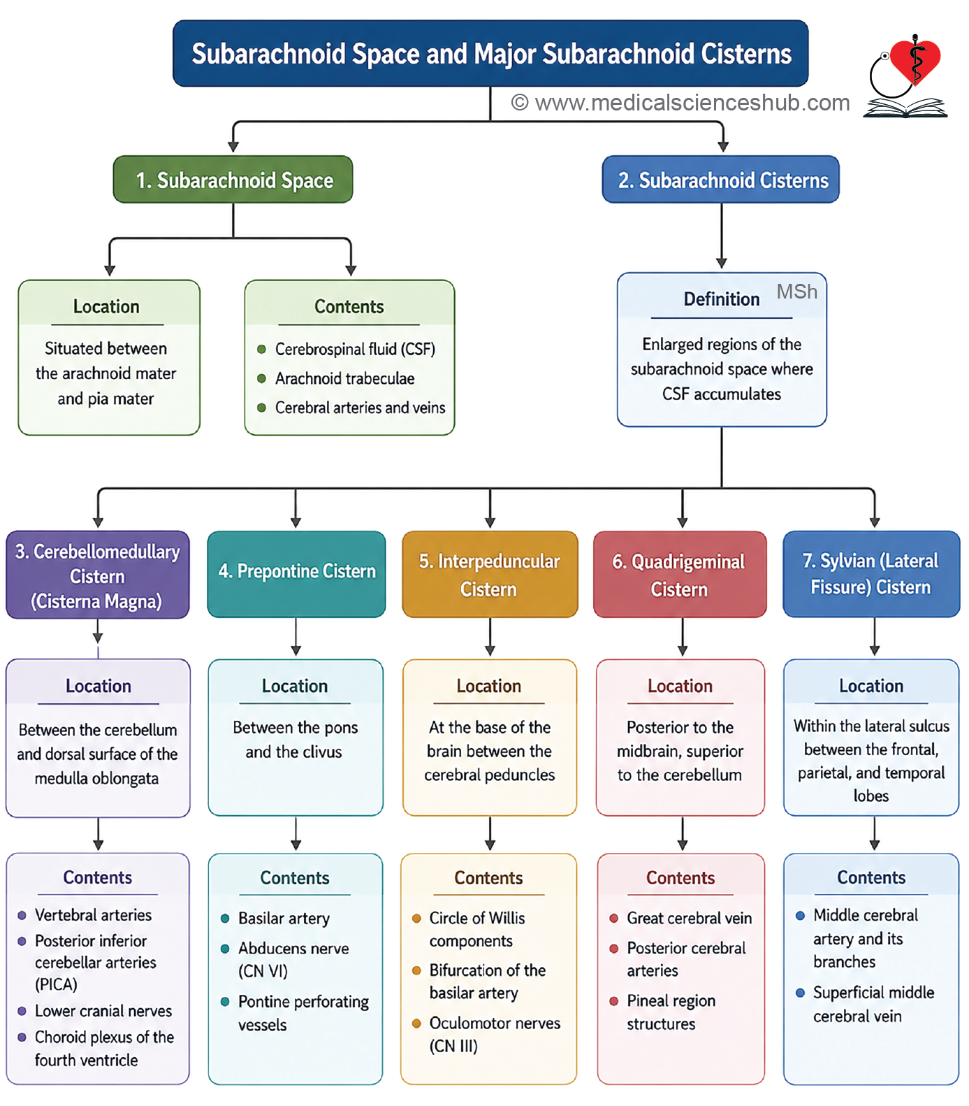
- Subarachnoid cisterns are enlarged regions of the subarachnoid space that contain significant pooled collections of CSF.
- They form where the arachnoid mater bridges over surface depressions of the brain (sulci and fissures) while the pia mater follows the contours closely, creating wide gaps between the two layers.
- They serve as a fluid cushion for the brain and act as CSF reservoirs.
Major Subarachnoid Cisterns
- Cisterna magna (cerebellomedullary cistern) — between the medulla oblongata and the inferior surface of the cerebellum; the largest cistern.
- Pontine cistern — on the ventral surface of the pons.
- Interpeduncular cistern (basal cistern) — between the two crura cerebri at the base of the brain.
- Cistern of the lateral sulcus (Sylvian cistern) — within the stem of the lateral sulcus.
- Cisterna ambiens (cistern of the great cerebral vein) — between the splenium of the corpus callosum and the superior surface of the cerebellum.
Cisterna Magma (Cerebellomedullary Cistern)
- The largest subarachnoid cistern, located between the medulla oblongata and the inferior surface of the cerebellum.
Communications
- Receives CSF from the fourth ventricle via:
- The foramen of Magendie (median aperture)
- The foramina of Luschka (two lateral apertures)
- Inferiorly, it is continuous with the spinal subarachnoid space.
Contents
- Vertebral artery
- Cranial nerves IX, X, and XI
- Choroid plexus
CLINICAL NEUROANATOMY
Cisternal puncture is performed to collect CSF when lumbar puncture is contraindicated. A needle is introduced into the cisterna magna through the posterior atlanto-occipital membrane. This procedure carries significant risk due to its proximity to the brainstem and its vital centers.
Table 18.2: Contents of subarachnoid cisterns
| Cistern | Important Structures Present |
|---|---|
| Cisterna Magna (Cerebellomedullary Cistern) | • Vertebral arteries • Cranial nerves IX, X, and XI • Cerebellar arteries |
| Pontine Cistern | • Basilar artery and its branches • Abducens nerve (CN VI) |
| Interpeduncular Cistern | • Circle of Willis • Oculomotor nerve (CN III) • Mammillary bodies |
| Sylvian (Lateral Fissure) Cistern | • Middle cerebral artery and branches |
| Cistern of the Vein of Galen (Quadrigeminal Cistern) | • Great cerebral vein (vein of Galen) • Pineal gland • Trochlear nerve (CN IV) |
Cerebrospinal Fluid
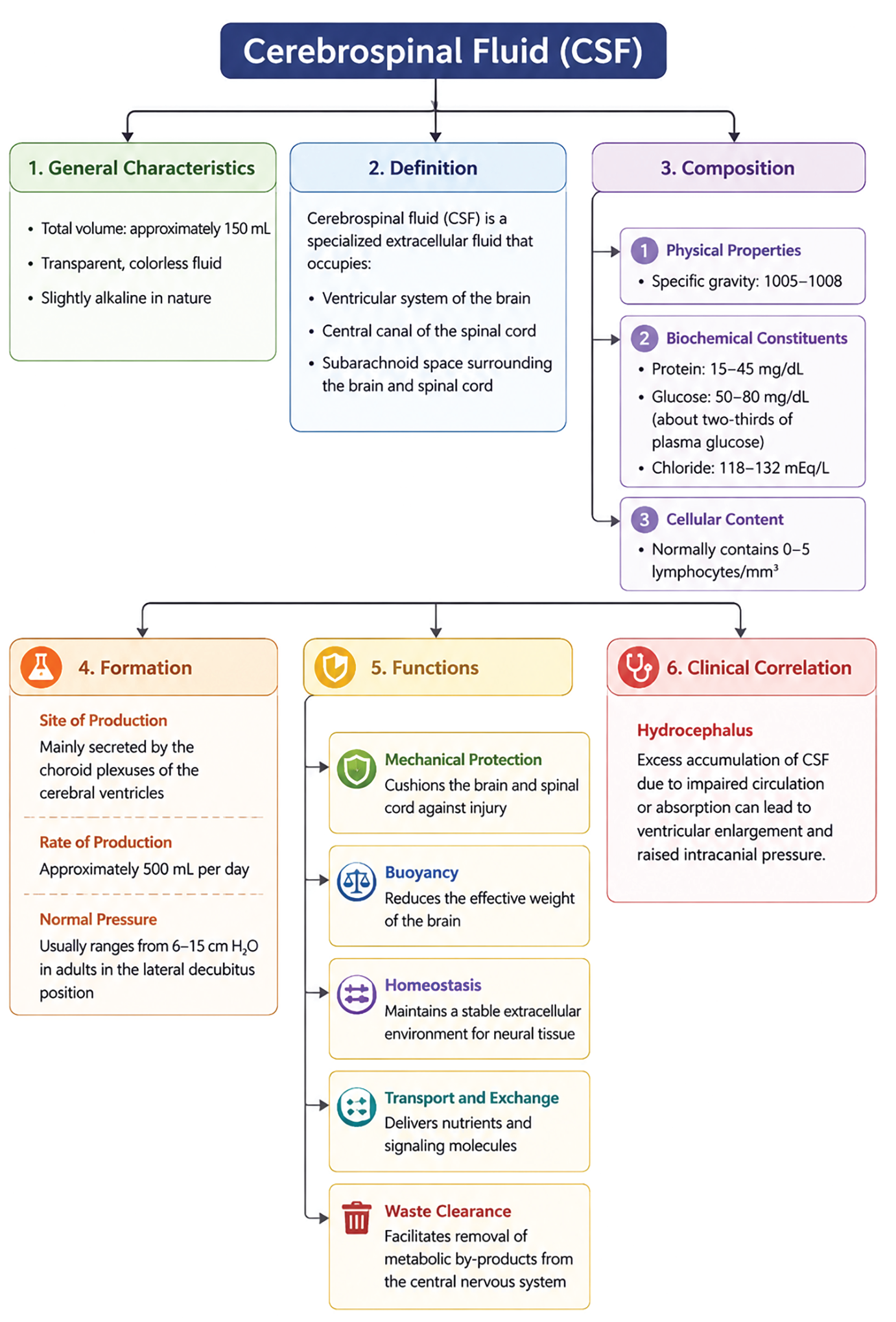
- CSF is a clear, colorless, odorless, modified tissue fluid with a specific gravity of 1005–1008 and a slightly alkaline pH.
- It is found in the ventricles of the brain, the central canal of the spinal cord, and the subarachnoid space.
- Total volume is approximately 150 mL, of which about 30 mL is within the ventricular system.
Composition
- CSF contains proteins, glucose, chloride, sodium, potassium ions, and a small number of lymphocytes.
Production
- CSF is produced by the choroid plexuses of the ventricles at a rate of approximately 500 mL/day.
- 80–90% is produced by the choroid plexuses of the lateral ventricles, with the remaining 10–20% produced by the choroid plexuses of the third and fourth ventricles.
Table 18.3: Composition of CSF
| Parameter | Typical Value | Key Point |
|---|---|---|
| Protein | 15-45 mg/dL | Much lower than plasma protein concentration. Elevated levels may indicate infection, inflammation, or hemorrhage. |
| Glucose | 50-80 mg/dL (about two-thirds of blood glucose) | Decreased in bacterial and tuberculous meningitis; usually normal in viral infections. |
| Chloride | 118-132 mEq/L | Important for maintaining osmotic balance; may decrease in some forms of meningitis. |
| Lymphocytes | 0-5 cells/mm³ | Normal CSF contains very few cells. Increased cell count suggests CNS pathology. |
| Volume | 90-150 mL (adults) | Total CSF volume is continuously renewed several times daily. |
| Opening Pressure | 60-150 mm H₂O (lateral recumbent position) | Reflects intracranial pressure; increased in hydrocephalus, tumors, and meningitis. |
CSF Circulation
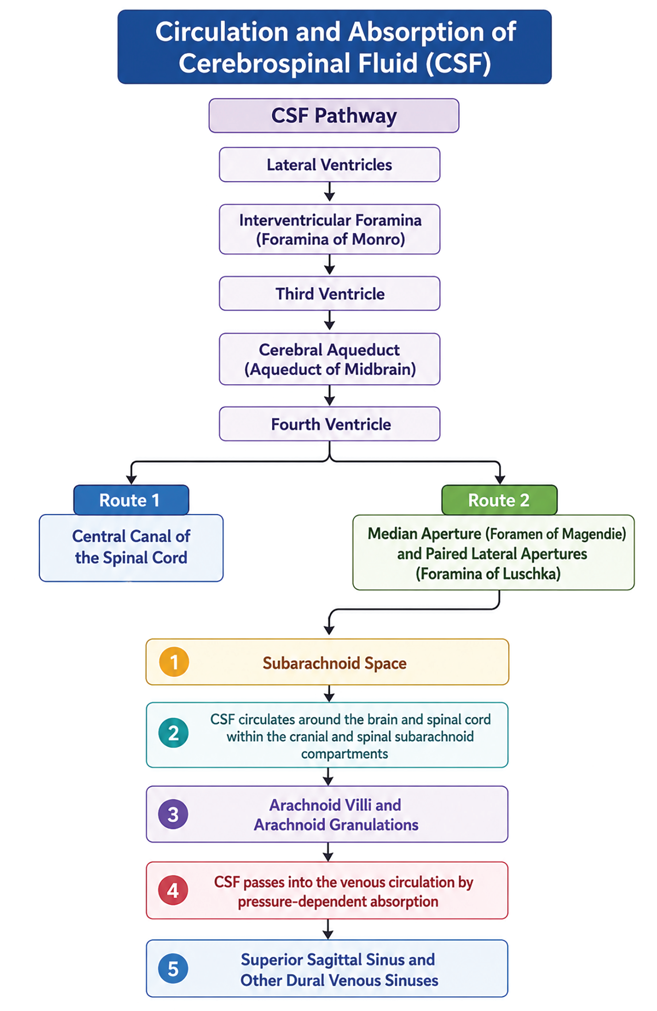
- CSF follows a defined pathway through the central nervous system:
Lateral ventricles → (viainterventricular foramina of Monro) → Third ventricle → (viacerebral aqueduct of Sylvius) → Fourth ventricle → (viamedian and lateral apertures) → Subarachnoid space → (viaarachnoid villi and granulations) → Dural venous sinuses → Bloodstream
- CSF also flows through the central canal of the spinal cord from the fourth ventricle.
Functions of CSF
- Mechanical protection: CSF acts as a shock absorber, allowing the brain and spinal cord to float within it. The brain weighs approximately 1500 g in air but only ~50 g when suspended in CSF, significantly reducing mechanical stress on neural tissue.
- Structural protection: CSF prevents brain structures from self-compression and shields the CNS from direct contact with blood via the blood–CSF and brain–CSF barriers.
- Nutrition: CSF provides a continuous supply of electrolytes and glucose to the brain and spinal cord.
- Metabolic waste removal: CSF serves as a medium for transporting metabolic waste products away from neural tissue.
- Transport medium: CSF carries pineal gland secretions to the pituitary gland.
CLINICAL NEUROANATOMY
CSF examination
CSF analysis provides diagnostically useful information:
- Tuberculosis: reduced chloride concentration.
- Pyogenic infections: reduced glucose, elevated lymphocyte count.
- Trauma: presence of blood in CSF.
Hydrocephalus
- Hydrocephalus is an abnormal increase in CSF volume, caused either by excessive CSF production or obstruction of CSF circulation.
- Sites of Obstruction and Their Effects
- Interventricular foramen of Monro: dilatation of the lateral ventricles.
- Cerebral aqueduct of Sylvius: distension of the lateral and third ventricles.
- Median and lateral apertures of the fourth ventricle: distension of all ventricles.
- Meningeal adhesions in the subarachnoid space: impaired CSF circulation and absorption.
- Classification
- Non-communicating (obstructive) hydrocephalus: blockage within the ventricular system at one of the CSF exit sites.
- Communicating hydrocephalus: blockage at the absorption site (arachnoid villi and granulations), often due to meningeal adhesions.
- Clinical Features in Infants and Children
- Abnormally enlarged head
- Frontal bossing
- Enlarged, tense fontanelle with widely separated cranial sutures
- Dilated scalp veins
- Sunset sign — downward deviation of the eyes
- Cracked-pot sound on skull percussion
- Progressive motor dysfunction
Dural Venous Sinuses
- The dural venous sinuses are endothelium-lined venous spaces within the dura mater, formed either by the separation of the two dural layers or by folding of the meningeal layer upon itself.
- They receive both venous blood from the brain and CSF via the arachnoid granulations, and communicate with veins outside the skull through emissary veins, helping to regulate intracranial venous pressure.
Peculiarities of Dural Sinuses
- Lined by endothelium but lack a muscular wall, unlike typical veins.
- They are valveless, allowing bidirectional blood flow.
- They receive CSF in addition to venous blood, distinguishing them from peripheral veins.
Classification
There are 21 dural venous sinuses in total, classified as either paired or unpaired:
Paired Sinuses (one on each side)
- Cavernous sinus
- Superior petrosal sinus
- Inferior petrosal sinus
- Transverse sinus
- Sigmoid sinus
- Sphenoparietal sinus
- Petrosquamous sinus
Unpaired Sinuses (single, midline)
- Superior sagittal sinus
- Inferior sagittal sinus
- Straight sinus
- Occipital sinus
- Anterior intercavernous sinus
- Posterior intercavernous sinus
- Basilar plexus of veins
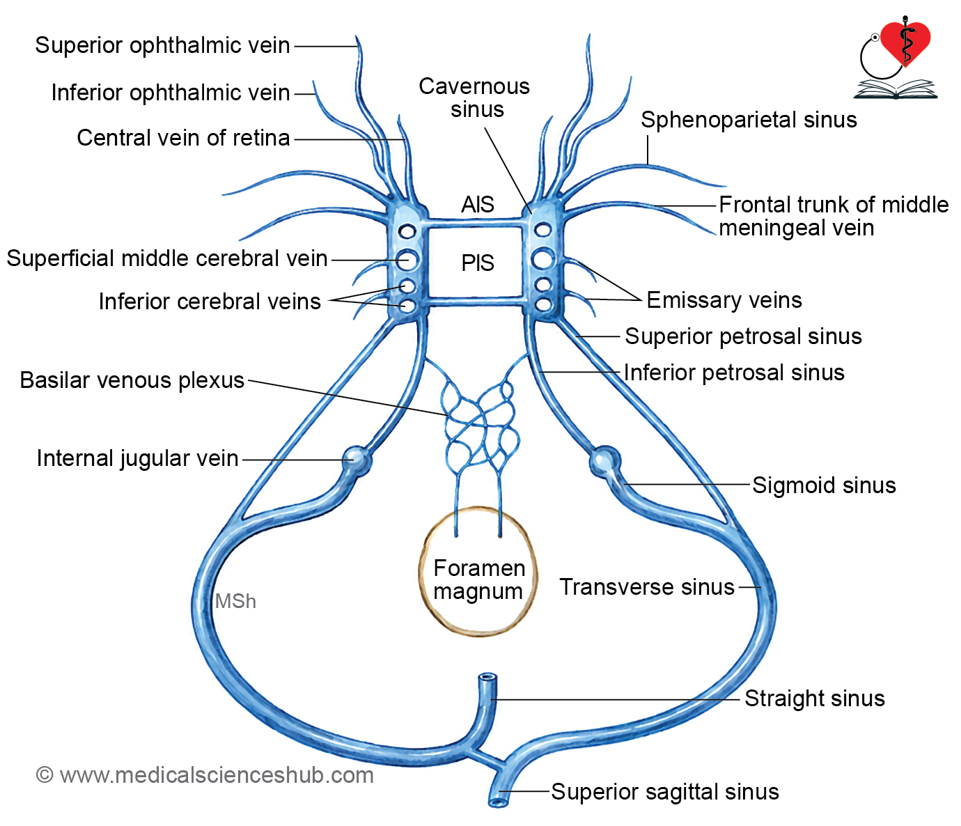
Cavernous Sinus
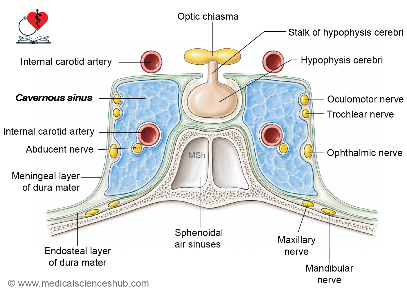
- The cavernous sinus is a large dural venous sinus located in the middle cranial fossa, on either side of the body of the sphenoid bone.
- Its interior contains trabeculae, giving it a cave-like appearance — hence the name cavernous.
Relations
- Superior: optic chiasma, optic tract, olfactory tract, and internal carotid artery
- Inferior: foramen lacerum
- Medial: pituitary gland and body of sphenoid
- Lateral: temporal lobe
Structures Within the Walls and Lumen
- Within the lateral wall (from superior to inferior):
- Oculomotor nerve (CN III)
- Trochlear nerve (CN IV)
- Ophthalmic nerve (CN V1) and maxillary nerve (CN V2)
- Passing through the sinus itself:
- Internal carotid artery
- Abducent nerve (CN VI)
Tributaries The cavernous sinus receives venous drainage from the orbit, brain, and meninges via:
- Superior and inferior ophthalmic veins
- Central vein of the retina (occasionally)
- Superficial middle cerebral vein
- Inferior cerebral veins
- Sphenoparietal sinus
- Anterior trunk of the middle meningeal vein
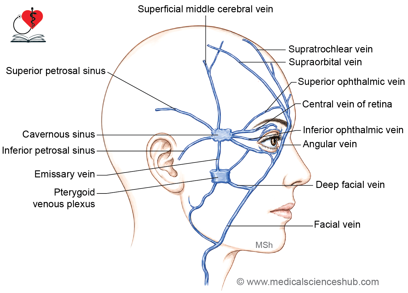
Communications
- With the transverse sinus via the superior petrosal sinus
- With the internal jugular vein via the inferior petrosal sinus
- With the pterygoid venous plexus via emissary veins
- With the facial vein via the superior ophthalmic and angular veins, and indirectly via the pterygoid plexus and deep facial vein
- With the superior sagittal sinus via the superficial middle cerebral and anastomosing veins
Regulation of Blood Flow Blood flow through the cavernous sinus is influenced by:
- Pulsations of the internal carotid artery
- Gravity
- Changes in head position
Note: The valveless communications of the cavernous sinus with facial and nasal veins allow infections from the dangerous area of the face, nasal cavities, and paranasal sinuses to spread intracranially, potentially causing cavernous sinus thrombosis — a serious condition affecting the cranial nerves and vessels running through and within its walls.
Other Sinuses and Veins
Superior Sagittal Sinus
- Lies between the two layers of the falx cerebri along its upper convex attached margin; triangular in cross-section.
- Begins at the crista galli by union of small meningeal veins; terminates at the internal occipital protuberance, usually continuing as the right transverse sinus.
- Arachnoid granulations project into its lumen, and numerous fibrous bands cross its lumen at the inferior angle.
- Tributaries: superior cerebral veins, parietal emissary veins, small nasal cavity veins, and veins of the frontal air sinus.
- Note: Thrombosis may result from spread of infection from the dangerous area of the face, scalp, or diploë.
Inferior Sagittal Sinus
- A small channel lying within the lower free concave margin of the falx cerebri.
- Terminates by joining the great cerebral vein of Galen to form the straight sinus.
Straight Sinus
- Lies in the median plane along the junction of the falx cerebri and tentorium cerebelli.
- Formed by the union of the inferior sagittal sinus and the great cerebral vein of Galen.
- Terminates at the internal occipital protuberance, usually continuing as the left transverse sinus; receives a few superior cerebellar veins.
Transverse Sinuses
- Each sinus lies along the posterior attached margin of the tentorium cerebelli, lodged in a groove from the internal occipital protuberance to the mastoid angle of the parietal bone.
- The right transverse sinus is usually a continuation of the superior sagittal sinus, while the left continues from the straight sinus.
Confluence of Sinuses (Torcular Herophili)
- Located at the internal occipital protuberance, it is the junction of the superior sagittal sinus, straight sinus, and occipital sinus.
Sigmoid Sinuses
- Each is an S-shaped continuation of the transverse sinus at the mastoid angle of the parietal bone, terminating at the jugular foramen as the superior bulb of the internal jugular vein.
- Tributaries: mastoid and condylar emissary veins, cerebellar veins, and labyrinthine vein.
- Note: The sigmoid sinus is separated from the mastoid antrum and air cells by only a thin plate of bone. Infections such as mastoiditis or otitis media can spread directly to the sigmoid sinus, causing thrombosis. Care must be taken to avoid injury to the sigmoid sinus during mastoid surgery.
Occipital Sinus
- A small sinus along the attached margin of the falx cerebelli, beginning at the posterior margin of the foramen magnum and draining into the confluence of sinuses.
Basilar Venous Plexus
- A network of veins lying on the clivus (sloping part of the basiocciput), anterior to the pons and medulla oblongata.
- Connects the two inferior petrosal sinuses and the internal vertebral venous plexus.
Sphenoparietal Sinuses
- Lie along the posterior free margin of the lesser wing of the sphenoid bone, draining into the anterior part of the cavernous sinus.
Superior Petrosal Sinuses
- Each lies along the upper border of the petrous part of the temporal bone, along the attached margin of the tentorium cerebelli.
- Connects the cavernous sinus with the transverse sinus; receives inferior cerebral and cerebellar veins.
Inferior Petrosal Sinuses
- Each lies along the petro-occipital fissure, connecting the cavernous sinus with the superior bulb of the internal jugular vein.
Petrosquamous Sinuses
- Each lies within the petrosquamous suture and drains into the transverse sinus.
Intercavernous Sinuses and Circular Sinus
- The anterior and posterior intercavernous sinuses connect the right and left cavernous sinuses, passing through the diaphragma sellae anterior and posterior to the infundibulum of the pituitary gland, respectively.
- Together with the cavernous sinuses, they form the circular sinus.
Middle Meningeal Veins
- Form two trunks, both draining into the pterygoid venous plexus:
- Frontal (anterior) trunk — via the foramen ovale
- Parietal (posterior) trunk — via the foramen spinosum
- Each trunk runs alongside the corresponding branch of the middle meningeal artery.
Superficial Middle Cerebral Vein
- Lies within the lateral sulcus, draining anteriorly into the cavernous sinus.
- Communicates posteriorly with the superior sagittal sinus via the superior anastomotic vein of Trolard and with the transverse sinus via the inferior anastomotic vein of Labbé.
Great Cerebral Vein of Galen
- Formed behind the midbrain, below the splenium of the corpus callosum, by the union of the two internal cerebral veins and the basal vein of Rosenthal.
- Drains into the straight sinus.
CLINICAL NEUROANATOMY
Cavernous Sinus Thrombosis
- Usually results from the spread of infection from the face, scalp, orbit, or paranasal sinuses through valveless veins.
- Causes ophthalmoplegia due to involvement of cranial nerves III, IV, and VI.
- Produces orbital pain, eyelid edema, proptosis, and impaired venous drainage from the orbit.
- May be associated with headache, fever, and visual disturbances.
Pulsating Exophthalmos
- Characterized by rhythmic protrusion of the eyeball synchronized with the arterial pulse.
- Commonly caused by a carotid-cavernous fistula following skull base fracture or vascular injury.
- Results from high-pressure arterial blood entering the cavernous sinus.
- May cause conjunctival congestion, diplopia, and visual impairment.
Sigmoid Sinus Thrombosis
- Usually occurs due to spread of infection from the middle ear or mastoid air cells.
- Leads to impaired venous drainage and intracranial complications.
- Common features include fever, headache, and raised intracranial pressure.
Some Interesting Facts
- Oculomotor trigone: The anterior ends of inner free margin cross the attached margin, pass laterally to get attached at anterior clinoid processes. The area in front of the crossing between the free and attached margins of the tentorium cerebelli is known as oculomotor tigon. It is pierced by oculomotor and trochlear nerves.
- Anterior petroclinoid ligament is an extension of inner margin of tentorium cerebelli between posterior to anterior clinoid processes. This ligament marks the junction of roof and lateral wall of the cavernous sinus.
- Posterior petroclinoid ligament is a rounded ridge of attached margin of tentorium cerebelli near the posterior clinoid process. It extends from apex of the petrous part of temporal bone to anterior clinoid process.
- Structural NA: Layers and spaces from outside inward:
Endosteal dura – Dural sinuses – Meningeal dura – Subdural space – Arachnoid mater – Subarachnoid space – blood vessels – Brain tissue
- Trigeminal or Meckel’s cave: It is a recess of dura mater present in relation to the attached margin of the tentorium (Fig. 21.4).MCQ, Viva
Formation: It is formed by evagination of the meningeal layer of dura mater over the trigeminal impression of the petrous temporal bone. Trigeminal cave lies below the superior petrosal sinus.
Contents: It contains trigeminal ganglion and CSF.
Important Questions
- Why are the dura mater called pachymeninx, arachnoid and pia mater as leptomeninges?
- Write a short note on CSF.
- Describe CSF circulation.
- Why is there a spread of infection from dangerous area of face to cavernous sinus?
- Why does the infection from one cavernous sinus spread to another?
📝 Test Your Knowledge – Practice MCQs
Attempt the chapter MCQ quiz and assess your understanding of key concepts.
Click to see: Medical Disclaimer and Copyright Notice