
Competencies
- AN60.1: Describe and demonstrate external and internal features of cerebellum.
- AN60.2: Describe connections of cerebellar cortex and intracerebellar nuclei.
- AN64.1: Describe and identify the microanatomical features of spinal cord, cerebellum, and cerebrum.
- AN60.3: Describe anatomical basis of cerebellar dysfunction
Introduction
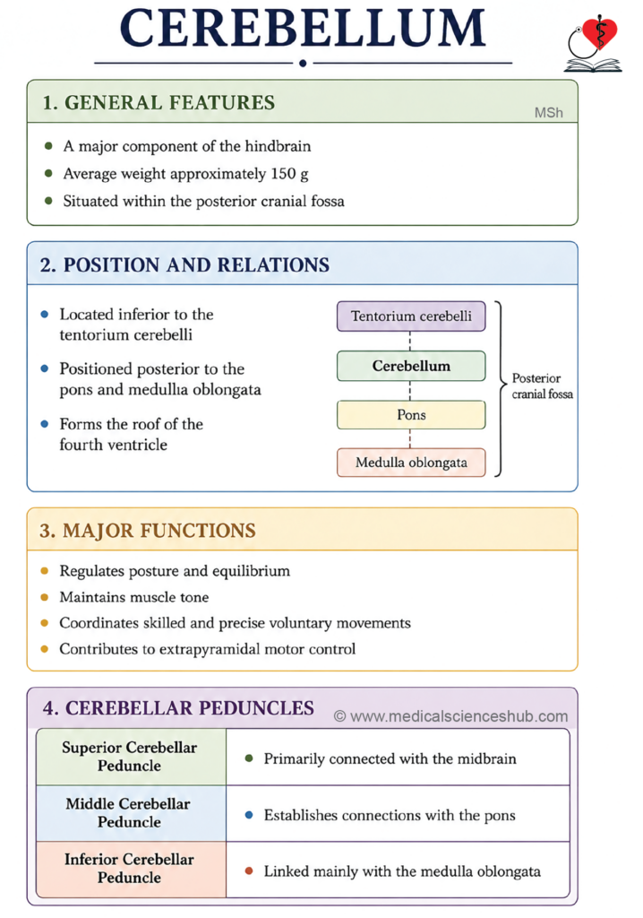
- The cerebellum coordinates voluntary muscle activity and helps maintain posture, balance, and muscle tone.
- It regulates the timing, precision, and smooth execution of body movements.
- Damage to the cerebellum may result in impaired coordination, tremors, gait instability, reduced muscle tone, and difficulty performing skilled movements.
- Such dysfunction can interfere with routine activities and increase the risk of falls.
Location and Relations
- The cerebellum is situated in the posterior cranial fossa, below the tentorium cerebelli.
- It lies posterior to the pons and medulla oblongata.
- The fourth ventricle separates the cerebellum from the posterior surfaces of the pons and upper medulla.
Coverings
- The cerebellum is covered externally by dura mater, including the tentorium cerebelli.
- Its surface is protected internally by the pia mater, which closely follows the cerebellar folds.
Connections of the Cerebellum
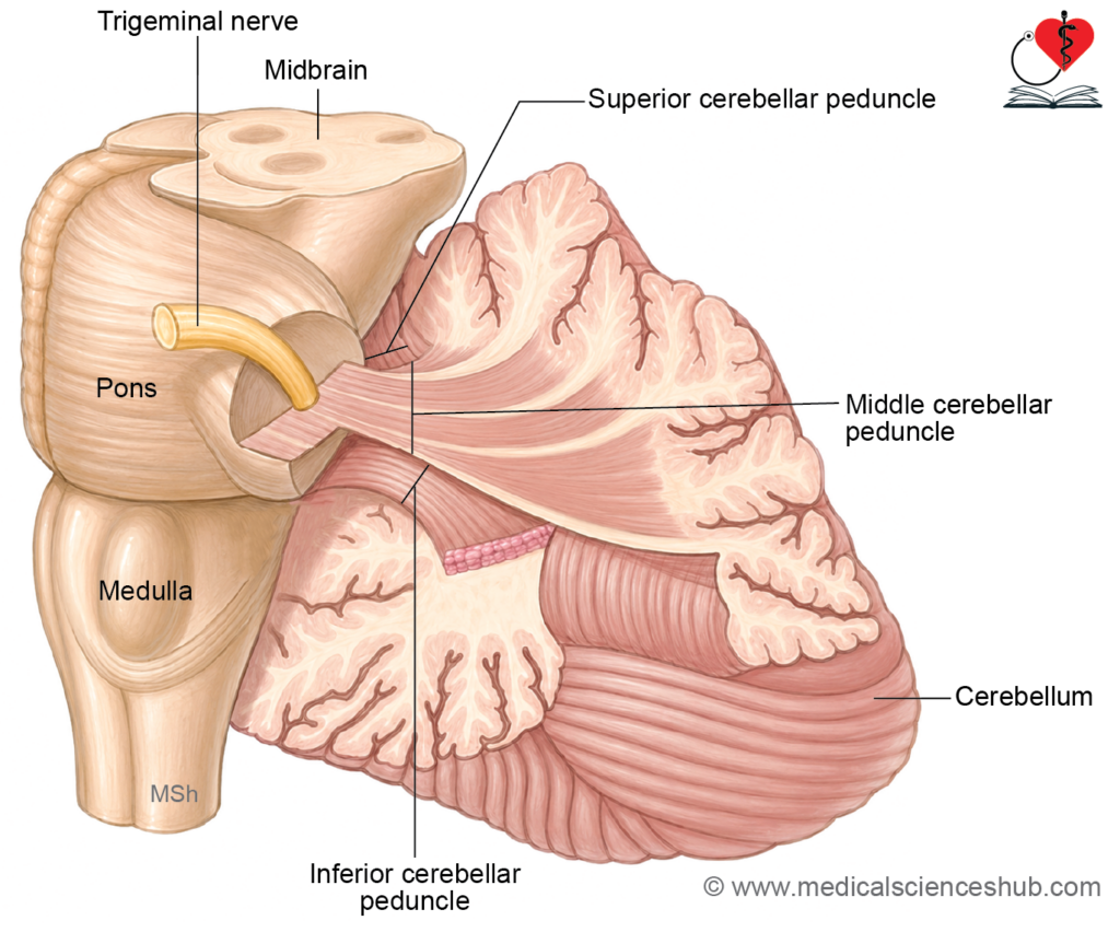
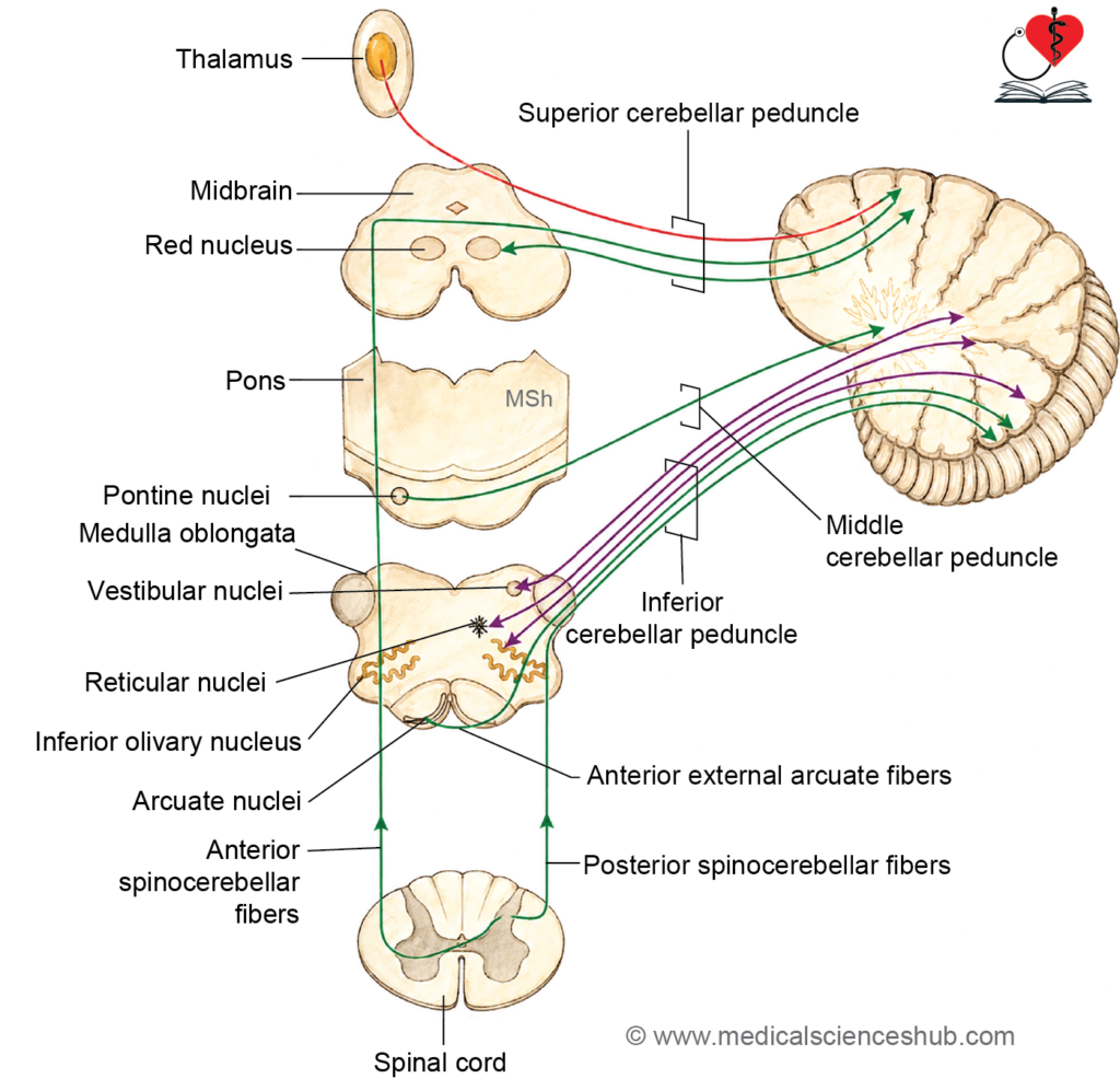
- The cerebellum is connected to the brainstem by three paired bundles of nerve fibers called cerebellar peduncles.
- These peduncles establish communication between the cerebellum and different parts of the brainstem.
- Superior cerebellar peduncle connects the cerebellum with the midbrain.
- Middle cerebellar peduncle connects the cerebellum with the pons.
- Inferior cerebellar peduncle connects the cerebellum with the medulla oblongata.
Functions
- The cerebellum does not initiate voluntary movements, but it plays an essential role in regulating and refining motor activity.
- It ensures that body movements are smooth, coordinated, and accurately timed.
- The cerebellum also helps maintain equilibrium and supports proper posture during movement and rest.
Major Functions
- The cerebellum coordinates voluntary muscular movements and improves their precision.
- It helps maintain normal muscle tone by regulating motor activity.
- It contributes to the maintenance of balance and posture through integration of sensory and motor inputs.
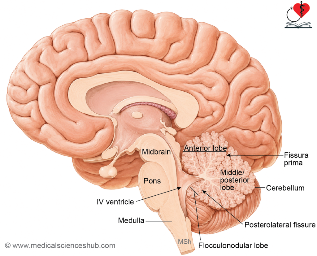
External Features of Cerebellum
- The cerebellum is oval in shape and flattened from above downward.
- Its transverse diameter is greater than its vertical diameter.
- The outer surface shows numerous narrow, parallel folds called folia, which increase the surface area of the cerebellar cortex.
- The cerebellum presents:
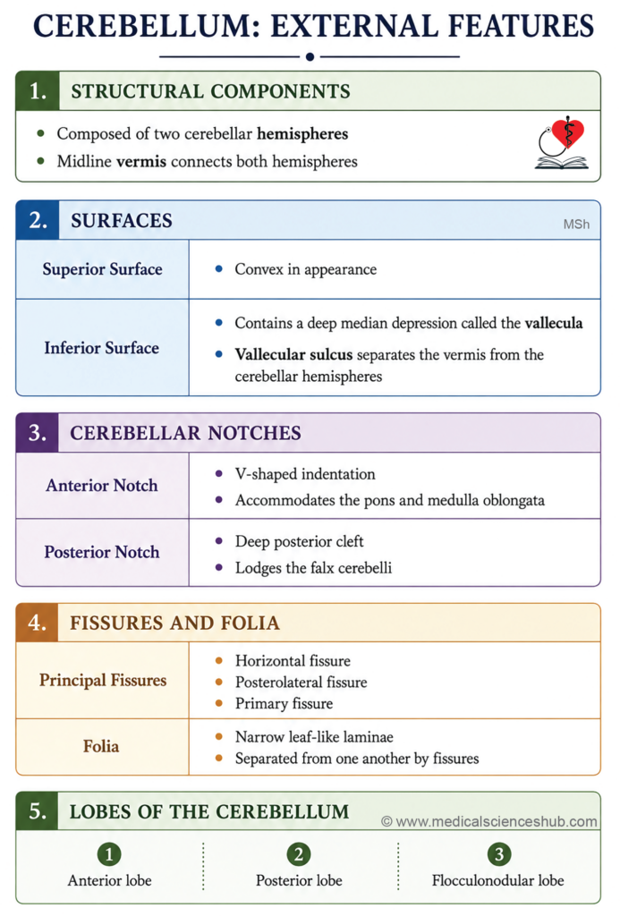
- Two surfaces: superior and inferior
- Two borders: anterior and posterior
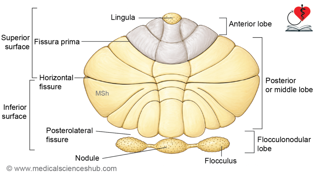
- Three prominent fissures: horizontal fissure, posterolateral fissure, and fissura prima
Parts of the Cerebellum
- Structurally, the cerebellum consists of:
- Two large cerebellar hemispheres located laterally
- A narrow median part called the vermis, which connects the two hemispheres
Surfaces
- The superior surface is convex and forms the upper aspect of the cerebellum.
- It is related superiorly to the tentorium cerebelli.
- The inferior surface is irregular and contains a deep median depression known as the vallecula cerebelli.
- The vermis lies within the vallecula and is visible mainly on the inferior surface.
Notches
- The anterior cerebellar notch is a broad indentation on the anterior aspect of the cerebellum.
- It accommodates the pons and upper medulla oblongata.
- The posterior cerebellar notch is a deep median groove lodging the falx cerebelli.
Folia
- The surface of the cerebellum shows numerous narrow, leaf-like folds called folia.
- These folds are arranged transversely and vary in depth and length.
- Each folium contains a central core of white matter covered externally by gray matter forming the cerebellar cortex.
Major Fissures
- Deep grooves called fissures separate the cerebellar folia.
- Three major fissures are of particular anatomical importance:
- Horizontal fissure
- It runs along the outer margin of the cerebellum.
- It partially separates the superior and inferior surfaces.
- Posterolateral fissure
- It is located on the inferior surface of the cerebellum.
- It separates the flocculonodular lobe from the posterior lobe.
- Primary fissure (fissura prima)
- It appears as a V-shaped groove on the superior surface.
- It separates the anterior lobe from the posterior lobe.
Lobes of the Cerebellum
- The cerebellum is divided into three lobes by two major fissures.
- These lobes are identified according to their anatomical position and developmental significance.
1. Anterior Lobe
- The anterior lobe lies on the superior surface of the cerebellum.
- It is located anterior to the primary fissure (fissura prima).
- This lobe is mainly associated with the regulation of muscle tone and posture.
2. Posterior Lobe
- The posterior lobe is the largest subdivision of the cerebellum.
- It occupies most of the superior surface and a major part of the inferior surface.
- It lies posterior to the primary fissure and superior to the posterolateral fissure.
- This lobe plays an important role in the coordination of skilled voluntary movements.
3. Flocculonodular Lobe
- The flocculonodular lobe is the smallest cerebellar lobe.
- It is situated on the inferior surface of the cerebellum, anterior to the posterolateral fissure.
- Functionally, it is closely related to maintenance of balance and control of eye movements.
Tonsils
- The cerebellar tonsils are rounded masses situated on the inferior surface of the cerebellar hemispheres.
- In conditions causing raised intracranial pressure, the tonsils may descend through the foramen magnum, producing tonsillar herniation.
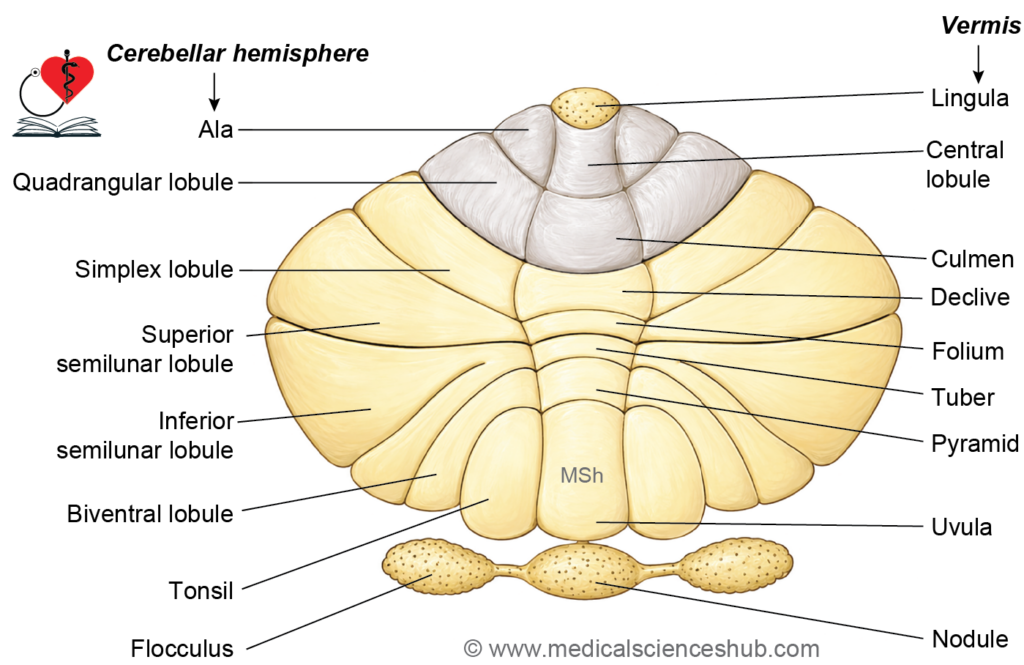
Table 9.1: Subdivisions or lobules of cerebellum
| Major Cerebellar Lobe | Subdivision of Vermis | Corresponding Hemisphere / Cerebellar Lobule | Functional Significance |
|---|---|---|---|
| Anterior Lobe | Lingula | Alae (Ala) | Participates in regulation of posture and muscle tone. |
| Central Lobule | Wing of Central Lobule | Contributes to coordination of trunk and proximal limb movements. | |
| Culmen | Quadrangular Lobule | Involved in control of voluntary motor activity and maintenance of posture. | |
| Posterior Lobe | Declive | Simple Lobule | Assists in coordination and precision of skilled movements. |
| Folium | Superior Semilunar Lobule | Plays a role in fine motor control and cerebellar integration. | |
| Tuber | Inferior Semilunar Lobule | Participates in coordination of complex voluntary actions. | |
| Pyramid | Biventral Lobule | Helps regulate posture and coordinated limb movements. | |
| Uvula | Tonsil | Associated with equilibrium and vestibular functions. | |
| Flocculonodular Lobe | Nodule | Flocculus | Maintains balance, equilibrium, and coordination of eye movements through vestibular connections. |
Phylogenetic Subdivisions of Cerebellum
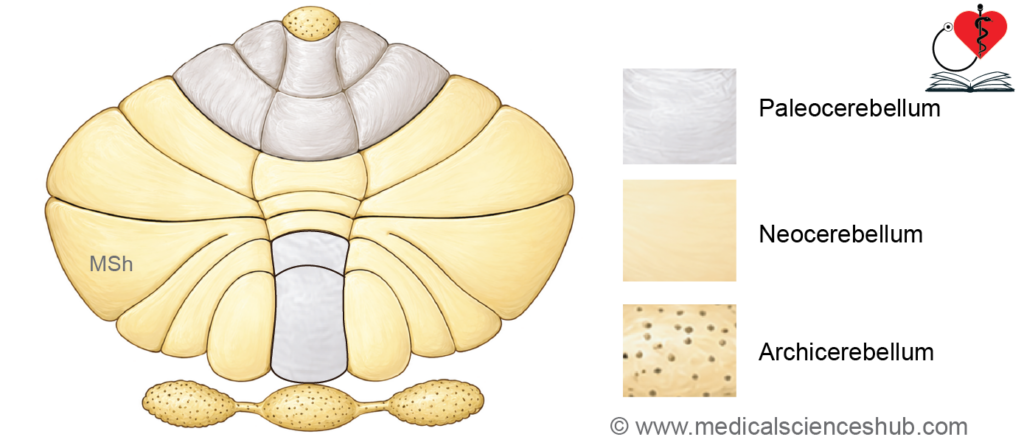
- Based on evolutionary development and functional specialization, the cerebellum is divided into three major parts:
- Archicerebellum (Vestibulocerebellum)
- Paleocerebellum (Spinocerebellum)
- Neocerebellum (Cerebrocerebellum)
- Each subdivision has distinct anatomical components and specific functional roles.
- Lesions affecting these regions produce characteristic disturbances of posture, balance, muscle tone, and coordinated movement.
1. Archicerebellum (Vestibulocerebellum)
- The archicerebellum is the oldest phylogenetic part of the cerebellum.
- It mainly consists of the flocculonodular lobe and the lingula.
- Functionally, it is closely related to the vestibular system.
Functions
- Maintains body equilibrium and posture.
- Regulates balance and coordination of eye movements.
- Helps maintain muscle tone of the trunk.
Effects of Lesions
- Damage may cause disturbances of balance, vertigo, and difficulty maintaining equilibrium.
2. Paleocerebellum (Spinocerebellum)
- The paleocerebellum developed later in evolution and is associated mainly with spinal cord connections.
- It includes:
- Most of the anterior lobe except the lingula
- The uvula and pyramid of the vermis
Functions
- Receives proprioceptive input through spinocerebellar tracts.
- Maintains posture, muscle tone, and coordination of limb movements.
- Assists in execution of coarse voluntary movements.
Effects of Lesions
- Lesions may produce hypotonia, postural instability, and uncoordinated limb movements.
3. Neocerebellum (Cerebrocerebellum)
- The neocerebellum is the most recently evolved and largest functional subdivision.
- It consists mainly of the posterior lobe, excluding the pyramid and uvula.
- It has extensive connections with the cerebral cortex.
Functions
- Coordinates skilled and precise voluntary movements.
- Regulates timing, planning, and smooth execution of motor activity.
Effects of Lesions
Damage may lead to ataxia, intention tremor, inaccurate movements, and impaired fine motor control.
Table 9.2: Cells of cerebellum
| Cell Type | Location | Structural Features | Main Connections | Principal Function |
|---|---|---|---|---|
| Stellate Cells | Superficial part of the molecular layer | Small inhibitory interneurons with short dendritic branches | Synapse mainly with dendrites of Purkinje cells | Modulate cerebellar cortical activity by inhibiting Purkinje cells. |
| Basket Cells | Deeper region of the molecular layer | Small multipolar inhibitory neurons; axons form basket-like endings around Purkinje cell bodies | Synapse with the cell bodies and initial axon segments of Purkinje cells | Provide strong inhibitory control over Purkinje cell output. |
| Purkinje Cells | Purkinje cell layer between molecular and granular layers | Large flask-shaped neurons with highly branched dendritic trees | Receive input from climbing fibers and parallel fibers; axons project to deep cerebellar nuclei | Form the sole output pathway of the cerebellar cortex and regulate coordinated voluntary movements. |
| Granule Cells | Granular layer | Small, densely packed excitatory neurons | Receive input from mossy fibers; axons ascend to form parallel fibers in molecular layer | Transmit excitatory impulses to Purkinje cells and interneurons for cerebellar processing. |
| Golgi Cells | Mainly in the superficial part of the granular layer | Large inhibitory interneurons with extensive dendritic branching | Synapse with granule cells within cerebellar glomeruli | Regulate and modulate granule cell activity through inhibitory feedback. |
Internal Features of Cerebellum
- The cerebellum has a highly folded structure that allows a large number of neurons to be packed within a limited space.
- These folds are known as folia and increase the surface area of the cerebellar cortex.
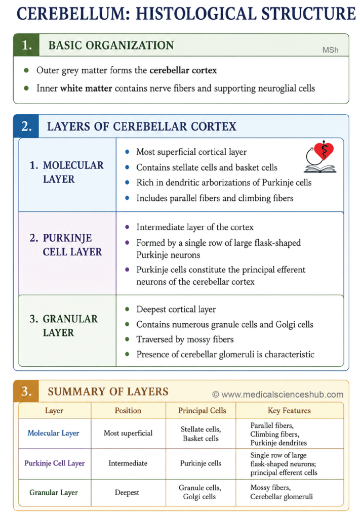
Layers of the Cerebellum
- The cerebellum consists of three main components:
- Outer gray matter forming the cerebellar cortex
- Inner white matter called the medulla
- Deep masses of gray matter known as the cerebellar nuclei
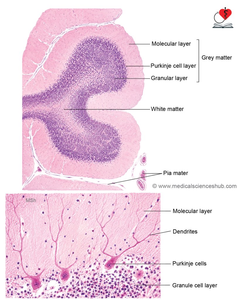
- The cerebellar cortex is organized into three distinct layers:
- Molecular layer
- Purkinje cell layer
- Granular layer
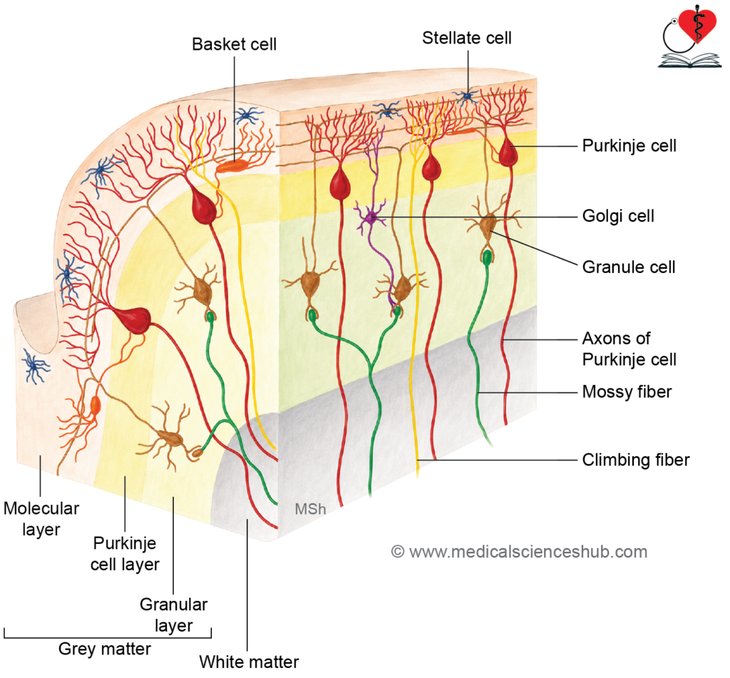
1. Molecular Layer
- The molecular layer is the outermost and relatively cell-poor layer.
- It mainly contains nerve fibers and scattered neurons.
Main Components
- Stellate cells are small inhibitory neurons located superficially.
- Basket cells are deeper neurons whose axons form basket-like networks around Purkinje cell bodies.
- Numerous dendritic branches of Purkinje cells extend into this layer.
- Parallel fibers derived from granule cells are also present.
2. Purkinje Cell Layer
- This layer consists of a single row of large Purkinje cells situated between the molecular and granular layers.
- Purkinje cells are flask-shaped neurons with extensively branched dendrites projecting into the molecular layer.
- Their axons pass through the granular layer into the white matter and terminate in the deep cerebellar nuclei.
- Purkinje cells provide inhibitory output from the cerebellar cortex.
- They play a major role in coordination and fine regulation of body movements.
Functional Importance
3. Granular Layer
- The granular layer is the deepest and most densely packed cortical layer.
- It contains numerous small neurons and specialized synaptic complexes.
Main Components
- Granule cells are small excitatory neurons whose axons ascend into the molecular layer and divide into parallel fibers.
- Golgi cells are inhibitory interneurons located within this layer.
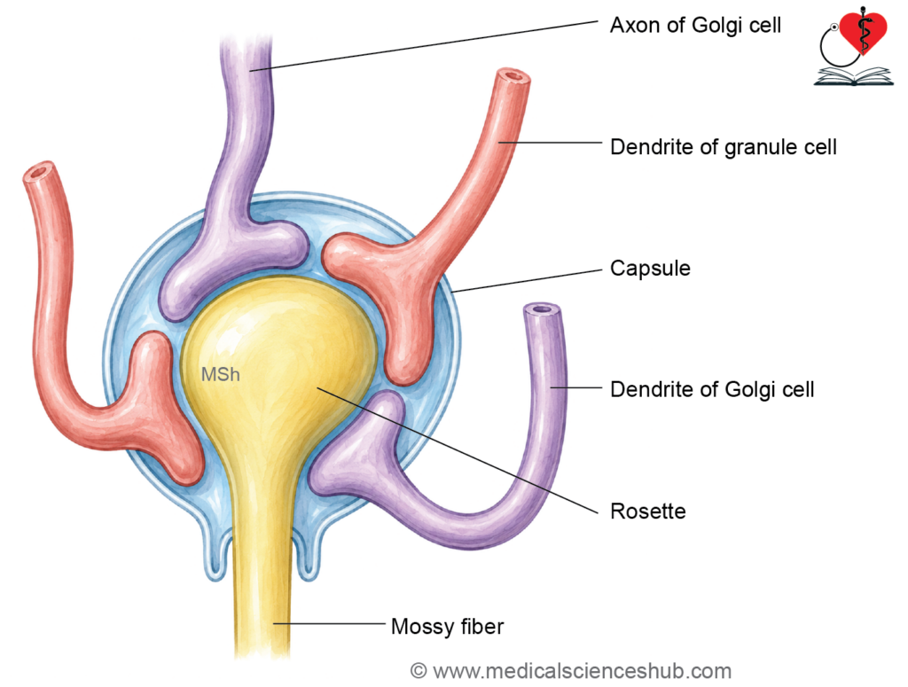
- Synaptic complexes called cerebellar glomeruli are formed by mossy fibers, granule cell dendrites, and Golgi cell processes.
Functional Importance
- The granular layer receives and processes sensory input carried by mossy fibers before transmitting signals to Purkinje cells.
White Matter
- The cerebellar white matter consists mainly of myelinated nerve fibers.
- It contains incoming and outgoing fiber tracts that connect the cerebellum with other parts of the nervous system.
Afferent Fibers of Cerebellum
- The cerebellum receives sensory and motor information through two major types of afferent fibers:
- Climbing fibers
- Mossy fibers
1. Climbing Fibers
- Climbing fibers arise mainly from the inferior olivary nucleus of the medulla oblongata.
- These fibers ascend into the cerebellar cortex and form extensive synaptic connections with the dendrites of Purkinje cells.
- A single climbing fiber establishes strong functional connections with only a limited number of Purkinje cells.
Functional Importance
- Climbing fibers play an important role in motor coordination and motor learning.
- They provide powerful excitatory input to Purkinje cells and help regulate precise movements.
2. Mossy Fibers
- Mossy fibers constitute the majority of cerebellar afferent fibers.
- They originate from several sources, including the vestibulocerebellar, pontocerebellar, and spinocerebellar pathways.
- Within the granular layer, mossy fibers terminate by forming synaptic complexes called cerebellar glomeruli.
- These fibers synapse mainly with the dendrites of granule cells and Golgi cells.
Functional Importance
- Mossy fibers transmit sensory and proprioceptive information to the cerebellum.
- They assist in maintaining posture, balance, and coordination of voluntary movements.
Cerebellar glomerulus
The cerebellar glomerulus is a synaptic complex where a mossy fiber rosette forms the central excitatory input, synapsing onto granule cell dendrites and Golgi cell axon terminals, all enclosed within a glial sheath, enabling precise modulation of cerebellar input signals.
Cerebellar Nuclei
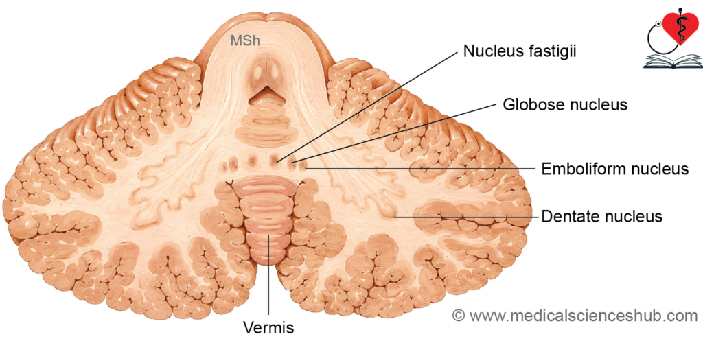
- The cerebellar nuclei are masses of gray matter located deep within the white matter of the cerebellum.
- There are four paired nuclei present on each side of the cerebellum.
- These nuclei serve as the major output centers of the cerebellum and are involved in coordination of movement, posture, and balance.
Types of Cerebellar Nuclei
1. Dentate Nucleus
- The dentate nucleus is the largest and most lateral cerebellar nucleus.
- It has a folded, sac-like appearance with an opening directed medially.
- Functionally, it is associated with the neocerebellum.
Functions
- Coordinates skilled and precise voluntary movements.
- Plays an important role in planning and timing of motor activity, especially movements of the hands and fingers.
2. Emboliform Nucleus
- The emboliform nucleus lies medial to the dentate nucleus.
- It is functionally related to the spinocerebellum.
Functions
- Helps regulate muscle tone and posture.
- Assists in coordination and fine adjustment of limb movements.
3. Globose Nucleus
- The globose nucleus is situated medial to the emboliform nucleus.
- Together, the globose and emboliform nuclei are often referred to as the interposed nuclei.
Functions
- Contributes to control of posture and ongoing limb movements.
- Participates in coordination of voluntary motor activity.
4. Fastigial Nucleus
- The fastigial nucleus lies close to the midline within the vermis, near the roof of the fourth ventricle.
- It is primarily associated with the vestibulocerebellum.
Functions
- Maintains balance and body posture.
- Helps coordinate movements of the trunk and eyes.
White Matter of Cerebellum
- The white matter forms the central core, or medulla, of the cerebellum.
- In sagittal section, it shows a characteristic branching pattern called the arbor vitae because of its tree-like appearance.
Components of Cerebellar White Matter
- The white matter contains three major categories of nerve fibers:
- Intrinsic fibers
- Afferent fibers
- Efferent fibers
1. Intrinsic Fibers
- Intrinsic fibers remain entirely within the cerebellum.
- They connect different regions of the cerebellar cortex and cerebellar nuclei.
- These fibers help integrate cerebellar activity and coordinate internal processing.
2. Projection Fibers
- Projection fibers include both afferent and efferent fibers.
- They connect the cerebellum with the brainstem, spinal cord, and other parts of the central nervous system.
Cerebellar Peduncles
- Projection fibers pass through three paired bundles known as cerebellar peduncles:
- Superior cerebellar peduncle
- Middle cerebellar peduncle
- Inferior cerebellar peduncle
- These peduncles serve as major pathways for communication between the cerebellum and the rest of the nervous system.
Connections of Cerebellum
Inferior Cerebellar Peduncle
- The inferior cerebellar peduncle connects the cerebellum with the medulla oblongata.
- It is also known as the restiform body.
- This peduncle carries mainly afferent fibers transmitting sensory and proprioceptive information to the cerebellum.
- It also contains some efferent fibers arising from cerebellar nuclei.
Afferent Fibers of the Inferior Cerebellar Peduncle
1. Posterior Spinocerebellar Fibers
- These fibers arise from neurons of the posterior thoracic nucleus (Clarke’s column) in the spinal cord.
- They carry unconscious proprioceptive information from muscles and joints of the lower limb and trunk.
- Their main function is maintenance of posture and coordination of movement.
2. Vestibulocerebellar Fibers
- These fibers originate from the vestibular nerve and vestibular nuclei.
- They terminate mainly in the flocculonodular lobe.
- They convey information related to equilibrium and head position.
Functions
- Maintain balance and posture.
- Assist in coordination of eye movements.
3. Olivocerebellar Fibers
- These fibers arise from the inferior olivary nucleus of the medulla.
- They enter the cerebellum as climbing fibers and synapse with Purkinje cells.
Functions
- Participate in motor learning and fine coordination of movements.
- Help regulate timing and precision of voluntary activity.
4. Reticulocerebellar Fibers
- These fibers originate from the reticular formation of the brainstem.
- They influence cerebellar control of muscle tone and motor activity.
5. Cuneocerebellar Fibers
- These fibers arise from the accessory cuneate nucleus.
- They carry unconscious proprioceptive impulses from the upper limb and neck.
- They assist in coordination and positioning of upper limb movements.
6. Arcuatocerebellar Fibers
- These fibers originate from nuclei of the medulla oblongata.
- They are involved in regulation of posture and muscle tone.
7. Trigeminocerebellar Fibers
- These fibers arise from sensory nuclei of the trigeminal nerve.
- They transmit proprioceptive sensations from the face and oral structures to the cerebellum.
Efferent Fibers of the Inferior Cerebellar Peduncle
1. Cerebellovestibular Fibers
- These fibers arise mainly from the fastigial nucleus and project to the vestibular nuclei.
Functions
- Help maintain equilibrium and posture.
- Regulate coordinated eye and head movements.
2. Cerebelloreticular Fibers
- These fibers project from cerebellar nuclei to the reticular formation.
Functions
- Influence muscle tone and postural reflexes.
- Assist in control of voluntary motor activity.
3. Cerebello-olivary Fibers
- These fibers extend from cerebellar nuclei to the inferior olivary nucleus.
- They form part of feedback pathways involved in motor coordination and motor learning.
Middle Cerebellar Peduncle
- The middle cerebellar peduncle is the largest cerebellar peduncle and connects the cerebellum with the pons.
- It consists almost entirely of afferent fibers that transmit information from the cerebral cortex to the cerebellum through pontine nuclei.
- These fibers are important for coordination and planning of voluntary movements.
Afferent Fibers of the Middle Cerebellar Peduncle
1. Pontocerebellar Fibers
- Pontocerebellar fibers arise from the pontine nuclei of the opposite side.
- They carry impulses from the cerebral cortex to the cerebellum through the corticopontocerebellar pathway.
Functions
- Help coordinate skilled voluntary movements.
- Assist in planning, timing, and smooth execution of motor activity.
2. Reticulocerebellar Fibers
- These fibers originate from the reticular formation of the pons and medulla.
- They convey information related to posture and ongoing motor activity.
Functions
- Contribute to maintenance of balance and muscle tone.
- Help regulate body movements during posture and locomotion.
Efferent Fibers
- The middle cerebellar peduncle does not contain significant efferent fibers.
Superior Cerebellar Peduncle
- The superior cerebellar peduncle connects the cerebellum with the midbrain.
- It contains predominantly efferent fibers, although a few important afferent tracts are also present.
- This peduncle serves as the principal output pathway of the cerebellum for regulation of voluntary motor activity.
Afferent Fibers of the Superior Cerebellar Peduncle
1. Anterior Spinocerebellar Tract
- These fibers arise mainly from neurons of the spinal cord.
- They carry unconscious proprioceptive information from the trunk and lower limbs.
- Most fibers cross within the spinal cord and many recross before terminating in the cerebellum.
Functions
2. Tectocerebellar Fibers
- Provide feedback regarding ongoing limb movements.
- Assist in coordination of posture and locomotion.
- These fibers originate from the superior and inferior colliculi of the midbrain tectum.
Functions
- Convey visual and auditory reflex information to the cerebellum.
- Help coordinate head and eye movements in response to sensory stimuli.
3. Trigeminocerebellar Fibers
- These fibers arise from sensory nuclei of the trigeminal nerve.
- They transmit proprioceptive impulses from facial muscles, the jaw, and oral structures.
Functions
- Assist in coordination of facial and jaw movements.
- Contribute to regulation of chewing and speech-related motor activity.
4. Rubrocerebellar Fibers
- These fibers originate from the red nucleus of the midbrain.
Functions
- Influence cerebellar control of limb movements.
- Participate in motor coordination and motor learning.
5. Corticocerebellar Fibers
- These fibers indirectly relay impulses from the cerebral cortex to the cerebellum.
Functions
- Help integrate cortical motor planning with cerebellar coordination.
- Contribute to smooth execution of skilled voluntary movements.
Efferent Fibers of the Superior Cerebellar Peduncle
1. Cerebellorubral Fibers
- These fibers arise mainly from the dentate, emboliform, and globose nuclei and terminate in the red nucleus.
Functions
- Influence voluntary motor control through connections with descending motor pathways.
- Help coordinate fine movements of the limbs.
2. Dentatorubral and Dentatothalamic Fibers
- These fibers originate from the dentate nucleus.
- They project to the red nucleus and thalamus, respectively.
Functions
- Connect the cerebellum with the motor areas of the cerebral cortex.
- Regulate initiation, planning, and precision of voluntary movements.
3. Cerebello-olivary Fibers
- These fibers pass from the cerebellar nuclei to the inferior olivary nucleus.
Functions
- Form part of feedback circuits involved in motor learning.
- Help refine and correct ongoing movements through continuous cerebellar modulation.
Blood Supply of Cerebellum
Arterial Supply
- The cerebellum receives arterial blood through three paired cerebellar arteries.
- These arteries arise from the vertebrobasilar system and supply different regions of the cerebellum and brainstem.
Major Cerebellar Arteries
- Superior cerebellar artery (SCA)
- Usually arises from the terminal part of the basilar artery.
- Supplies the superior surface of the cerebellum and parts of the midbrain.
- Anterior inferior cerebellar artery (AICA)
- Arises from the basilar artery.
- Supplies the anterior and inferior regions of the cerebellum and adjacent brainstem structures.
- Posterior inferior cerebellar artery (PICA)
- Commonly arises from the vertebral artery.
- Supplies the inferior surface of the cerebellum and parts of the medulla oblongata.
Venous Drainage
Venous blood from the cerebellum drains through superior and inferior cerebellar veins.
Superior Cerebellar Veins
- These veins drain mainly into the straight sinus, transverse sinus, and superior petrosal sinus.
Inferior Cerebellar Veins
- These veins drain into the transverse sinus, inferior petrosal sinus, and nearby venous channels.
CLINICAL NEUROANATOMY
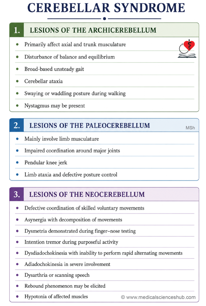
- Cerebellar syndrome refers to a group of clinical signs and symptoms caused by lesions of the cerebellum.
- The manifestations depend on the specific functional part of the cerebellum involved.
- Functionally, cerebellar lesions may involve:
- Archicerebellum (vestibulocerebellum)
- Paleocerebellum (spinocerebellum)
- Neocerebellum (cerebrocerebellum)
Lesions of the Archicerebellum (Flocculonodular Lobe)
- The archicerebellum maintains equilibrium, posture, and coordination of eye movements.
- It receives major input from the vestibular nuclei.
- Damage mainly affects balance and control of body posture.
Clinical Features
1. Disequilibrium
- The patient develops an unsteady, staggering gait known as cerebellar ataxia.
- Swaying or falling may occur in any direction while standing or walking.
2. Broad-Based or Waddling Gait
- The patient walks with feet widely separated to improve stability.
- This wide-based gait acts as a compensatory mechanism to reduce falls.
3. Nystagmus
- Nystagmus consists of involuntary rhythmic eye movements.
- It occurs because coordination between head and eye movements is impaired.
Lesions of the Paleocerebellum (Spinocerebellum)
- The paleocerebellum regulates muscle tone, posture, and simple limb movements.
- Lesions interfere with proprioceptive control of limb muscles.
Major Effects
1. Hypotonia
- Hypotonia refers to reduced muscle tone.
- Limbs become soft, floppy, and less resistant to passive movement.
2. Asthenia
- Asthenia is muscular weakness with inability to sustain prolonged contractions during voluntary activity.
3. Flail Joints
- Poor muscular support causes joints to appear loose and unstable.
- Limb movements become excessive and poorly controlled.
4. Pendular Knee Jerk
- After eliciting the patellar reflex, the leg swings repeatedly like a pendulum.
- This occurs due to reduced muscle tone and impaired damping of movement.
5. Cerebellar Ataxia
- The patient shows impaired coordination and difficulty maintaining balance during standing or walking.
- Gait becomes irregular, staggering, and unstable.
Lesions of the Neocerebellum (Cerebrocerebellum)
- The neocerebellum coordinates skilled and precise voluntary movements.
- Lesions mainly disturb planning, timing, and smooth execution of motor activity.
Clinical Features
1. Asynergia
- There is loss of coordinated action between different muscle groups.
- Movements become fragmented and jerky instead of smooth.
2. Dysmetria
- Dysmetria is inability to judge the range and force of movement accurately.
- Movements may overshoot or undershoot the intended target.
Past-Pointing Test
- During the finger–nose test, the patient misses the target because of impaired movement control.
3. Intention Tremor
- Tremor appears during voluntary movement and increases near the target.
- It disappears at rest.
4. Dysdiadochokinesia
- The patient cannot perform rapid alternating movements smoothly.
- Movements become slow, irregular, and poorly coordinated.
5. Dysarthria
- Speech becomes slow, slurred, and irregular due to incoordination of speech muscles.
- This produces characteristic scanning speech.
6. Rebound Phenomenon
- The patient cannot abruptly stop movement after sudden removal of resistance.
- The limb moves excessively because corrective control is impaired.
7. Hypotonia
- Reduced cerebellar influence on stretch reflexes causes decreased muscle tone and floppy limbs.
Lesions of the Vermis and Paravermal Zone
- The vermis and paravermal zone control posture and coordination of trunk and proximal limb muscles.
- These regions receive input from spinal and vestibular pathways.
Clinical Features
1. Truncal Ataxia
- The trunk sways during sitting or standing due to impaired postural control.
2. Abnormal Stance and Gait
- The patient stands or walks with feet widely separated.
- Gait becomes lurching and unstable.
3. Nystagmus
- Involuntary rhythmic eye movements occur because of defective vestibular coordination.
Lesions of the Cerebellar Hemisphere
- Lesions of a cerebellar hemisphere produce symptoms mainly on the ipsilateral side of the body.
Common Manifestations
- Ataxia
- Intention tremor
- Dysmetria
- Dysdiadochokinesia
- Dysarthria
- Hypotonia
- Delayed initiation of movement
- Abnormal stance and gait
- Nystagmus and rebound phenomenon
Important Questions
- Describe the gross features of cerebellum.
- Write a short note on phylogenetic or morphological subdivisions of the cerebellum.
- Draw a well-labeled diagram of histology of cerebellum.
- List the cerebellar nuclei.
- Write a short note on connection of cerebellum.
- Write a note on applied aspects of cerebellum.
📝 Test Your Knowledge – Practice MCQs
Attempt the chapter MCQ quiz and assess your understanding of key concepts.
Click to see: Medical Disclaimer and Copyright Notice