
Competencies
- AN57.1: Identify external features of spinal cord.
- AN57.2: Describe extent of spinal cord in children and adults with its clinical implications.
- AN57.3: Draw and label transverse section of spinal cord at midcervical and midthoracic level.
- AN57.5: Describe anatomical basis of syringomyelia. AN64.2: Describe the development of neural tube, spinal cord, …
Introduction
The spinal cord is an elongated, cylindrical structure of the central nervous system that occupies the upper two-thirds of the vertebral canal. It represents the caudal continuation of the brain and gives rise to 31 pairs of spinal nerves, which emerge segmentally to supply the trunk and limbs.
Functionally, the spinal cord serves two primary roles. First, it acts as a bidirectional conduit, transmitting sensory information from the body to the brain via ascending tracts and relaying motor commands from the brain to peripheral effectors via descending tracts. Second, it integrates and executes spinal reflex arcs — rapid, stereotyped responses to sensory stimuli that do not require direct cortical involvement. These reflexes underlie essential functions such as the maintenance of posture, coordination of movement, and protective withdrawal responses, allowing the nervous system to react to stimuli with minimal processing delay.
Size and Shape
- The spinal cord is cylindrical in form and slightly flattened in the anteroposterior (front-to-back) plane.
Measurements in Adults
- Length: approximately 45 cm in males and 42 cm in females.
- Width: approximately 1.25 cm at its widest point.
- Weight: approximately 30 g.
Extent
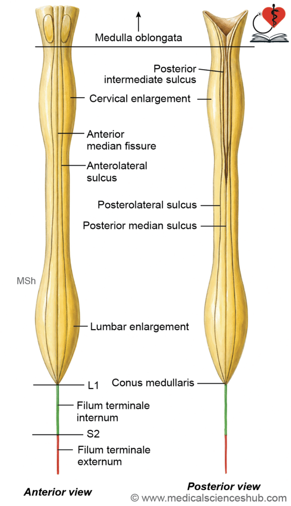
- The spinal cord extends from the upper border of the posterior arch of the first cervical vertebra (C1) superiorly to the lower border of the first lumbar vertebra (L1) inferiorly in adults.
- This relationship is not fixed at birth; in neonates, the cord extends to approximately L3, gradually ascending to its adult level as the vertebral column grows disproportionately faster than the spinal cord.
Conus Medullaris
- The spinal cord tapers inferiorly to form a cone-shaped terminus known as the conus medullaris, situated at the level of L1 in most adults.
Filum Terminale
- Extending from the tip of the conus medullaris is a slender, fibrous strand called the filum terminale.
- It descends through the subarachnoid space and anchors the spinal cord to the posterior surface of the first coccygeal vertebral segment (Co1), providing longitudinal support and limiting excessive movement of the cord.
Coverings of Spinal Cord
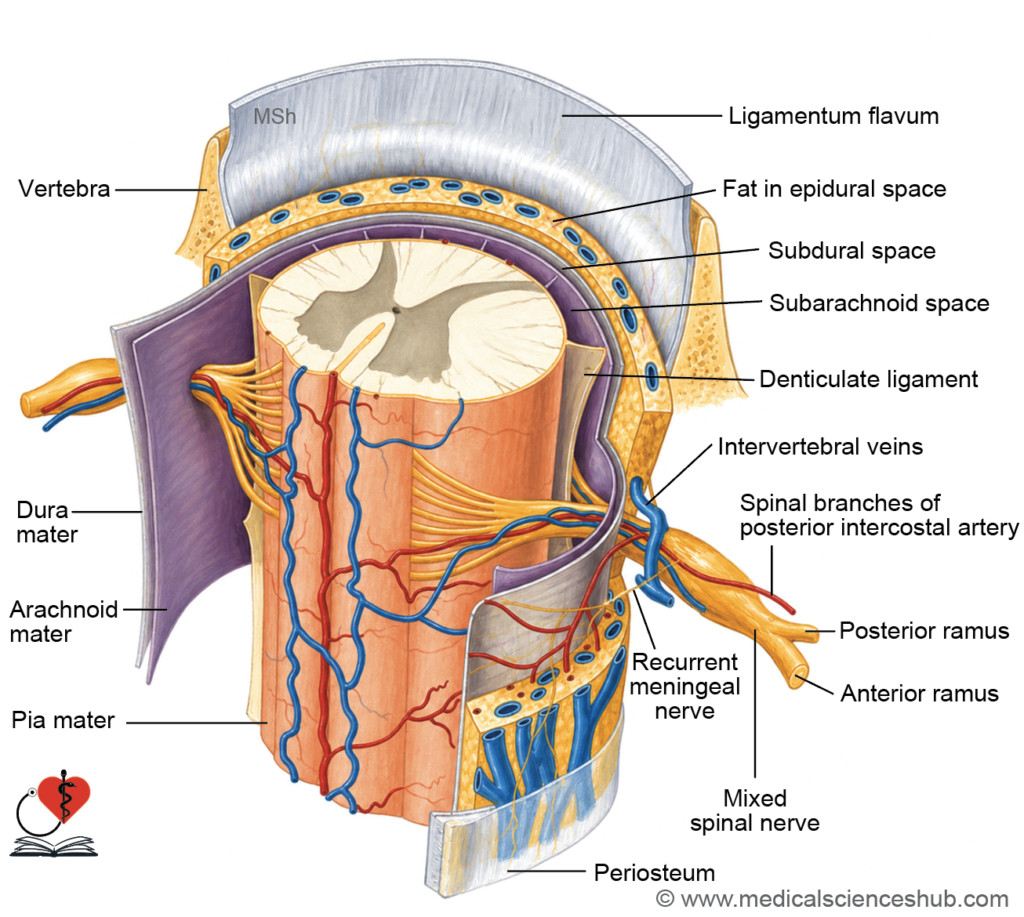
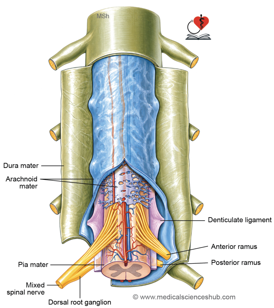
- The spinal cord is enclosed by three concentric membranous layers collectively termed the spinal meninges: the outermost dura mater, the middle arachnoid mater, and the innermost pia mater.
- Together, these layers protect the spinal cord, enclose the cerebrospinal fluid (CSF), and provide structural support against displacement during movement.
Dura Mater
- The dura mater is the outermost and toughest of the three meningeal layers, composed predominantly of dense fibrous connective tissue.
- It extends from the foramen magnum superiorly to the lower border of the second sacral vertebra (S2) inferiorly, where it fuses with the filum terminale to form the coccygeal ligament.
Arachnoid Mater
- The arachnoid mater is a thin, delicate, and translucent membrane that loosely surrounds the spinal cord, separated from the pia mater below by the subarachnoid space, which contains circulating cerebrospinal fluid.
- Like the dura mater, it extends inferiorly to the level of S2, forming the lower boundary of the lumbar cistern.
Pia Mater
- The pia mater is the innermost meningeal layer — a thin, highly vascular membrane that intimately adheres to the surface of the spinal cord and its nerve roots.
Modifications of Spinal Pia Mater
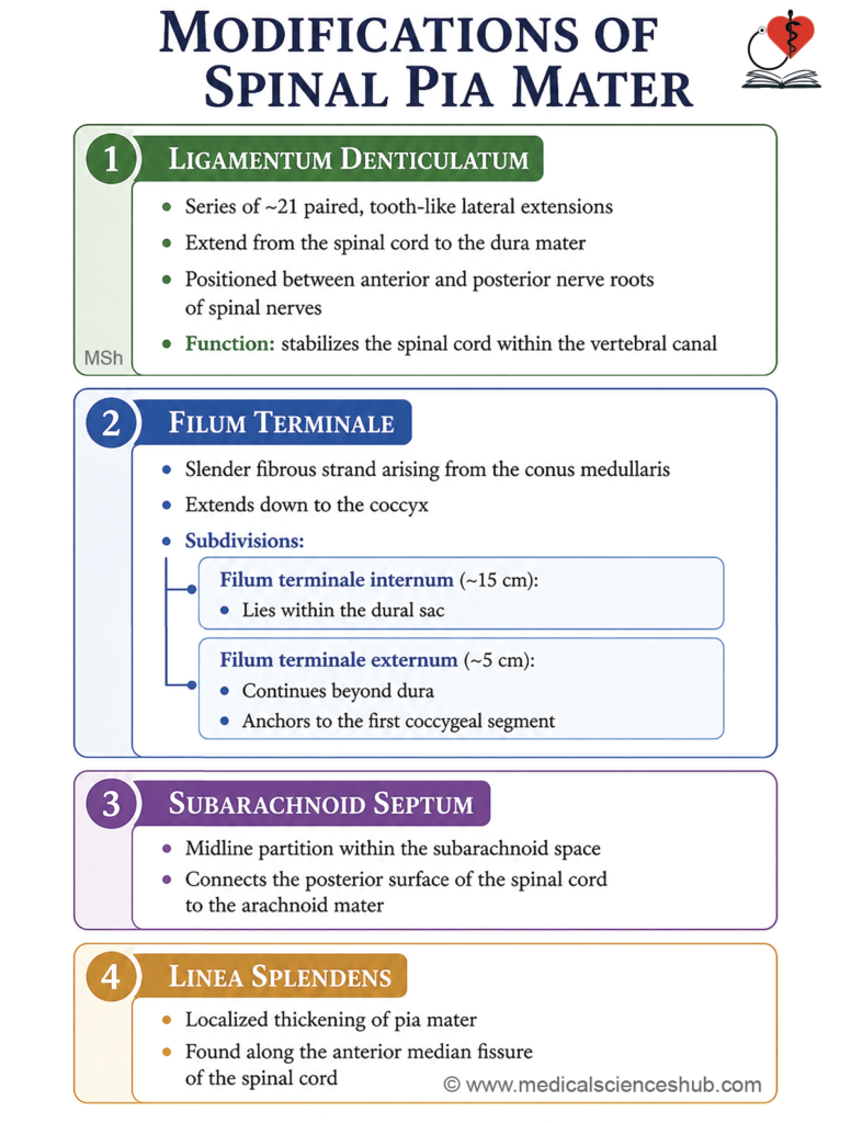
The spinal pia mater gives rise to several specialized structural modifications that stabilize the spinal cord within the vertebral canal, preventing excessive displacement during body movements and protecting the associated vessels and neural tissue.
1. Ligamentum Denticulatum (Denticulate Ligament)
- These are 21 pairs of tooth-like lateral extensions of the pia mater that project from the lateral surface of the spinal cord.
- Each projection pierces the arachnoid mater and attaches firmly to the inner surface of the dura mater, anchoring the cord between the anterior and posterior nerve roots of adjacent spinal segments.
- The first pair is located at the level of the foramen magnum, and the last pair lies between the T12 and L1 spinal nerve roots.
- The denticulate ligaments serve as important surgical landmarks, particularly during anterolateral cordotomy, where they guide the surgeon’s orientation relative to the anterior and posterior nerve roots.
2. Filum Terminale
- The filum terminale is a slender, fibrous extension of the pia mater that descends from the tip of the conus medullaris to the first coccygeal segment, providing longitudinal support to the cord.
- It measures approximately 20 cm in total length and exits the vertebral canal through the sacral hiatus.
- It is divided into two anatomically distinct parts:
- Filum terminale internum: The upper segment, approximately 15 cm in length, enclosed within the dura mater and arachnoid mater as it traverses the lumbar cistern.
- Filum terminale externum: The lower segment, approximately 5 cm in length, which lies outside the dural sac and attaches to the posterior surface of the first coccygeal vertebral segment (Co1).
3. Subarachnoid Septum
- The subarachnoid septum is a midline sheet of pial tissue that connects the posterior surface of the spinal cord to the arachnoid mater.
- It partially subdivides the subarachnoid space in the posterior midline and contributes to the positional stability of the cord.
4. Linea Splendens
- The linea splendens is a longitudinal thickening of the pia mater located in the anterior median plane, overlying the anterior median fissure of the spinal cord.
- It reinforces the pial covering in this region and provides structural protection to the underlying blood vessels running along the anterior surface of the cord.
CLINICAL NEUROANATOMY
Ligamentum Denticulatum as a Surgical Landmark
- The ligamentum denticulatum serves as a critical anatomical reference point during cordotomy, a neurosurgical procedure performed to interrupt pain-conducting pathways in the spinal cord.
- Pain relief (anterolateral cordotomy): The spinothalamic tract, which transmits pain and temperature signals, lies anterolateral to the denticulate ligament. Surgical sectioning of the cord posterior to the ligamentum denticulatum interrupts these ascending pain pathways, providing relief in patients with intractable pain.
- Motor pathway interruption: The corticospinal (pyramidal) tract, which carries voluntary motor commands, lies anterior to the denticulate ligament. Sectioning the cord anterior to the ligamentum denticulatum disrupts this descending tract, resulting in ipsilateral muscle paralysis.
- Nerve root identification: The last (most inferior) pair of denticulate ligaments is consistently located just above the first lumbar (L1) spinal nerve root. This relationship provides a reliable intraoperative reference for identifying and confirming the level of specific spinal nerve roots during surgery, reducing the risk of inadvertent injury to adjacent structures.
Space Surrounding The Spinal Cord
- The spinal cord is surrounded by three distinct spaces, arranged from outermost to innermost: the epidural space, the subdural space, and the subarachnoid space. Each has a unique anatomical boundary and clinical significance.
1. Epidural Space
- The epidural space (also called the extradural space) is situated between the inner wall of the vertebral canal and the outer surface of the spinal dura mater.
- It contains loose areolar connective tissue, adipose tissue, small arterial branches, the internal vertebral venous plexus, and the proximal portions of the spinal nerve roots as they exit through the intervertebral foramina.
- This space is clinically significant as the target site for epidural anesthesia and epidural steroid injections.
2. Subdural Space
- The subdural space is a narrow potential space located between the inner surface of the dura mater and the outer surface of the arachnoid mater.
- Under normal conditions, it contains only a minimal film of serous fluid sufficient to moisten the opposing surfaces; it is not a true fluid-filled cavity.
3. Subarachnoid Space
- The subarachnoid space lies between the arachnoid mater and the pia mater and is filled with cerebrospinal fluid (CSF), which cushions the spinal cord against mechanical trauma and maintains a stable ionic environment.
- It is traversed by delicate fibrous strands called arachnoid trabeculae and is continuous superiorly with the cranial subarachnoid space.
Lumbar Cistern
- Inferior to the conus medullaris (approximately at the L1 vertebral level), the spinal cord proper ends but the subarachnoid space continues, forming an expanded CSF-filled reservoir known as the lumbar cistern.
- The lumbar cistern extends from approximately L1 to S2 and contains the cauda equina — the descending lumbosacral nerve roots suspended within the CSF.
- This cistern is the preferred site for lumbar puncture (spinal tap), as needle insertion below L1–L2 avoids direct injury to the spinal cord while allowing safe access to the CSF.
CLINICAL NEUROANATOMY
Epidural Anesthesia
- Epidural anesthesia involves the injection of a local anesthetic agent into the epidural space, located between the inner wall of the vertebral canal and the outer surface of the spinal dura mater.
- The anesthetic agent diffuses across the dural sleeve to block spinal nerve roots at the targeted segmental levels, producing regional loss of sensation and pain.
- It is widely used for lower limb surgeries, gynecological procedures, cesarean sections, and postoperative pain management, offering the advantage of regional analgesia while preserving consciousness.
Lumbar Puncture (Spinal Tap)
Indications
- Lumbar puncture is performed to obtain a sample of cerebrospinal fluid (CSF) for the following purposes:
- Diagnostic: Investigation of meningitis, subarachnoid hemorrhage, encephalitis, demyelinating disorders, and other neurological conditions where CSF analysis provides critical information.
- Therapeutic: Controlled removal of CSF to reduce raised intracranial pressure in conditions such as idiopathic intracranial hypertension.
Contraindications
- Lumbar puncture should be avoided in the following situations:
- Active skin or soft tissue infection at the proposed puncture site, due to the risk of introducing organisms into the subarachnoid space.
- Clinical or radiological evidence of raised intracranial pressure, as sudden CSF withdrawal may precipitate transtentorial herniation.
- Severe thrombocytopenia or other significant coagulopathies, owing to the risk of epidural hematoma formation.
Patient Positioning
- The patient is placed either in the lateral decubitus position (lying on one side) with the knees drawn up toward the chest, or in the seated position leaning forward.
- Both positions maximally flex the lumbar spine, widening the interspinous spaces and facilitating needle insertion into the subarachnoid space.
Procedure
- The skin over the lower back is prepared under strict aseptic conditions, and local anesthesia is infiltrated into the skin and deeper tissues at the chosen site.
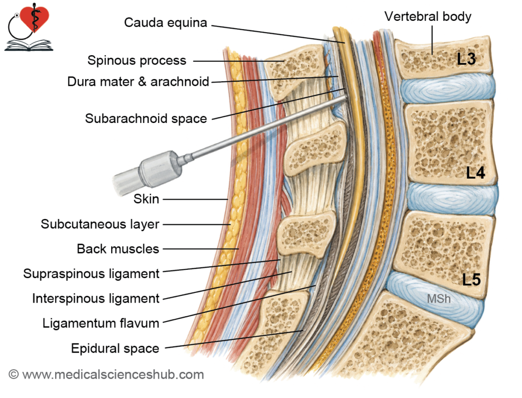
- A spinal needle is inserted in the midline between the L3–L4 or L4–L5 vertebral interspaces into the subarachnoid space, and the required volume of CSF is collected before the needle is gently withdrawn.
- The L4 spinous process serves as a reliable surface landmark, as it lies approximately on the intercristal line — a horizontal line connecting the highest points of the two iliac crests.
Rationale for Site Selection
- In adults, the spinal cord terminates at the lower border of L1, below which the vertebral canal contains only the cauda equina — the descending lumbar, sacral, and coccygeal nerve roots — suspended freely in CSF within the lumbar cistern.
- Needle insertion below the L1–L2 level therefore avoids direct injury to the spinal cord; nerve roots of the cauda equina are mobile and tend to deflect away from an advancing needle rather than being transected.
- The L3–L4 interspace is preferred because the lumbar cistern is widest at this level, providing the greatest volume of accessible CSF and the highest margin of procedural safety.
External Features of Spinal Cord
The external features of the spinal cord comprise four principal components: fissures and sulci, spinal nerve attachments, enlargements, and the cauda equina.
Fissures and Sulci
- The spinal cord is slightly flattened in the anteroposterior plane and presents several longitudinal grooves on its surface that serve as important anatomical landmarks and attachment sites.
- Anterior median fissure: A deep, midline groove running along the entire anterior surface of the cord, lined by pia mater and housing the anterior spinal artery. It partially subdivides the cord anteriorly but does not extend to the central canal.
- Posterior median sulcus: A shallow midline groove on the posterior surface from which a thin glial partition, the posterior median septum, projects inward toward the central canal, partially dividing the posterior cord.
- Ventrolateral (anterolateral) sulcus: A poorly defined groove on the anterolateral surface through which the ventral (anterior) rootlets of the spinal nerves emerge in a series of small fascicles.
- Dorsolateral (posterolateral) sulcus: A more consistently identifiable groove located on the posterolateral surface of the cord, approximately 2 mm lateral to the posterior median sulcus, marking the entry zone of the dorsal (posterior) rootlets of the spinal nerves.
- Dorsointermediate (posterointermediate) sulcus: A shallow groove situated between the posterior median and posterolateral sulci, present only at cervical and upper thoracic levels. It demarcates the fasciculus gracilis medially from the fasciculus cuneatus laterally within the posterior funiculus.
Attachment of Spinal Nerve
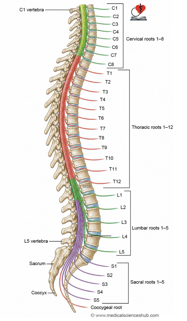
- The spinal cord gives rise to 31 pairs of spinal nerves, distributed as follows: 8 cervical, 12 thoracic, 5 lumbar, 5 sacral, and 1 coccygeal pair.
- Each spinal nerve is formed by the union of two roots:
- Ventral (anterior) root: Carries efferent (motor) fibers originating from the anterior horn cells of the spinal cord gray matter to skeletal muscles and autonomic effectors.
- Dorsal (posterior) root: Carries afferent (sensory) fibers conveying sensory information from the periphery to the spinal cord. It bears an oval enlargement called the dorsal root ganglion (spinal ganglion), which contains pseudounipolar neurons.
- Each pseudounipolar neuron in the dorsal root ganglion has a single process that divides in a T-shaped configuration into:
- A central process that enters the spinal cord via the dorsolateral sulcus to synapse within the posterior horn or ascend in the posterior funiculus.
- A peripheral process that travels distally to join the ventral root, forming the mixed spinal nerve proper.
Spinal Segments
- A spinal segment is defined as the portion of the spinal cord that gives attachment to one pair of spinal nerves — one on each side.
- The spinal cord comprises 31 spinal segments in total: 8 cervical, 12 thoracic, 5 lumbar, 5 sacral, and 1 coccygeal.
- Each spinal nerve exits the vertebral canal through its corresponding intervertebral foramen.
- Because the spinal cord in adults terminates at the lower border of L1, the spinal segments do not correspond directly to the vertebral levels of the same number. Upper spinal nerves (cervical) run nearly horizontally to reach their foramina, whereas lower spinal nerves (lumbar, sacral, coccygeal) descend at increasingly oblique angles before exiting, forming the cauda equina.
- Accurate knowledge of spinal segment–vertebral level relationships is clinically essential for lesion localization, planning lumbar puncture, administering spinal anesthesia, and guiding surgical approaches.
Enlargements of Spinal Cord
- The spinal cord presents two fusiform enlargements that correspond to regions supplying the upper and lower limbs, where a substantially greater number of motor and sensory neurons are concentrated.
- Cervical enlargement:
- Formed by the large volume of neural tissue required to innervate the upper limbs.
- Extends from the C3 to T2 spinal segments (Gray’s Anatomy, 42nd edition).
- Reaches its maximum diameter of approximately 3.8 cm at the level of the sixth cervical (C6) spinal segment.
- Lumbar enlargement:
- Formed by the neural tissue required to innervate the lower limbs.
- Extends from the L1 to S3 spinal segments (Gray’s Anatomy, 42nd edition).
- Reaches its maximum diameter of approximately 3.5 cm at the level of the twelfth thoracic (T12) vertebra, even though it represents lumbar cord segments — a reflection of the discrepancy between vertebral and segmental levels in the lower spinal cord.
Table 4.1: Vertebral level of spinal cord segments
| Spinal Cord Segment | Corresponding Vertebral Level |
|---|---|
| C1, C2 | Same vertebral level |
| C3 – C6 | Approximately one vertebra above the corresponding segment |
| C7, C8, T1 – T5 | Approximately two vertebrae above the corresponding segment |
| T6 – T10 | Approximately three vertebrae above the corresponding segment |
| T11, T12 | Three to five vertebrae above the corresponding segment |
| L1, L2 | At the level of the T10 vertebra |
| L3, L4 | At the level of the T11 vertebra |
| L5 | At the level of the T12 vertebra |
| All sacral and coccygeal segments | At the level of the L1 vertebra |
Cauda Equina
Definition
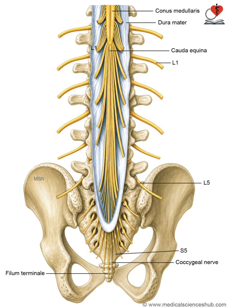
- The cauda equina (Latin: horse’s tail) is a collection of descending spinal nerve roots that surround the conus medullaris and filum terminale within the lumbar cistern of the subarachnoid space.
- It derives its name from its gross anatomical resemblance to a horse’s tail, formed by the parallel descent of multiple nerve roots through the CSF-filled lumbar cistern before exiting at their respective intervertebral foramina.
Components
- The cauda equina is composed of the following structures:
- The ventral (anterior) and dorsal (posterior) nerve roots of the lower four lumbar segments (L2–L5), all five sacral segments (S1–S5), and the single coccygeal segment (Co1).
- The filum terminale, the fibrous pial extension that descends centrally among the nerve roots to anchor the cord to the coccyx.
Course of the Nerve Roots
- Below the conus medullaris, the constituent nerve roots of the cauda equina descend vertically within the lumbar cistern, passing obliquely downward and laterally to reach their corresponding intervertebral foramina.
- At each foramen, the ventral and dorsal roots unite to form a mixed spinal nerve, which then exits the vertebral canal to supply its respective peripheral territory.
- Because the nerve roots are freely suspended in cerebrospinal fluid and are mobile, they tend to be displaced rather than injured by a lumbar puncture needle — an important factor underlying the safety of performing lumbar puncture below the L1–L2 level.
Table 4.2: Enlargement of spinal cord
| Feature | Cervical Enlargement | Lumbar Enlargement |
|---|---|---|
| Extent | C3 to T2 spinal segments | L1 to S3 spinal segments |
| Maximum diameter | 3.8 cm at the C6 spinal segment | 3.5 cm at the T12 vertebral level |
| Primary function | Provides the increased neural circuitry required for motor and sensory innervation of the upper limb | Provides the increased neural circuitry required for motor and sensory innervation of the lower limb |
| Reason for enlargement | Greater concentration of motor neurons (anterior horn) and sensory relay neurons to support complex, fine-skilled movements of the upper limb | Greater concentration of motor neurons and sensory relay neurons to support weight-bearing and locomotion of the lower limb |
CLINICAL NEUROANATOMY
Cauda Equina Syndrome
- Cauda equina syndrome (CES) results from injury to or compression of the nerve roots constituting the cauda equina within the lumbar cistern. Common causes include large intervertebral disc herniations, spinal stenosis, tumors, hematomas, and traumatic fractures. It constitutes a neurosurgical emergency requiring prompt decompression to prevent permanent deficits.
Clinical Features
- Lower motor neuron (LMN) paralysis of the lower limbs:
- Damage to the motor nerve roots of the cauda equina produces flaccid paralysis or weakness of the lower limbs, characterised by reduced muscle tone, absent or diminished reflexes, and progressive muscle wasting (atrophy) over time — consistent with a lower motor neuron lesion pattern.
- Loss of tendon reflexes:
- Involvement of the peripheral motor and sensory roots abolishes deep tendon reflexes in the lower limbs, including the ankle jerk (S1) and knee jerk (L3–L4), reflecting interruption of the reflex arc at the level of the nerve roots.
- Radicular pain:
- Compression of sensory nerve roots produces severe, sharp, radiating pain along the dermatomal distribution of the affected roots — commonly resembling sciatica in its distribution along the posterior thigh and leg. Pain characteristically worsens with movement, coughing, or straining due to transient increases in intraspinal pressure.
- Saddle anaesthesia:
- Loss of sensation occurs in the perineal region, including the inner thighs, buttocks, scrotum or labia, and perianal skin — the area that would contact a saddle when seated on a horse. This pattern reflects involvement of the S3–S5 sensory roots and is a hallmark feature of cauda equina syndrome.
- Sexual dysfunction:
- Compression of autonomic and somatic sensory fibers within the sacral roots impairs sexual function, producing erectile dysfunction in males and reduced genital sensation in both sexes, due to disruption of both the parasympathetic (S2–S4) and sensory pathways.
- Bladder and bowel dysfunction:
- Involvement of the sacral parasympathetic roots (S2–S4) disrupts voluntary control of the bladder and bowel, resulting in urinary retention, difficulty initiating micturition, or overflow incontinence, alongside constipation or faecal incontinence.
- Bladder and bowel dysfunction may appear later in the clinical course but, when present, indicates significant sacral root involvement and underscores the urgency of surgical decompression.
Internal Structure of Spinal Cord
Spinal cord consists of:
- Central grey matter
- Peripheral white matter
- Central canal containing CSF.
Grey Matter of Spinal Cord
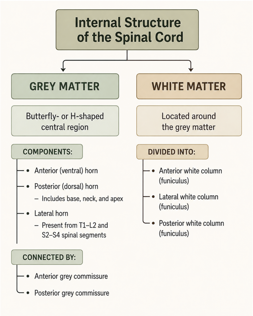
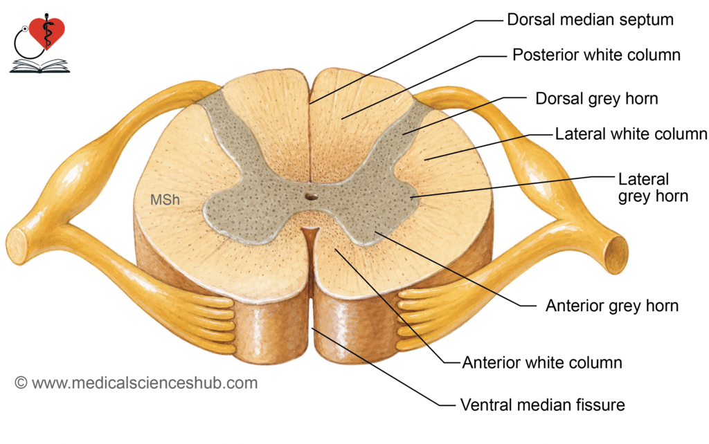
- The gray matter surrounds the central canal and contains neuronal cell bodies, their processes, and supporting neuroglial cells. On cross-section, it presents a characteristic H-shaped (butterfly-shaped) appearance.
- The two symmetric halves are joined across the midline by the gray commissure, which bridges the cord on either side of the central canal.
Anterior Horn (Ventral Gray Column)
- The anterior horn projects anterolaterally from the gray commissure and is separated from the cord surface by white matter.
- It contains the cell bodies of lower motor neurons (LMNs), whose axons form the ventral roots of spinal nerves and innervate skeletal muscle.
Posterior Horn (Dorsal Gray Column)
- The posterior horn projects posterolaterally and extends almost to the posterolateral sulcus, separated from the cord surface by the dorsolateral tract (Lissauer’s tract).
- It is subdivided into four parts: the base (continuous with the gray commissure), the neck (a constricted isthmus), the head (an expanded fusiform region), and the apex (the posteriormost tip).
- The apex is capped by the substantia gelatinosa, a translucent region of small interneurons that plays a key role in modulating pain and temperature transmission.
- The posterior horn receives the central processes of pseudounipolar neurons from the dorsal root ganglion, conveying afferent sensory signals into the cord.
Lateral Horn (Lateral Gray Column)
- The lateral horn is a small, wedge-shaped projection of gray matter present only at T1–L2 (thoracolumbar) and S2–S4 (sacral) spinal segments.
- Neurons in the T1–L2 lateral horn give rise to preganglionic sympathetic fibers, forming the thoracolumbar outflow of the autonomic nervous system.
- Neurons in the S2–S4 lateral horn give rise to preganglionic parasympathetic fibers, forming the sacral outflow that innervates the pelvic viscera, bladder, and bowel.
Substantia Gelatinosa Centralis
- Surrounding the central canal is a collar of glial and neural tissue termed the substantia gelatinosa centralis (central gelatinous substance), composed mainly of neuroglial cells with sparse nerve fibers, which maintains the structural integrity of the central canal lining.
Regional Characteristics of Spinal Cord
- The proportion and configuration of gray matter vary at different spinal levels, reflecting the volume and complexity of the tissue each segment innervates.
- Gray matter is most abundant at cervical and lumbar levels, corresponding to the enlargements that serve the upper and lower limbs respectively.
- Conversely, white matter is greatest in volume at cervical levels, where it carries the cumulative ascending and descending fiber tracts for the entire body below, and progressively diminishes toward the sacral segments.
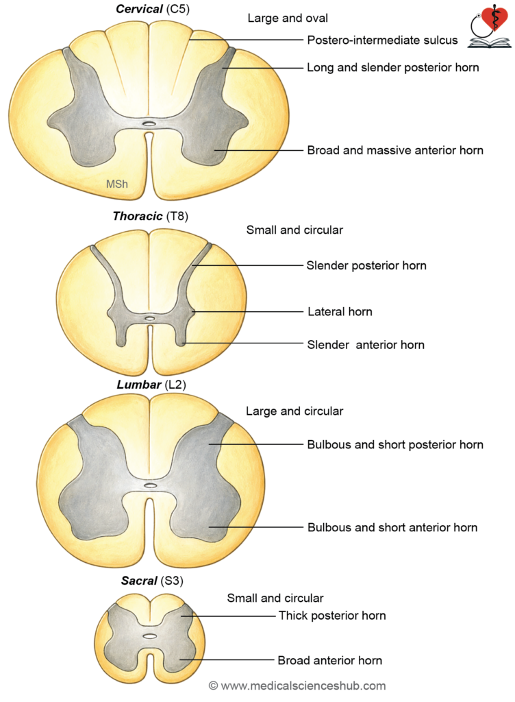
Table 4.3: Regional characteristics of spinal grey matter
| Feature | Cervical | Thoracic | Lumbar | Sacral |
|---|---|---|---|---|
| Cross-sectional shape | Oval | Small, circular | Large, circular | Small, circular |
| Anterior horn | Narrow at C1–C3; broad and prominent at C4–C8 | Slender throughout, except at T1 | Broad and prominent | Broad |
| Lateral horn | Absent | Present at T1–T12 (preganglionic sympathetic neurons) | Present only at L1–L2 (lower sympathetic outflow) | Present at S2–S4 (preganglionic parasympathetic neurons) |
| Posterior horn | Slender | Slender | Broad | Thick |
| White matter volume | Very large (carries tracts for entire body below) | Large | Reduced | Minimal |
| Posterointermediate sulcus | Present (separates fasciculus gracilis and cuneatus) | Present in upper thoracic segments | Absent | Absent |
Structure of Grey Matter of Spinal Cord
- The gray matter of the spinal cord consists of neuronal cell bodies and their processes, neuroglial cells, and blood vessels.
- All neurons within the gray matter are multipolar and are functionally classified into three groups: motor neurons, sensory neurons, and interneurons.
Types of Neurons
- Motor neurons (located in the anterior and lateral horns) are of two subtypes:
- Alpha (α) motor neurons: Large cells (≥25 μm in diameter) that innervate extrafusal muscle fibers of skeletal muscle, directly generating contractile force.
- Gamma (γ) motor neurons: Smaller multipolar cells (15–25 μm in diameter) that innervate intrafusal fibers of muscle spindles, regulating spindle sensitivity and contributing to muscle tone.
- Sensory neurons are small neurons situated in the posterior horn that receive synaptic input from dorsal root ganglion neurons. Their axons contribute to ascending sensory tracts or intersegmental pathways.
- Interneurons (internuncial neurons) are small associative neurons distributed throughout the gray matter that integrate and relay signals between motor, sensory, and autonomic neurons.
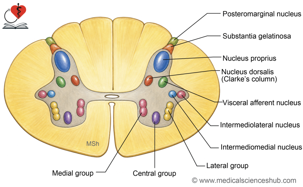
Neuronal Cell Groups in the Anterior Horn
- Medial group: Extends along most of the cord’s length; innervates axial musculature of the neck and trunk. Subdivided into ventromedial and dorsomedial parts.
- Lateral group: Confined to the cervical and lumbosacral enlargements; innervates the limb musculature.
- Central group: Comprises discrete, localised nuclei:
- Phrenic nucleus (C3–C5): Innervates the diaphragm.
- Lumbosacral nucleus (L2–S3): Function not yet fully established.
Neuronal Cell Groups in the Posterior Horn
- Substantia gelatinosa (Rexed lamina II): Located at the apex of the posterior horn and extends the full length of the cord. Composed of small Golgi type II interneurons; receives afferent input primarily carrying pain and temperature signals from the lateral division of dorsal roots. Continues superiorly into the nucleus of the spinal trigeminal tract.
- Nucleus proprius (Rexed laminae III–IV): Situated immediately anterior to the substantia gelatinosa and extends throughout the cord. Receives input from the posterior funiculus and processes proprioception, two-point discrimination, and vibration sense.
- Nucleus dorsalis (Clarke’s column, Rexed lamina VII): Located at the medial base of the posterior horn, extending from C8 to L2–L3. Receives proprioceptive input from muscles and joints and exteroceptive input (touch and pressure) from the trunk and lower limbs; gives rise to the posterior spinocerebellar tract.
- Visceral afferent nucleus: Situated lateral to Clarke’s column; present at T1–L2 and S2–S4 segments. Receives visceral sensory input conveyed via the dorsal roots.
Neuronal Cell Groups in the Lateral Horn
- Intermediolateral nucleus: Present at T1–L2 segments; contains preganglionic neurons that give rise to thoracolumbar sympathetic outflow, with axons exiting via the anterior nerve roots.
- Intermediomedial nucleus: Present at S2–S4 segments; contains preganglionic neurons that give rise to sacral parasympathetic outflow, with axons exiting via the anterior roots of the corresponding sacral nerves.
Rexed Laminae
- Rexed laminae represent a systematic laminar organization of the grey matter of the spinal cord, described by Bror Rexed in 1952.
- Neuronal cell bodies are arranged into 10 distinct laminae (I–X) based on structure and function.
- Laminae I–VI are located in the dorsal horn and are primarily involved in sensory processing.
- Lamina I (posteromarginal nucleus) and Lamina II (substantia gelatinosa) are important for pain and temperature modulation.
- Laminae III and IV form the nucleus proprius, which processes touch and pressure sensations.
- Lamina VII occupies the intermediate zone and includes the nucleus dorsalis (Clarke’s column) and intermediolateral cell column for autonomic output.
- Laminae VIII and IX lie in the ventral horn, containing motor neuron pools for skeletal muscle control.
- Lamina X surrounds the central canal and contains interneurons involved in visceral integration.
White Matter of Spinal Cord
- The white matter surrounds the grey matter and appears white due to myelinated nerve fibers.
- It conducts ascending sensory pathways and descending motor pathways, enabling communication between the brain and spinal segments.
Divisions of White Matter
- It is divided into right and left halves by the anterior median fissure and posterior median septum.
- Each half contains three funiculi (columns):
- Anterior (ventral) funiculus: Between anterior median fissure and ventral root exit zone.
- Lateral funiculus: Between ventral root exit zone and posterolateral sulcus.
- Posterior (dorsal) funiculus: Between posterior median sulcus and posterolateral sulcus.
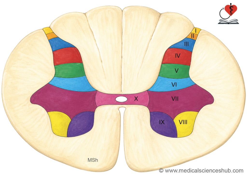
Table 4.4: Neuronal columns of grey matter of spinal cord
| Column | Extent | Key Functions |
|---|---|---|
| Anterior horn (motor) | Entire cord (regional specialization) | Contains motor neuron pools supplying skeletal muscles of trunk and limbs |
| Medial group | Entire cord | Muscles of back and axial skeleton |
| Lateral group | Cervical & lumbar enlargements | Muscles of limbs (proximal to distal control) |
| Central group | C3–C5, C1–C5 | Phrenic nucleus (diaphragm), spinal accessory nucleus |
| Posterior horn (sensory) | Entire cord (with regional nuclei) | Processes sensory inputs |
| Substantia gelatinosa | Entire cord | Pain and temperature modulation |
| Marginal nucleus | Entire cord | Pain and crude touch relay |
| Nucleus proprius | Entire cord | Touch and pressure processing |
| Dorsal nucleus (Clarke’s column) | C8–L3 | Proprioception to cerebellum |
| Visceral afferent nucleus | S1–S4 | Visceral sensory input |
| Lateral horn (autonomic) | T1–L2, S2–S4 | Autonomic outflow |
| Intermediolateral column | T1–L2 | Preganglionic sympathetic neurons |
| Sacral parasympathetic nucleus | S2–S4 | Preganglionic parasympathetic neurons |
Types of Fibers in White Matter
The white matter contains three main fiber types:
- Sensory (afferent) fibers: Central processes of dorsal root ganglion neurons and ascending tract fibers from dorsal horn cells.
- Motor (efferent) fibers: Descending fibers from higher centers and axons of anterior horn cells.
- Association (intersegmental) fibers: Fibers that originate and terminate within the spinal cord, coordinating segmental activity.
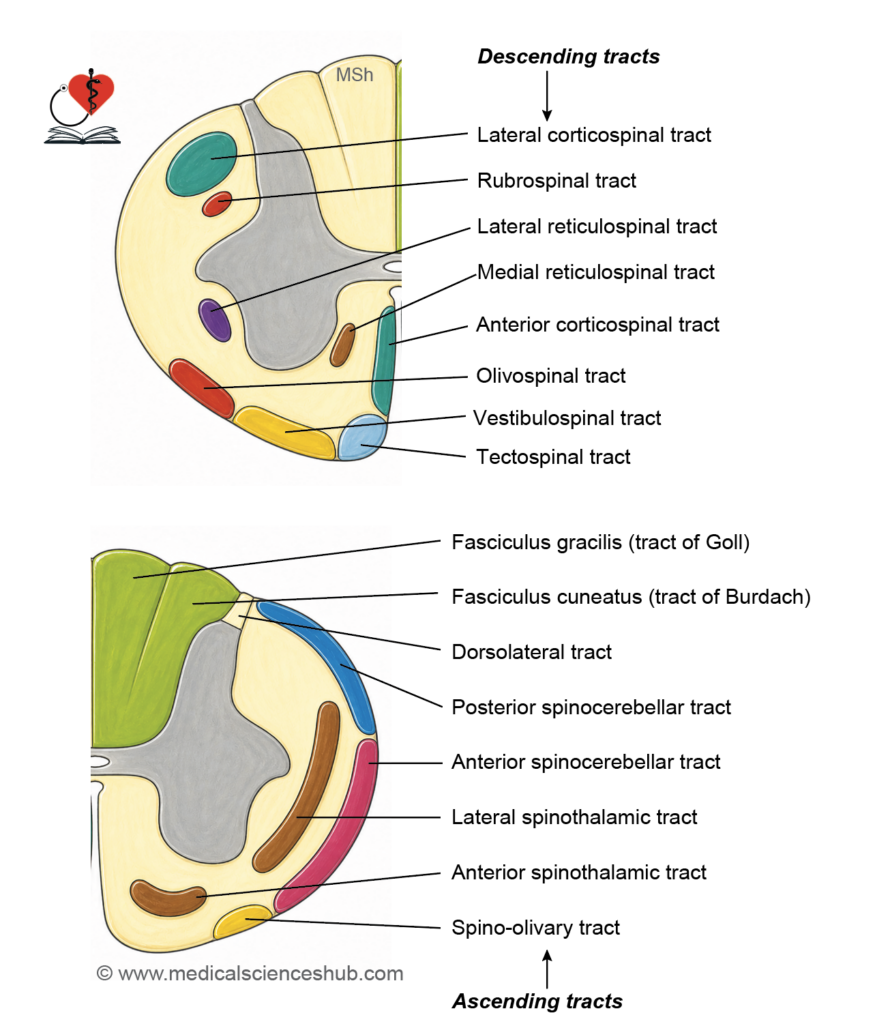
Tracts of Spinal Cord
- A tract is a bundle of nerve fibers within the central nervous system that share a common origin, course, termination, and function.
- It is also referred to as a fasciculus or lemniscus, depending on its anatomical context.
Classification of Tracts
- Spinal cord tracts are grouped into three main types:
- Ascending tracts: Carry sensory information from peripheral receptors to higher centers in the brain.
- Descending tracts: Convey motor commands from the brain to spinal cord neurons that control muscles.
- Intersegmental tracts: Consist of short association fibers that connect different spinal segments and coordinate reflex activity.
Blood Supply of Spinal Cord
- Adequate vascular supply is essential to prevent ischemic injury, which can disrupt motor and sensory functions.
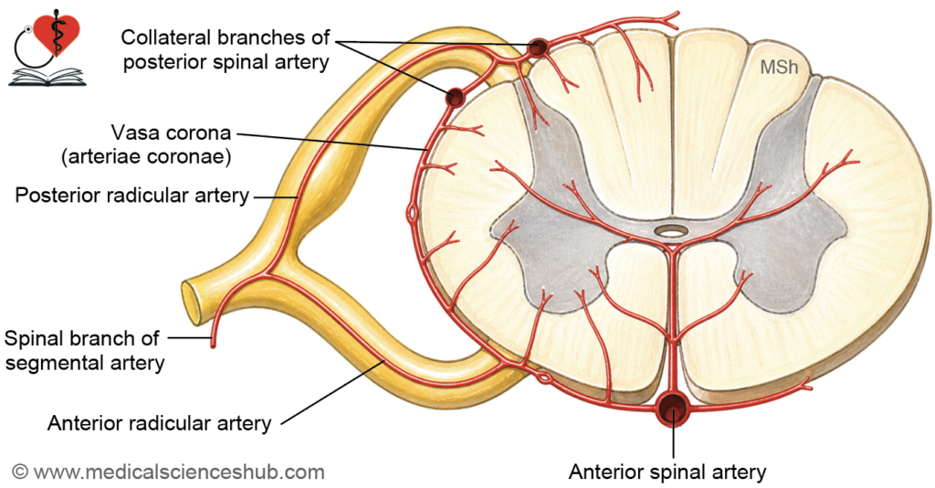
- The spinal cord receives blood from longitudinal arteries reinforced by segmental (radicular) arteries.
Arterial Supply
- The arterial supply consists of:
- Anterior spinal artery (ASA)
- Posterior spinal arteries (PSA) (right and left)
- Segmental spinal branches from vertebral, deep cervical, posterior intercostal, lumbar, and lateral sacral arteries
Anterior Spinal Artery
- Formed by the union of two branches from the vertebral arteries at the level of the medulla oblongata.
- Descends in the anterior median fissure.
- Branches include:
- Central (intramedullary) arteries supplying the inner cord
- Vasa corona forming a pial arterial network
- Supplies approximately the anterior two-thirds of the spinal cord, including:
- Anterior and lateral horns
- Base of the posterior horn
- Anterior and part of lateral white columns
Posterior Spinal Arteries
- Usually arise from the vertebral artery or posterior inferior cerebellar artery.
- Run along the posterior aspect near the dorsal roots.
- Supply:
- Most of the posterior horn
- Posterior (dorsal) columns
- Peripheral parts of the lateral white column
Radicular (Segmental) Arteries
- Enter through intervertebral foramina and accompany spinal nerve roots.
- Reinforce the longitudinal spinal arteries, especially in lower segments.
- Functionally important as end arteries, making the cord vulnerable to ischemia if occluded.
- The largest radicular artery is the arteria radicularis magna (artery of Adamkiewicz), typically arising on the left side between T9 and L2 (commonly around T11).
- It is the principal supply to the lumbar enlargement; its injury may cause severe neurological deficits, including paralysis.
Table 4.5: Arterial Distribution
| Artery | Origin | Course | Major Supply |
|---|---|---|---|
| Anterior spinal arteryDefinition | Vertebral arteries | Along anterior median fissure | Anterior 2/3 of cord (motor regions, anterior and lateral columns) |
| Posterior spinal arteries | Vertebral or PICA | Along posterior surface near dorsal roots | Posterior 1/3 (sensory regions, dorsal columns) |
| Radicular arteries | Segmental arteries | Along nerve roots | Reinforce spinal arteries; segmental supply |
Venous Drainage
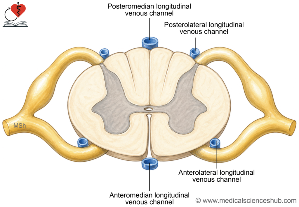
- The spinal cord is drained by a network of longitudinal veins within the pia mater, forming a rich venous plexus.
- Six main channels include:
- Anteromedian vein (along anterior median fissure)
- Posteromedian vein (along posterior median sulcus)
- Paired anterolateral veins (near ventral roots)
- Paired posterolateral veins (near dorsal roots)
- These veins interconnect and drain into the internal vertebral venous plexus (Batson’s plexus).
- This plexus communicates with segmental veins (vertebral, intercostal, lumbar, sacral), providing collateral pathways. · Clinical relevance: The valveless vertebral venous plexus allows potential spread of infection or metastasis between pelvic, thoracic, and cranial regions.
- Clinical relevance: The valveless vertebral venous plexus allows potential spread of infection or metastasis between pelvic, thoracic, and cranial regions.
CLINICAL NEUROANATOMY
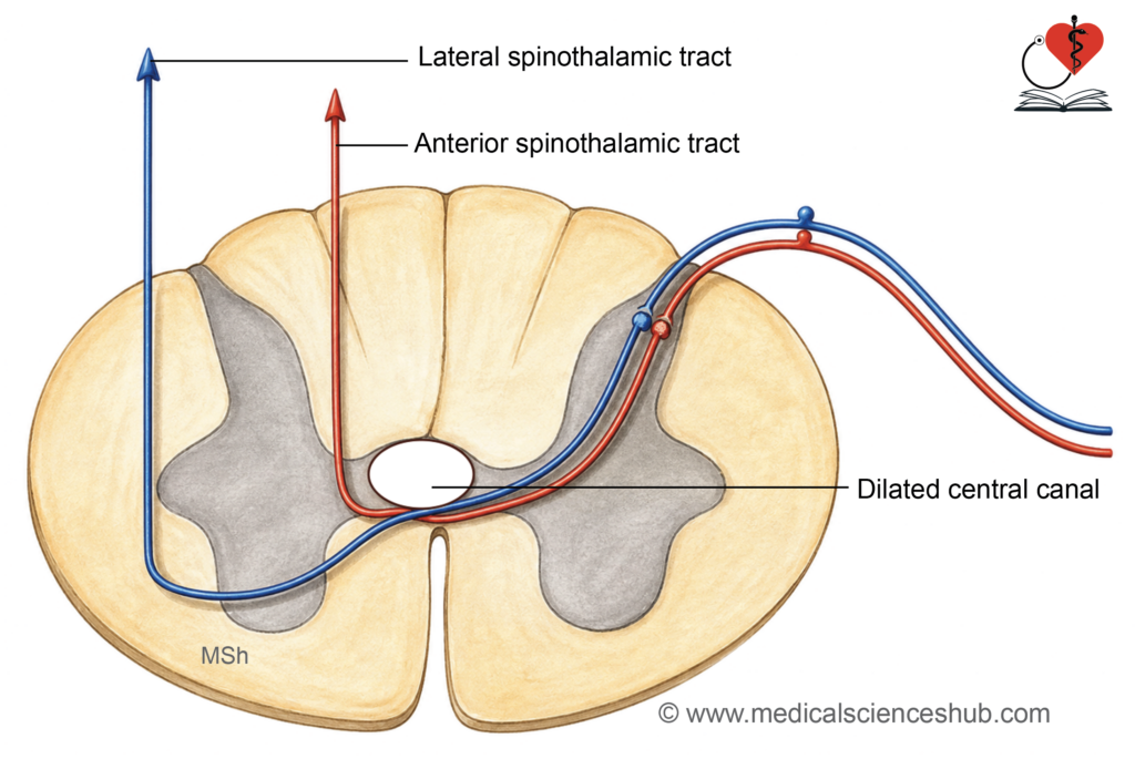
- Syringomyelia (central cord syndrome)
- It is characterized by formation of a CSF-filled cavity (syrinx) within the central spinal cord, most commonly in the cervical region.
- Expansion compresses adjacent fibers, especially crossing spinothalamic fibers.
- Results in bilateral loss of pain and temperature at the affected segments (often “cape-like” distribution).
- With progression, light touch may also be affected.
- Dissociated sensory loss is typical: proprioception, vibration, and fine touch remain intact due to preservation of posterior columns.
- Anterior spinal artery syndrome
- Occurs due to thrombosis, embolism, or compression of the anterior spinal artery, which supplies the anterior two-thirds of the cord.
- Affects:
- Corticospinal tracts (motor pathways)
- Anterior horn cells
- Spinothalamic tracts
- Clinical features:
- Motor deficits: Weakness or paralysis below the lesion with combined upper motor neuron signs (spasticity) and lower motor neuron signs at the level of lesion (flaccidity, atrophy).
- Sensory deficits: Bilateral loss of pain and temperature below the lesion.
- Posterior columns (fasciculus gracilis and cuneatus) are spared; therefore, proprioception, vibration, and fine touch remain preserved.
- Conus medullaris syndrome
- Results from damage or compression of the conus medullaris (terminal spinal cord).
- Key features include:
- Sudden onset
- Perianal (saddle) anesthesia
- Severe low back pain
- Early involvement of bladder and bowel function
- Frequent sexual dysfunction
- Mixed upper and lower motor neuron signs
- A lesion refers to any area of tissue damage caused by injury or disease, leading to functional impairment.
Important Questions
- Enumerate the specialized adaptations of the pia mater in the spinal cord.
- Write a short note on the filum terminale.
- Identify the anatomical spaces surrounding the spinal cord and mention their clinical relevance.
- Write a short note on the cauda equina.
- Outline the arterial and venous supply of the spinal cord.
📝 Test Your Knowledge – Practice MCQs
Attempt the chapter MCQ quiz and assess your understanding of key concepts.
Click to see: Medical Disclaimer and Copyright Notice