
Competencies
- AN61.1: Identify external and internal features of midbrain.
- AN61.2: Describe internal features of midbrain at the level of superior and inferior colliculus.
- AN61.3: Describe anatomical basis and effects of Benedikt’s and Weber’s syndromes.
Introduction
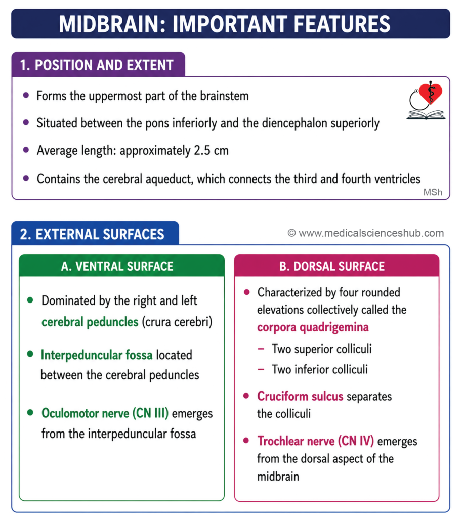
The midbrain (mesencephalon) is the uppermost part of the brainstem, connecting the pons and cerebellum inferiorly with the forebrain (diencephalon and cerebral hemispheres) superiorly.
- It measures approximately 2.5 cm in length and 2.5 cm in width.
- Functionally, the midbrain coordinates visual and auditory reflexes, contributes to the control of voluntary movement, regulates alertness and arousal, and helps maintain body posture and muscle tone. Damage to the midbrain may produce disorders of eye movement, contralateral hemiplegia, rigidity, tremors, or loss of consciousness in severe cases.
Location
- The midbrain ascends from the upper border of the pons, passing anterosuperiorly through the tentorial notch — the gap in the tentorium cerebelli — to reach the undersurface of the diencephalon. At this level, it is flanked on either side by the cerebral hemispheres, which overhang and largely conceal it when viewed from above.
Cavity
- The midbrain contains a narrow internal channel called the cerebral aqueduct (aqueduct of Sylvius), named after Dutch physician Franciscus Sylvius (1614–1672). It connects the third ventricle superiorly with the fourth ventricle inferiorly, and is the narrowest part of the ventricular system, making it a common site of obstructive hydrocephalus.
External Features of Midbrain
The midbrain presents two surfaces: a ventral (anterior) surface and a dorsal (posterior) surface.
Ventral Surface
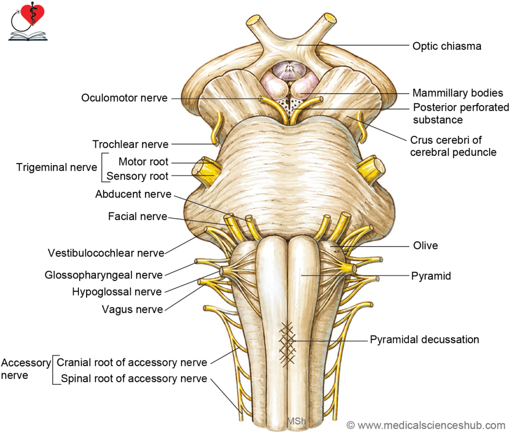
- The ventral surface is dominated by two large, rounded fiber bundles called the crura cerebri (singular: crus cerebri), also referred to collectively as the cerebral peduncles. They emerge from the undersurface of the cerebral hemispheres, converge toward each other as they descend, and meet the upper border of the pons inferiorly.
- The crura cerebri form the posterolateral boundaries of the interpeduncular fossa, a diamond-shaped space between the two crura on the ventral midbrain surface.
- The oculomotor nerve (CN III) emerges from the medial aspect of each crus cerebri, in the groove between the crus and the midbrain tegmentum, within the interpeduncular fossa.
Dorsal Surface
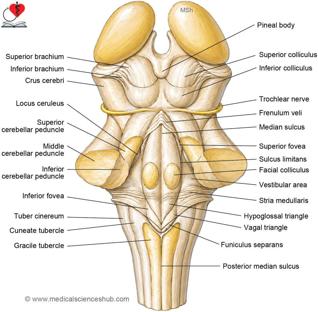
- The dorsal surface presents four rounded elevations collectively termed the corpora quadrigemina (or tectum), arranged in two pairs: the superior colliculi (upper pair) and the inferior colliculi (lower pair).
- The four colliculi are separated from one another by a cross-shaped groove called the cruciform sulcus. A small median elevation extending superiorly from this sulcus, called the frenulum veli, marks the point of attachment of the superior medullary velum.
- The trochlear nerve (CN IV) is the only cranial nerve to emerge from the dorsal surface of the brainstem. Its fibers decussate within the superior medullary velum before emerging on either side of the frenulum veli. After exiting, each nerve curves around the lateral aspect of the cerebral peduncle and passes anteriorly to enter the orbit, supplying the superior oblique muscle of the eye.
Internal Structure of Midbrain
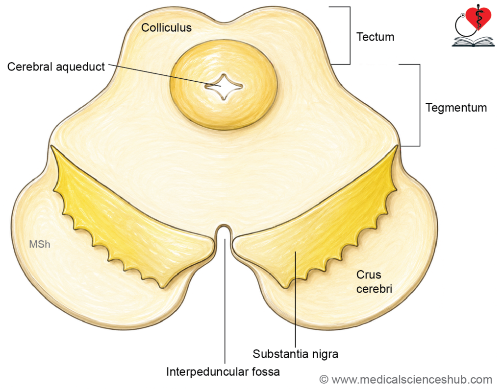
• In transverse section, the midbrain is organized into three principal divisions:
- Cerebral peduncles (anterior), comprising the crus cerebri, substantia nigra, and tegmentum
- Periaqueductal grey matter surrounding the cerebral aqueduct
Tectum (posterior), comprising the superior and inferior colliculi and pretectal nuclei.
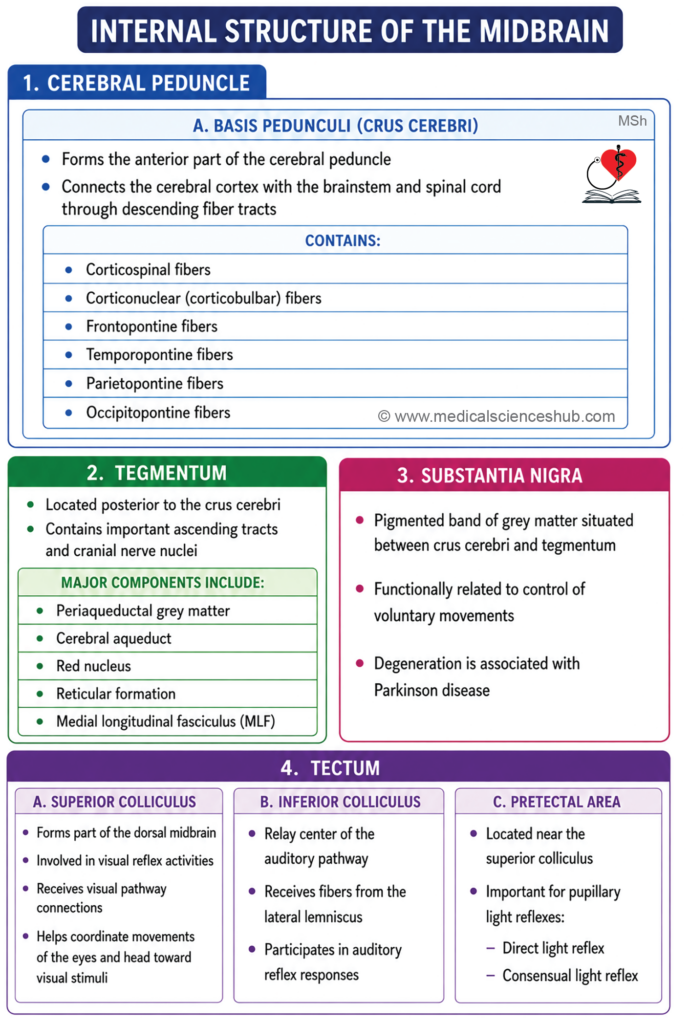
Crus Cerebri (Basis Pedunculi)
- Paired structures (one on each side) situated anterior to the substantia nigra, composed of descending white matter fiber tracts linking the cerebral cortex to the brainstem and spinal cord.
- Fiber arrangement within each crus follows a consistent topographic organization:
- Corticospinal and corticonuclear fibers occupy the middle two-thirds, conveying voluntary motor signals to the spinal cord and cranial nerve nuclei respectively.
- Frontopontine fibers occupy the medial one-sixth.
- Temporopontine, parietopontine, and occipitopontine fibers occupy the lateral one-sixth.
- Unilateral damage to the crus cerebri produces contralateral hemiplegia due to interruption of descending motor pathways.
Tegmentum
- Situated dorsal to the substantia nigra and ventral to the tectum.
- Its internal composition varies at the levels of the superior and inferior colliculi respectively.
Tectum (Corpora Quadrigemina)
- Forms the dorsal surface of the midbrain, presenting four rounded elevations collectively termed the corpora quadrigemina.
- The superior pair are the superior colliculi; the inferior pair are the inferior colliculi.
- The four colliculi are demarcated from one another by a cruciform sulcus on their posterior aspect.
Superior Colliculus
- Structurally organized into seven concentric alternating laminae of grey and white matter.
- Layers 1–3 are part of the visual system, receiving afferents from the retina and visual cortex.
- Layers 4–7 are associated with the auditory system (receiving input from the inferior colliculus) and the somatosensory system (receiving spinotectal fibers).
- Functions as a multimodal sensory integration center, generating reflex orientation of the eyes, head, and neck in response to visual, auditory, and somatic stimuli. It also coordinates eye-hand tracking movements.
- Principal efferent projections include:
- Motor nuclei of cranial nerves III, IV, VI, VII, and XI via the tectobulbar tract
- Anterior horn cells of the spinal cord via the tectospinal tract
- Components of the extrapyramidal system — including the red nucleus, substantia nigra, reticular formation, and cerebellum
- The contralateral superior colliculus and the medial longitudinal fasciculus
- Damage impairs visual tracking, reflex eye movements, and head-eye coordination.
Inferior Colliculus
- Composed of a compact central nucleus.
- Serves as a critical relay in the ascending auditory pathway, receiving afferents primarily from the lateral lemniscus.
- Involved in sound localization and audiovisual reflexes.
- Efferent fibers project predominantly to the ipsilateral medial geniculate body of the thalamus, with additional projections to the contralateral inferior colliculus, ipsilateral superior colliculus, and cerebellum.
- Damage results in hearing impairment, defective sound localization, and disrupted auditory reflexes.
Pretectal Nucleus
- A discrete neuronal group located ventral to the superior colliculus, at the midbrain–diencephalon junction.
- Receives visual afferents via the optic tract and superior brachium, as well as from the visual cortex through the optic radiation.
- Sends bilateral efferent projections to the Edinger–Westphal nucleus (the parasympathetic nucleus of cranial nerve III), forming the afferent limb of the pupillary light reflex and consensual light reflex.
- Additional efferents contribute to the tectospinal and tectobulbar tracts.
- Selective damage abolishes the light reflex while sparing the accommodation reflex — a dissociation characteristic of Parinaud’s syndrome.
Substantia Nigra
- A crescent-shaped, brown-pigmented band of grey matter interposed between the crus cerebri (ventrally) and the tegmentum (dorsally).
- Divided into two functionally and cytologically distinct zones:
- Pars compacta (dorsal zone): Contains densely packed, melanin-pigmented neurons that synthesize dopamine.
- Pars reticularis (ventral zone): Contains fewer neurons that synthesize GABA.
- Dopaminergic axons project to the corpus striatum via nigrostriatal fibers, modulating basal ganglia activity and facilitating smooth, coordinated, and purposeful motor execution.
- Reciprocal connections exist with the corpus striatum, red nucleus, and reticular formation.
- Functionally, the substantia nigra contributes to extrapyramidal motor control, including fine ocular movements, motor planning, and reward-driven behavior.
- Progressive degeneration of the pars compacta results in Parkinson’s disease, characterized by resting tremor, bradykinesia, rigidity, and postural instability.
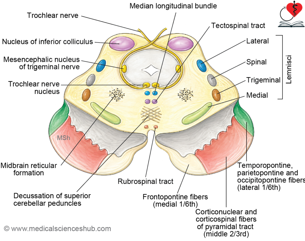
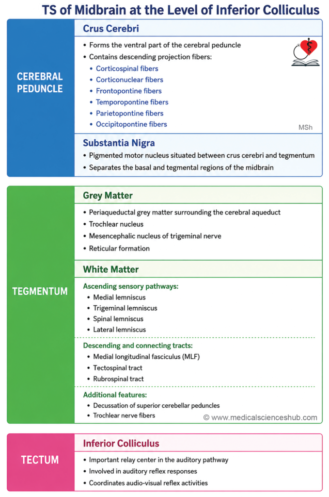
Midbrain at the Level of the Inferior Colliculus
- At this level, the midbrain is organized into two main regions:
- Cerebral peduncles (anterior), comprising the crus cerebri, substantia nigra, and tegmentum.
- Tectum (posterior), represented at this level exclusively by the inferior colliculi.
Crus Cerebri
- Contains the following descending fiber tracts arranged in a consistent topographic pattern:
- Corticospinal and corticonuclear tracts in the middle two-thirds.
- Frontopontine, temporopontine, parietopontine, and occipitopontine fibers in the medial and lateral one-sixth respectively.
Substantia Nigra
- A crescent-shaped, brown-pigmented band of grey matter separating the crus cerebri from the tegmentum.
- Its dopaminergic neurons give rise to nigrostriatal fibers that project to the corpus striatum, regulating basal ganglia activity and coordinated motor function.
Tectum
- At this level, the tectum is represented solely by the inferior colliculi.
- Serves as a key relay station in the ascending auditory pathway and mediates audiovisual reflexes.
Tegmentum
- Lies between the substantia nigra anteriorly and the inferior colliculus posteriorly.
- Contains both grey matter and white matter components.
Grey Matter Components
1. Periaqueductal Grey Matter
Contains the following nuclei:
- Nucleus of the trochlear nerve (CN IV):
- A somatic motor nucleus situated in a paramedian position within the periaqueductal grey matter.
- Axons from this nucleus pass dorsally around the periaqueductal grey matter, cross to the opposite side dorsal to it, and decussate at the level of the superior medullary velum.
- The nerve emerges on the dorsal surface of the midbrain on either side of the frenulum veli, making it the only cranial nerve to exit dorsally.
- It then winds around the superior cerebellar peduncle and cerebral peduncle to reach its target — the superior oblique muscle, which intorts and depresses the adducted eye.
- Damage produces vertical diplopia, difficulty with downward gaze (notably during reading or descending stairs), and a compensatory contralateral head tilt.
- Mesencephalic nucleus of the trigeminal nerve (CN V):
- A sensory nucleus located in the lateral part of the periaqueductal grey matter, composed of unipolar pseudounipolar neurons.
- Functionally unique in that it is the only sensory ganglion situated within the central nervous system rather than outside it.
- Transmits proprioceptive signals from the muscles of mastication, periodontal ligaments, temporomandibular joint, and extraocular and facial muscles.
- Damage disrupts the jaw jerk reflex and impairs the fine coordination of masticatory movements.
2. Reticular Formation
- Nuclei of the reticular formation are distributed diffusely throughout the tegmentum and contribute to arousal, autonomic regulation, and motor modulation.
White Matter Components
1. Lemniscal System
- The lemnisci form a curved, compact band of ascending sensory fibers arranged from medial to lateral in the following consistent order:
- Medial lemniscus → trigeminal lemniscus → spinal lemniscus → lateral lemniscus
- The lateral lemniscus partially terminates in the nucleus of the inferior colliculus; remaining fibers pass via the inferior brachium to reach the medial geniculate body of the thalamus.
- The medial, trigeminal, and spinal lemnisci continue ascending medial to the lateral lemniscus in the upper midbrain.
2. Trochlear Nerve (CN IV)
- The intraparenchymal course of the trochlear nerve runs posteriorly around the periaqueductal grey matter before decussating and exiting on the dorsal midbrain surface, as described above.
3. Decussation of the Superior Cerebellar Peduncles
- The superior cerebellar peduncles enter the tegmentum through its dorsolateral aspect.
- The majority of their fibers decussate with those of the contralateral peduncle in the central tegmentum, ventral to the periaqueductal grey matter.
- Following decussation, fibers ascend to the red nucleus and thalamus or descend as the rubrospinal tract.
4. Medial Longitudinal Fasciculus (MLF)
- A paramedian fiber bundle coordinating conjugate eye movements by interconnecting the nuclei of cranial nerves III, IV, and VI.
5. Tectospinal Tract
- Originates from the superior colliculus, decussates in the dorsal tegmentum, and descends to cervical spinal cord levels to mediate reflex head and neck movements in response to visual and auditory stimuli.
6. Rubrospinal Tract Originates from the red nucleus in the tegmentum, decussates immediately in the ventral tegmental decussation, and descends to influence contralateral limb musculature as part of the extrapyramidal motor system.
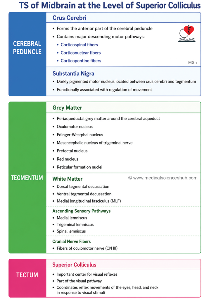
Midbrain at the Level of the Superior Colliculus
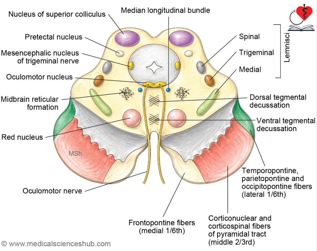
- At this level, the midbrain is organized into two principal regions:
- Cerebral peduncles (anterior), comprising the crus cerebri, substantia nigra, and tegmentum.
- Tectum (posterior), represented at this level by the superior colliculi.
- The superior colliculus mediates reflex orientation of the eyes, head, and neck in response to visual stimuli.
Tegmentum
- Lies between the substantia nigra anteriorly and the tectum posteriorly.
- Contains both grey matter nuclei and organized white matter tracts.
Grey Matter Components
1. Periaqueductal Grey Matter
- Surrounds the cerebral aqueduct and houses several important nuclei at this level.
2. Oculomotor Nucleus (CN III)
- A somatic motor nucleus situated in the ventral periaqueductal grey matter, close to the midline.
- The nuclei of the two sides are fused at the midline.
- Innervates all extraocular muscles except the lateral rectus and superior oblique, and also supplies the levator palpebrae superioris.
- Axons pass ventrally through the tegmentum and substantia nigra, emerging on the medial aspect of the crus cerebri within the interpeduncular fossa.
- Damage produces ptosis (drooping eyelid), ophthalmoplegia, and diplopia, with the eye typically deviated laterally and downward.
3. Edinger–Westphal Nucleus
- Forms the dorsal component of the oculomotor nuclear complex.
- Contains preganglionic parasympathetic neurons whose axons travel with CN III to synapse in the ciliary ganglion.
- Postganglionic fibers innervate the sphincter pupillae (mediating pupillary constriction) and the ciliary muscle (mediating accommodation for near vision).
- Damage results in a fixed, dilated pupil, loss of the pupillary light reflex, and impaired accommodation.
4. Mesencephalic Nucleus of the Trigeminal Nerve
- Located in the lateral periaqueductal grey matter, transmitting proprioceptive signals from the muscles of mastication, periodontal ligaments, and extraocular and facial muscles.
5. Pretectal Nucleus
- Situated at the midbrain–diencephalon junction, ventral to the superior colliculus.
- Mediates the pupillary light reflex and consensual light reflex via bilateral projections to the Edinger–Westphal nucleus.
6. Red Nucleus
- A cigar-shaped mass of grey matter located in the anterior tegmentum, close to the midline and posterior to the substantia nigra, at the level of the superior colliculus.
- Approximately 0.5 cm in diameter; appears pink in fresh specimens due to its rich vascularity and the iron-containing pigment within its neurons.
- Divided into a magnocellular (caudal) portion and a parvocellular (rostral) portion.
Afferent connections:
- Cerebellorubral fibers from the contralateral dentate nucleus of the cerebellum (via the superior cerebellar peduncle).
- Corticorubral fibers from the ipsilateral motor cortex.
- Pallidorubral fibers from the globus pallidus.
- Additional inputs from the subthalamus, superior colliculus, and substantia nigra.
Efferent connections:
- Rubrospinal tract → decussates immediately and descends to contralateral anterior horn cells of the spinal cord, influencing limb musculature.
- Rubrobulbar tract → projects to cranial nerve nuclei in the brainstem.
- Rubroreticular tract → projects to the reticular formation.
- Additional projections via rubro-olivary, rubrothalamic, rubrocerebellar, and rubronigral fibers.
Functions:
- Serves as an important relay within the extrapyramidal motor system, contributing to the maintenance of posture, regulation of muscle tone, and coordination of limb movements — particularly through its cerebellar and spinal cord connections.
- Damage produces contralateral intention tremor, limb ataxia, and involuntary movements.
7. Nuclei of the Reticular Formation
- Scattered throughout the tegmentum, contributing to arousal, autonomic regulation, and modulation of motor and sensory activity.
White Matter Components
1. Dorsal Tegmental Decussation (Decussation of Meynert)
- Tectobulbar fibers originating from the superior colliculus curve anteriorly around the periaqueductal grey matter and decussate in the midline, ventral to the central grey matter.
- This crossing constitutes the dorsal tegmental decussation, forming the origin of the tectospinal and tectobulbar tracts that coordinate reflex head and neck responses.
2. Ventral Tegmental Decussation (Decussation of Forel)
- Rubrospinal fibers arising from the red nucleus decussate immediately in the anterior tegmentum as the ventral tegmental decussation.
- Following decussation, fibers descend through the pons, medulla, and lateral funiculus of the spinal cord, terminating on anterior horn cells to regulate posture and muscle tone as part of the extrapyramidal system.
- Damage to this pathway causes contralateral weakness, poor coordination, and impaired fine motor control, particularly of the upper limb.
3. Medial Longitudinal Fasciculus (MLF)
- A paramedian tract coordinating conjugate eye movements by interconnecting the nuclei of cranial nerves III, IV, and VI, as well as vestibular nuclei.
4. Lemniscal System
- Three ascending sensory lemnisci are arranged in a consistent medial-to-lateral sequence at this level:
- Medial lemniscus → trigeminal lemniscus → spinal lemniscus
- The lateral lemniscus, prominent at the inferior collicular level, is no longer present as a distinct bundle at this level.
5. Fibers of the Oculomotor Nerve (CN III) Pass ventrally through the tegmentum and substantia nigra before emerging in the interpeduncular fossa, as described above.
Blood Supply of Midbrain
- The midbrain receives its arterial supply from multiple sources, all derived from the vertebrobasilar system.
- The principal arterial contributors are branches of the following vessels:
- Posterior cerebral artery — the primary source, supplying the tegmentum and tectum via its paramedian and circumferential branches.
- Superior cerebellar artery — contributes branches to the lateral tegmentum and structures near the isthmus.
- Posterior communicating artery — supplies the anterior and medial aspects of the midbrain, including portions of the cerebral peduncle.
- Anterior choroidal artery — contributes to the supply of the cerebral peduncle, including the crus cerebri and parts of the substantia nigra.
- All of the above vessels are branches of, or connect with, the basilar artery and its terminal divisions, forming part of the circle of Willis at the base of the brain.
CLINICAL NEUROANATOMY
Weber’s Syndrome
- Synonyms: Superior alternating hemiplegia; midbrain stroke syndrome (described by Sir Herman David Weber, 1823–1918).
- Cause: Occlusion of a paramedian branch of the posterior cerebral artery, producing ischemic damage to the ventral midbrain involving the oculomotor nerve (CN III) fascicles and the crus cerebri.
Clinical Features and Anatomical Basis
- Damage to CN III fascicles → ipsilateral ptosis (levator palpebrae superioris paralysis) and the eye adopts a “down-and-out” position due to unopposed action of the lateral rectus and superior oblique muscles.
- Damage to Edinger–Westphal nucleus fibers (parasympathetic component of CN III) → ipsilateral mydriasis (fixed, dilated pupil) with loss of both the direct pupillary light reflex and the accommodation reflex.
- Damage to the corticospinal tract in the crus cerebri → contralateral hemiplegia, affecting the upper and lower limbs of the opposite side.
- Damage to corticonuclear fibers → contralateral lower facial weakness and contralateral tongue deviation, due to interruption of upper motor neuron input to the relevant cranial nerve nuclei.
- The combination of ipsilateral CN III palsy with contralateral hemiplegia constitutes the classic alternating hemiplegia pattern, which localizes the lesion to the ventral midbrain.
Benedikt’s Syndrome
- Synonym: Paramedian midbrain syndrome.
- Cause: Occlusion of a branch of the posterior cerebral artery producing ischemic damage to the paramedian tegmentum of the midbrain — a region situated dorsal to the crus cerebri and substantia nigra.
Clinical Features and Anatomical Basis
- Damage to CN III fascicles → ipsilateral oculomotor palsy, comprising ptosis, lateral deviation of the eye, and ipsilateral mydriasis with loss of the light reflex — identical to the CN III involvement seen in Weber’s syndrome.
- Damage to the medial lemniscus → contralateral loss of tactile discrimination, proprioception, and vibration sense, as this tract carries dorsally decussated fibers conveying these modalities from the opposite side of the body.
- Damage to the trigeminal and spinal lemnisci → contralateral loss of pain and temperature sensation.
- Damage to the red nucleus and superior cerebellar peduncle → contralateral intention tremor and involuntary movements of the limbs, resulting from disruption of cerebellorubral connections.
- Benedikt’s syndrome is distinguished from Weber’s syndrome by the presence of contralateral sensory deficits and involuntary movements, reflecting tegmental rather than purely ventral peduncular involvement.
Parinaud’s Syndrome
- Synonyms: Dorsal midbrain syndrome; pretectal syndrome.
- Cause: Compression or ischemic damage to the dorsal midbrain, particularly affecting the superior colliculi, pretectal area, and posterior commissure.
- Common etiologies include:
- Pineal gland tumors (most frequent cause, due to direct dorsal compression of the midbrain).
- Obstructive hydrocephalus from aqueductal stenosis.
- Multiple sclerosis, vascular lesions, midbrain infarction, and arteriovenous malformations.
Clinical Features and Anatomical Basis
- Upward gaze palsy — the most characteristic feature, caused by damage to the vertical gaze centers near the rostral interstitial nucleus of the medial longitudinal fasciculus (riMLF) and the posterior commissure. Voluntary upward gaze is lost, while downward gaze is typically preserved.
- Convergence–retraction nystagmus — on attempted upward gaze, the eyes exhibit synchronous convergent movements accompanied by posterior retraction into the orbits. This results from disordered input to the ocular motor nuclei producing co-contraction of extraocular muscles.
- Light–near dissociation — the pupils fail to constrict in response to direct light but constrict normally during the near (accommodation) reflex. This dissociation occurs because the pretectal nuclei mediating the light reflex are damaged, while the accommodation pathway through the Edinger–Westphal nucleus remains functionally intact.
- Collier’s sign (eyelid retraction) — the upper eyelids are pathologically elevated, producing a fixed “staring” appearance, due to involvement of fibers modulating the levator palpebrae superioris.
- Downward deviation of gaze at rest — with voluntary upward gaze lost, the eyes tend to rest in a slightly downward position, sometimes described as the “setting sun” sign, particularly in cases with associated hydrocephalus.
Important Questions
- Write a short note on substantia nigra.
- Write a short note on transverse section of midbrain at the level of inferior colliculi.
- Draw a well-labeled diagram of TS midbrain at the level of superior colliculus or at the level of red nucleus.
- Write a short note on red nucleus.
📝 Test Your Knowledge – Practice MCQs
Attempt the chapter MCQ quiz and assess your understanding of key concepts.
Click to see: Medical Disclaimer and Copyright Notice