
Competencies
- AN25.1 Identify, draw and label a slide of trachea and lung.
- AN43.2 Identify, describe and draw the microanatomy of epiglottis.
- AN43.2 Identify, describe and draw the microanatomy of olfactory epithelium.
Introduction
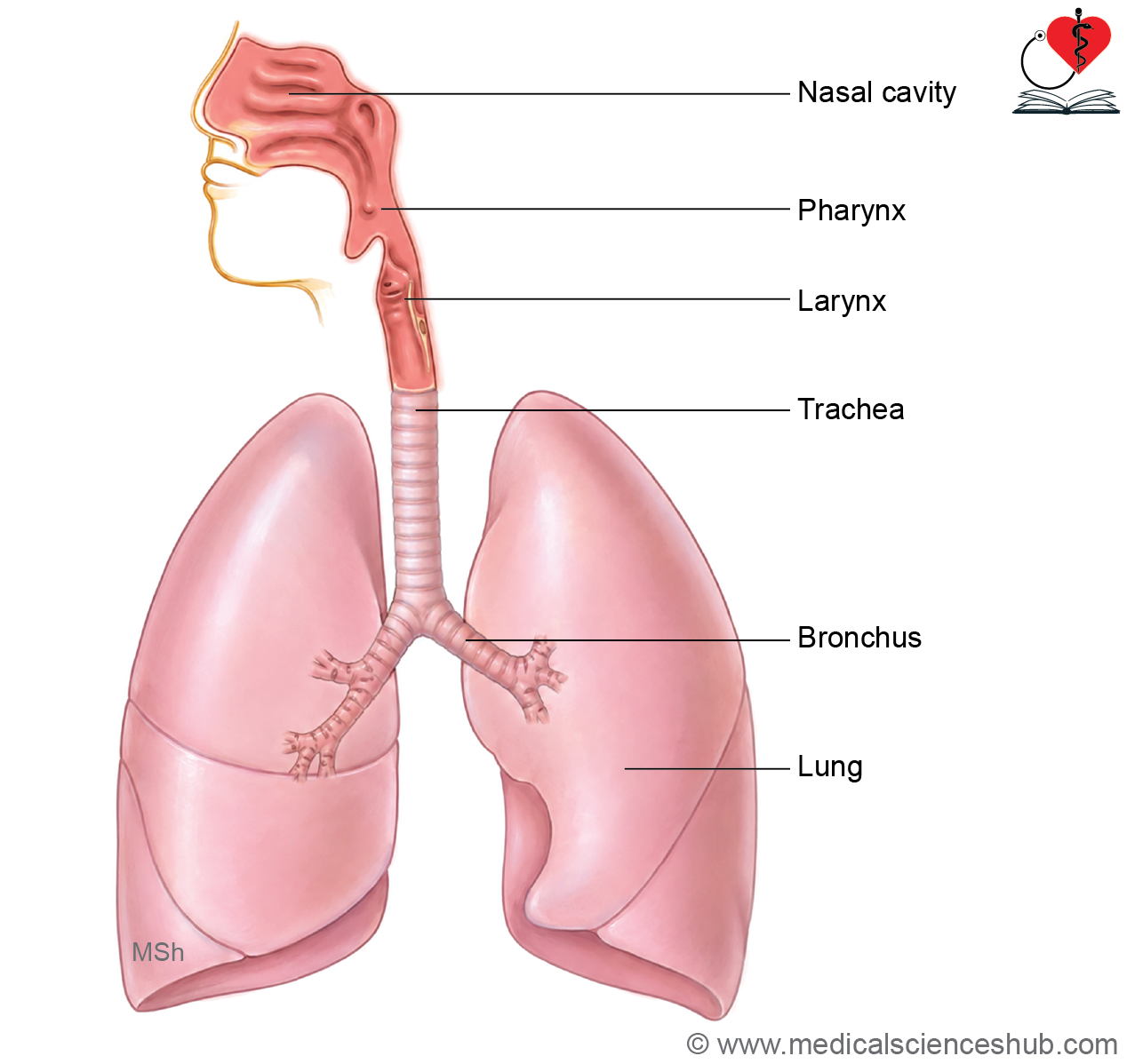
- The respiratory system is responsible for the transport of air and the exchange of oxygen and carbon dioxide between the atmosphere and blood.
- It is divided into a conducting portion and a respiratory portion.
- The conducting portion transports air and includes the nasal cavities, pharynx, larynx, trachea, bronchi, and terminal bronchioles. It does not participate in gas exchange.
- The respiratory portion consists of respiratory bronchioles, alveolar ducts, alveolar sacs, and alveoli, where gaseous exchange occurs.
- Anatomically, the upper respiratory tract includes the nasal cavity, paranasal sinuses, and nasopharynx, whereas the lower respiratory tract comprises the larynx, trachea, bronchi, and lungs.
- Inspired air undergoes conditioning, including warming, humidification, and filtration.
- Goblet cells secrete mucus that traps inhaled particles and helps maintain moisture within the respiratory passages.
Nasal Cavities
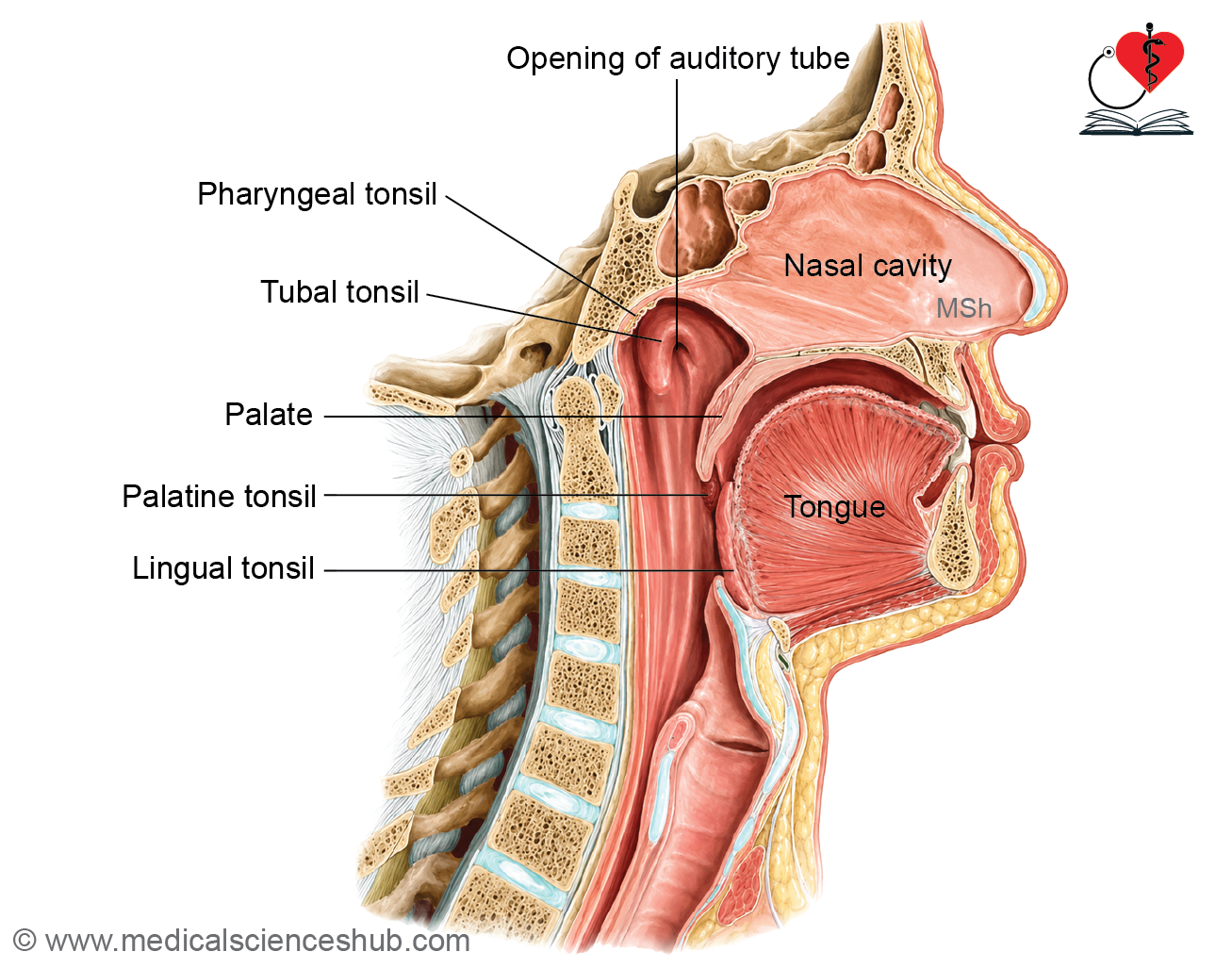
- The nasal cavities are paired air passages separated by the nasal septum.
- They extend from the external nostrils (nares) to the posterior choanae, which open into the nasopharynx.
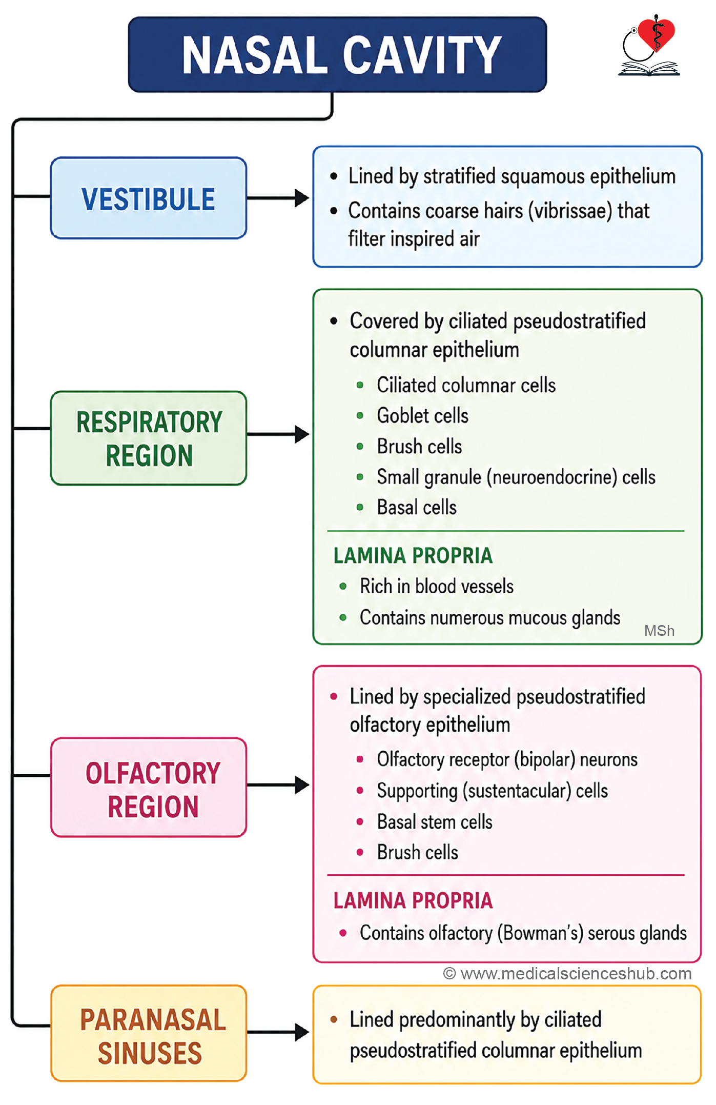
- The nasal mucosa is divided into three regions: the vestibular region, respiratory region, and olfactory region, each with distinct structural and functional characteristics.
Vestibule of Nasal Cavity
- The nasal vestibule is the anterior expanded part of the nasal cavity.
- It is lined by stratified squamous epithelium, which gradually transitions to respiratory epithelium posteriorly.
- The vestibule contains coarse hairs called vibrissae, which filter and trap large inhaled particles, providing an initial protective barrier to the respiratory tract.
Respiratory Zone of Nasal Cavity
- he respiratory region forms the largest part of the nasal cavity and is lined by ciliated pseudostratified columnar epithelium (respiratory epithelium).
- Its lamina propria is firmly attached to the underlying bone and cartilage.
- Medially, this region is related to the nasal septum, while laterally it contains the nasal conchae (turbinates), which increase the surface area for air conditioning.
Cell Types of the Respiratory Epithelium
- Ciliated columnar cells move mucus and trapped particles toward the pharynx.
- Goblet cells secrete mucus that traps dust and microorganisms.
- Brush cells are nonciliated columnar cells with microvilli and sensory functions.
- Small granule (neuroendocrine) cells contain secretory granules and participate in local regulatory functions.
- Basal cells act as stem cells and replace damaged epithelial cells.
Lamina Propria
- The lamina propria contains a rich vascular plexus that warms inspired air.
- Numerous seromucous glands produce secretions that humidify the respiratory passages.
- It also contains immune cells, including lymphocytes, plasma cells, and eosinophils, which contribute to local defense.
- In allergic rhinitis, eosinophils increase in number.
- During viral infections such as the common cold, edema of the lamina propria may obstruct airflow and cause nasal congestion.
Olfactory Zone of Nasal Cavity
- The olfactory region occupies a small area in the superior part of the nasal cavity and is specialized for the sense of smell.
- The olfactory mucosa appears yellowish due to pigments within its supporting structures, whereas the respiratory mucosa is pink because of its rich vascularity.
Olfactory Epithelium
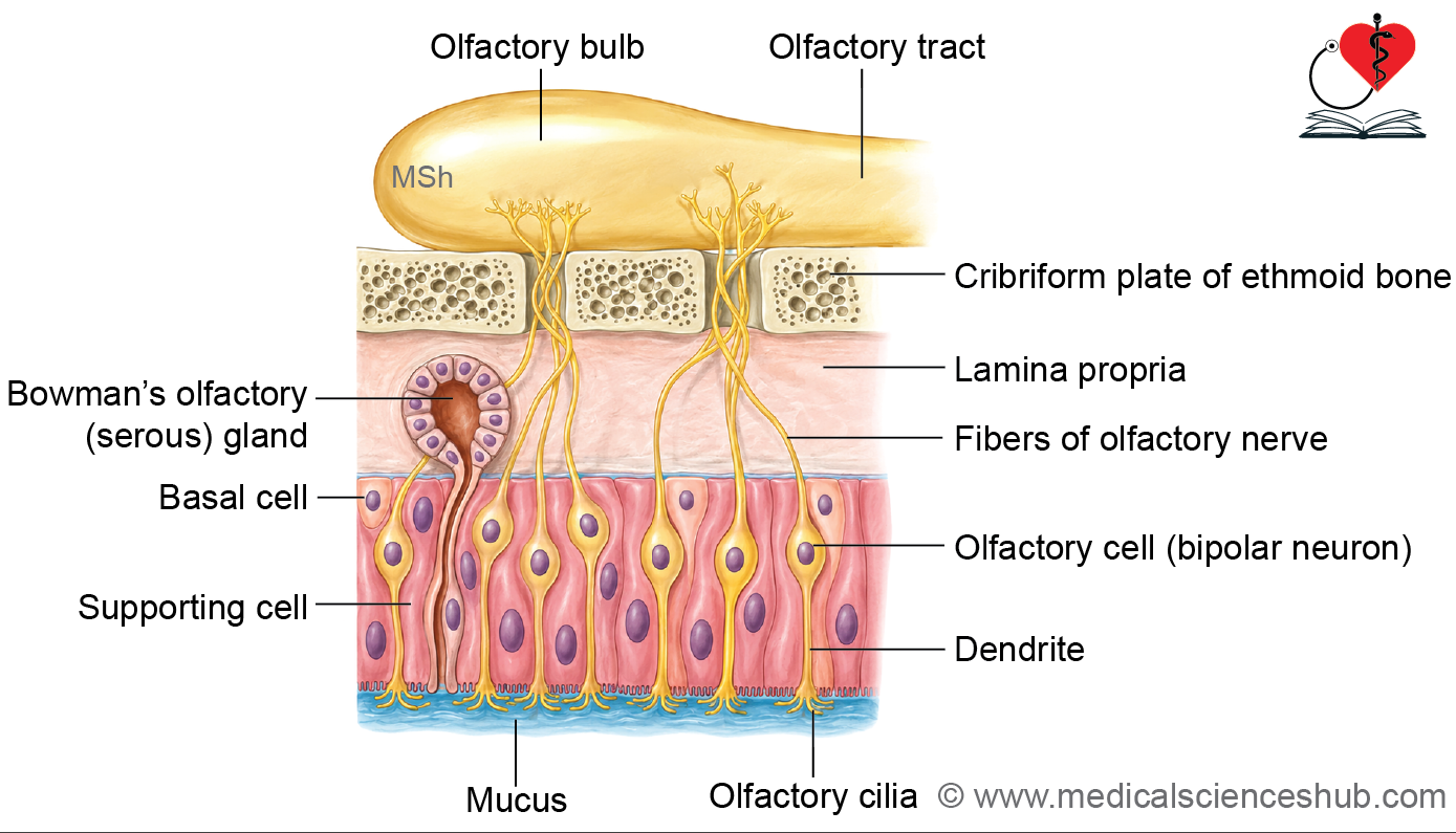
- The epithelium is pseudostratified columnar, but unlike respiratory epithelium, it lacks goblet cells and motile cilia.
- It contains four principal cell types:
- Olfactory receptor cells (bipolar neurons)
- Sustentacular (supporting) cells
- Basal cells
- Brush cells
Olfactory Receptor Cells
- These are specialized bipolar neurons that function as sensory receptors for smell.
- Their apical ends expand into an olfactory vesicle, from which several nonmotile cilia project into the mucus layer.
- These cilia contain odorant receptors that bind dissolved odor molecules and initiate nerve impulses.
- Axons of olfactory receptor cells bundle together to form the olfactory nerve (cranial nerve I), which carries signals to the brain.
- Olfactory receptor neurons are among the few neurons in the body capable of continuous regeneration throughout life.
Supporting and Basal Cells
- Sustentacular cells are tall columnar cells with microvilli that provide structural and metabolic support to olfactory neurons.
- They contain lipofuscin pigment, which contributes to the yellow coloration of the olfactory mucosa.
- Basal cells serve as stem cells and replace olfactory receptor and supporting cells.
Brush Cells
- Brush cells possess blunt microvilli and form synaptic connections with sensory fibers of the trigeminal nerve.
- They mediate general sensory sensations, such as irritation, from the nasal mucosa.
Bowman’s (Olfactory) Glands
- Bowman’s glands are branched serous glands located in the lamina propria.
- Their secretions dissolve and continuously remove odorant molecules from the epithelial surface, allowing detection of new odors.
Paranasal Sinuses
- The paranasal air sinuses are lined by thin respiratory epithelium consisting of ciliated pseudostratified columnar cells with goblet cells that help maintain mucosal moisture and mucus clearance.
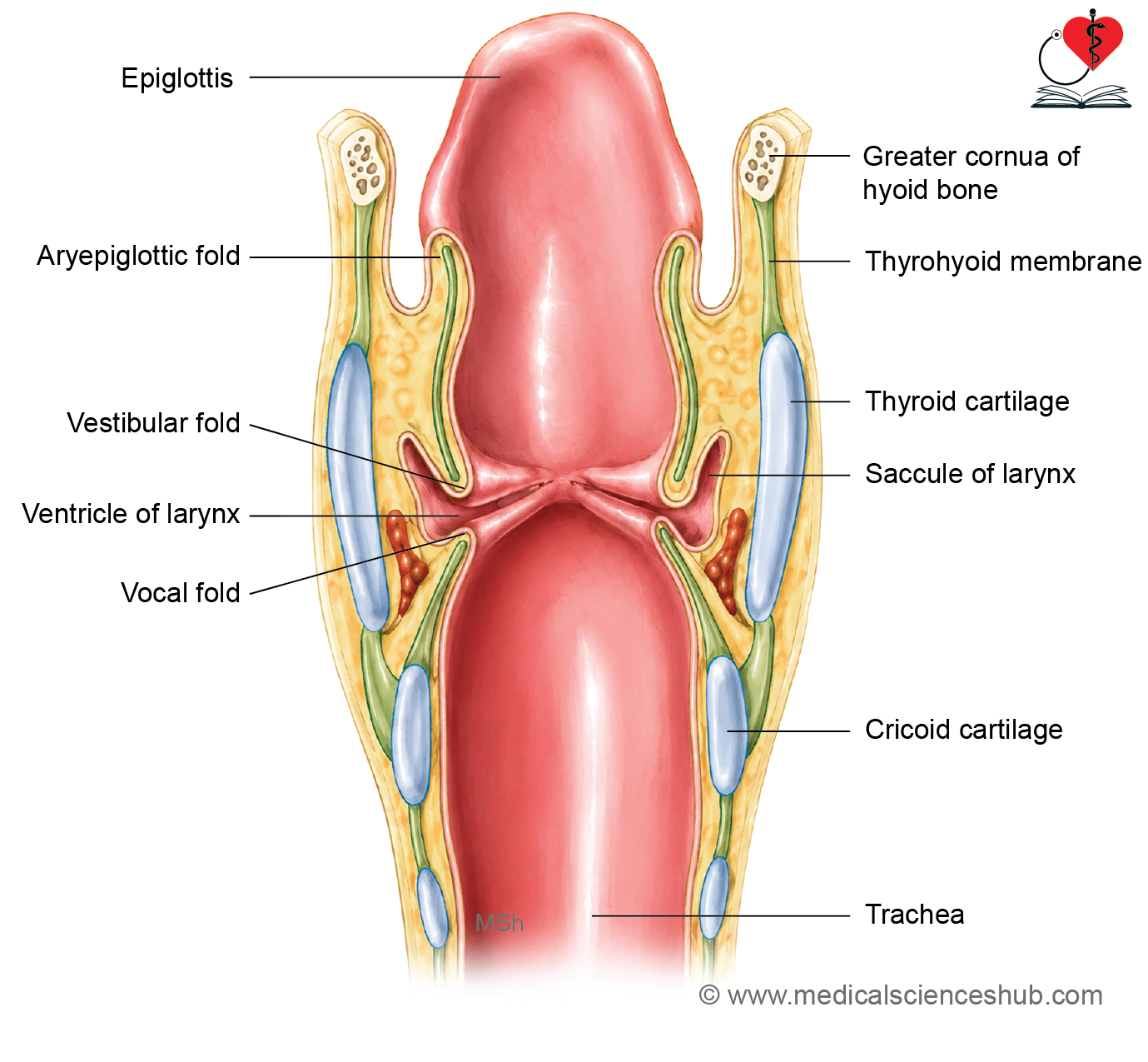
Epiglottis
- The epiglottis is a leaf-shaped structure that protects the airway by covering the laryngeal inlet during swallowing and directing food toward the esophagus.
- It contains a central core of elastic cartilage covered by mucous membrane.
- The epiglottis has anterior and posterior surfaces with a free superior margin.
- Embryologically, it develops primarily from the fourth pharyngeal arch region.
Histology of Epiglottis
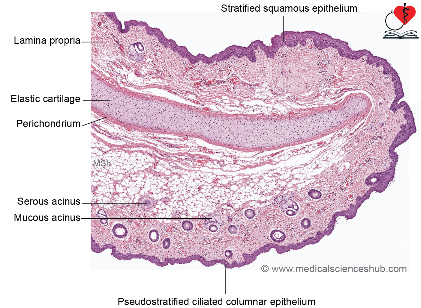
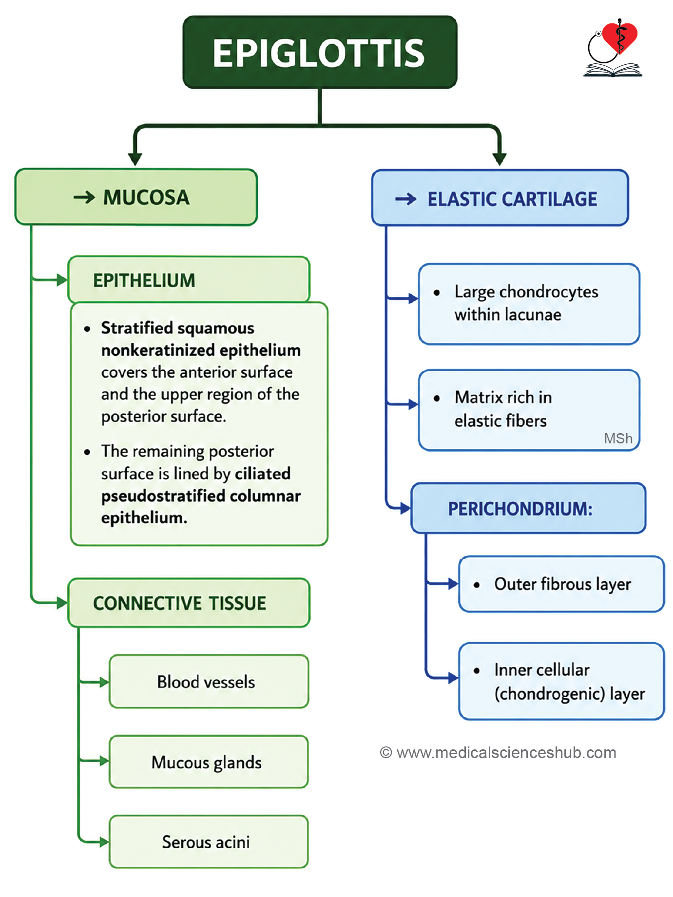
The epiglottis consists of a central core of elastic cartilage covered by a mucosal lining.
Mucosa
- The anterior (lingual) surface, free margin, and upper part of the posterior surface are lined by nonkeratinized stratified squamous epithelium, which provides protection against mechanical stress during swallowing.
- The remaining posterior surface is lined by ciliated pseudostratified columnar epithelium containing goblet cells, characteristic of respiratory mucosa.
- A few taste buds may be present on the posterior surface.
- The mucosa is separated from the cartilage by a loose, highly vascular connective tissue layer (lamina propria).
- The lamina propria contains numerous mucous glands, particularly on the posterior surface, along with occasional serous glands.
- These glands help maintain moisture and lubrication of the epiglottic surface.
Elastic Cartilage
- The core of the epiglottis is composed of elastic cartilage, which provides flexibility and resilience during swallowing.
- The cartilage contains chondrocytes located within lacunae and embedded in a matrix rich in elastic fibers.
- It is surrounded by a perichondrium consisting of an outer fibrous layer and an inner cellular (chondrogenic) layer.
Trachea
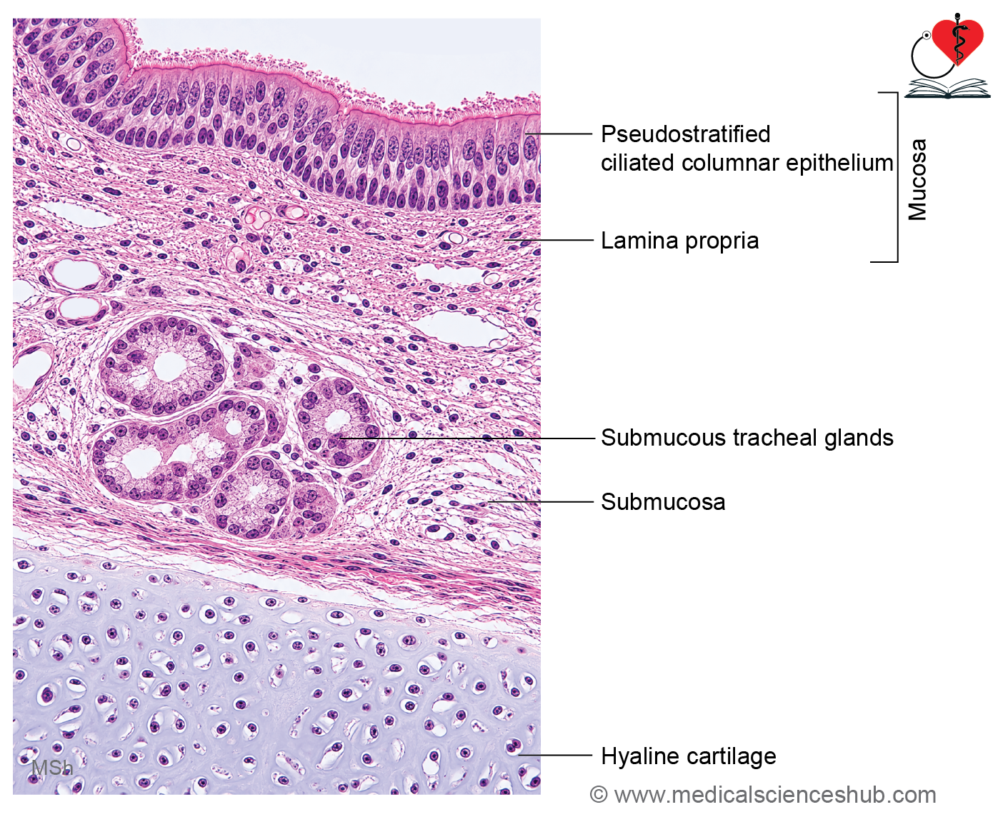
- The trachea is a cartilaginous airway that conducts air from the larynx to the lungs.
- It is approximately 10–12 cm long and about 2 cm in diameter in adults.
- Inferiorly, it bifurcates into the right and left primary bronchi.
- Its lumen remains patent due to supporting C-shaped hyaline cartilage rings.
- The presence of these hyaline cartilages is a characteristic histological feature of the trachea.
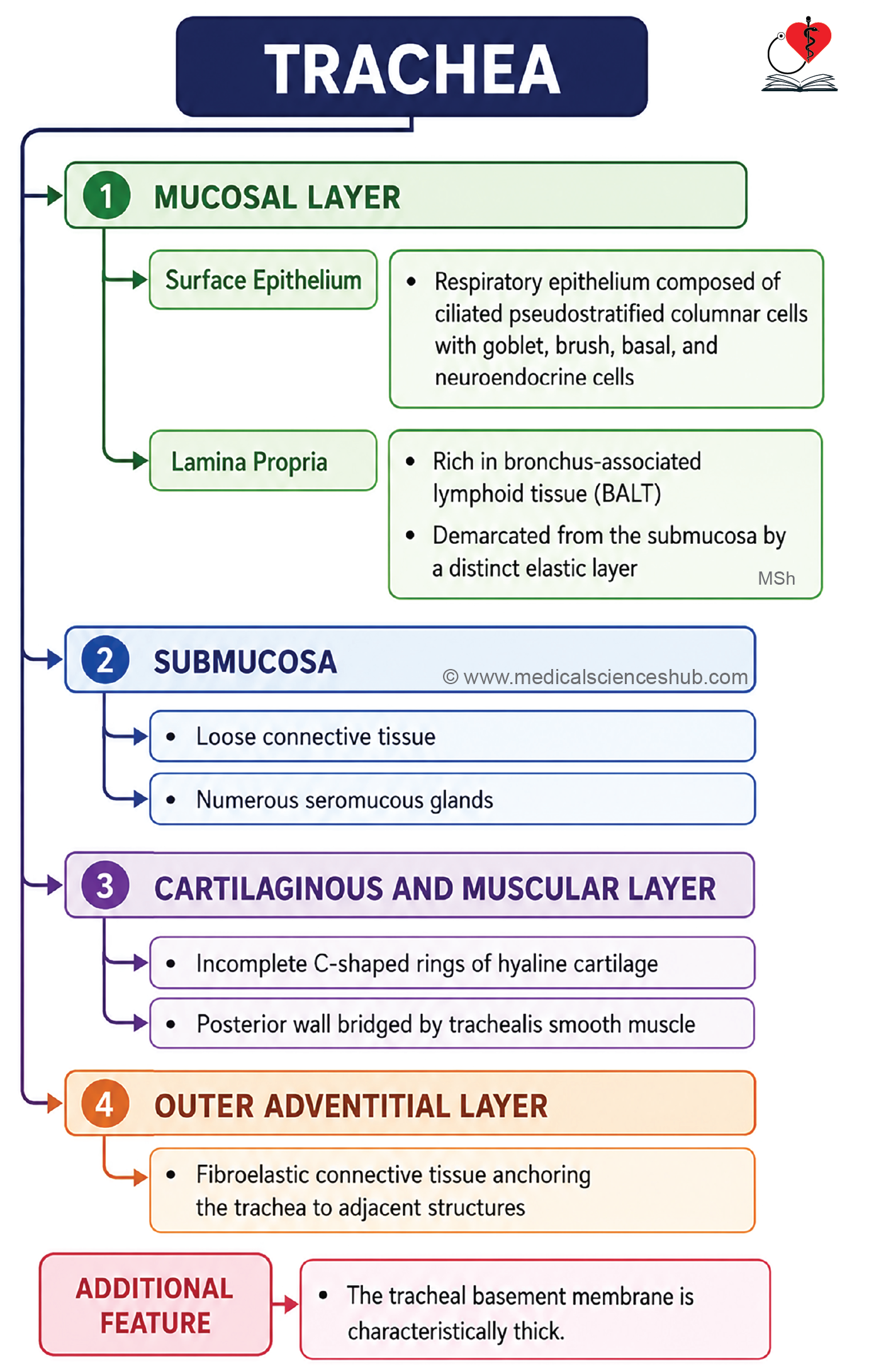
Histology of Trachea
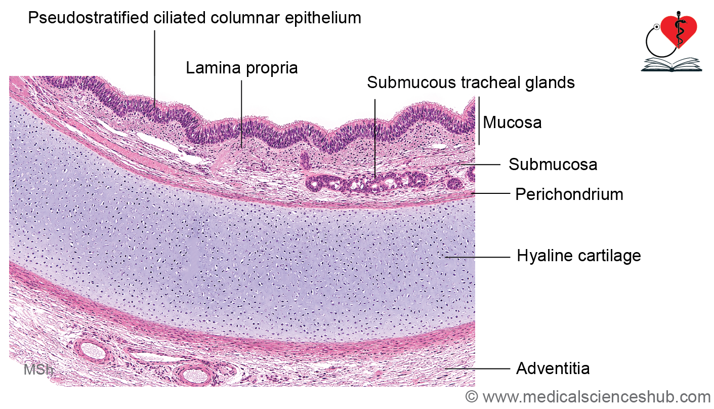
Wall of trachea shows:
- Mucosa
- Submucosa
- Cartilage–muscular layer
- Adventitia
Mucosa
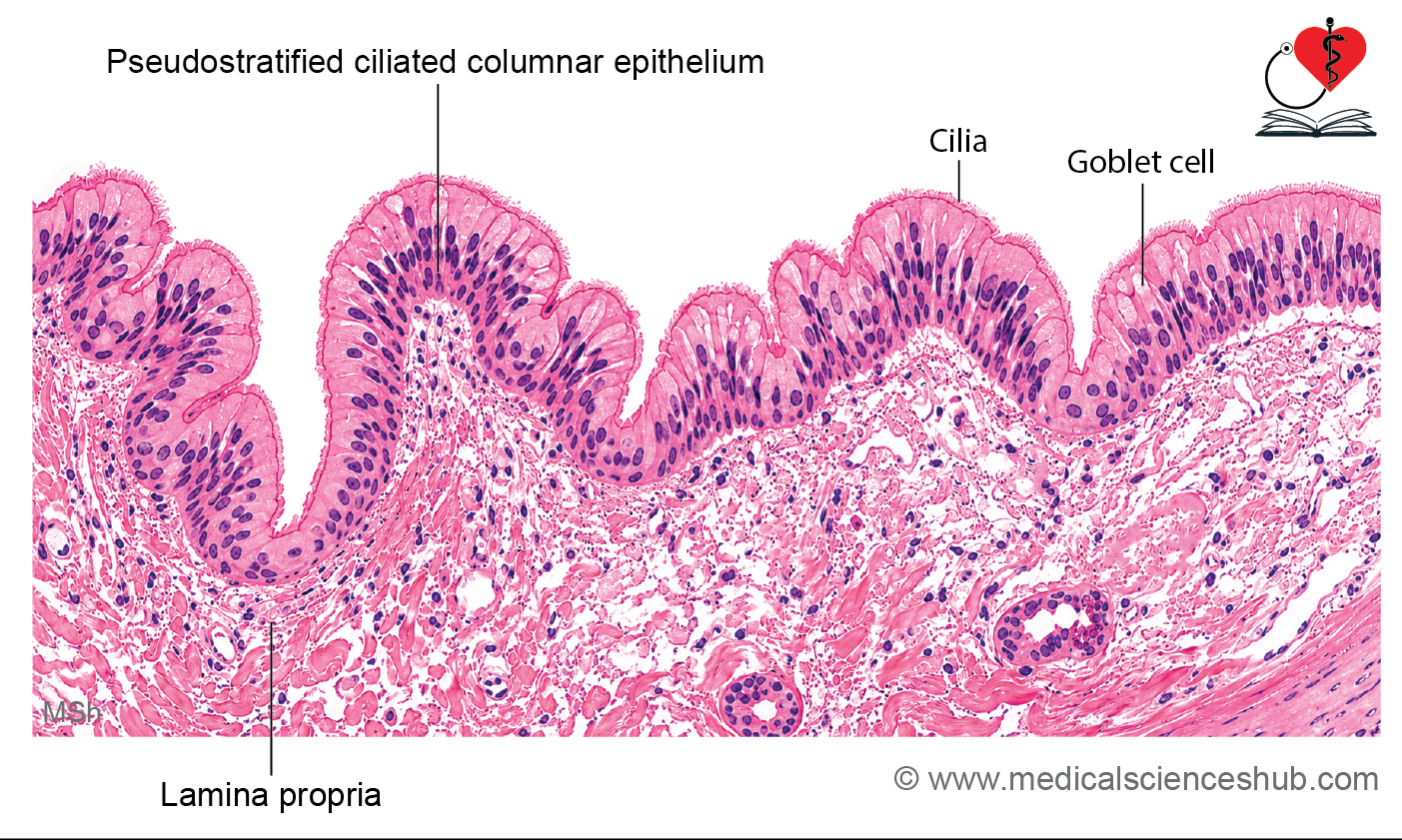
- The trachea is lined by ciliated pseudostratified columnar epithelium, commonly known as respiratory epithelium.
Cells of the Tracheal Epithelium
- Ciliated columnar cells are the most abundant cells and extend through the full thickness of the epithelium. Their cilia propel mucus and trapped particles toward the pharynx, forming the mucociliary escalator that protects the lower respiratory tract.
- Goblet (mucous) cells secrete mucus that traps dust, microorganisms, and other inhaled particles. These cells lack cilia and typically show pale cytoplasm in routine histological sections.
- Brush cells are columnar cells with microvilli that establish contact with sensory nerve endings and function as chemoreceptor-like cells.
- Small granule cells (Kulchitsky cells) belong to the diffuse neuroendocrine system. They contain secretory granules and release biologically active substances that regulate local airway function.
- Basal cells are stem cells located adjacent to the basal lamina and are responsible for regeneration of the respiratory epithelium.
Basement Membrane
- A thick basement membrane is a characteristic histological feature of the trachea.
- In routine staining, it appears as a homogeneous eosinophilic layer beneath the epithelium.
Lamina Propria
- The lamina propria consists of loose connective tissue rich in blood vessels and elastic fibers.
- It contains lymphocytes and lymphoid aggregates that contribute to local immune defense as part of bronchus-associated lymphoid tissue (BALT).
- Elastic fibers become particularly concentrated near the junction of the lamina propria and submucosa, forming an elastic layer that helps maintain airway flexibility and recoil.
Submucosa
- The submucosa lies beneath the lamina propria, and the boundary between the two layers is often indistinct in routine histological sections.
- It contains numerous seromucous glands, their ducts lined by cuboidal epithelium, as well as blood vessels, lymphatics, and lymphoid tissue.
- These glands help maintain moisture and protect the respiratory mucosa.
Tracheal Cartilage and Trachealis Muscle
- The trachea contains 16–20 C-shaped rings of hyaline cartilage, which maintain airway patency and prevent collapse.
- The posterior gap between the cartilage rings is bridged by fibroelastic tissue and the trachealis muscle, a band of smooth muscle.
- With advancing age, tracheal cartilage may undergo partial calcification.
- Hyaline cartilage consists of chondrocytes within lacunae embedded in a homogeneous matrix and is surrounded by a perichondrium.
Adventitia
- The outermost layer, the adventitia, is composed of fibroelastic connective tissue containing blood vessels and nerves that support the tracheal wall.
Bronchial Tree
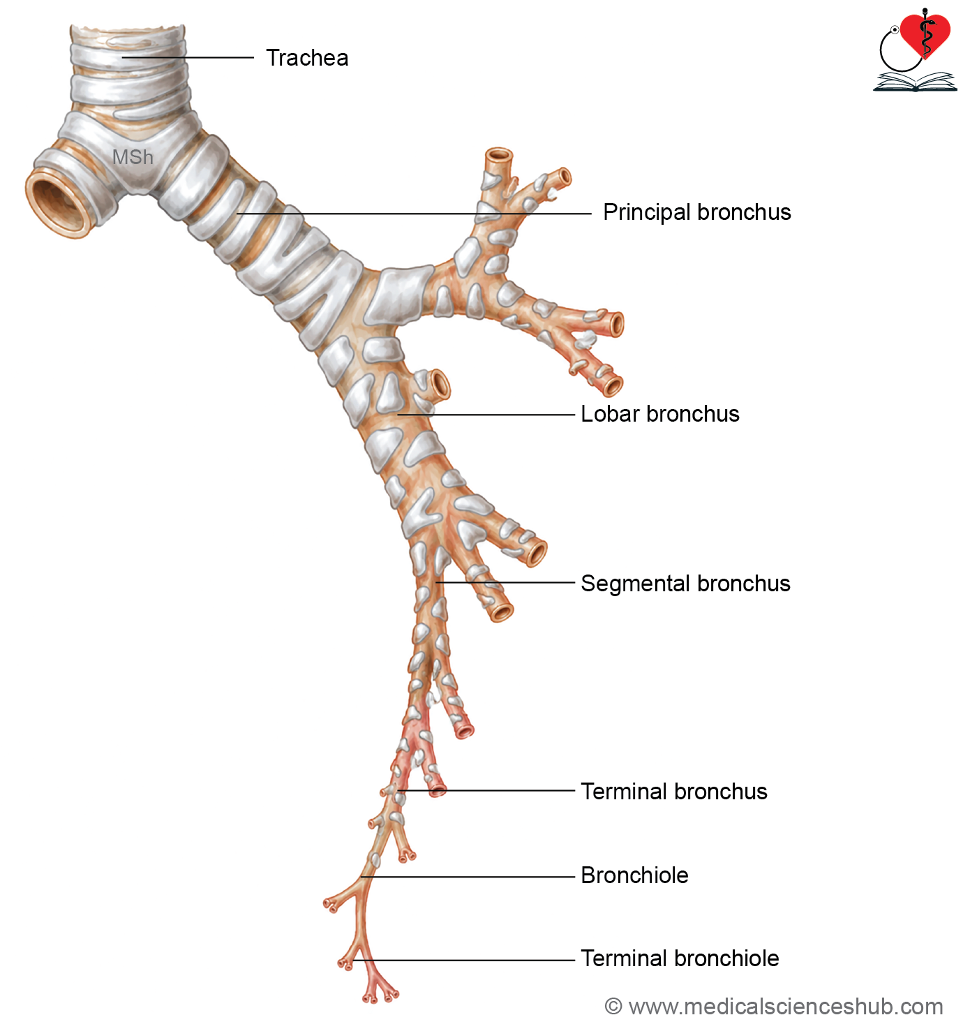
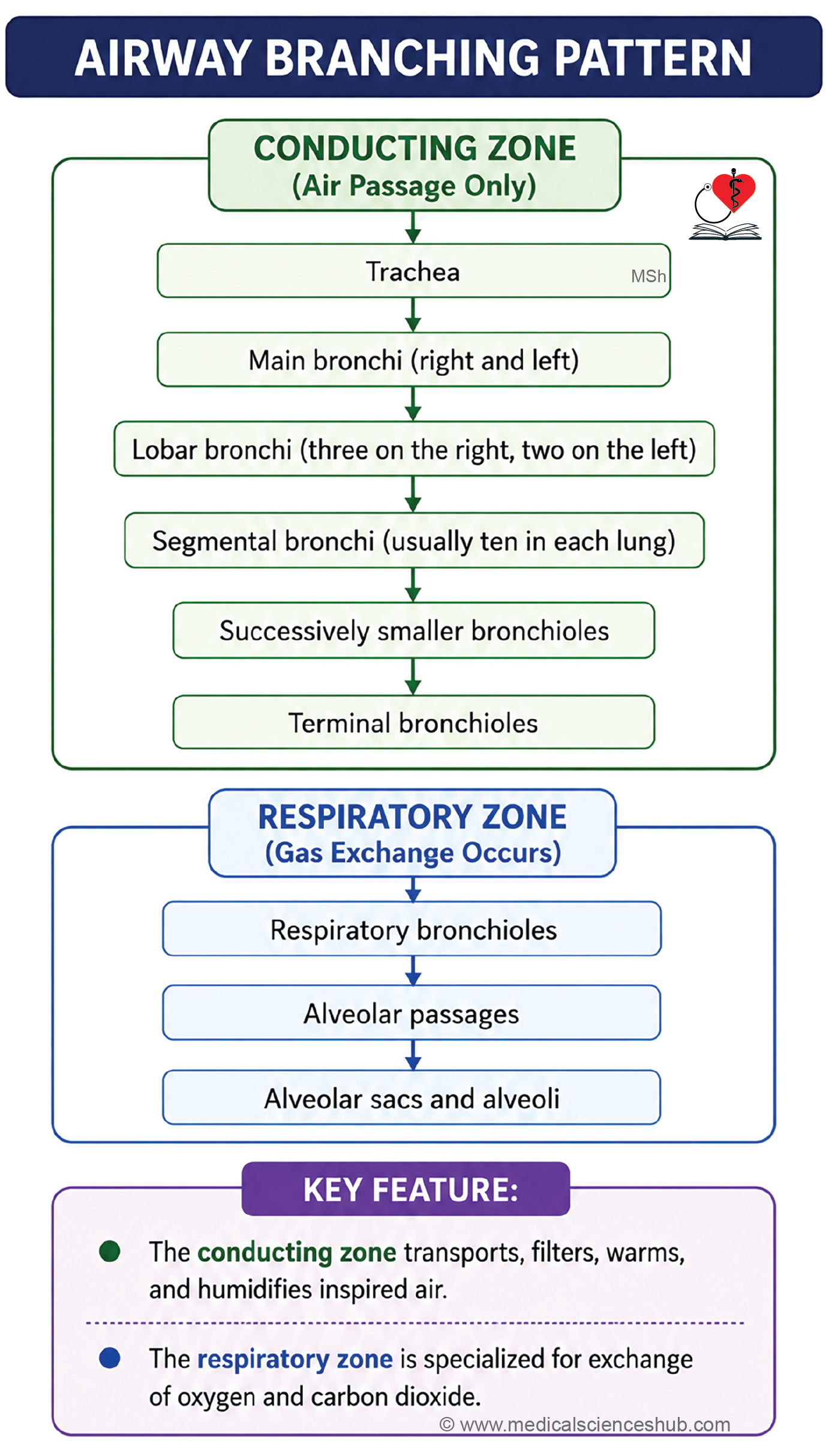
- The trachea bifurcates into the right and left primary (main) bronchi, which enter the corresponding lungs.
- The right main bronchus is wider, shorter, and more vertical than the left bronchus.
- The right bronchus divides into three lobar (secondary) bronchi, while the left bronchus divides into two lobar bronchi, corresponding to the lobes of each lung.
- Secondary bronchi further divide into segmental (tertiary) bronchi, which supply the bronchopulmonary segments.
- These bronchi undergo repeated branching to form progressively smaller bronchioles.
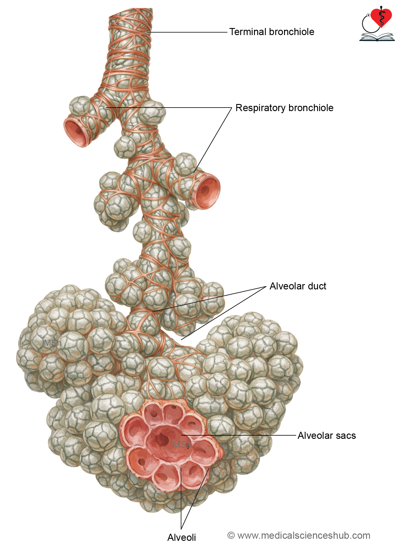
- The smallest conducting airways are the terminal bronchioles.
- Terminal bronchioles give rise to respiratory bronchioles, which continue as alveolar ducts, alveolar sacs, and finally alveoli.
- The airway from the trachea to the terminal bronchioles constitutes the conducting portion, responsible for air transport and conditioning.
- The respiratory bronchioles and structures distal to them form the respiratory portion, where gas exchange occurs between air and blood.
Lung
- The lungs are paired respiratory organs and are the primary sites of gas exchange.
- Histologically, the principal bronchi closely resemble the trachea.
- Each lung consists of a pleural covering, connective tissue septa, and functional lung parenchyma.
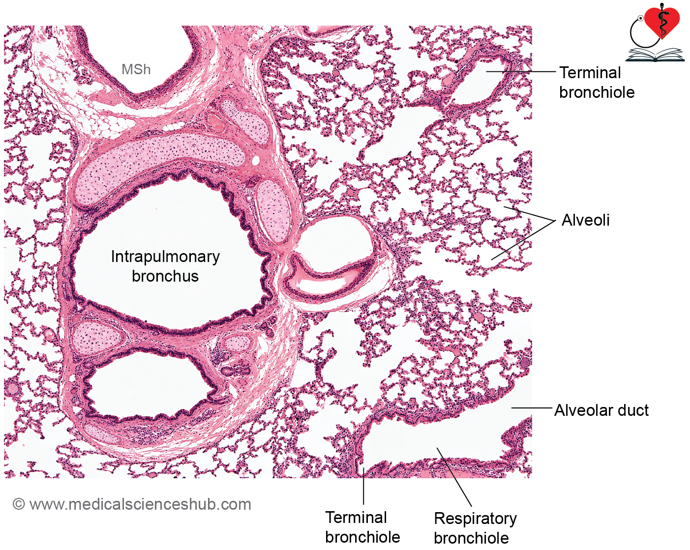
- The principal bronchus divides into lobar (secondary) bronchi, which enter the lung and continue as intrapulmonary bronchi.
- Lung parenchyma contains the branching airways and alveoli.
Intrapulmonary Bronchi
- Intrapulmonary bronchi include the lobar and segmental (tertiary) bronchi.
- Repeated branching of segmental bronchi produces bronchioles, which are airways less than 1 mm in diameter and characteristically lack cartilage in their walls.
Histology of Intrapulmonary Bronchi
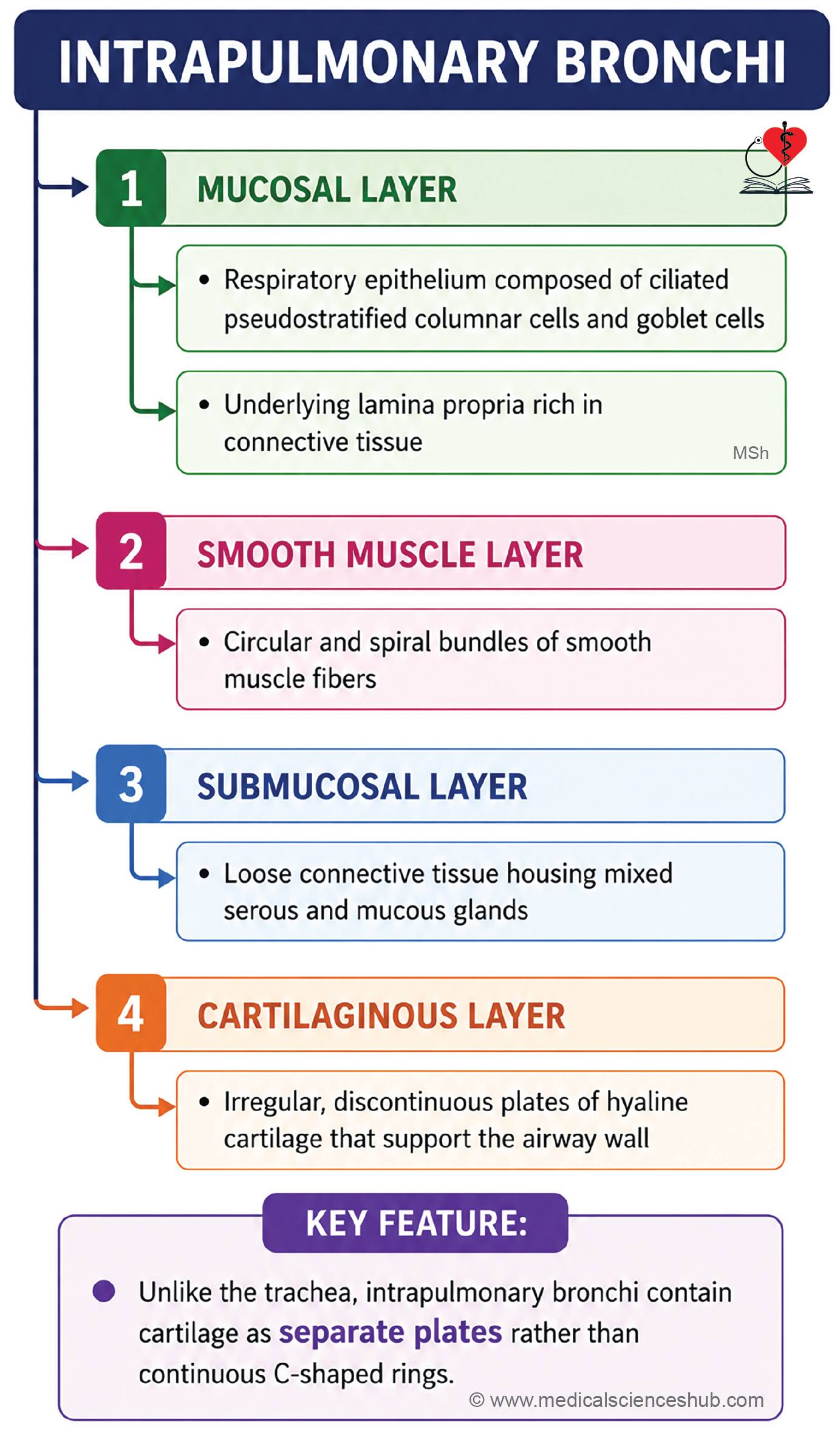
- Intrapulmonary bronchi have the following layers:
- Mucosa
- Muscular layer
- Submucosa
- Cartilage
- Adventitia
Mucosa
- The mucosa is lined by ciliated pseudostratified columnar epithelium containing goblet cells.
- The height of the epithelium gradually decreases as the bronchial diameter becomes smaller.
- The lamina propria is rich in elastic fibers and supports mucociliary function.
Muscular layer
- A continuous layer of smooth muscle surrounds the mucosa.
- The muscle fibers are arranged in a spiral pattern and regulate airway caliber by bronchoconstriction and bronchodilation.
Submucosa
- The submucosa consists of loose connective tissue containing numerous seromucous glands.
- These glands contribute secretions that help humidify and protect the airway.
Cartilage layer
- Unlike the trachea and main bronchi, intrapulmonary bronchi contain irregular discontinuous plates of hyaline cartilage rather than C-shaped rings.
- These cartilage plates provide structural support and prevent airway collapse.
Adventitia
- The outermost adventitia is composed of connective tissue that blends with the surrounding lung parenchyma.
- It contains blood vessels, nerves, and supporting connective tissue.
- The presence of a continuous smooth muscle layer and discontinuous cartilage plates is a key histological feature of intrapulmonary bronchi.
Bronchioles
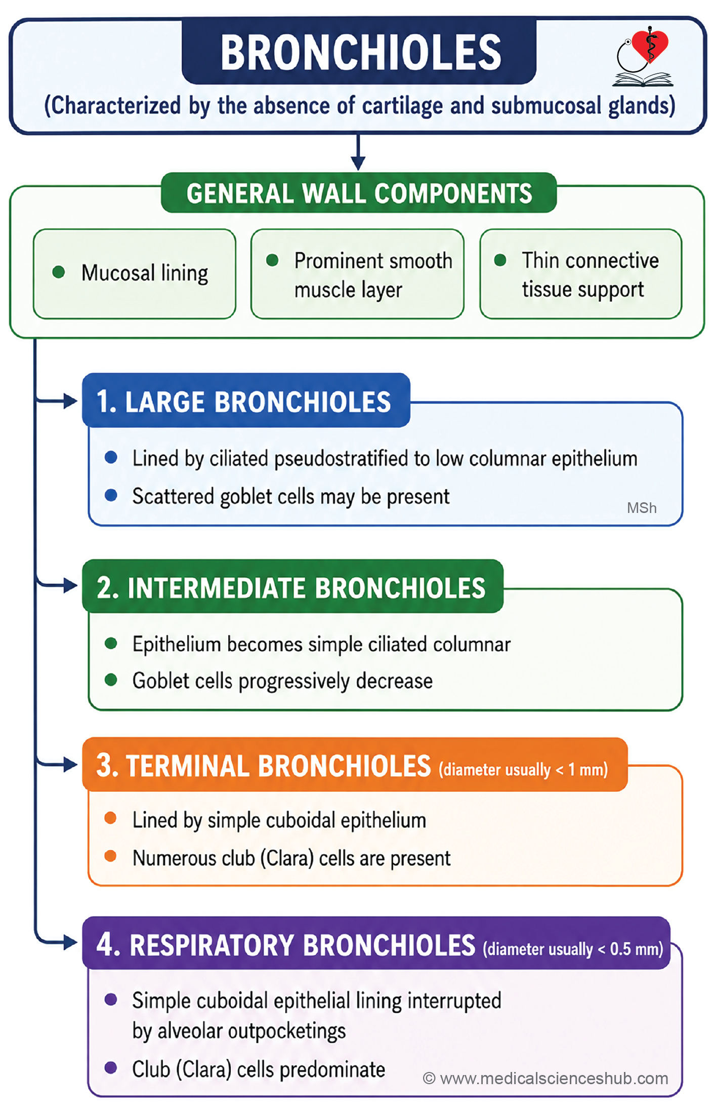
- Bronchioles are small conducting airways with a diameter generally less than 1 mm.
- Unlike bronchi, their walls lack both cartilage and submucosal glands.
- A pulmonary lobule is a subdivision of a bronchopulmonary segment supplied by a single terminal bronchiole and its branches.
- Bronchioles branch repeatedly to form terminal bronchioles, the last part of the conducting airway system.
- Each terminal bronchiole gives rise to respiratory bronchioles, which lead to alveolar ducts, alveolar sacs, and alveoli.
- A pulmonary acinus comprises all structures distal to a terminal bronchiole and represents the gas-exchanging portion of the lung.
- The respiratory bronchiolar unit, consisting of a respiratory bronchiole and its associated alveoli, functions as a basic unit of pulmonary gas exchange.
- Airway branching follows the sequence: segmental bronchus → bronchiole → terminal bronchiole → respiratory bronchiole → alveolar duct → alveoli.
Structure of Bronchiole
- Bronchioles are small airways composed of three layers: mucosa, smooth muscle layer, and an outer connective tissue layer.
- Unlike bronchi, bronchioles lack both cartilage and submucosal seromucous glands.
Mucosa
- The epithelial lining changes with decreasing airway size:
- Larger bronchioles: Ciliated pseudostratified columnar epithelium with goblet cells.
- Medium bronchioles: Simple ciliated columnar epithelium with fewer goblet cells.
- Terminal bronchioles: Simple cuboidal epithelium containing Club (Clara) cells and no goblet cells.
- Club cells are nonciliated secretory cells that become prominent in terminal bronchioles.
- These cells possess apical microvilli and secrete surfactant-like substances that reduce surface tension and help maintain airway patency.
- Club cells also produce Club Cell Secretory Protein (CC16), a useful marker of lung epithelial injury.
- They detoxify inhaled harmful substances, contribute to epithelial repair, and function as progenitor (stem) cells for bronchiolar epithelium.
Smooth muscle layer
- The smooth muscle layer is the most prominent component of the bronchiolar wall.
- Its contraction and relaxation, regulated by the autonomic nervous system, control airway diameter and airflow resistance.
Connective tissue layer
- A thin outer layer of connective tissue surrounds each bronchiole and separates it from adjacent lung tissue.
- This layer is rich in elastic fibers, which facilitate lung expansion and recoil during respiration.
Key histological features
- Absence of cartilage.
- Absence of submucosal glands.
- Absence of goblet cells in terminal bronchioles.
- Presence of Club (Clara) cells in terminal bronchioles.
Respiratory Bronchiolar Unit
- The respiratory bronchiolar unit is the smallest functional unit of the lung involved in gas exchange.
- It consists of a respiratory bronchiole and the alveoli supplied by it.
Respiratory Bronchiole
- Respiratory bronchioles are small airways, typically less than 0.5 mm in diameter.
- They represent the transition between the conducting and respiratory portions of the respiratory tract.
- Their lining is simple cuboidal epithelium, which gradually loses cilia toward the distal segments.
- The epithelium contains Club (Clara) cells, which contribute to airway protection and epithelial regeneration.
- A thin layer of smooth muscle surrounds the epithelium and helps regulate airflow.
- An outer layer of delicate connective tissue provides structural support.
- The walls of respiratory bronchioles contain scattered alveoli, allowing limited gas exchange.
Alveolar Ducts and Alveolar Sacs
- Each respiratory bronchiole branches into two or more alveolar ducts.
- Alveolar ducts are elongated passages whose walls are lined predominantly by alveoli.
- Each alveolar duct opens into an alveolar sac, a common chamber surrounded by clusters of alveoli.
- Multiple alveoli communicate with the alveolar sac, providing a large surface area for efficient gas exchange.
- The airflow pathway is: Respiratory bronchiole → Alveolar duct → Alveolar sac → Alveoli.
Alveoli
- Alveoli are thin-walled, sac-like terminal air spaces of the lungs and are the primary sites of gas exchange.
- Each lung contains approximately 150–250 million alveoli, providing a total respiratory surface area of about 70–100 m².
- Individual alveoli measure roughly 200 μm in diameter.
Alveolar Septum
- Adjacent alveoli are separated by a delicate alveolar septum containing capillaries, elastic fibers, fibroblasts, and immune cells.
- The septum provides structural support and facilitates efficient diffusion of respiratory gases.
- Pores of Kohn (interalveolar pores) connect neighboring alveoli, allowing collateral ventilation and equalization of air pressure.
Cells of Alveolar Epithelium
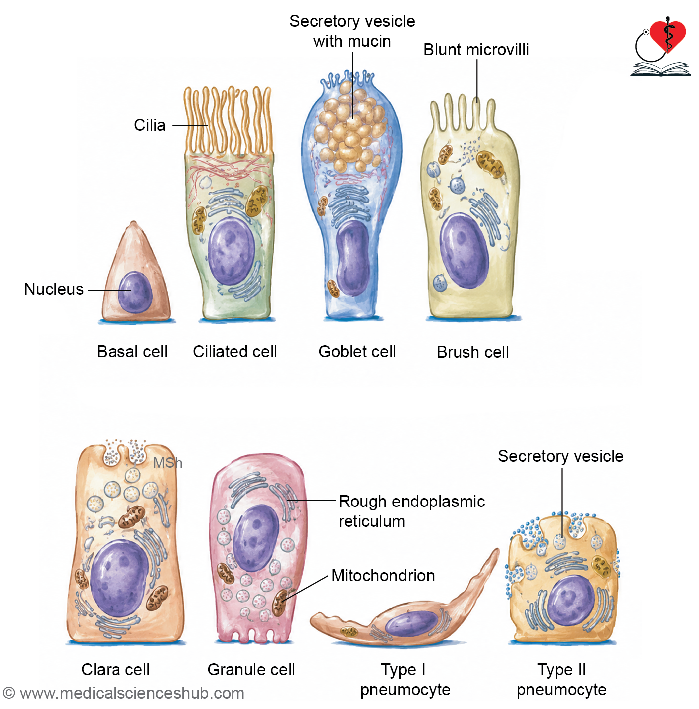
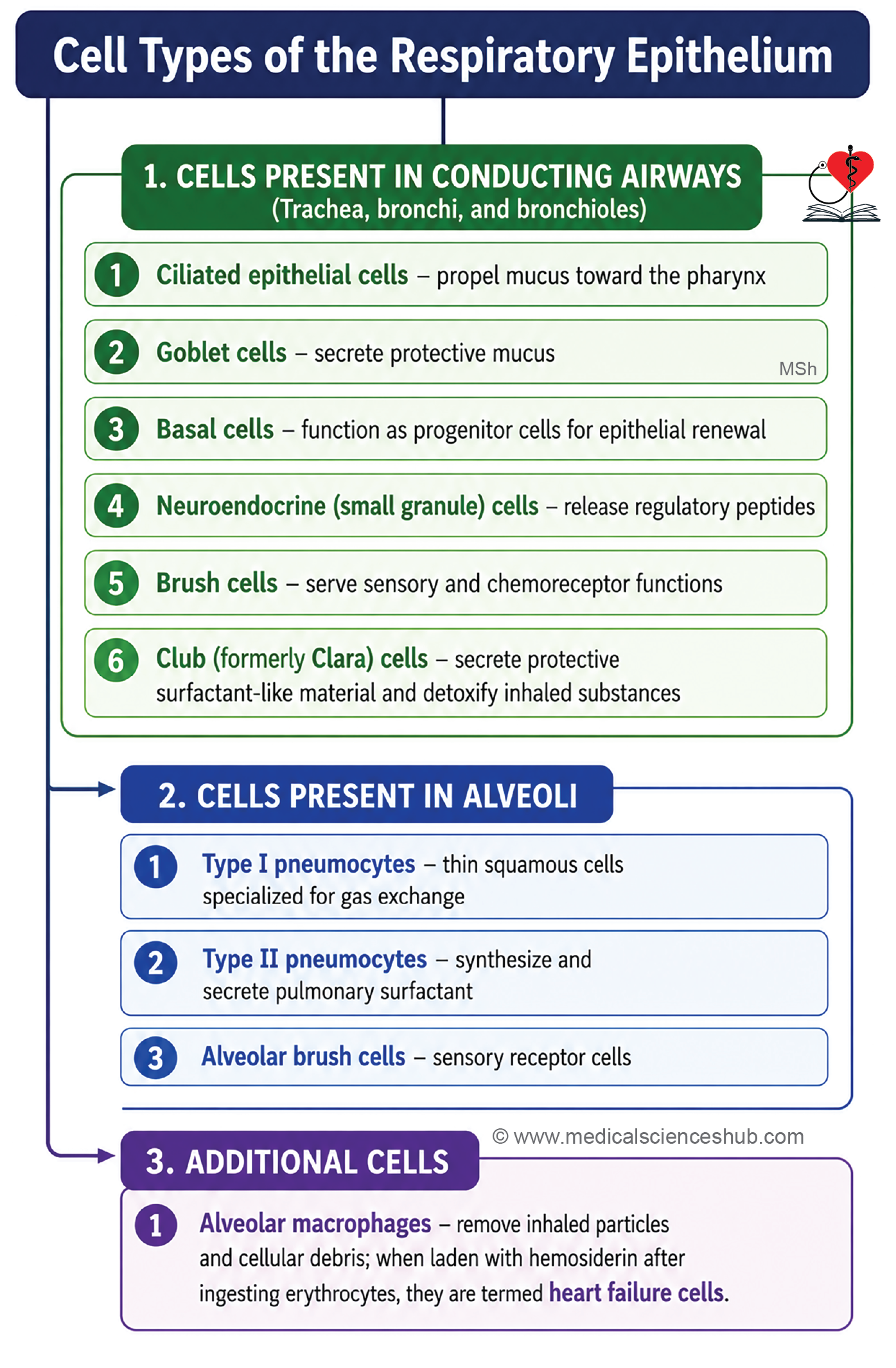
- Alveolar wall is lined by type I pneumocytes, type II pneumocytes, and occasional brush cells.
H4: Type I Pneumocytes (95%)
- Type I pneumocytes are extremely thin simple squamous cells that cover about 95% of the alveolar surface.
- They are joined by tight junctions and form the principal component of the blood–air barrier.
- These cells have limited regenerative capacity and are not actively mitotic.
Type II Pneumocytes (5%)
- Type II pneumocytes are cuboidal epithelial cells located between type I cells.
- They contain characteristic lamellar bodies, which store pulmonary surfactant.
- Surfactant, rich in dipalmitoylphosphatidylcholine (DPPC), reduces surface tension and prevents alveolar collapse during expiration.
- Type II pneumocytes serve as progenitor cells and can replace damaged type I and type II alveolar cells.
Brush Cells
- A small number of brush cells are present within the alveolar epithelium.
- These cells possess microvilli and function as sensory receptors that monitor the composition of inspired air.
- Together, these specialized cells maintain alveolar integrity and ensure efficient pulmonary gas exchange.
CLINICAL CORRELATION
- Pneumonia is inflammation of the lung parenchyma, particularly the alveoli, and may result from infections, allergens, chemical irritants, or certain medications.
- Lung cancer is strongly associated with tobacco smoke exposure. Chronic irritation can induce squamous metaplasia, in which normal respiratory epithelium is replaced by stratified squamous epithelium, increasing the risk of malignant transformation.
- Metaplasia refers to the reversible replacement of one mature cell type by another better adapted to a persistent stimulus.
- Acute Respiratory Distress Syndrome (ARDS) is a severe inflammatory condition causing respiratory failure and impaired gas exchange. In neonates, respiratory distress syndrome commonly results from surfactant deficiency due to immature lungs.
- Bronchitis is inflammation of the bronchi and may occur as an acute or chronic condition.
- Primary Ciliary Dyskinesia (Kartagener syndrome) is an autosomal recessive disorder characterized by defective ciliary motility. Impaired mucociliary clearance leads to recurrent respiratory infections and may also cause infertility due to abnormal ciliary or sperm function.
- Emphysema is characterized by permanent enlargement of air spaces distal to the terminal bronchioles, accompanied by destruction of alveolar walls. Cigarette smoking is the most important risk factor, although air pollution and genetic factors may also contribute.
Alveolar Macrophages
- Alveolar macrophages are resident immune cells found within the alveolar lumen and the connective tissue of alveolar septa.
- They constitute an important first line of defense against inhaled pathogens and foreign particles.
- Their primary function is phagocytosis, which helps maintain the cleanliness and sterility of the alveolar environment.
Functions
- They engulf and remove inhaled dust particles, earning the name dust cells.
- They phagocytose accumulated carbon particles and tar in the lungs of smokers.
- They help eliminate inhaled allergens, including pollen grains and other particulate matter.
- In chronic left-sided heart failure, red blood cells may leak into alveoli from congested pulmonary capillaries. Macrophages ingest these erythrocytes and accumulate hemosiderin, becoming heart failure cells (siderophages).
Clinical Significance
- Mycobacterium tuberculosis can survive within macrophages by evading intracellular destruction.
- Persistent infection stimulates a chronic immune response, leading to the formation of granulomas that contain multinucleated giant cells derived from fused macrophages.
- Thus, alveolar macrophages play essential roles in pulmonary defense, debris clearance, and immune surveillance.
Blood–Air Barrier
- The blood–air barrier is the thin interface through which oxygen and carbon dioxide diffuse between alveolar air and pulmonary capillary blood.
- Efficient gas exchange depends on the minimal thickness of this barrier.
- From the alveolar lumen to the capillary lumen, the thinnest part of the barrier consists of:
- A thin layer of pulmonary surfactant.
- The type I pneumocyte lining the alveolus (simple squamous epithelium).
- The basal lamina of the alveolar epithelium.
- The basal lamina of the capillary endothelium, usually fused with the alveolar basal lamina.
- The continuous (non-fenestrated) capillary endothelium.
- ·This specialized structure provides a very short diffusion distance, enabling rapid and efficient gaseous exchange between air and blood.
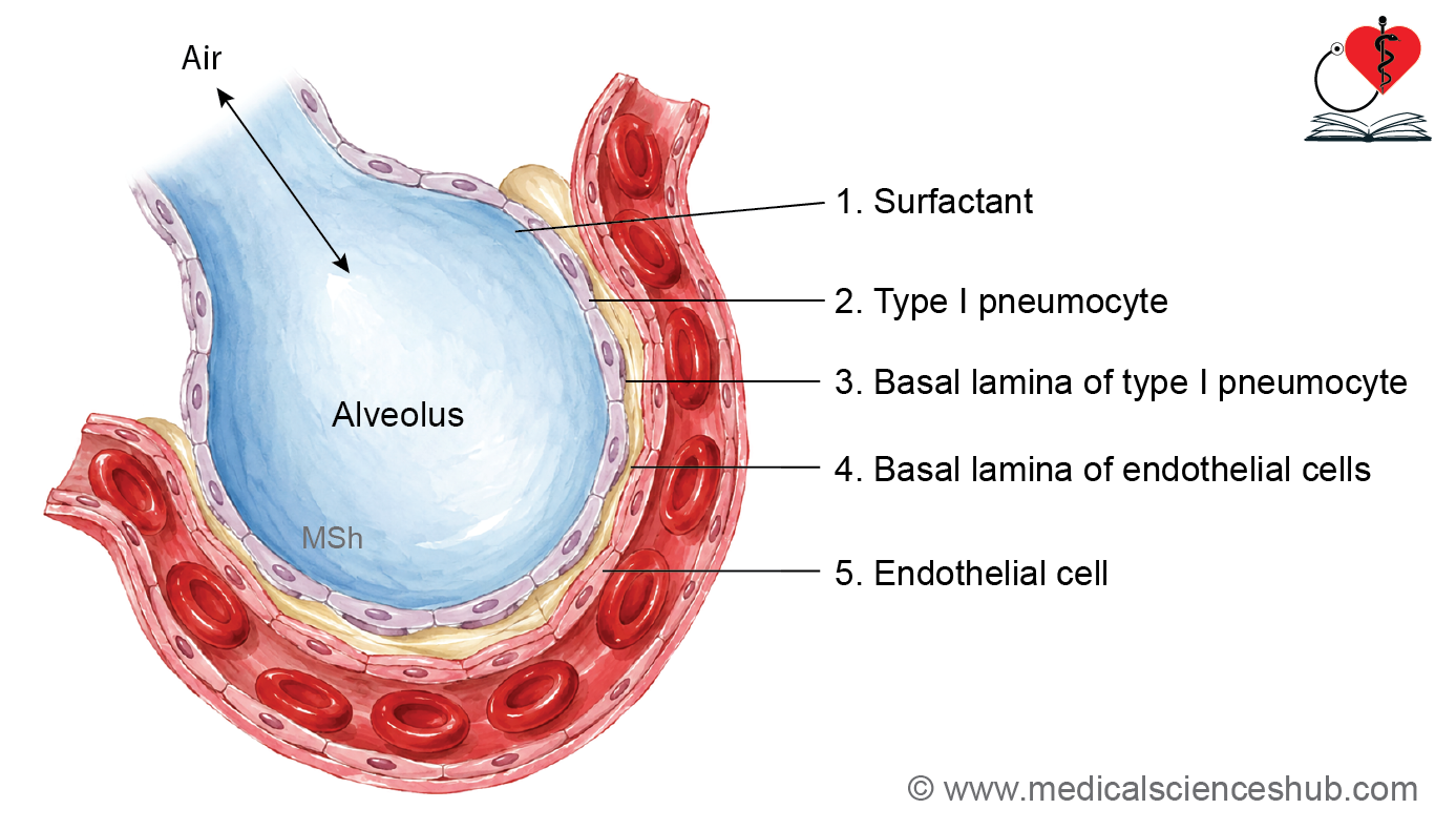
Table 12.1: Differences between bronchus and bronchiole
| Feature | Bronchus | Bronchiole |
|---|---|---|
| Diameter | Usually greater than 1 mm | Usually less than 1 mm |
| Epithelial lining | Ciliated pseudostratified columnar epithelium with numerous goblet cells | Larger bronchioles are lined by simple ciliated columnar epithelium with few goblet cells; terminal bronchioles are lined by simple cuboidal epithelium with Club (Clara) cells and lack goblet cells |
| Smooth muscle | Present as a distinct layer beneath the mucosa and internal to cartilage plates | Forms a prominent circumferential layer beneath the mucosa and external to it lies connective tissue |
| Cartilage | Hyaline cartilage plates are present in the wall | Cartilage is absent |
| Submucosal glands | Seromucous glands are present in the submucosa | Glands are absent |
| Goblet cells | Commonly present throughout the epithelium | Decrease in number with airway size and are absent in terminal bronchioles |
| Supporting cells | Club cells are generally absent | Club (Clara) cells are prominent, especially in terminal bronchioles |
| Histological identification | Presence of cartilage plates and seromucous glands | Absence of cartilage and glands, with a prominent smooth muscle layer |
Important Questions
- Write a short note on histology of epiglottis.
- Write a short note on histology of trachea.
- Write a short note on histology of lung.
- List the cells lining respiratory passage.
- List the microscopic differences between bronchus and bronchiole.
- Write a short note on blood–air barrier.
📝 Test Your Knowledge – Practice MCQs
Attempt the chapter MCQ quiz and assess your understanding of key concepts.
Click to see: Medical Disclaimer and Copyright Notice