
Competency
- AN70.1 Identify exocrine gland under the microscope and distinguish between serous, mucous and mixed acini.
Introduction
- A gland is a specialized group of cells that synthesizes and secretes specific substances, including hormones. Glands are embryologically derived from epithelial tissue.
- Note: Mucus is a noun; mucous is an adjective — for example, mucous cells secrete mucus.
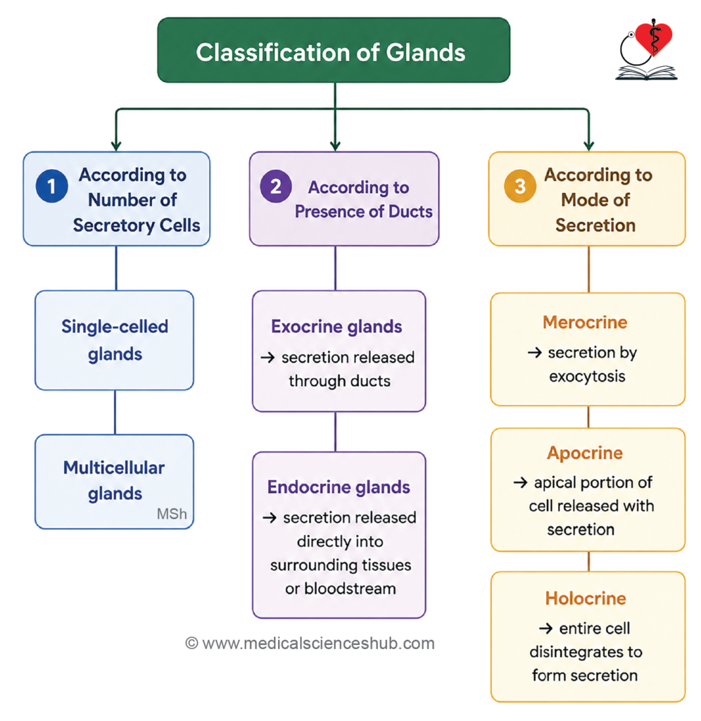
Classification of Glands
- Based on cell number:
- Unicellular glands consist of a single secretory cell.
- Multicellular glands are composed of a group of secretory cells.
- Based on presence of a duct:
- Exocrine glands release secretions through a duct onto an epithelial surface.
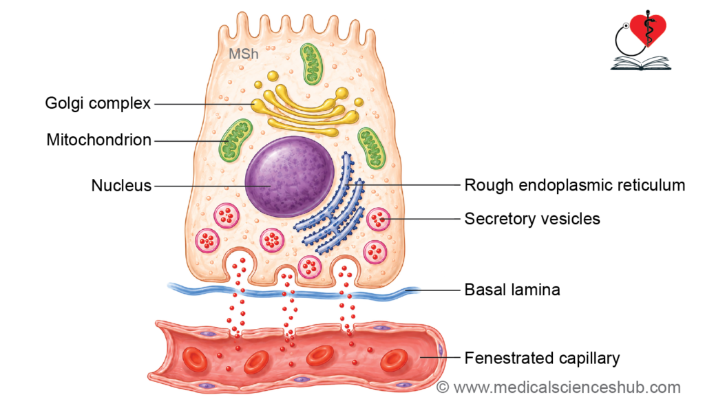
- Endocrine glands are ductless and release hormones directly into the bloodstream.
- Based on secretory mechanism:
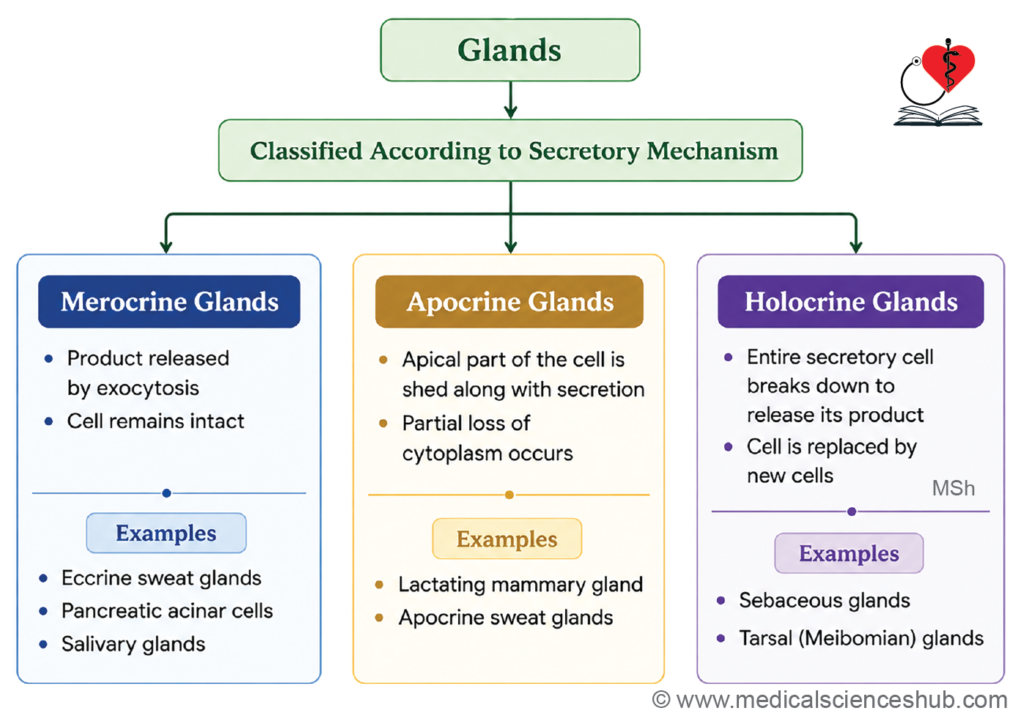
- Merocrine glands release secretions via exocytosis, leaving the cell intact.
- Apocrine glands secrete by shedding the apical cytoplasm of the cell.
- Holocrine glands release secretions through complete disintegration of the secretory cell.
Exocrine Glands
- Exocrine glands deliver their secretions to a target site via a duct.
- They are classified based on:
- Duct branching: simple (unbranched) or compound (branched)
- Secretory unit shape: tubular, acinar, or alveolar
- Nature of secretion: serous, mucous, or mixed
- Secretory mechanism: merocrine, apocrine, or holocrine (included by some authors as a fourth classification criterion)
Classification Based on Branching Pattern of Duct
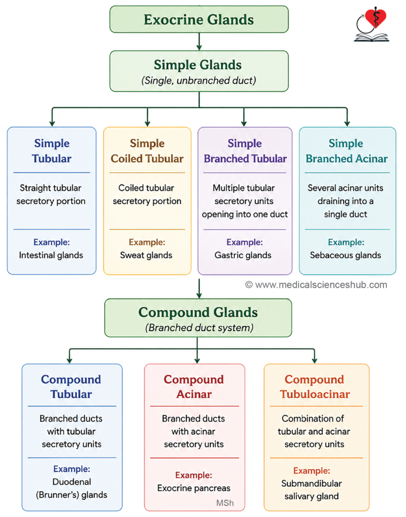
- Exocrine glands are classified according to the branching pattern of their ducts.
- Simple glands possess a single unbranched duct. Examples include the crypts of Lieberkühn, sweat glands, fundic glands of the stomach, urethral mucous glands, and Meibomian glands.
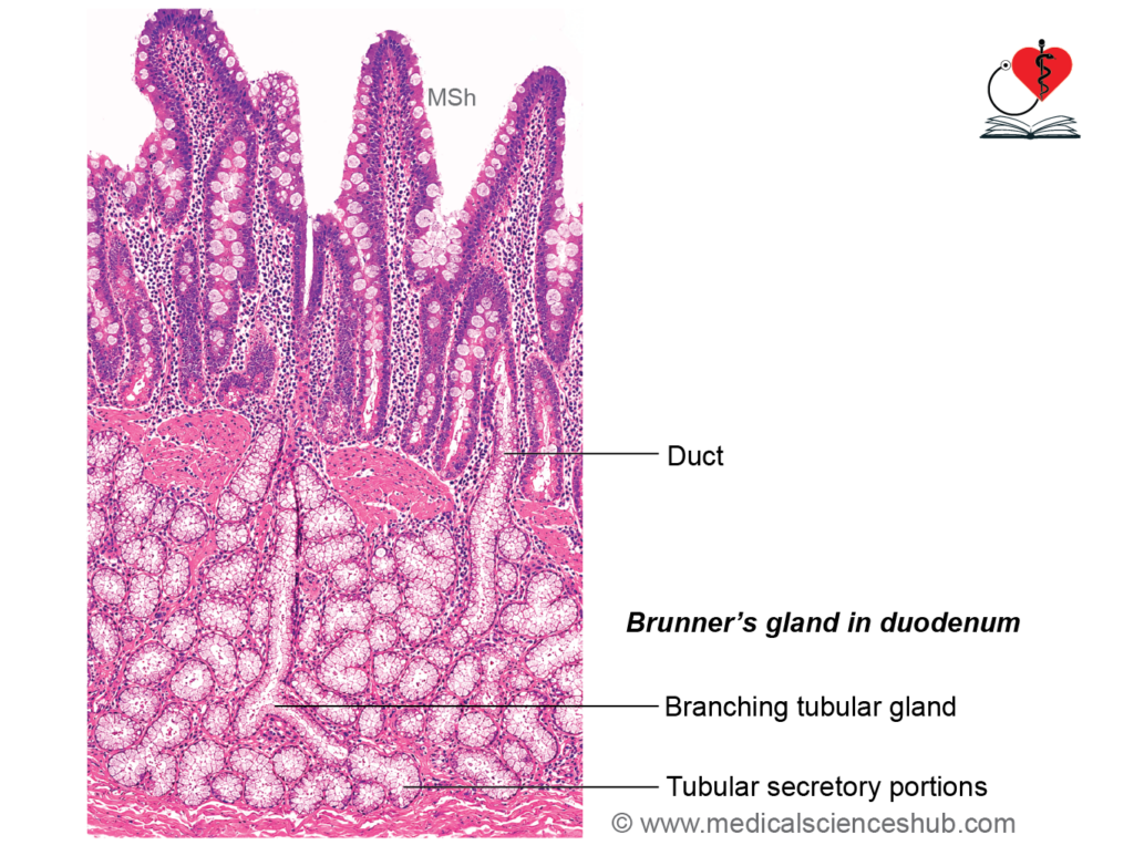
- Compound glands have a branched duct system that drains multiple secretory units. Examples include Brunner’s glands, the mammary gland, and the submandibular gland.
Classification Based on Shape of Secretory Unit
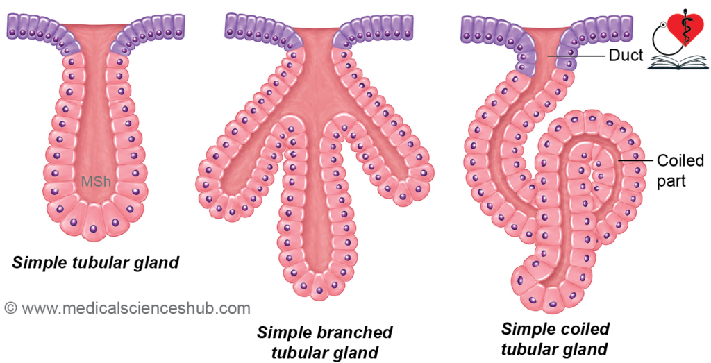
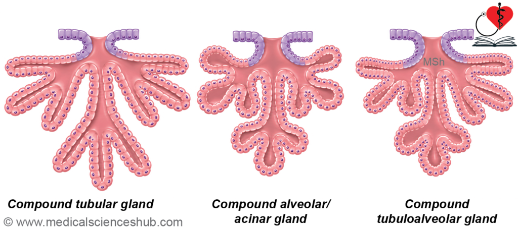
- Exocrine glands can be grouped into three groups based on the shape of secretory unit as follows:
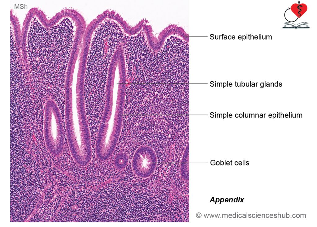
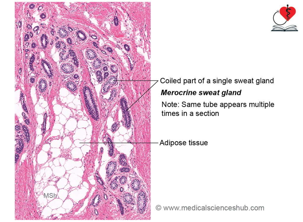
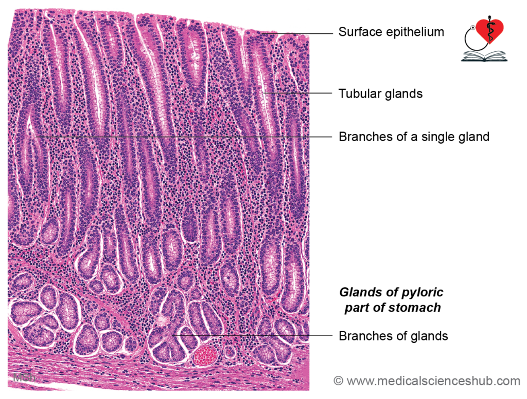
- Tubular glands have a tube-shaped secretory unit, which may be straight, coiled, or branched. Examples: Intestinal glands, eccrine sweat glands (simple coiled tubular), gastric glands, and Brunner’s glands.
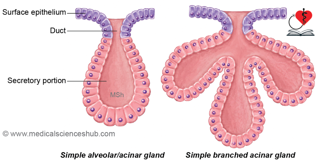
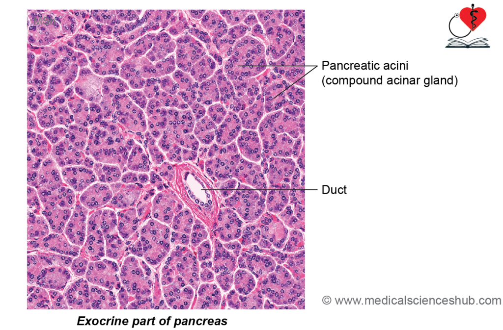
- Acinar glands have a spherical secretory unit, appearing round in cross-section. Examples: Exocrine pancreas and salivary glands.
- Alveolar (saccular) glands have a flask-shaped or distended secretory unit. Acinar glands may adopt an alveolar shape when maximally active or distended. Examples: Mammary glands and exocrine pancreas.
- In practice, glands are most commonly classified by combining secretory unit shape with ductal branching pattern — for example, “simple coiled tubular” or “compound acinar” — providing a more precise and informative description.
Classification Based on Nature of Secretions
- Based on the nature of secretions, the exocrine glands are classified as serous, mucous, and mixed glands.
Serous Glands
- Serous glands produce a thin, protein-rich watery secretion.
- Their secretory units, called serous acini, are lined by pyramidal simple columnar epithelial cells.
- The cells contain a rounded basal nucleus and abundant rough endoplasmic reticulum in the basal cytoplasm, resulting in basophilia with hematoxylin staining.
- The apical cytoplasm contains numerous secretory granules (zymogen granules), which impart eosinophilia with eosin staining.
- Serous acini typically possess a relatively small lumen compared with mucous acini.
- Examples: Parotid gland, exocrine pancreas, lacrimal gland, and von Ebner’s glands of the tongue.
Mucous Glands
- Mucous glands produce thick, viscous mucus through their secretory units called mucous acini.
- Mucous acini are lined by pyramidal simple columnar epithelial cells and possess a wider lumen compared to serous acini.
- Characteristic features of mucous cells include:
- A flattened, basally positioned nucleus.
- An apparently empty cytoplasm on H&E staining — because mucus is stored as water-soluble mucinogen granules, which dissolve during routine tissue processing, leaving vacuolated spaces.
- Mucous cells stain positively with Periodic Acid–Schiff (PAS) stain, producing a characteristic dark magenta color, confirming the presence of glycoproteins.
- Example: The sublingual salivary gland is a predominantly mucous gland.
Mixed Gland
- Mixed glands produce both viscous mucus and enzyme-rich serous secretions.
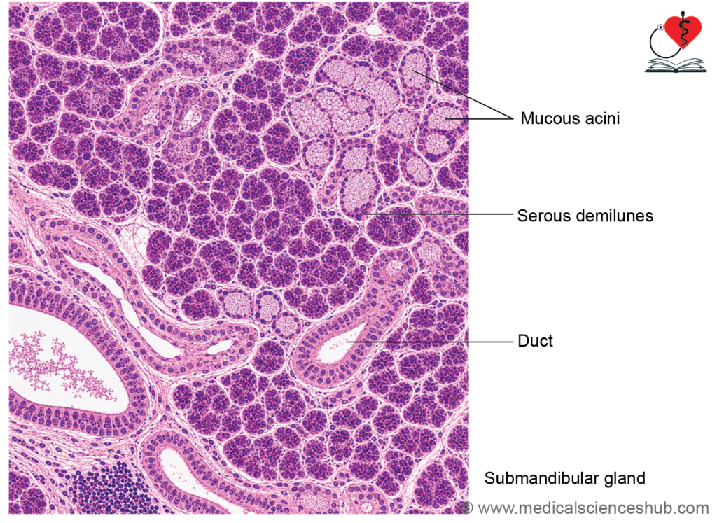
- Their secretory units consist of mucous acini associated with serous demilunes — crescent-shaped clusters of serous cells that appear to cap the mucous acinus.
- Serous cells of the demilune deliver their secretions into the acinar lumen via fine intercellular canaliculi.
- Updated concept: Serous demilunes are now recognized as a preparation artifact. During routine tissue processing, mucous cells swell and displace serous cells peripherally, creating the false demilune appearance. Studies using rapid freezing techniques demonstrate that both serous and mucous cells are actually intermixed within the same acinus.
- Example: The submandibular salivary gland is the classic example of a mixed gland.
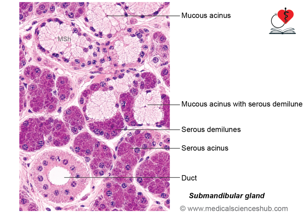
Table 3.1: Differences between serous and mucous cells
| Feature | Serous Cells | Mucous Cells |
|---|---|---|
| Shape and Size | Small pyramidal cells | Large pyramidal cells |
| H&E Staining | Basal basophilia; apical eosinophilia | Cytoplasm appears empty due to dissolution of mucus during tissue processing |
| Nucleus | Round, basally positioned | Flattened, displaced toward the basement membrane |
| Secretory Granules | Zymogen granules | Mucinogen granules |
Table 3.2: Differences between serous and mucous acini
| Feature | Serous Acini | Mucous Acini |
|---|---|---|
| Size | Smaller than mucous acini | Larger than serous acini |
| Acini Type | Compound alveolar | Compound tubular or tubulo-alveolar |
| Lumen | Narrow | Wide |
| Constituent Cells | Serous cells | Mucous cells |
| Nature of Secretion | Thin, watery, enzyme-rich fluid | Thick, viscous mucoid secretion |
| Example | Parotid gland | Sublingual gland |
Classification Based on Mode of Secretion
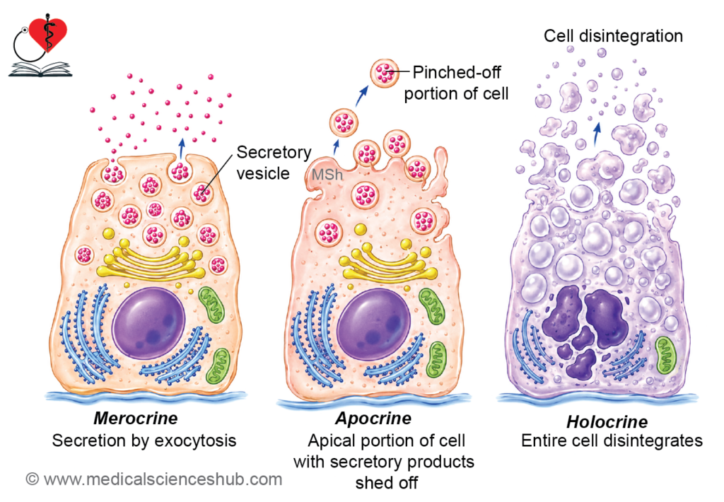
Exocrine glands are classified into three types based on their secretory mechanism: merocrine, apocrine, and holocrine.
Merocrine/eccrine Glands
- Secretory products are packaged into membrane-bound vesicles, which fuse with the apical cell membrane and release their contents by exocytosis, leaving the cell structurally intact.
- This is the most common secretory mechanism among exocrine glands.
- Examples: Pancreatic acinar cells and eccrine sweat glands.
Apocrine Glands
- Secretory products accumulate within the apical cytoplasm, which is subsequently shed along with its contents as the secretory mechanism.
- Examples: Lactating mammary glands, apocrine sweat glands, and ceruminous glands of the external ear canal.
Holocrine Glands
- Secretory products accumulate within the cell, which ultimately undergoes programmed cell death (apoptosis) upon maturation, releasing its entire contents as the secretion.
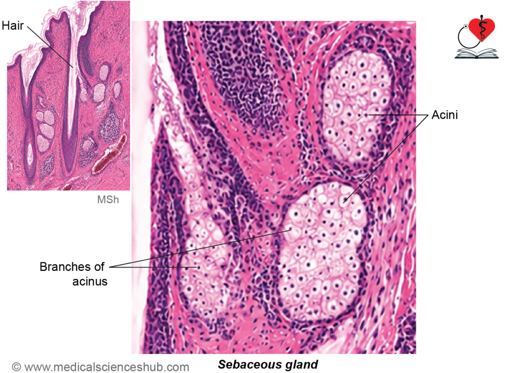
- Examples: Sebaceous glands and Meibomian (tarsal) glands of the eyelids.
- Note: Endocrine glands are ductless and secrete hormones directly into the bloodstream.
Paracrine and Autocrine Glands
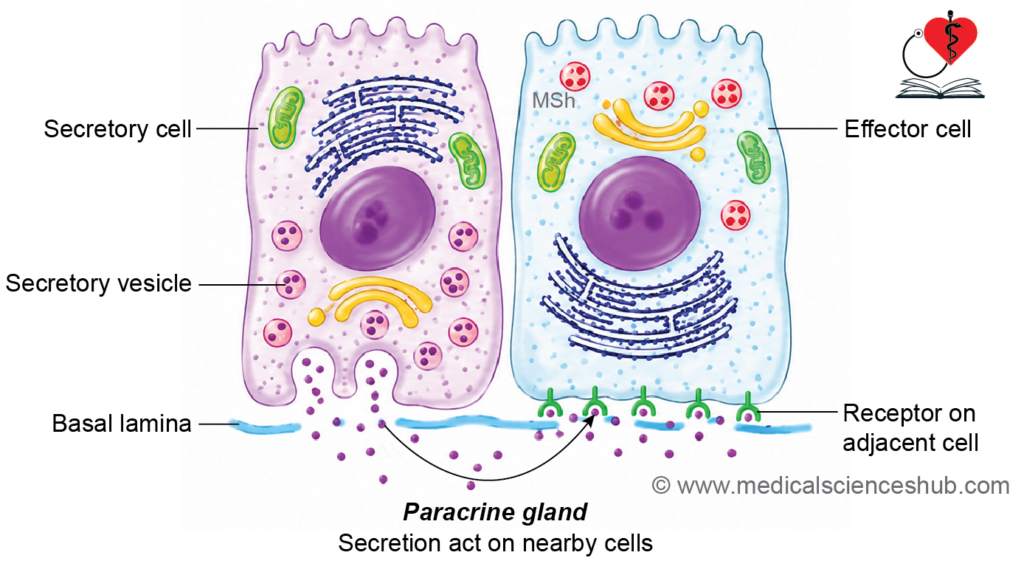
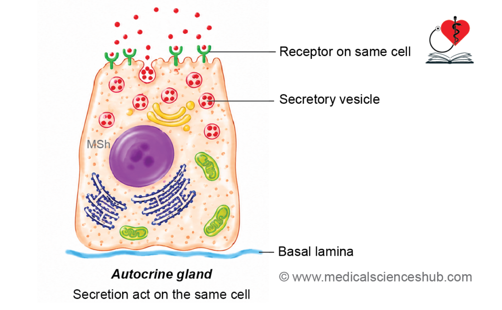
- Certain epithelial cells, functioning as unicellular glands, employ paracrine or autocrine signaling mechanisms in addition to classical endocrine and exocrine secretion.
Paracrine Signaling
- Cells release signaling molecules into the extracellular matrix, which diffuse locally to influence neighboring cells without entering the bloodstream.
- Example: Vasodilators released by vascular endothelial cells diffuse to adjacent smooth muscle cells, inducing vascular wall relaxation.
Autocrine Signaling
- Cells secrete molecules that bind to receptors on the same cell, thereby regulating their own activity.
- Autocrine signaling can produce either positive feedback (self-activation) or negative feedback (self-inhibition), depending on the signaling context.
- Example: Interleukins secreted by immune cells that act back on the same cell to amplify or regulate the immune response.
Goblet cell – as a gland
Goblet Cells as Unicellular Glands
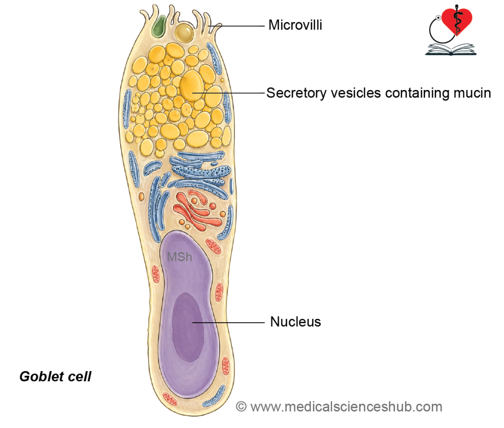
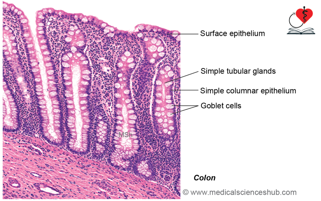
- Goblet cells are specialized simple columnar epithelial cells that function as unicellular exocrine glands, distributed singly among the lining epithelium or within larger glands.
- Their secretory mode is primarily merocrine, occasionally apocrine.
- Their characteristic goblet shape features a distended apical region and a narrow basal stem, resembling a wine glass.
Structure:
- The apical cytoplasm contains numerous mucus-filled secretory vesicles, which appear empty or foamy on H&E staining due to dissolution of mucus during tissue processing.
- The basal portion contains a flattened, elongated nucleus.
- PAS staining renders mucin a magenta color; mucicarmine staining highlights goblet cells in deep red.
Function:
- Goblet cells secrete mucus, a gel-forming substance composed of large glycoproteins (mucins) that bind water molecules, creating a protective coating over the underlying epithelium.
Locations
- Respiratory epithelium
- Gastrointestinal tract
- Conjunctiva of upper eyelid.
CLINICAL INTEGRATION
- Allergic asthma triggers excessive mucus production by goblet cells, contributing to airway obstruction.
- Goblet cell carcinoid (goblet cell adenocarcinoma) is a rare tumor of the gastrointestinal tract arising from the proliferation of both goblet cells and neuroendocrine cells, most commonly occurring in the appendix.
Important Questions
- Draw a well-labelled diagram of serous gland.
- Draw a well-labeled diagram of mucous gland.
- List the differences between serous and mucous cells.
- List the differences between serous and mucous acini.
- What are paracrine and autocrine glands.
- Write a short note on goblet cell.
📝 Test Your Knowledge – Practice MCQs
Attempt the chapter MCQ quiz and assess your understanding of key concepts.
Click to see: Medical Disclaimer and Copyright Notice