
Competency
- AN62.4: Enumerate parts and major connections of basal ganglia
Introduction
- The limbic system is a network of interconnected brain structures involved in emotion, motivation, learning, memory, and behavior. Working closely with the hypothalamus and cerebral cortex, it links emotional experiences with autonomic and endocrine responses, influencing both mental and physiological functions.
Limbic System
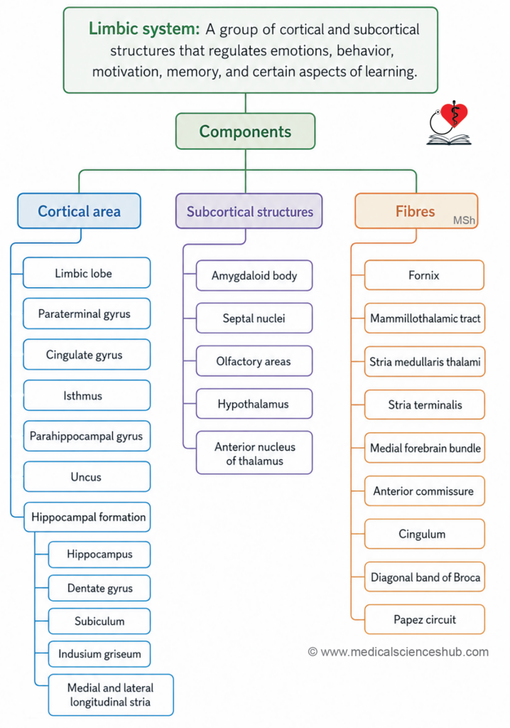
- The limbic system is a group of cortical and subcortical structures that regulates emotions, behavior, motivation, memory, and certain aspects of learning.
- It links emotional experiences with physiological responses.
- Damage to these structures can lead to memory loss, emotional instability, anxiety, or behavioral changes — as seen in conditions such as Alzheimer’s disease and temporal lobe epilepsy.
- The term limbus means border or ring, reflecting the anatomical position of these structures around the brainstem.
- The limbic system also plays a role in primary biological drives, including feeding and reproductive behavior.
Historical and Structural Context
- The limbic system was previously called the rhinencephalon (smell brain), as it was historically associated with olfactory processing; it is comparatively smaller in humans than in other mammals.
- The cortical component of the limbic system is classified as allocortex, which is organized into three neuronal layers — fewer than the six layers found in the neocortex.
- A transitional zone, called juxtallocortex, lies between the allocortex and the neocortex and contains three to six neuronal layers.
Functions of Limbic System
- The limbic system serves three broad functions: generating and regulating emotions, integrating multiple sensory inputs, and supporting learning and memory.
1. Emotional Regulation
- The limbic system generates and modulates emotions such as fear, anger, pleasure, and affection.
- It also coordinates the expression of these emotions through facial expressions and body language.
2. Integration of Olfactory, Visceral, and Somatic Inputs
- The limbic system integrates signals from the olfactory system, internal organs, and body sensations.
- This allows the brain to associate sensory experiences with appropriate emotional and physiological responses.
3. Learning and Memory
- The hippocampus, a key limbic structure, is essential for forming new memories and consolidating short-term memories into long-term storage.
- Damage to the hippocampus typically results in difficulty retaining recently acquired information.
Emotional Responses Related to Survival
The limbic system drives emotional reactions necessary for the survival of the individual and the species:
- Feeding behavior — Sensations such as hunger and satiety motivate food-seeking and regulate eating patterns.
- Reproductive behavior — Sexual motivation promotes reproduction and continuation of the species.
- Parental behavior — Affective bonding between a parent and offspring encourages caregiving and increases offspring survival.
- Defensive responses — Emotions such as fear and anger prepare the body to confront or avoid threats; olfactory-triggered responses such as nausea or salivation also serve a protective role.
- Visceral responses — Emotional states directly influence autonomic organ function; for example, heart rate increases during fear, and gastrointestinal discomfort may arise during anxiety.
Because of its strong influence over internal organ activity, the limbic system is also referred to as the visceral brain.
Components of Limbic System
The limbic system comprises several cortical and subcortical structures, with the hypothalamus serving as its primary regulatory center.
Cortical Areas
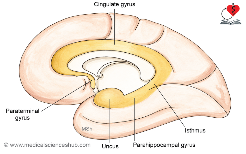
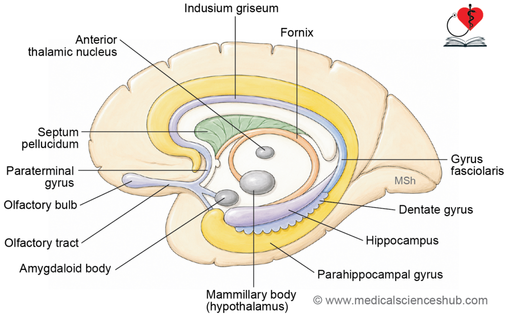
- Limbic lobe — A ring-shaped cortical region (limbus = border) consisting of the paraterminal gyrus, cingulate gyrus, isthmus, parahippocampal gyrus, uncus, and the collateral and rhinal sulci.
- Note: The term “limbic lobe” is less commonly used in current literature.
- Hippocampal formation — Comprises the hippocampus, dentate gyrus, gyrus fasciolaris, and indusium griseum.
Subcortical Structures
- Amygdaloid body
- Septal nuclei
- Olfactory areas
- Hypothalamus — the primary regulatory center of the limbic system
- Anterior nucleus of the thalamus
Fiber Connections
The limbic system communicates through the following fiber tracts:
- Fornix
- Mammillothalamic tract
- Stria medullaris thalami
- Stria terminalis
- Medial forebrain bundle
- Anterior commissure
- Cingulum
- Diagonal band of Broca
- Papez circuit
Areas Concerned with Sense of Smell
The limbic system functions largely in response to olfactory and other sensory stimuli. The key areas involved in olfactory processing are listed below.
Olfactory Nerve, Bulb, and Tracts
- The olfactory nerve (first cranial nerve) is purely sensory and carries smell information from the olfactory epithelium, which lines the roof of the nasal cavity, the upper nasal septum, and the superior nasal concha.
- Specialized olfactory receptor neurons in this epithelium each have:
- A dendrite ending in an olfactory knob bearing cilia that are bathed in mucus and sensitive to odor molecules.
- A central axon that joins others to form fine filaments, collectively forming the olfactory nerve.
- These filaments pass through the cribriform plate of the ethmoid bone to reach the olfactory bulb, located on the inferior surface of the frontal lobe.
- Within the olfactory bulb, axons synapse with mitral and tufted cells, forming olfactory glomeruli.
- Second-order neurons from the olfactory bulb form the olfactory tract, which projects posteriorly to the piriform cortex, amygdaloid body, and entorhinal area.
- Unlike other sensory pathways, the olfactory pathway reaches the cortex without relaying through the thalamus.
Olfactory Trigone
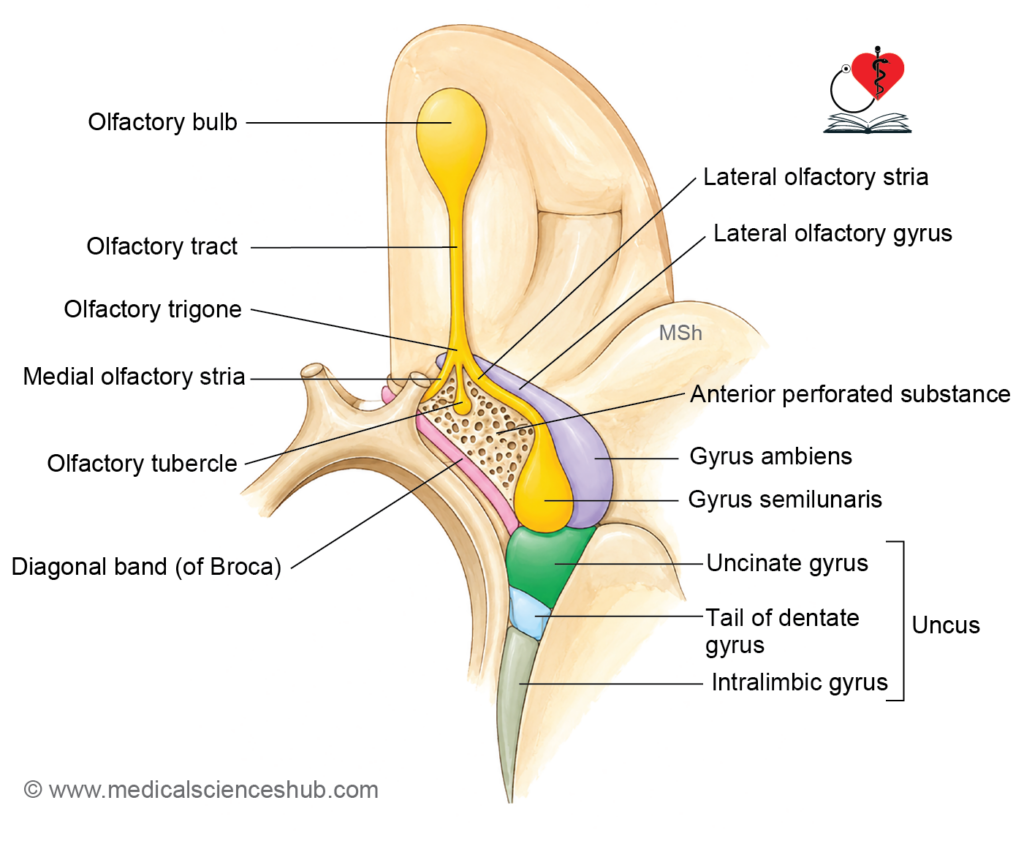
- The olfactory trigone is the flattened posterior end of the olfactory tract, dividing into three striae:
- Medial olfactory stria — continues into the paraterminal gyrus, anterior to the lamina terminalis.
- Lateral olfactory stria — runs laterally into the limen insulae, then turns medially to form the gyrus semilunaris (periamygdaloid area).
- Intermediate olfactory stria — occasionally present; ends as the olfactory tubercle in the anterior perforated substance.
- Medial and lateral olfactory gyri are thin layers of grey matter covering their respective striae.
Anterior Perforated Substance
- A small, depressed area of grey matter perforated by central branches of the anterior and middle cerebral arteries.
Limen Insulae
- A small ridge connecting the anterior perforated substance to the apex of the insula.
Parahippocampal Gyrus
- Located on the tentorial surface of the cerebral hemisphere, medial to the collateral sulcus.
- Continues posteriorly with the isthmus and lingual gyrus.
- Involved in processing both taste and smell.
Uncus
- The anterior 2.5 cm of the parahippocampal gyrus, hook-shaped in appearance.
- Separated from the temporal pole by the rhinal notch.
- Lies just posterior to the anterior perforated substance.
Primary Olfactory Area
- Responsible for the reception and perception of smell.
- Comprises the lateral olfactory gyrus, gyrus ambiens, gyrus semilunaris, limen insulae, uncus, and the dorsomedial part of the amygdaloid body.
Entorhinal Cortex (Secondary Olfactory Area)
- Also called the entorhinal area or Brodmann’s area 28; performs olfactory association.
- Located ventral to the amygdaloid body and anterior to the hippocampal formation, corresponding to the anterior part of the parahippocampal gyrus.
- Provides the most prominent afferent input to the dentate gyrus.
Isthmus
- Connects the cingulate gyrus to the parahippocampal gyrus.
- Lies posterior to the splenium of the corpus callosum.
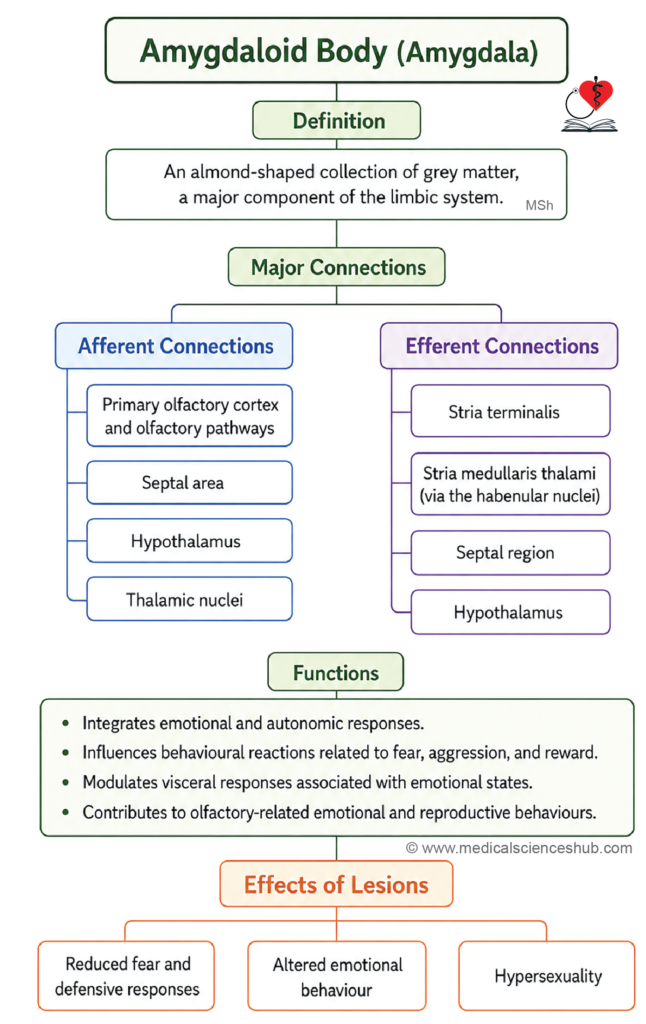
Amygdaloid Body
The amygdaloid body (also called the amygdaloid nuclear complex) is an almond-shaped mass of grey matter that regulates emotional behavior — particularly fear, anger, and pleasure. It links emotions with memories and influences autonomic and endocrine responses. Overactivity of the amygdala is associated with anxiety, phobias, and post-traumatic stress disorder (PTSD).
Location
- Situated in the anterior part of the parahippocampal gyrus, above the tip of the inferior horn of the lateral ventricle and beneath the lentiform nucleus.
- Posteriorly, it is continuous with the tail of the caudate nucleus and the stria terminalis.
Subdivisions
The amygdala is organized into three subnuclear groups:
- Basolateral (ventrolateral) group — receives afferent fibers
- Corticomedial (dorsomedial) group — receives afferent fibers
- Central group — gives rise to efferent fibers
Connections
- Afferent input — from the primary olfactory area
- Efferent output — primarily via the stria terminalis, which follows a C-shaped course alongside the caudate nucleus and terminates in the:
- Septal area
- Medial preoptic area of the hypothalamus
- Bed nucleus of the stria terminalis
- Some efferent fibers travel posteriorly as the stria medullaris thalami, connecting the amygdala with the habenular nucleus, septal nuclei, and anterior hypothalamus.
Functions
- Regulates somatic responses to internal biological needs.
- Controls olfactory-mediated reproductive behavior.
- Modulates autonomic nervous system (ANS) activity based on prior experiences.
- Modulates hypothalamic activity via two principal pathways:
- Stria terminalis
- Ventral amygdalofugal pathway
Effects of Lesion
- Bilateral damage to the amygdaloid body results in reduced fear responses and increased sexual activity.
Septal Area and Septal Nuclei
The septal area and septal nuclei play a key role in emotional behavior, reward, and pleasure, and help regulate autonomic functions through their connections with the limbic system. Electrical stimulation of this region produces sensations of calmness or satisfaction, while lesions may cause irritability, loss of emotional control, or reduced experience of pleasure.
Components of the Septal Area
- Paraterminal gyrus — a narrow strip of cortex along the anterior surface of the lamina terminalis.
- Paraolfactory gyrus (subcallosal gyrus) — a narrow cortical lamina on the medial surface of the cerebral hemisphere, anterior to the lamina terminalis; continuous around the genu of the corpus callosum with the indusium griseum (supracallosal gyrus).
- Paraolfactory area (subcallosal area) — a small triangular region on the medial surface of the cerebral hemisphere, anterior to the paraolfactory gyrus; continuous inferiorly with the olfactory trigone.
Function
- The septal area is regarded as a pleasure zone and serves as a key reward center within the limbic system.
Hippocampal Formation
The hippocampal formation is located in the medial part of the temporal lobe and is essential for learning, memory consolidation, and spatial navigation. It converts short-term memories into long-term storage and links emotions with experiences. Lesions produce memory loss — particularly for recent events — as seen in Alzheimer’s disease and temporal lobe epilepsy.
Components
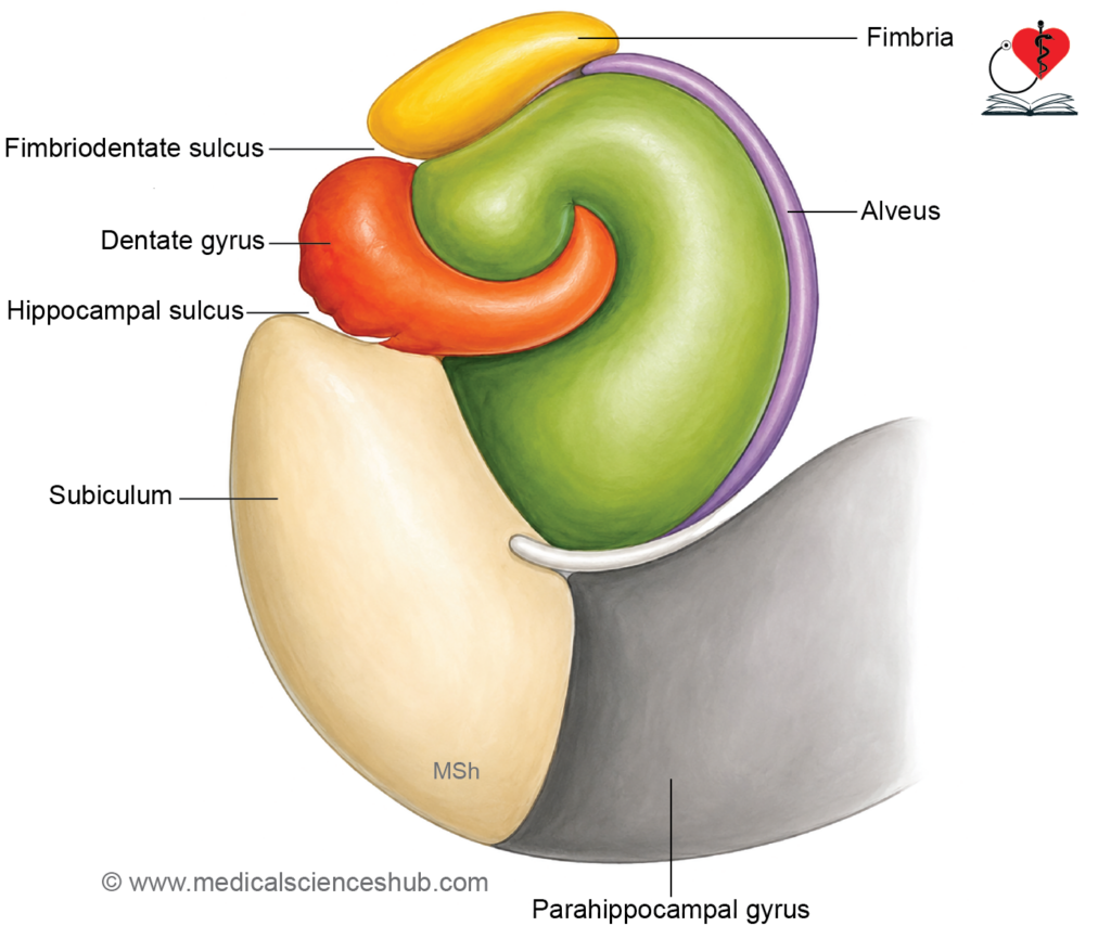
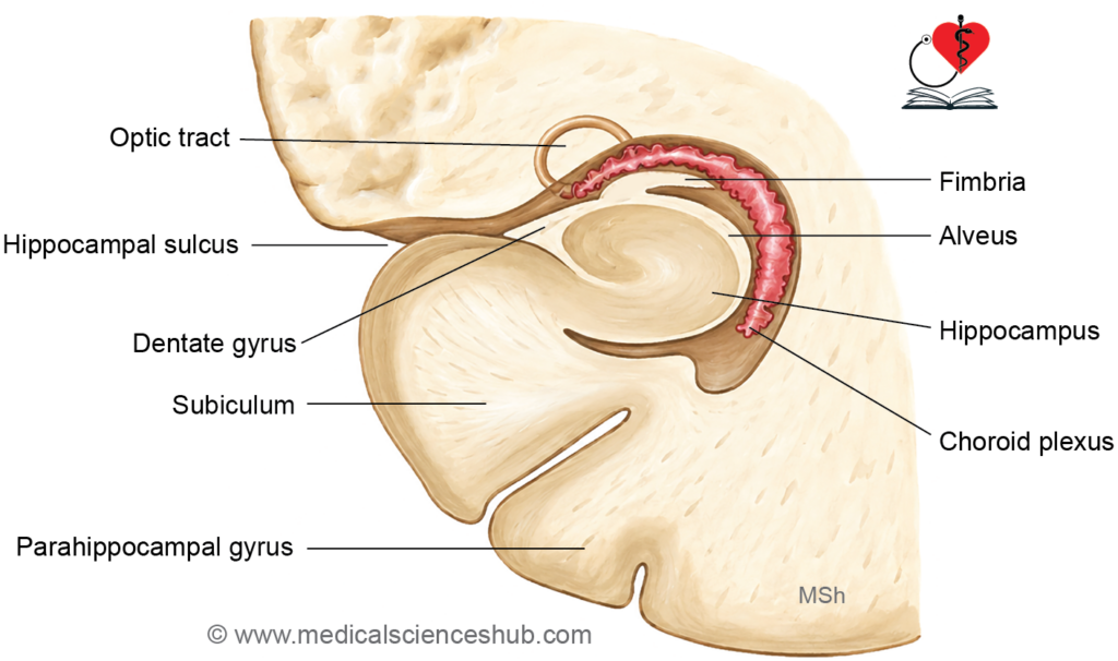
- Hippocampus proper — a C-shaped, phylogenetically ancient cortical structure (archicortex) located in the floor of the inferior horn of the lateral ventricle; also called Ammon’s horn. Its anterior end bears 2–3 digit-like ridges and is called the pes hippocampi.
- Alveus — a thin layer of white matter covering the ventricular surface of the hippocampus, formed by its efferent fibers.
- Fimbria — a flattened band of white matter formed by convergence of efferent fibers from the alveus; lies superior to the dentate gyrus.
- Crus of fornix — the direct posterior continuation of the fimbria.
- Subiculum — a transitional cortical zone (3–6 neuronal layers; juxtallocortex) interposed between the hippocampus proper and the parahippocampal gyrus.
- Dentate gyrus — a narrow, toothed strip of grey matter on the upper surface of the parahippocampal gyrus; continuous posteriorly with the gyrus fasciolaris through the indusium griseum. Its tooth-like appearance is created by blood vessels perforating the ventricular surface. The dentate gyrus runs medially across the inferior surface of the uncus, where it forms the tail of the dentate gyrus.
- Indusium griseum (supracallosal gyrus) — a thin layer of grey matter covering the superior surface of the corpus callosum; continuous anteriorly with the paraterminal gyrus and posteriorly with the gyrus fasciolaris.
- Medial and lateral longitudinal striae — two thin longitudinal strands of white fibers embedded within the indusium griseum.
- Gyrus fasciolaris — a transitional cortical zone connecting the dentate gyrus with the indusium griseum.
- Anterior commissure — carries decussating fibers transmitting olfactory information between the olfactory bulbs and between the parahippocampal gyri.
- Diagonal band of Broca — fiber bundle arising from the paraolfactory area and terminating in the periamygdaloid area of the temporal lobe.
Connections
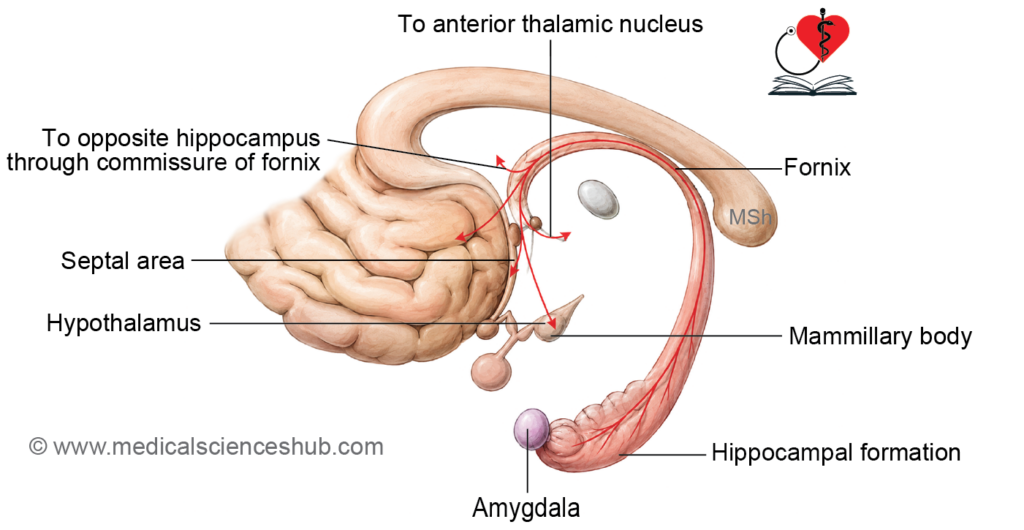
- Afferent input — from the indusium griseum, cingulate gyrus, septal nuclei, entorhinal cortex, dentate gyrus, parahippocampal gyrus, and contralateral hippocampus.
- Efferent output — via the fornix to the contralateral hippocampus, septal area, anterior hypothalamic regions, and mammillary body; efferent fibers arise mainly from pyramidal cells of the hippocampal pyramidal cell layer.
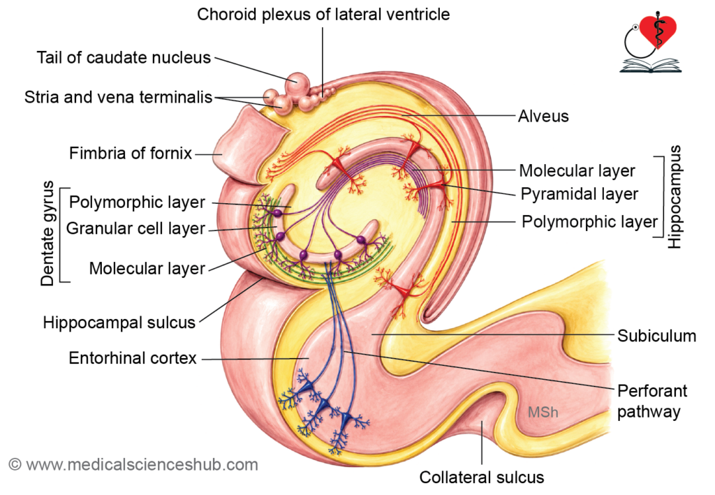
Histology of the Hippocampus
The hippocampal archicortex consists of three layers:
- Molecular layer (deepest) — contains apical dendritic trees of pyramidal cells and axon terminals from granule cells, the perforant pathway, the septohippocampal tract, and pyramidal cell axon collaterals; merges with the molecular layer of the dentate gyrus and neocortex.
- Pyramidal layer (middle) — the most prominent layer; contains pyramidal cells whose dendrites extend into the molecular layer and whose axons travel through the alveus, fimbria, and fornix. Axon collaterals, called Schaffer collaterals, synapse with dendrites of neighboring pyramidal neurons within the molecular layer.
- Polymorphic layer (superficial) — contains interneurons, pyramidal cell dendrites, and axon collateral branches; shares structural features with the deepest layer of the neocortex.
Histology of the Dentate Gyrus
The dentate gyrus also comprises three archicortical layers:
- Molecular layer (outer) — contains a sparse population of nerve cell bodies and granule cell dendrites.
- Granule cell layer (middle) — contains cell bodies of granule cells, the predominant cell type of the dentate gyrus. Granule cell dendrites extend into the molecular layer, where they synapse with terminals of the perforant pathway. Their axons, called mossy fibers, project exclusively to the pyramidal cells of the hippocampus.
- Polymorphic layer (deep) — contains interneurons.
Functions
- Integral component of the limbic system.
- Regulates endocrine and visceral functions and emotional states.
- Essential for recent memory consolidation.
CLINICAL NEUROANATOMY
Effects of Lesion
- Damage to the hippocampus results in loss of recent memory and may cause psychomotor epilepsy.
Table 19.1: Connections of hippocampus
| Pathway / Structure | Connected Region | Functional Significance |
|---|---|---|
| Pathway / Structure | Septal nuclei | Principal efferent pathway of the hippocampus; involved in memory processing and limbic integration. |
| Alveus → Fimbria → Fornix | Cingulate gyrus | Provides communication between the hippocampal formation and limbic cortex, contributing to emotional and cognitive functions. |
| Cingulum | Septal area | Modulate hippocampal activity and play a role in learning and memory mechanisms. |
| Septohippocampal Fibers | Lateral hypothalamic area | Link memory-related functions of the hippocampus with autonomic and behavioral responses. |
| Hypothalamic Connections | Mammillary bodies | Important component of the Papez circuit, essential for memory consolidation. |
| Fornix Fibers | Anterior nucleus of thalamus | Relay limbic information to the thalamus, supporting memory and emotional processing. |
| Hippocampothalamic Fibers | Hippocampus | Major cortical input pathway carrying information from association areas to the hippocampal formation. |
| Parahippocampal Gyrus | Midbrain tegmentum | Influence arousal, attention, and limbic-brainstem interactions. |
| Tegmental Connections | Opposite hippocampus | Interconnect the two hippocampi, allowing bilateral coordination of hippocampal activity. |
| Commissural Fibers | Habenular nuclei | Provide a link between limbic structures and brainstem centers involved in emotional behavior. |
Fornix
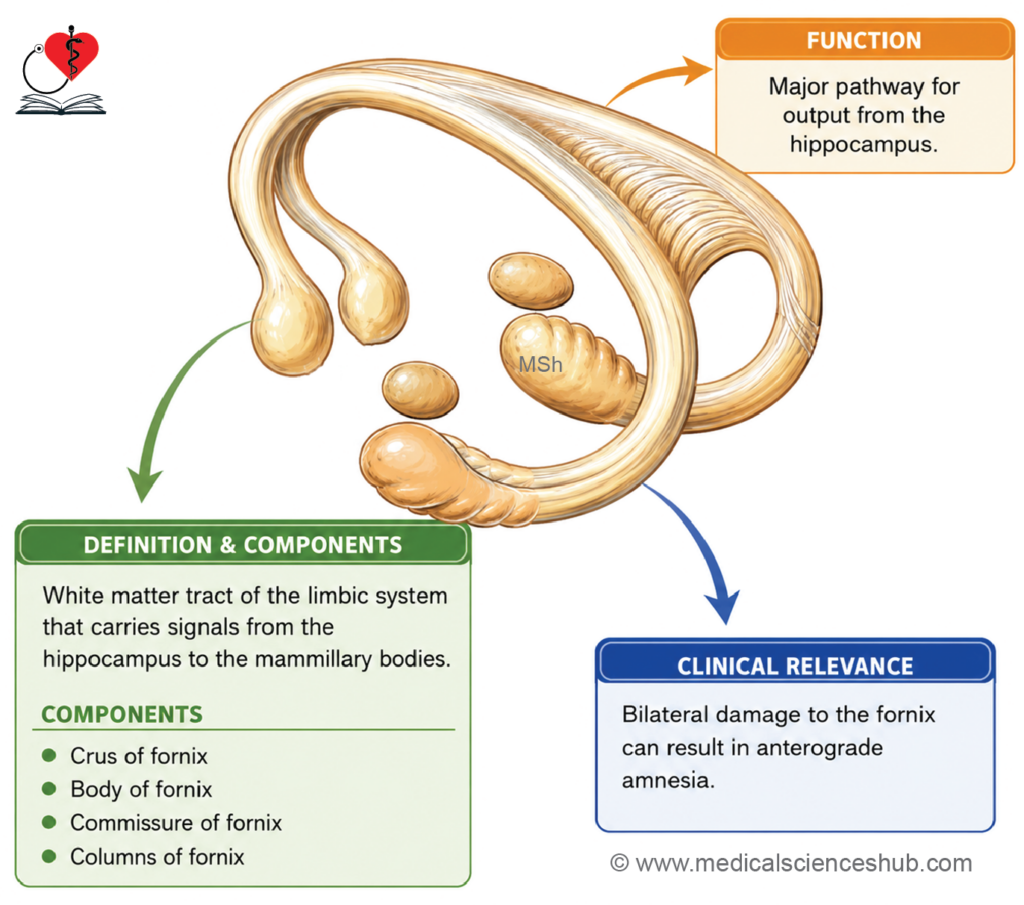
The fornix is an arched bundle of projection fibers that carries signals related to memory and emotion between the hippocampus and other brain regions. Lesions can cause memory loss, disorientation, and difficulty forming new memories, as seen in head injuries and degenerative brain disorders.
Definition
The fornix is the principal efferent fiber tract of the hippocampus, projecting primarily to the mammillary body.
Formation
Efferent axons of hippocampal pyramidal cells follow this sequential path:
Pyramidal cell axons → alveus → fimbria → crus of fornix
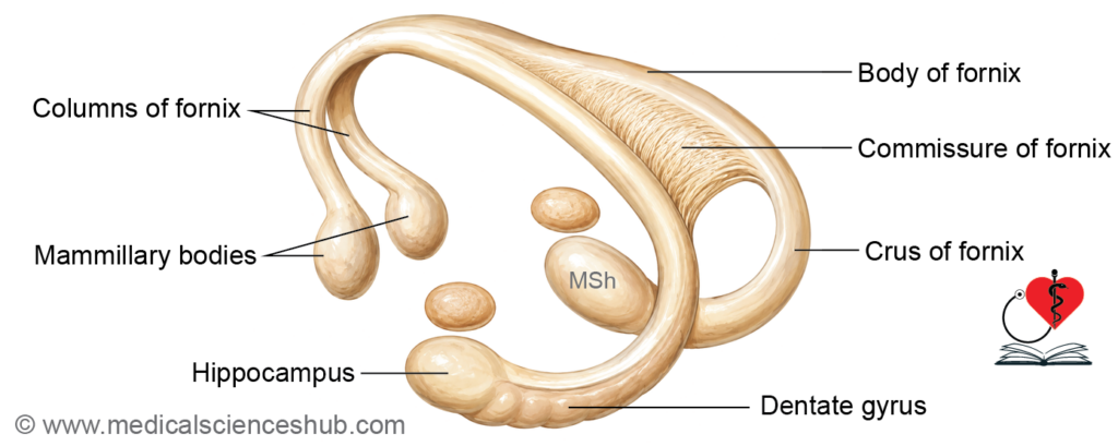
Parts
- Crus of fornix (posterior column) — curves upward posterior to the thalamus; represents the direct continuation of the fimbria.
- Body of fornix — formed by convergence of the right and left crura in the midline; lies above the roof of the third ventricle and below the body and splenium of the corpus callosum.
- Commissure of fornix (hippocampal commissure) — a small bundle of decussating fibers from the crura, located just posterior to the body of the fornix; connects the two hippocampi.
- Anterior columns of fornix — formed as the body of the fornix divides anteriorly; each column curves downward past the anterior commissure, passes through the hypothalamus, and terminates in the mammillary body; a small number of fibers end in the septal area.
Function
- The fornix serves as the primary efferent tract of the hippocampus, relaying memory-related signals to the mammillary body and other limbic structures.
CLINICAL NEUROANATOMY
- Bilateral damage to the fornix disrupts the consolidation of short-term memory into long-term memory, a condition referred to as acute amnestic syndrome.
Papez Circuit
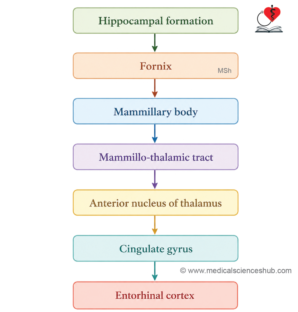
The Papez circuit is an intraneuronal communication loop within the limbic system that forms the structural and functional basis for emotional experience and memory consolidation. Lesions within this circuit can lead to memory loss, emotional instability, and behavioral changes, as seen in Alzheimer’s disease and temporal lobe epilepsy.
Components
- Hippocampal formation
- Fornix
- Mammillary body
- Anterior nucleus of the thalamus
- Cingulate gyrus
- Entorhinal cortex
Circuit Pathway
The circuit flows in a loop:
Hippocampal formation → Fornix → Mammillary body → Anterior nucleus of thalamus → Cingulate gyrus → Entorhinal cortex → Hippocampal formation
Functions
- Mediates emotional experience by linking limbic structures with higher cortical centers.
- Essential for memory consolidation, particularly the transfer of short-term memories into long-term storage.
Brainstem Centers Associated with Limbic System Function
Several brainstem and diencephalic structures work in close association with the limbic system to regulate emotional and visceral responses.
Components
- Hypothalamus — mediates autonomic nervous system (ANS) responses that accompany emotional expression, serving as the primary visceral output center of the limbic system.
- Thalamus — receives projections from the amygdala and hypothalamus to its limbic nuclei, which include the anterior nuclear group and the lateral dorsal and medial dorsal nuclei.
- Habenular nuclei — serve as a relay station through which the limbic system projects to the reticular formation of the mesencephalon.
- Ventral tegmental area — a midbrain region that modulates memory processing via dopaminergic fibers terminating in cortical areas associated with the limbic system.
- Locus ceruleus and dorsal raphe — the locus ceruleus sends noradrenergic fibers, while the dorsal raphe sends serotonergic fibers to the hippocampal formation, limbic lobe, and amygdala, where both pathways modulate memory processing.
Table 19.2: Brainstem centers associated with limbic system function
| Structure | Major Functions | Clinical Importance |
|---|---|---|
| Hypothalamic Nuclei and Mammillary Bodies | Regulate autonomic activities, emotional expression, endocrine functions, feeding behavior, and memory pathways. | Form an important link between the limbic system and autonomic nervous system. |
| Thalamic Nuclei (Especially Anterior Nucleus) | Receive limbic inputs from the hypothalamus and mammillary bodies and relay them to the cerebral cortex. | Essential component of the Papez circuit involved in memory and emotional behavior. |
| Habenular Nuclei | Act as relay stations transmitting limbic information to midbrain reticular and autonomic centers. | Participate in emotional responses, motivation, and behavioral regulation. |
| Hippocampus and Adjacent Temporal Lobe Structures | Responsible for memory formation, learning, spatial orientation, and consolidation of short-term memories into long-term memories. | Damage results in memory impairment and anterograde amnesia. |
Important Questions
- List the components of limbic system.
- Write a short note on amygdaloid body.
- Write a short note on hippocampal formation.
- Write a short note on fornix.
- Write a short note on Papez circuit.
📝 Test Your Knowledge – Practice MCQs
Attempt the chapter MCQ quiz and assess your understanding of key concepts.
Click to see: Medical Disclaimer and Copyright Notice