
Competency
- AN62.6: Describe and identify formation, branches, and major areas of distribution of circle of Willis.
Introduction
The brain receives blood from the internal carotid and vertebral arteries, which together form the circle of Willis. Major branches include the anterior, middle, and posterior cerebral arteries, supplying oxygen and nutrients essential for normal neural activity. Blockage or rupture of these vessels can cause stroke, ischemia, or neurological deficits affecting movement, sensation, speech, or cognition.
Features
- The brain requires a continuous and uninterrupted blood supply as it cannot tolerate hypoxia or ischemia.
- The brain receives approximately 750 mL of blood per minute, representing nearly 20% of total cardiac output.
- Interruption of blood supply for 10–15 seconds results in loss of consciousness.
- Interruption lasting 5 minutes initiates irreversible brain damage.
- Arterial occlusion by thrombosis is a common pathological event in the brain, leading to infarction of neural tissue.
Arteries of Brain
- The brain is supplied by:
- Two vertebral arteries
- Two internal carotid arteries.
- The arterial arcade is formed by a pair of vertebral and a pair of carotid arteries – circle of Willis – maintains continuous blood supply.
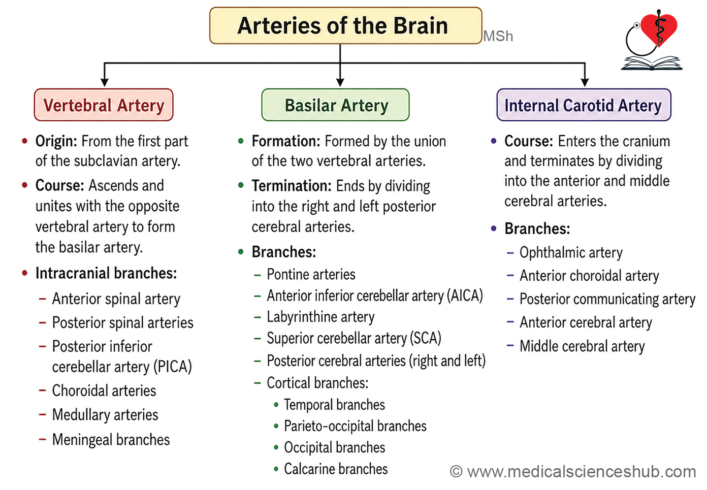
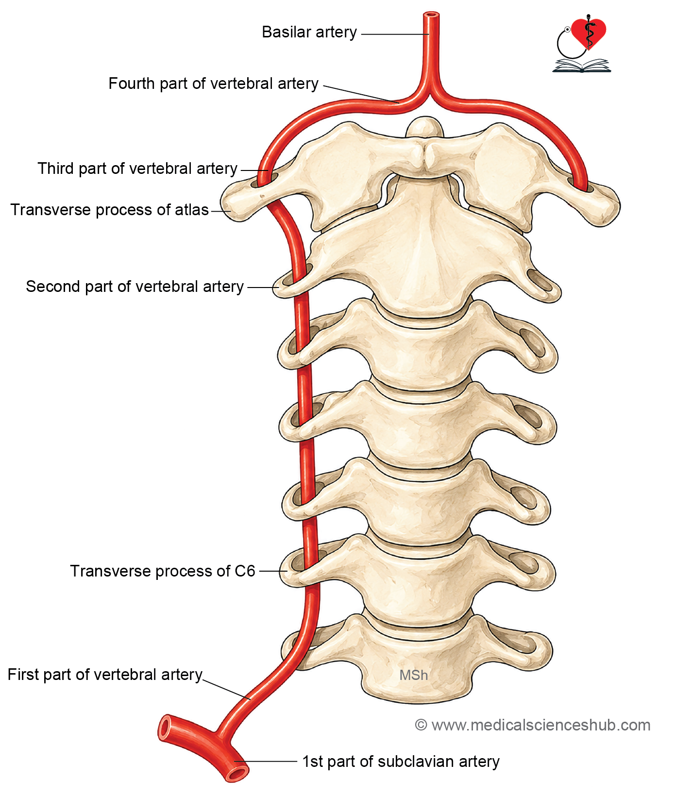
Vertebral Artery
The vertebral artery supplies blood to the brainstem, cerebellum, and posterior brain. Its occlusion or injury can cause dizziness, visual disturbances, or posterior circulation stroke.
Origin and Termination
- Arises as a branch of the first part of the subclavian artery.
- Ascends through the foramina transversaria of the cervical vertebrae.
- Above C1, it pierces the posterior atlantooccipital membrane to enter the cranial cavity.
- At the lower border of the pons, both vertebral arteries fuse in the midline to form the basilar artery.
Parts
- First part (prevertebral) — runs upward and backward from the subclavian artery, between the longus colli (medially) and scalenus anterior (laterally); enters the foramen transversarium of C6.
- Second part (foraminal) — ascends through the foramina transversaria from C6 to C1; lies anterior to the cervical spinal nerves and is surrounded by a sympathetic nerve plexus and veins.
- Third part (suboccipital) — extends from the foramen transversarium of the atlas to the foramen magnum; curves medially and posteriorly around the lateral mass of the atlas within the suboccipital triangle, covered by the semispinalis capitis and obliquus capitis superior muscles.
- Fourth part (intracranial) — pierces the dura mater and arachnoid mater through the foramen magnum; joins its counterpart at the lower border of the pons to form the basilar artery.
Intracranial Branches
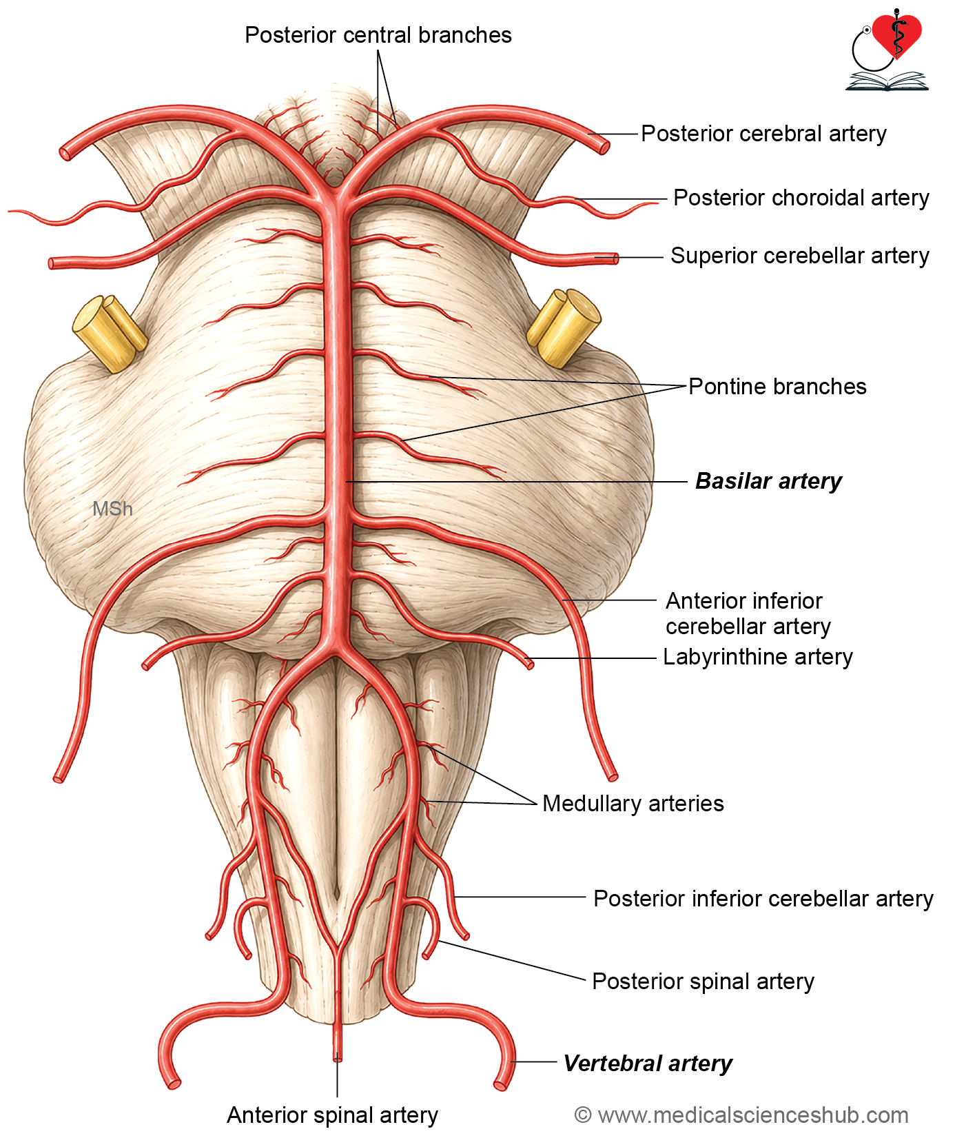
- Anterior spinal artery — formed by a small branch from each vertebral artery; descends along the anterior longitudinal fissure of the spinal cord.
- Posterior spinal artery — may arise from the posterior inferior cerebellar artery; passes inferiorly on the medulla and divides into two branches along the medial and posterior aspects of the dorsal roots of spinal nerves.
- Posterior inferior cerebellar artery (PICA) — the largest branch of the fourth part of the vertebral artery; winds around the medulla oblongata at the lower end of the olive and at the pontomedullary junction; supplies the lateral half of the medulla and the cerebellum; gives a choroidal branch to the choroid plexus of the fourth ventricle.
- Medullary branches — supply the medulla oblongata.
- Meningeal branch — supplies the dura mater of the posterior cranial fossa.
Basilar Artery
The basilar artery supplies the brainstem, cerebellum, and occipital lobes, which are essential for balance, coordination, and vision. Its occlusion or rupture can cause brainstem stroke, presenting as dizziness, dysphagia, paralysis, or life-threatening complications.
Origin and Termination
- Formed by the union of the two vertebral arteries at the lower border of the pons.
- Terminates at the upper border of the pons by dividing into the two posterior cerebral arteries.
Branches
- Pontine branches — numerous small vessels that pierce and supply the pons.
- Anterior inferior cerebellar artery (AICA) — arises at the lower border of the pons and runs posterolaterally; supplies the sixth, seventh, and eighth cranial nerves and the anteroinferior part of the cerebellum.
- Labyrinthine artery — accompanies the eighth cranial nerve into the internal acoustic meatus; supplies the internal ear; functions as an end artery with no collateral supply.
- Superior cerebellar artery — arises near the superior border of the pons and passes posteriorly between the oculomotor and trochlear nerves; supplies the superior surface of the cerebellum.
- Posterior cerebral artery — follows a curved course around the midbrain to reach the medial surface of the cerebral hemisphere; gives rise to:
- Posterior central branches — pierce the posterior perforated substance in the interpeduncular fossa.
- Posterior choroidal artery — supplies the choroid plexus of the lateral and third ventricles.
- Cortical branches — temporal, parieto-occipital, occipital, and calcarine branches.
internal carotid artery
The internal carotid arteryenters the cranial cavity through the carotid canal and has two intracranial parts:
- Cavernous part — passes through the cavernous sinus; gives rise to cavernous branches supplying the trigeminal ganglion and the superior and inferior hypophyseal arteries.
- Cerebral part — lies on the base of the brain.
The carotid siphon is a U-shaped bend of the intracranial ICA that helps dampen pulsatile blood flow.
The ICA terminates at the stem of the lateral sulcus by dividing into the anterior and middle cerebral arteries.
Branches
- Ophthalmic artery — enters the orbit through the optic canal.
- Anterior choroidal artery — passes posterolaterally; supplies the crus cerebri and the choroid plexus of the inferior horn of the lateral ventricle.
- Posterior communicating artery — passes posteriorly to join the posterior cerebral artery; supplies the pituitary gland, optic tract, hypothalamus, and midbrain.
- Anterior cerebral artery — due to its long course and small lumen, this vessel is most commonly affected by cerebral thrombosis.
- Middle cerebral artery
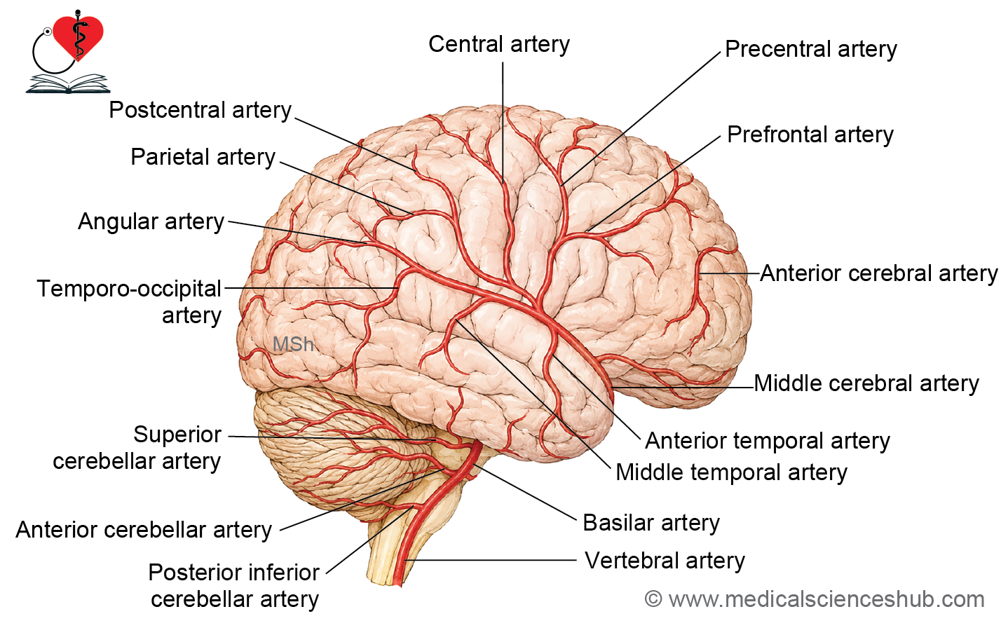
Middle Cerebral Artery
The middle cerebral artery (MCA) is the largest branch of the internal carotid artery. It runs laterally through the Sylvian (lateral) fissure between the frontal and temporal lobes.
Branches
The MCA gives rise to two main sets of branches:
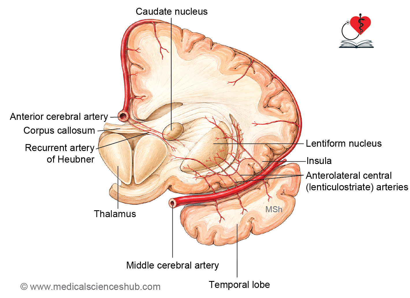
1. Central (Perforating) Branches — Lenticulostriate Arteries
- Supply the basal ganglia, internal capsule, and deep white matter.
- Critical for the integrity of motor and sensory pathways.
2. Cortical (Pial) Branches
Frontal lobe:
- Prefrontal branches — supply the lateral prefrontal cortex, involved in decision-making, planning, and executive functions.
- Precentral (motor) branches — supply the primary motor cortex for face and upper limb movements.
Parietal lobe:
- Postcentral (sensory) branches — supply the primary somatosensory cortex, processing sensation from the face, hand, and arm.
- Angular and supramarginal branches — supply the supramarginal and angular gyri, essential for language processing, reading, and spatial awareness.
Temporal lobe:
- Superior temporal branches — supply the superior temporal gyrus, including Wernicke’s area for language comprehension.
- Middle temporal branches — supply the middle temporal gyrus, involved in semantic memory and auditory processing.
- Inferior temporal branches — supply the inferior temporal gyrus, important for visual object recognition.
Insular cortex:
- Short and long insular branches — supply the insula, which contributes to taste, visceral sensation, and emotional processing.
Functional Significance
The MCA supplies blood to key cortical areas responsible for movement, sensation, and speech, including the motor and sensory cortices of the face and upper limb, Broca’s and Wernicke’s areas, and portions of the auditory and visual association cortices.
CLINICAL NEUROANATOMY
- MCA occlusion is one of the most common causes of ischemic stroke.
- Depending on the hemisphere involved, patients may develop:
- Contralateral paralysis or sensory loss of the face and upper limb.
- Speech disturbances (if the dominant hemisphere is affected).
- Hemispatial neglect (if the non-dominant hemisphere is affected).
- Early diagnosis and intervention are essential to prevent permanent neurological damage.
Anterior Cerebral Artery
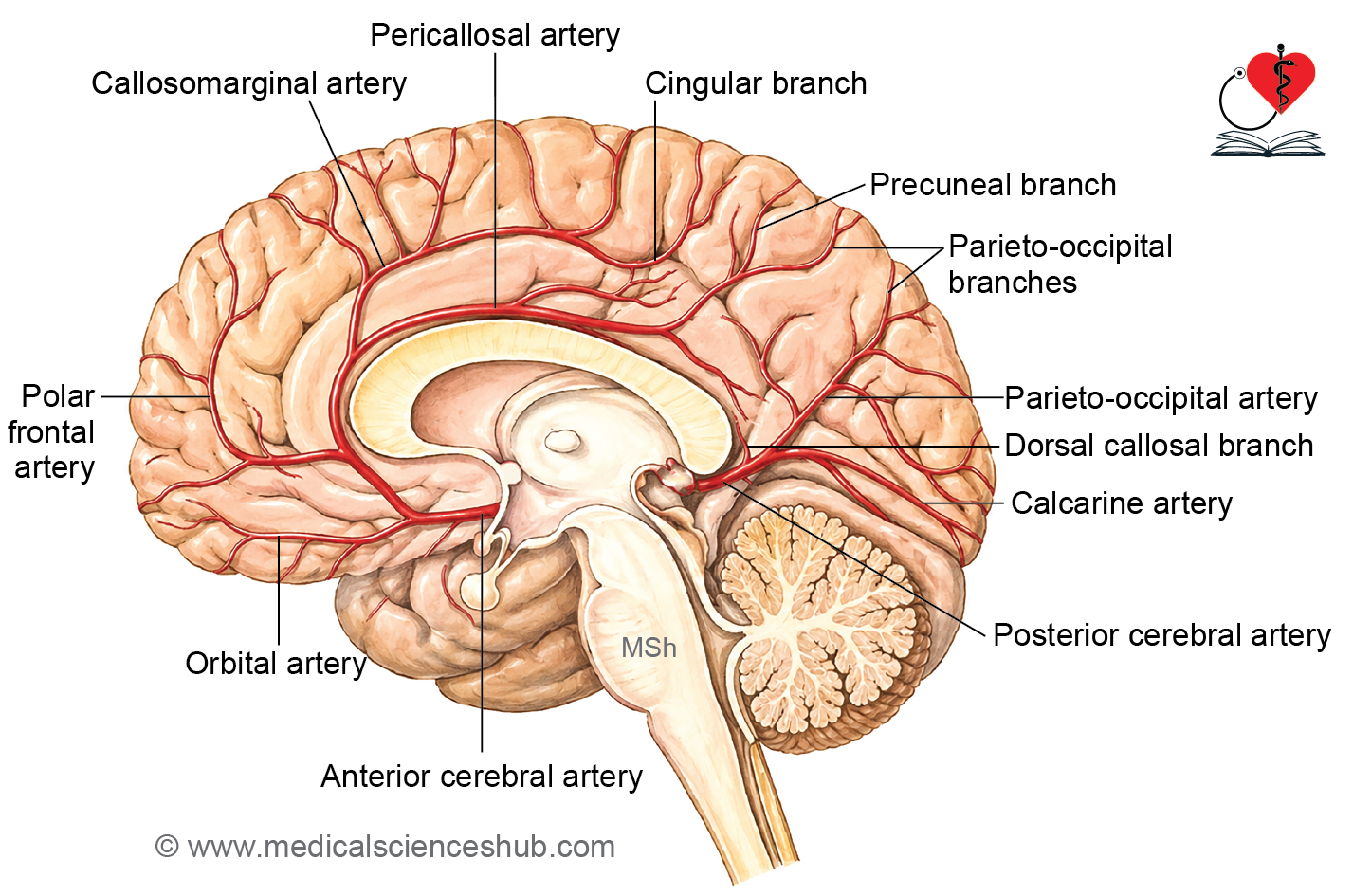
The anterior cerebral artery (ACA) is a branch of the internal carotid artery that arches forward and medially above the optic chiasm, running along the longitudinal fissure. It supplies the medial portions of the frontal and parietal lobes. The right and left ACAs are connected by the anterior communicating artery.
Branches
1. Central (Perforating) Branches
- Medial striate artery (recurrent artery of Heubner) — arises from the proximal ACA; supplies the head of the caudate nucleus, anterior limb of the internal capsule, and adjacent basal ganglia. Occlusion may cause contralateral motor deficits, particularly in the lower limb.
2. Cortical (Pial) Branches
- Orbitofrontal branches — supply the medial and inferior frontal cortex, involved in decision-making and behavior.
- Frontopolar branches — supply the frontal pole, affecting planning and personality.
- Paracentral branches — supply the paracentral lobule, including the motor and sensory areas for the lower limb.
- Pericallosal artery — continues along the upper surface of the corpus callosum; gives rise to the callosomarginal artery, which runs in the cingulate sulcus and supplies the medial frontal and parietal lobes, including the supplementary motor area.
CLINICAL NEUROANATOMY
- ACA stroke characteristically causes contralateral weakness and sensory loss in the lower limb, along with abulia (reduced motivation) or personality changes.
- The face and upper limb are typically spared, as these regions are predominantly supplied by the middle cerebral artery.
Circle of Willis (Circulus arteriosus)
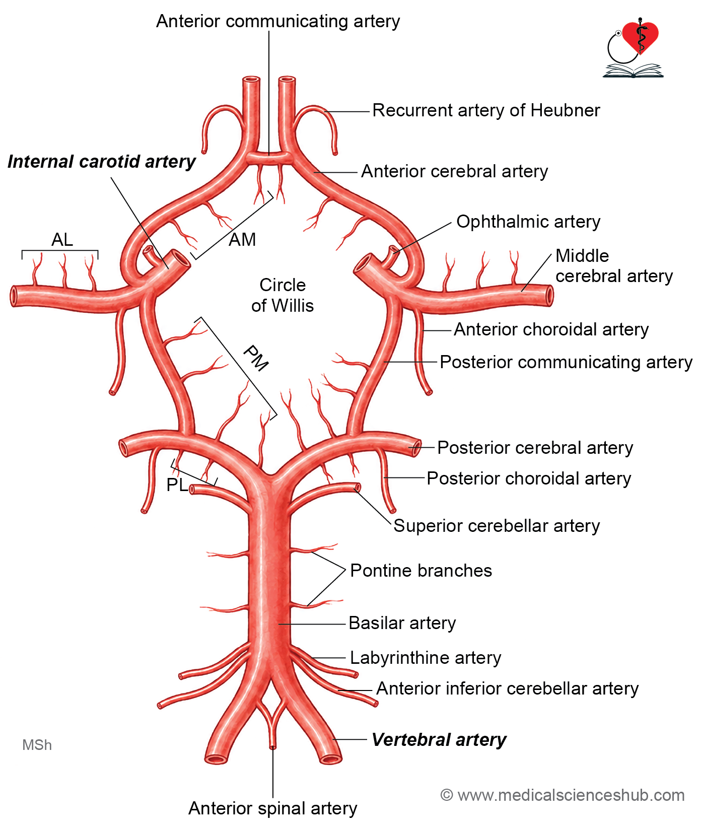
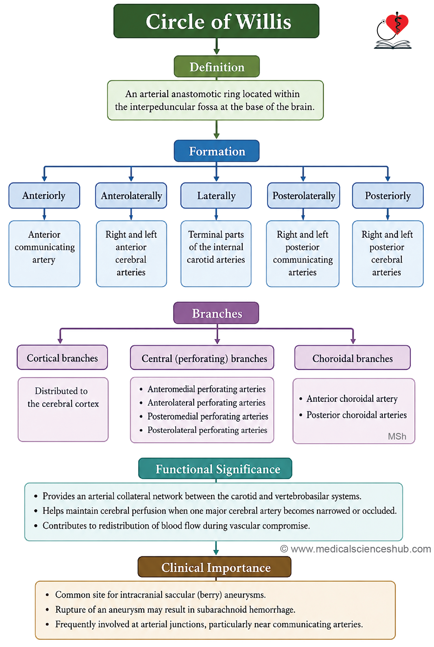
- The circle of Willis is a hexagonal arterial anastomosis located at the base of the brain within the interpeduncular fossa.
Formation
The circle is formed by the following vessels:
- Anteriorly: anterior communicating artery
- Anterolaterally: anterior cerebral arteries
- Laterally: internal carotid arteries
- Posterolaterally: posterior communicating arteries
- Posteriorly: posterior cerebral arteries
Branches
- Cortical branches course over the surface of the cerebral hemispheres.
- Central (perforating) branches penetrate the brain substance to supply the thalamus, basal ganglia, and internal capsule, and are grouped as anteromedial, anterolateral, posteromedial, and posterolateral.
- Choroidal branches include the anterior choroidal artery (from the internal carotid artery) and the posterior choroidal artery (from the posterior cerebral artery).
Functional Significance
- Under normal conditions, blood from the right and left sides does not mix within the circle; the right cerebral hemisphere is supplied by the right internal carotid and right vertebral arteries, and vice versa on the left.
- When one major artery is occluded, the circle of Willis provides collateral circulation, helping to maintain blood flow to the affected territory and protect brain tissue from ischemia.
CLINICAL NEUROANATOMY
Congenital cerebral aneurysms most commonly involve vessels of the circle of Willis. These typically present as small, saccular outpouchings known as berry aneurysms. Rupture of a berry aneurysm is the most common cause of subarachnoid hemorrhage, a potentially life-threatening emergency requiring prompt diagnosis and intervention.
Arterial Supply of Different Areas
Superolateral Surface
- The middle cerebral artery supplies the majority of the superolateral surface, with the following exceptions:
- A narrow strip (~2 cm wide) along the superomedial border, from the frontal pole to the parieto-occipital sulcus, is supplied by the anterior cerebral artery.
- The occipital lobe and a narrow strip along the lower border of the temporal lobe are supplied by the posterior cerebral artery.
Medial and Tentorial Surfaces
- These surfaces are predominantly supplied by the anterior cerebral artery, except:
- The temporal pole is supplied by the middle cerebral artery.
- The occipital lobe is supplied by the posterior cerebral artery.
Inferior Surface
- The posterior cerebral artery supplies most of the inferior surface, except:
- The lateral part of the orbital surface and the temporal pole are supplied by the middle cerebral artery.
- The medial part of the orbital surface is supplied by the anterior cerebral artery.
Cerebellum
- The cerebellum receives its arterial supply from three vessels:
- Superior cerebellar artery
- Anterior inferior cerebellar artery (AICA)
- Posterior inferior cerebellar artery (PICA)
Blood–brain barrier
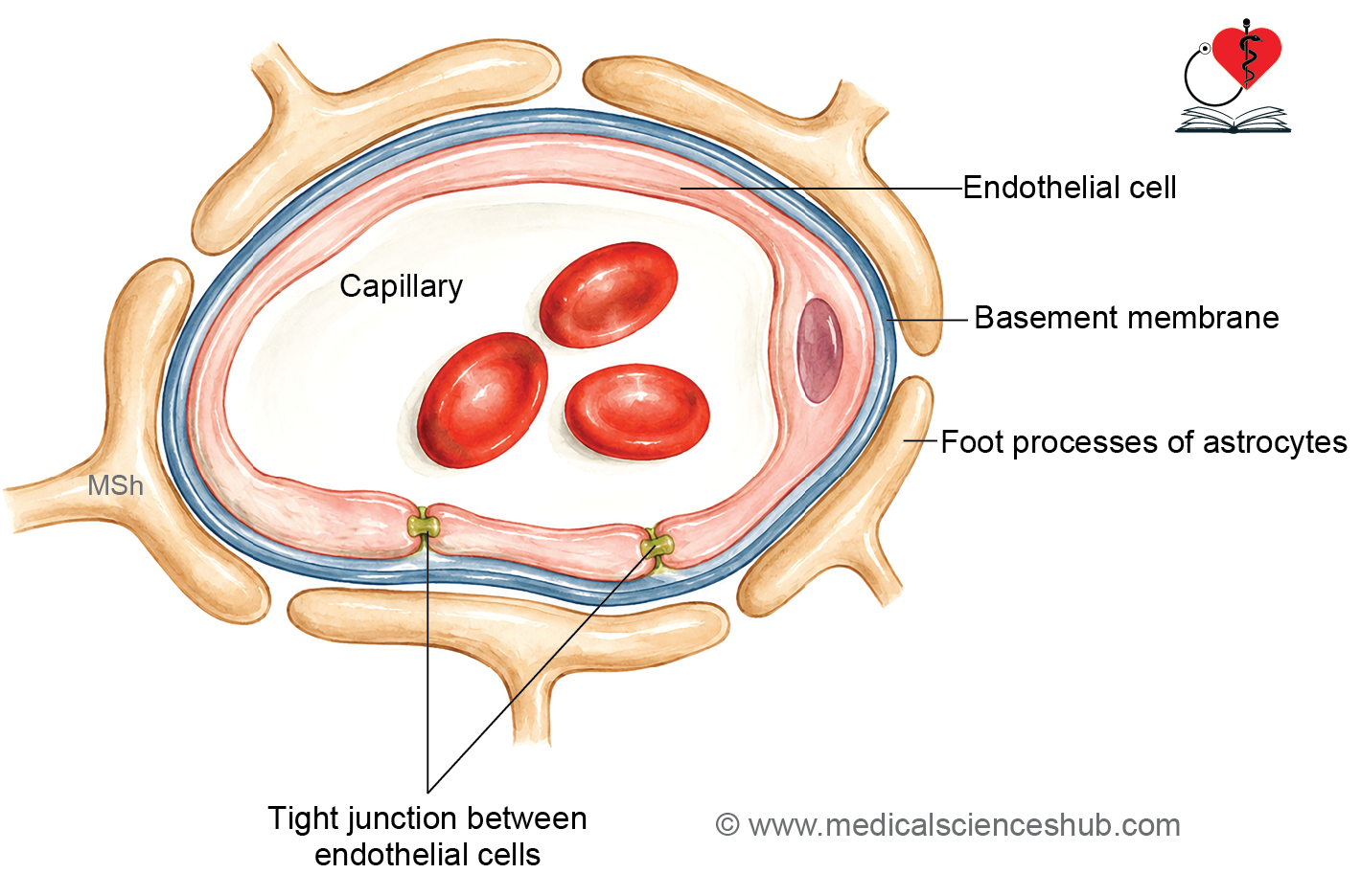
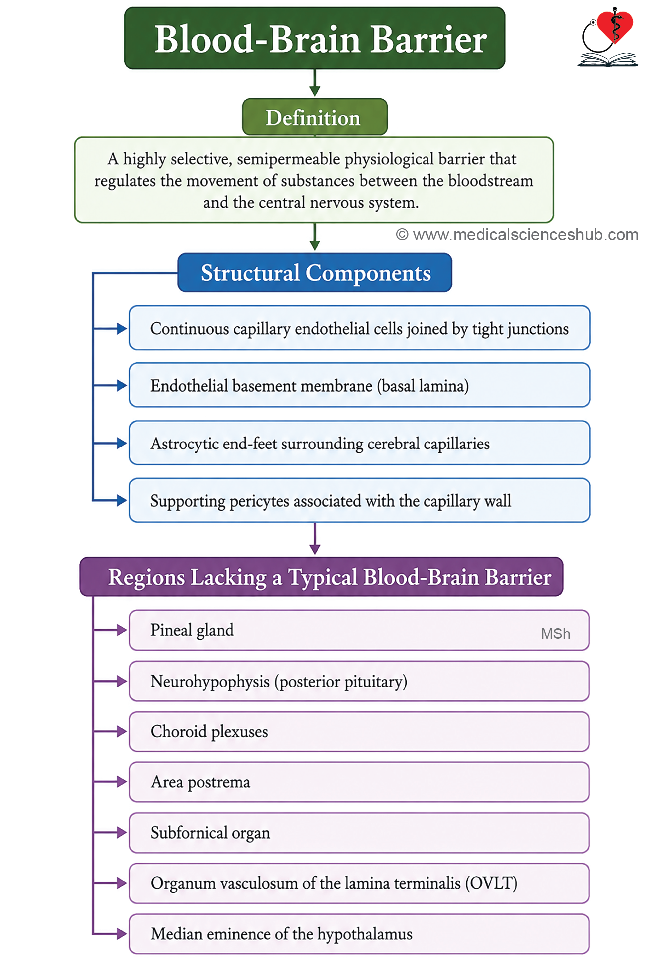
- The blood–brain barrier (BBB) is a highly selective semipermeable barrier that isolates brain tissue from circulating blood, regulating the passage of substances into the central nervous system.
Structural Components
- Capillary endothelial cells connected by tight junctions
- Basal lamina of the endothelial cells
- Astrocytic end-feet (perivascular processes of astrocytes) covering the basal lamina
Areas Lacking a Blood–Brain Barrier
Certain regions are deliberately devoid of a BBB, allowing direct exposure to circulating blood:
- Pineal gland
- Hypophysis cerebri (pituitary gland)
- Choroid plexuses
- Area postrema
- Tuber cinereum
- Interventricular foramen
Functions
- Selective permeability: allows free passage of glucose, respiratory gases, and amino acids while restricting lipid-insoluble substances.
- Neuroprotection: prevents entry of toxins, pathogens, large protein molecules, and most pharmacological agents into brain tissue.
CLINICAL NEUROANATOMY
Disruption of the BBB is implicated in neurological conditions including stroke, multiple sclerosis, and CNS infections. Its selective nature also presents a significant challenge in delivering therapeutic drugs to the brain.
Veins of Brain
Characteristic Features
- Cerebral veins are thin-walled and lack a muscular coat.
- They are valveless, allowing bidirectional flow.
- They do not accompany their corresponding arteries.
- They drain into the dural venous sinuses.
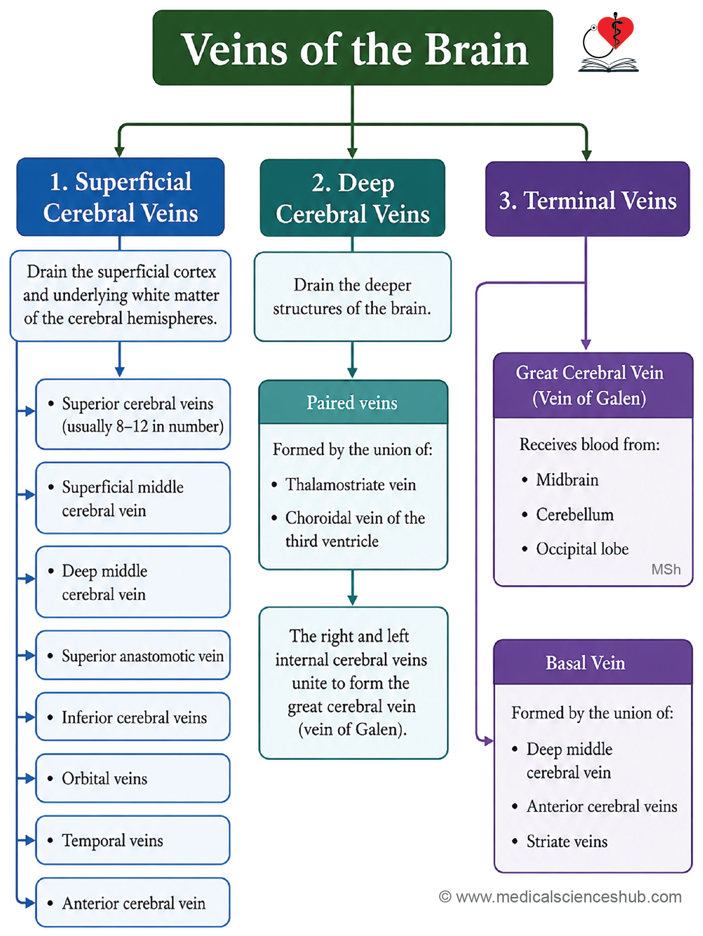
Classification of Cerebral Veins
External (Superficial) Cerebral Veins
- Superior cerebral veins
- Superficial middle cerebral vein
- Deep middle cerebral vein
- Inferior cerebral veins
- Anterior cerebral veins
Internal Cerebral Veins
- Drain the deep structures of the brain.
Terminal Veins
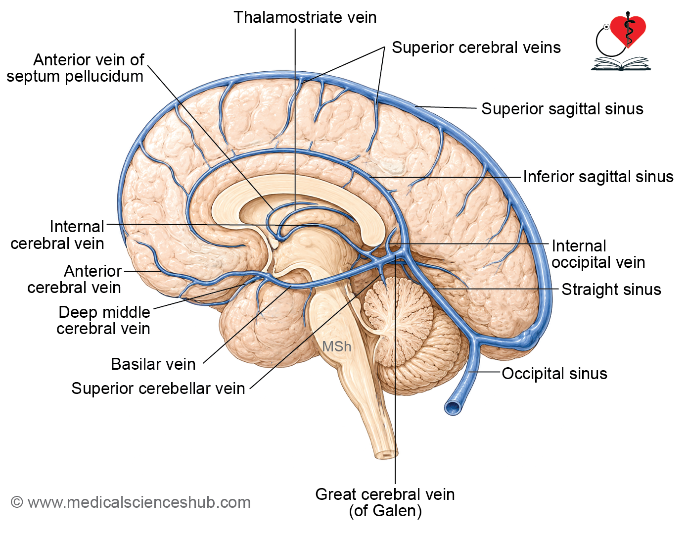
- Great cerebral vein of Galen — formed by the union of the two internal cerebral veins and the basal veins, draining into the straight sinus.
- Basal veins (of Rosenthal) — drain the deep basal regions of the brain into the great cerebral vein.
Superior Cerebral Veins
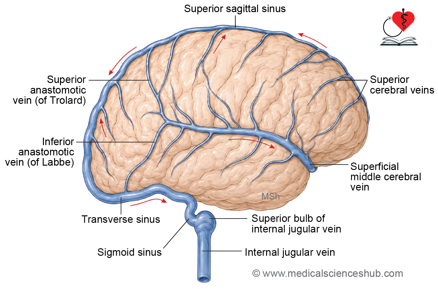
- The superior cerebral veins are a group of approximately 8-12 veins situated on the superolateral surface of the cerebral hemisphere.
- They drain blood from the lateral aspects of the frontal, parietal, and occipital lobes.
- These veins pass superiorly across the subarachnoid space and pierce the arachnoid mater and dura mater before terminating in the superior sagittal sinus.
- The largest superior cerebral vein usually courses near the central sulcus and is often referred to as the superior anastomotic vein (vein of Trolard).
- The number, size, and distribution of these veins show considerable anatomical variation among individuals.
- They play an important role in superficial cortical venous drainage and contribute significantly to cerebral venous outflow.
CLINICAL NEUROANATOMY
- The superior cerebral veins may become stretched or torn following head injury, particularly when sudden acceleration or deceleration forces are involved.
- Rupture of these veins can result in a subdural hematoma, caused by accumulation of blood between the dura mater and arachnoid mater.
- Large superficial cortical veins may occasionally serve as important collateral pathways when normal venous drainage is impaired.
- During neurosurgical procedures, preservation of major cortical veins is essential because injury may lead to venous congestion, cerebral edema, or venous infarction.
- Obstruction or thrombosis of cortical veins can impair venous drainage and may produce neurological deficits, seizures, headache, or raised intracranial pressure.
- Knowledge of their anatomy is important for interpreting neuroimaging studies and planning surgical approaches to the cerebral hemispheres.
Superficial Middle Cerebral Vein
- The superficial middle cerebral vein is located on the lateral surface of the cerebral hemisphere and courses within the lateral sulcus (Sylvian fissure).
- It drains blood from large areas of the frontal, parietal, and temporal lobes.
- The vein originates from numerous superficial cortical veins and collects venous blood from the adjacent cerebral cortex.
- It runs posteriorly within the lateral sulcus and then passes anteriorly toward the base of the brain.
- The vein commonly terminates in the cavernous sinus, either directly or through the sphenoparietal sinus.
- Considerable variation exists in its size, course, and pattern of termination.
- The superficial middle cerebral vein forms important anastomotic connections with the superior anastomotic vein (vein of Trolard) and inferior anastomotic vein (vein of Labbé), providing alternative pathways for venous drainage.
Functional Significance
- It serves as a major channel for superficial venous drainage of the lateral cerebral surface.
- It contributes significantly to venous outflow from the frontal, parietal, and temporal lobes.
CLINICAL NEUROANATOMY
- Injury to this vein may occur during neurosurgical procedures involving the lateral sulcus and surrounding regions.
- Damage or sacrifice of a major superficial middle cerebral vein can result in venous congestion, cerebral edema, or venous infarction.
- Thrombosis of this vein may present with headache, seizures, focal neurological deficits, or signs of raised intracranial pressure.
- Recognition of its anatomical variations is important during neuroimaging interpretation and surgical planning.
- Preservation of major venous channels is essential to maintain adequate cerebral venous drainage and reduce postoperative complications.
Deep Middle Cerebral Vein
- The deep middle cerebral vein, also known as the deep Sylvian vein, travels within the lateral sulcus, positioned deep to the superficial middle cerebral vein.
- It receives tributaries from the insula, the opercula, and neighboring cortical areas.
- The vein originates in the insular cortex, courses along the floor of the lateral sulcus in a posterior direction, and typically drains into the basal vein of Rosenthal or empties into the cavernous sinus through the sphenoparietal sinus.
- It maintains anastomotic connections with the superficial middle cerebral vein, forming part of a broader venous network of the lateral hemisphere.
- Its primary drainage territory includes the deep cortical and subcortical structures of the lateral cerebral hemisphere.
CLINICAL NEUROANATOMY
- Thrombosis or traumatic injury to this vein can result in deep cortical or subcortical venous infarction, manifesting clinically as contralateral hemiparesis, hemisensory deficits, or aphasia, the latter occurring when the dominant hemisphere is involved.
- Its deep anatomical position within the lateral sulcus places it at risk of injury during trans-sylvian surgical approaches, including clipping of middle cerebral artery aneurysms and resection of insular neoplasms.
Anterior Cerebral Veins
- The anterior cerebral veins accompany the anterior cerebral artery along the medial surface of each cerebral hemisphere.
- Each side usually consists of two main venous channels:
- Internal (deep) anterior cerebral vein
- External (superficial) anterior cerebral vein
- These veins drain blood from the medial aspects of the frontal lobe and adjacent cortical regions.
- They play an important role in venous drainage of structures supplied by the anterior cerebral artery.
Course
- The superficial anterior cerebral vein begins near the frontal pole and ascends along the medial surface of the cerebral hemisphere.
- It receives blood from the superior and medial parts of the frontal lobe, including the paracentral and cingulate regions.
- The deep anterior cerebral vein drains deeper white matter and subcortical structures located in the territory of the anterior cerebral artery.
- Both veins course posteriorly and eventually communicate with or drain into the deep venous system through connections near the anterior communicating artery region.
- Venous blood ultimately reaches larger deep cerebral veins and then drains into the straight sinus through the deep cerebral venous pathway.
Tributaries
The anterior cerebral veins commonly receive tributaries from:
- Frontal polar veins draining the frontal pole.
- Callosal veins draining the corpus callosum.
- Cingulate veins draining the cingulate gyrus.
- Medial frontal cortical veins draining the medial frontal cortex.
- Paracentral veins draining the paracentral lobule and adjacent cortex.
CLINICAL NEUROANATOMY
- These veins may serve as important collateral channels when normal cerebral venous drainage is compromised.
- Injury during interhemispheric surgical approaches can lead to venous congestion, cerebral edema, or venous infarction.
- Thrombosis involving anterior cerebral veins is uncommon but may produce headache, seizures, weakness of the lower limb, or other focal neurological deficits depending on the affected drainage territory.
- Detailed knowledge of their anatomy is important during neurosurgical procedures and interpretation of cerebral angiographic and venographic studies.
Inferior Cerebral Veins
- The inferior cerebral veins are a group of superficial veins situated on the inferior (basal) surface of the cerebral hemispheres.
- They are variable in number, size, and distribution.
- These veins drain venous blood from the orbital surface of the frontal lobe, the inferior aspect of the temporal lobe, and the inferior occipital region.
- They form an important part of the superficial venous drainage system of the brain.
- Depending on their location, the veins terminate in different dural venous sinuses and basal cerebral veins.
Course and Drainage
- Veins from the orbital surface of the frontal lobe commonly drain into the superior sagittal sinus, cavernous sinus, or adjacent venous channels.
- Veins from the inferior temporal region usually drain into the transverse sinus, superior petrosal sinus, or the basal vein.
- Veins from the inferior occipital surface generally terminate in the transverse sinus or nearby venous sinuses.
- Numerous anastomotic connections exist between inferior cerebral veins and other superficial cerebral veins, providing collateral pathways for venous drainage.
CLINICAL NEUROANATOMY
- The inferior cerebral veins may be injured during surgical procedures involving the skull base, temporal lobe, or inferior frontal region.
- Obstruction of venous drainage can result in venous congestion, cerebral edema, or venous infarction.
- These veins may serve as important collateral channels when major cerebral venous pathways are compromised.
- Their anatomy is highly variable; therefore, careful evaluation is necessary during neuroimaging and neurosurgical planning.
- Knowledge of their drainage patterns is essential for understanding the spread of venous thrombosis and the consequences of cerebral venous outflow obstruction.
Other Veins
Anastomotic Veins
- The cerebral venous system contains important communicating veins that connect major superficial venous channels and provide collateral pathways for venous drainage.
- The superior anastomotic vein (vein of Trolard) connects the superficial middle cerebral vein with the superior sagittal sinus.
- The inferior anastomotic vein (vein of Labbé) connects the superficial middle cerebral vein with the transverse sinus.
- These veins are highly variable in size and may become the dominant route of venous drainage in some individuals.
- They are of considerable surgical importance because injury may result in venous congestion or venous infarction.
Inferior Cerebral Veins
- Inferior cerebral veins on the basal surface of the brain are commonly formed by the union of small cortical and deep medullary veins.
- They drain the inferior surfaces of the frontal, temporal, and occipital lobes.
- Their blood ultimately reaches nearby dural venous sinuses or deep cerebral venous channels.
Great Cerebral Vein (Vein of Galen)
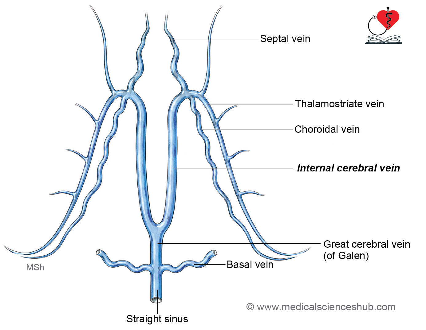
- The great cerebral vein is a short but important midline vein of the deep cerebral venous system.
- It is formed by the union of the two internal cerebral veins beneath the splenium of the corpus callosum.
- It receives tributaries from:
- Basal veins
- Veins of the midbrain
- Veins of the cerebellum
- Veins from the posterior parts of the cerebral hemispheres
- Posteriorly, it joins the inferior sagittal sinus to form the straight sinus.
- Obstruction or malformation of the vein of Galen can produce significant neurological and vascular abnormalities.
Internal Cerebral Veins
- The internal cerebral veins are paired deep veins located near the roof of the third ventricle.
- Each internal cerebral vein is formed near the interventricular foramen by the union of:
- Thalamostriate vein
- Superior choroidal vein
- The right and left internal cerebral veins run posteriorly beneath the corpus callosum and unite to form the great cerebral vein.
- They drain deep cerebral structures, including the thalamus, basal ganglia, choroid plexus, and adjacent white matter.
Thalamostriate Vein
- The thalamostriate vein arises near the junction of the thalamus and caudate nucleus.
- It courses posteriorly in the groove between these structures and receives numerous tributaries from the basal ganglia and surrounding deep white matter.
- Near the interventricular foramen, it joins the superior choroidal vein to form the internal cerebral vein.
- It represents one of the principal venous drainage pathways of the deep cerebral nuclei.
CLINICAL NEUROANATOMY
- Deep cerebral veins are essential for drainage of the ventricular system and deep brain structures.
- Thrombosis of the deep cerebral venous system may lead to bilateral thalamic involvement, altered consciousness, seizures, and raised intracranial pressure.
- Knowledge of these veins is crucial during ventricular, pineal, and deep midline neurosurgical procedures.
- Modern neuroimaging techniques frequently use these venous landmarks to identify deep cerebral structures and assess cerebral venous disorders.
CLINICAL NEUROANATOMY
- Cerebral angiography is a radiological technique used to visualize the arteries and veins of the brain.
- The procedure involves injecting a radiopaque contrast agent into a cerebral vessel, followed by a series of radiographic images to assess cerebral blood flow and vascular anatomy.
- A carotid angiogram is obtained by injecting contrast material into the common carotid artery or internal carotid artery.
- Thromboembolism refers to obstruction of a blood vessel by an embolus originating from a thrombus and is a common cause of cerebral ischemia and stroke.
- Cerebral venous sinus thrombosis occurs when a thrombus forms within a dural venous sinus, impairing venous drainage and increasing intracranial pressure.
- Subdural hematoma results from rupture of bridging cerebral veins, leading to accumulation of blood between the dura mater and arachnoid mater.
- A Charcot-Bouchard aneurysm is a small microaneurysm of penetrating cerebral arteries, particularly those supplying the basal ganglia, and is a recognized cause of intracerebral hemorrhage.
- Cerebral ischemia develops when cerebral blood flow falls below the level required to maintain normal neuronal function.
- Cerebral infarction occurs when severe or prolonged reduction of cerebral blood flow causes irreversible neuronal injury and tissue necrosis.
- Early recognition and management of cerebrovascular disorders are essential to reduce neurological damage and improve clinical outcomes.
Important Questions
- Write a short note on circle of Willis.
- Draw a well-labeled diagram of Circle of Willis.
📝 Test Your Knowledge – Practice MCQs
Attempt the chapter MCQ quiz and assess your understanding of key concepts.
Click to see: Medical Disclaimer and Copyright Notice