
Competencies
- AN59.1: Identify external features of pons.
- AN59.2: Draw and label transverse section of pons at the upper and lower levels.
- AN59.3: Enumerate cranial nerve nuclei in pons with their functional group.
Introduction
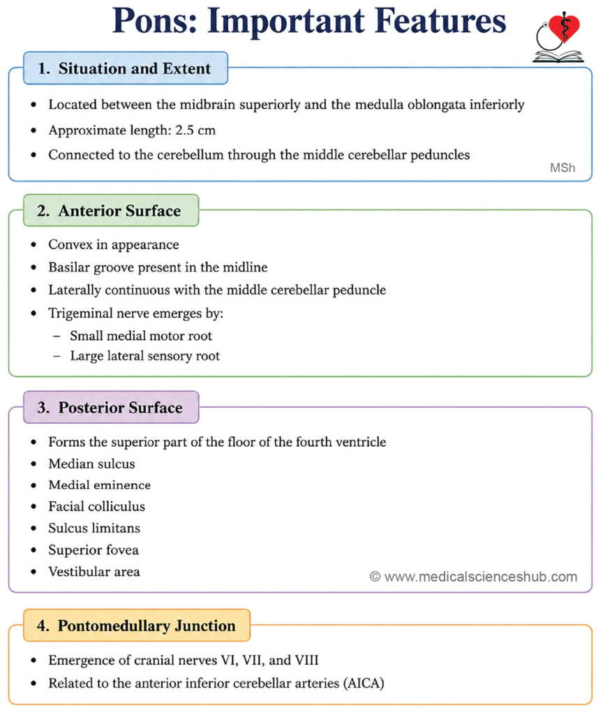
- The pons is the middle component of the brainstem, situated between the midbrain superiorly and the medulla oblongata inferiorly. The term pons is derived from the Latin word for “bridge,” reflecting its role in connecting these structures. It lies anterior to the cerebellum and the fourth ventricle.
- It measures approximately 2.5 cm in length.
- The pons performs several important functions, including the regulation of breathing rhythm, modulation of sleep and arousal, and control of facial movements and sensation via its cranial nerve nuclei.
- Damage to the pons can produce a range of serious neurological deficits, including respiratory disturbances, loss of motor coordination, dysphagia (difficulty swallowing), and facial or limb paralysis. Severe injury involving the reticular formation of the pons may impair consciousness and, in the most serious cases, result in coma.
External Features of Pons
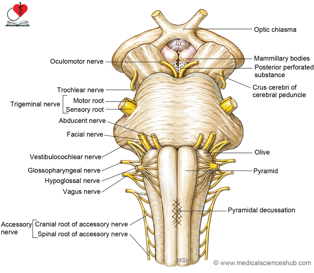
Anterior (Ventral) Surface
- The anterior surface is convex both transversely and vertically, and is characterized by prominent transverse striations produced by the horizontally coursing pontocerebellar fibers.
- The basilar sulcus is a shallow longitudinal groove running along the midline of the ventral surface. It accommodates the basilar artery. On either side of this sulcus, the surface is elevated by the underlying corticospinal fibers descending through the pons.
- Laterally, the anterior surface transitions smoothly into the middle cerebellar peduncles (brachium pontis), which connect the pons to the cerebellum.
- The trigeminal nerve (CN V) is attached to the lateral aspect of the ventral surface by two roots: a smaller motor root and a larger sensory root. The motor root lies superomedial to the sensory root.
Posterior (Dorsal) Surface
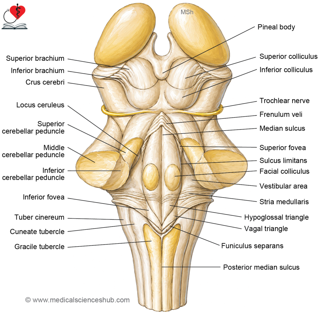
- The posterior surface of the pons forms the upper part of the floor of the fourth ventricle and is normally concealed by the overlying cerebellum. It has a triangular configuration and displays the following features:
- A median sulcus runs longitudinally along the midline, dividing the floor of the fourth ventricle into symmetrical halves.
- The medial eminence is a longitudinal elevation on either side of the median sulcus. In the pontine portion, it presents a rounded prominence called the facial colliculus, produced by the fibers of the facial nerve (CN VII) looping around the underlying abducens nucleus (CN VI).
- The sulcus limitans is a longitudinal groove lateral to the medial eminence, marking the embryological boundary between the alar plate (sensory) and basal plate (motor) derivatives.
- The superior fovea is a small depression in the sulcus limitans at the level of the pons, serving as an important surface landmark.
- The vestibular area lies lateral to the sulcus limitans and overlies the underlying vestibular nuclei.
Superior Border
- The superior border of the pons demarcates its junction with the cerebral peduncles of the midbrain. The superior cerebellar arteries curve around this border.
Inferior Border
- The inferior border corresponds to the pontomedullary junction. The rootlets of the abducens (CN VI), facial (CN VII), and vestibulocochlear (CN VIII) nerves emerge at this level. The anterior inferior cerebellar arteries (AICA) curve around this border.
Internal Structure of Pons
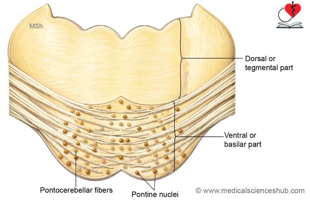
The pons is divided into two structurally and functionally distinct parts:
Basilar (Ventral) Part
- The basilar part forms the large, prominent ventral portion of the pons and maintains a relatively uniform internal organization throughout its extent.
- It contains two principal components: the pontine nuclei (nuclei pontis) and associated nerve fiber tracts.
- The pontine nuclei are scattered masses of grey matter distributed throughout the basilar pons. They function as relay stations in the corticopontocerebellar pathway, transmitting information from the cerebral cortex to the contralateral cerebellum to assist in the coordination of posture, balance, and the fine control of voluntary movements. Damage to these nuclei may result in ataxia, poor motor coordination, and impairment of smooth, purposeful movement.
- The nerve fibers of the basilar part are organized into two groups:
- Longitudinal fibers, which include the corticospinal, corticonuclear, and corticopontine tracts, descending from the cerebral cortex through the basilar pons.
- Transverse fibers, which arise from the pontine nuclei, cross the midline, and course horizontally as pontocerebellar fibers to reach the contralateral cerebellum via the middle cerebellar peduncle (brachium pontis).
Tegmental (Dorsal) Part
- The tegmental part forms the dorsal portion of the pons and contains the majority of the ascending and descending tracts traversing the pons, with the exception of the corticospinal and corticonuclear tracts, which are confined to the basilar part.
- It also houses the cranial nerve nuclei associated with the pons. The specific structures present within the tegmentum differ between the lower (caudal) and upper (rostral) levels of the pons, reflecting the changing organization of nuclei and fiber tracts at each level.
TS through Lower Part of Pons
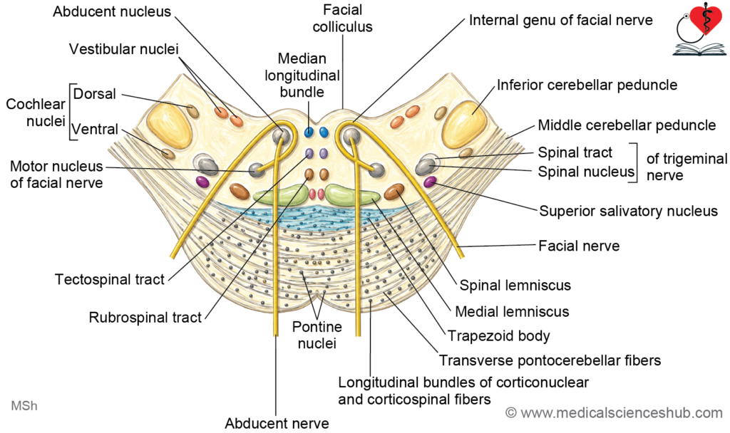
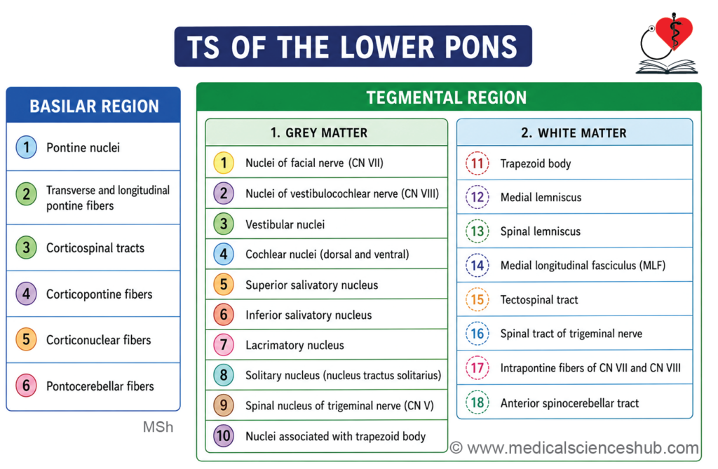
At the level of a transverse section through the lower (caudal) pons, passing through the facial colliculi, the following structures are identified:
Tegmentum — Grey Matter
Cranial Nerve Nuclei
- The abducens nucleus (CN VI) is located in the paramedian tegmentum, beneath the facial colliculus in the floor of the fourth ventricle. It provides motor innervation to the lateral rectus muscle, mediating abduction of the eye.
- The facial nerve nucleus (CN VII) lies ventrolateral to the abducens nucleus in the tegmentum. Its axons follow a distinctive intranuclear course, looping dorsally around the abducens nucleus to form the internal genu of the facial nerve, which produces the surface elevation of the facial colliculus, before turning anterolaterally to exit the pons at the pontomedullary junction.
- The vestibular nuclei (medial and inferior at this level) and cochlear nuclei are present, as described at the level of the upper medulla.
Autonomic and Visceral Nuclei
- The superior salivatory nucleus gives rise to preganglionic parasympathetic fibers that travel with the facial nerve (CN VII) to innervate the submandibular and sublingual salivary glands and the lacrimal gland.
- The inferior salivatory nucleus provides preganglionic parasympathetic fibers that travel with the glossopharyngeal nerve (CN IX) to innervate the parotid gland.
- The lacrimal nucleus (sometimes considered part of the superior salivatory nucleus) contributes parasympathetic fibers via the facial nerve to the lacrimal gland, regulating tear secretion.
- The nucleus tractus solitarius (NTS) is present, receiving gustatory and visceral afferent inputs from cranial nerves VII, IX, and X.
- The spinal nucleus of the trigeminal nerve continues at this level, processing pain and temperature information from the face and oral cavity.
- The nuclei of the trapezoid body are present within and adjacent to the trapezoid body fibers, serving as relay stations in the ascending auditory pathway.
Tegmentum — White Matter
- The trapezoid body is a prominent band of transversely oriented fibers crossing the midline in the ventral tegmentum. It carries decussating auditory fibers from the cochlear nuclei and contributes to the formation of the lateral lemniscus.
- The medial lemniscus is present as a vertically oriented band, carrying fine touch, vibration, and conscious proprioception from the contralateral body toward the thalamus.
- The spinal lemniscus (spinothalamic tract) carries pain, temperature, and crude touch sensations from the contralateral body, ascending toward the ventral posterolateral (VPL) nucleus of the thalamus.
- The medial longitudinal fasciculus (MLF) runs in its characteristic paramedian position, coordinating conjugate eye movements and vestibulo-ocular reflexes.
- The tectospinal tract descends from the superior colliculus of the midbrain, mediating reflex head and neck movements in response to visual and auditory stimuli.
- The spinal tract of the trigeminal nerve continues laterally, carrying descending pain and temperature fibers from the face toward the spinal nucleus below.
- The intranuclear course of the abducens and facial nerve fibers is a defining feature of this level, with facial nerve axons looping around the abducens nucleus before exiting the brainstem.
- The anterior spinocerebellar tract is present in the lateral tegmentum, transmitting unconscious proprioceptive information from the lower limb to the cerebellum.
Basilar Part
- The basilar part at this level contains the pontine nuclei, together with the longitudinal fiber tracts — corticospinal, corticonuclear, and corticopontine — and the transversely running pontocerebellar fibers projecting to the contralateral cerebellum via the middle cerebellar peduncle.
Key Description
Abducens Nucleus and Nerve (CN VI)
- The abducens nucleus lies in the floor of the fourth ventricle, just lateral to the medial longitudinal fasciculus (MLF), beneath the facial colliculus. It supplies the lateral rectus muscle, mediating abduction of the eyeball.
- Axons from the abducens nucleus pass ventrally and caudally through the tegmentum and basilar pons, emerging at the pontomedullary junction just superior to the pyramid.
- A lesion of the abducens nucleus or nerve results in ipsilateral lateral rectus palsy, causing the affected eye to deviate medially at rest (convergent squint) and inability to abduct the eye, producing horizontal diplopia (double vision).
Facial Nerve Nucleus and Nerve (CN VII)
- The motor nucleus of the facial nerve lies ventromedial to the abducens nucleus within the pontine tegmentum. It receives bilateral corticonuclear (corticobulbar) input, which explains why an upper motor neuron lesion typically spares the forehead muscles, as they receive input from both hemispheres.
- Axons from the facial nucleus follow a distinctive intranuclear course: they initially pass dorsally toward the floor of the fourth ventricle, loop around the abducens nucleus forming the internal genu of the facial nerve, and then course anterolaterally to exit at the pontomedullary junction, superior to the olive, between the abducens nerve and the nervus intermedius.
- The looping fibers around the abducens nucleus produce the surface elevation in the floor of the fourth ventricle known as the facial colliculus.
- The facial nerve (CN VII) controls the muscles of facial expression and the orbicularis oculi (responsible for eye closure and blinking). It also mediates lacrimation and salivation via parasympathetic fibers, and conveys taste sensation from the anterior two-thirds of the tongue via the chorda tympani.
- Damage to the facial nerve nucleus or its intrapontine fibers produces ipsilateral lower motor neuron facial palsy, characterized by paralysis of all ipsilateral facial muscles, inability to close the eye, drooping of the mouth, loss of the corneal blink reflex, and impaired taste and salivation.
Neurobiotaxis
- The intranuclear course of the facial nerve within the pons is a classic example of neurobiotaxis — the embryological tendency of a nucleus to migrate toward its principal source of afferent stimulation. During development, the facial nucleus migrates ventrally toward the spinal nucleus of the trigeminal nerve, and its axons trail behind, forming the characteristic loop around the abducens nucleus. This migration is thought to facilitate rapid corneal reflex responses.
Superior Salivatory Nucleus, Lacrimal Nucleus, and Nucleus Tractus Solitarius
- The superior salivatory nucleus lies in the lateral reticular formation of the lower pons. It gives rise to preganglionic parasympathetic fibers that travel sequentially via the facial nerve → chorda tympani → lingual nerve → submandibular ganglion, ultimately innervating the submandibular and sublingual salivary glands.
- The lacrimal nucleus is functionally a subdivision of the superior salivatory nucleus. Its preganglionic parasympathetic fibers travel via the facial nerve → greater petrosal nerve → pterygopalatine ganglion → lacrimal gland, regulating tear secretion.
- Damage to these nuclei results in reduced salivation (xerostomia) and reduced tear production (xerophthalmia), leading to difficulty swallowing, impaired speech, and ocular surface irritation.
- The nucleus tractus solitarius (NTS) lies lateral to the superior salivatory nucleus at this level, continuing its role in processing gustatory and visceral afferent inputs.
Vestibular Nuclei
- The vestibular nuclear complex comprises four nuclei — superior, inferior, medial, and lateral — located in the lateral floor of the fourth ventricle. The superior and lateral nuclei lie in the pons, while the inferior and medial nuclei extend into the medulla.
- Afferent inputs arrive from the vestibular division of CN VIII and from the cerebellum.
- Efferent projections include:
- Vestibulocerebellar fibers, projecting to the cerebellum for balance coordination.
- The vestibulospinal tracts: the lateral vestibulospinal tract arises from the lateral vestibular nucleus and facilitates extensor muscle tone; the medial vestibulospinal tract arises from the medial vestibular nucleus and coordinates head and neck posture.
- Projections to the MLF, which coordinates conjugate eye movements and vestibulo-ocular reflexes.
- Lesions of the vestibular nuclei produce vertigo, nausea, postural imbalance, and nystagmus (involuntary rhythmic eye movements).
Cochlear Nuclei, Trapezoid Body, and Lateral Lemniscus
- The dorsal and ventral cochlear nuclei lie on the corresponding surfaces of the inferior cerebellar peduncle and function as the second-order neurons of the ascending auditory pathway, receiving input from the spiral ganglion of the cochlea via the cochlear division of CN VIII.
- The trapezoid body is a band of transversely oriented fibers in the ventral tegmentum, carrying decussating auditory fibers from both cochlear nuclei toward the contralateral side. Embedded within these fibers are the nuclei of the trapezoid body, which receive cochlear input and project ascending fibers as the lateral lemniscus toward the inferior colliculus.
- The superior olivary complex lies in the anterolateral tegmentum. It receives bilateral input from the cochlear nuclei and serves as an important third-order relay in the auditory pathway, playing a central role in binaural sound localization by comparing interaural differences in timing and intensity.
- Damage to these structures may cause sensorineural hearing loss, difficulty localizing sounds, and impaired speech discrimination in noisy environments.
Lemnisci
- The medial lemniscus lies in the ventral tegmentum close to the midline, ascending to carry fine touch, vibration, and conscious proprioception from the contralateral body to the thalamus.
- The trigeminal lemniscus is positioned between the medial and spinal lemnisci, conveying discriminative touch and proprioception from the contralateral face via the principal sensory nucleus of CN V.
- The spinal lemniscus represents the ascending continuation of the lateral spinothalamic tract, carrying pain and temperature sensations from the contralateral body.
- The lateral lemniscus ascends laterally through the tegmentum, transmitting auditory signals from the cochlear nuclei and trapezoid body nuclei toward the inferior colliculus.
Other Tegmental Structures
- The spinal nucleus of the trigeminal nerve lies on the anteromedial aspect of the inferior cerebellar peduncle at this level, continuing to process pain and temperature from the face.
- The reticular formation occupies the central core of the tegmentum as a diffuse network of scattered neurons and interconnecting fibers, contributing to the regulation of consciousness, autonomic function, and motor coordination.
TS through the Upper Part of Pons
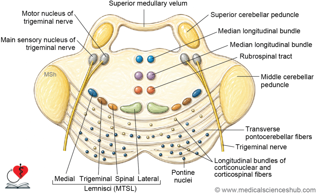
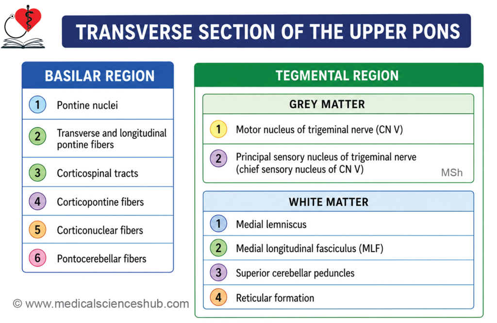
At the level of a transverse section through the upper (rostral) pons, passing through the trigeminal nuclei, the section reveals three principal regions: a central cavity, an anterior basilar part, and a posterior tegmentum.
Cavity
- At this level, the cavity of the fourth ventricle has become narrow and is bounded dorsally by the superior medullary velum and laterally by the superior cerebellar peduncles.
- The superior medullary velum is a thin lamina of white matter that bridges the two superior cerebellar peduncles, forming the roof of the upper fourth ventricle. Its anterior (inner) surface is lined by ependymal cells, while its posterior (outer) surface is covered by pia mater.
Basilar Part
- The basilar part at this level has the same organization as at lower pontine levels, containing the pontine nuclei and the four groups of longitudinal and transverse nerve fiber tracts: corticospinal, corticonuclear, corticopontine, and pontocerebellar fibers.
Tegmentum — Grey Matter
- The motor nucleus of the trigeminal nerve (CN V) is present at this level. It is a special visceral efferent (SVE) nucleus that supplies the muscles of mastication (temporalis, masseter, medial and lateral pterygoids), along with the mylohyoid, anterior belly of the digastric, tensor veli palatini, and tensor tympani — all derivatives of the first pharyngeal arch.
- The principal (main) sensory nucleus of the trigeminal nerve lies lateral to the motor nucleus. It receives discriminative touch and pressure sensations from the ipsilateral face and oral cavity, functioning as the pontine equivalent of the dorsal column nuclei for facial sensation. Its axons cross the midline and ascend as the trigeminal lemniscus to the ventral posteromedial (VPM) nucleus of the thalamus.
Trigeminal Nuclei
- The trigeminal nuclear complex consists of one motor nucleus and three sensory nuclei:
- The motor nucleus lies in the pons and innervates the muscles of the first pharyngeal arch derivatives.
- The mesencephalic nucleus extends into the midbrain and is unique in containing primary afferent (first-order) neuron cell bodies. It mediates proprioception from the muscles of mastication, the extraocular muscles, and the periodontium.
- The principal sensory nucleus lies in the pons and processes fine touch and pressure from the face.
- The spinal nucleus extends from the lower pons through the medulla oblongata to the level of the second cervical spinal segment (C2). It processes pain and temperature from the face, scalp, and oral cavity, and is functionally analogous to the dorsal horn of the spinal cord.
- Lesions of the trigeminal nuclei may produce ipsilateral facial sensory loss, weakness of the masticatory muscles, or, in cases of abnormal neural activity within the system, trigeminal neuralgia — a condition characterized by severe, paroxysmal facial pain.
Tegmentum — White Matter
- The four lemnisci — medial, trigeminal, spinal, and lateral — are all present in the tegmentum at this level, ascending toward their respective thalamic relay nuclei as described at lower pontine levels.
- The medial longitudinal fasciculus (MLF) continues in its paramedian position, coordinating conjugate eye movements and vestibulo-ocular reflexes.
- The superior cerebellar peduncles (brachia conjunctiva) are prominent at this level, lying dorsolateral to the tegmentum. They represent the principal efferent pathway of the cerebellum, carrying dentatorubrothalamic fibers from the dentate nucleus to the contralateral red nucleus and ventral lateral nucleus of the thalamus, and decussating in the caudal midbrain tegmentum.
- The reticular formation continues as a diffuse network in the central tegmentum, contributing to the regulation of arousal, autonomic function, and descending motor control.
Lemnisci
In the tegmentum of the upper pons, four lemnisci are arranged in a consistent sequence from medial to lateral:
- The medial lemniscus (band of Reil) is the most medially positioned. It carries second-order neurons originating from the nucleus gracilis (tract of Goll) and nucleus cuneatus (tract of Burdach), conveying conscious proprioception, discriminative (fine) touch, and vibration sense from the contralateral body to the ventral posterolateral (VPL) nucleus of the thalamus.
- The trigeminal lemniscus lies lateral to the medial lemniscus. It carries pain, temperature, and discriminative touch sensations from the contralateral face. Its fibers originate from the spinal nucleus (pain and temperature) and the principal sensory nucleus (fine touch) of the trigeminal nerve, and ascend to the ventral posteromedial (VPM) nucleus of the thalamus.
- The spinal lemniscus lies lateral to the trigeminal lemniscus and represents the ascending continuation of two spinothalamic tracts: the lateral spinothalamic tract, carrying pain and temperature from the contralateral body, and the anterior spinothalamic tract, carrying crude touch and pressure from the contralateral body. Both project to the VPL nucleus of the thalamus.
- The lateral lemniscus is the most laterally positioned and constitutes the principal ascending auditory pathway in the brainstem. It carries third-order auditory neurons derived from the superior olivary complex, which itself receives input sequentially from the cochlear nuclei → trapezoid body nuclei → superior olivary nucleus. The lateral lemniscus ascends to terminate in the inferior colliculus of the midbrain.
Blood Supply of Pons
- The pons receives its arterial supply exclusively from branches of the basilar artery and its derivatives:
- Pontine arteries (paramedian and circumferential branches) arise directly from the basilar artery and represent the primary arterial supply to the pons. They are further divided into short circumferential branches, supplying the anteromedial and anterolateral tegmentum, and long circumferential branches, supplying the lateral and dorsal tegmentum.
- Branches of the anterior inferior cerebellar artery (AICA), which arises from the lower basilar artery, supply the caudal and lateral pons as well as the anterior surface of the cerebellum.
The superior cerebellar artery, arising from the rostral basilar artery just proximal to its bifurcation, supplies the upper (rostral) pons and the superior surface of the cerebellum.
CLINICAL NEUROANATOMY
Pontine Hemorrhage
- Pontine hemorrhage is a catastrophic brainstem event resulting from rupture of branches of the basilar artery or the anterior inferior cerebellar artery (AICA). Due to the concentration of vital structures within the pons, it is almost invariably fatal.
- The clinical features reflect damage to the following structures:
- Damage to the facial nerve nucleus (CN VII) and the descending corticospinal tracts produces bilateral paralysis of the face and limbs, with loss of facial expression and voluntary limb movement.
- Damage to the reticular formation disrupts the ascending arousal system, resulting in deep coma.
- Damage to descending hypothalamospinal fibers controlling the thermoregulatory centers leads to loss of normal heat regulation, causing hyperpyrexia (abnormally elevated body temperature).
- Damage to the descending sympathetic fibers abolishes sympathetic tone to the eye, producing bilateral pinpoint pupils (miosis) — a hallmark sign of pontine hemorrhage.
Millard–Gubler Syndrome
- Millard–Gubler syndrome, also known as ventral pontine syndrome or crossed (alternating) hemiplegia, results from a lesion in the ventral pons, typically caused by occlusion of the paramedian branches of the basilar artery. It was described by French physicians Auguste Millard (1830–1915) and Adolphe-Marie Gubler (1821–1879).
- The clinical features result from damage to three structures:
- Damage to the abducens nerve (CN VI) produces ipsilateral lateral rectus palsy, causing the affected eye to deviate medially at rest (convergent squint) and inability to abduct the eye, resulting in horizontal diplopia. This presentation is also termed abducent alternating hemiplegia.
- Damage to the facial nerve (CN VII) produces ipsilateral lower motor neuron facial palsy, with inability to close the eye, loss of facial expression, and drooping of the mouth on the side of the lesion.
- Damage to the corticospinal tract in the basilar pons produces contralateral hemiplegia, as these fibers have not yet crossed the midline at this level.
- The combination of ipsilateral cranial nerve palsies with contralateral limb paralysis is the defining characteristic of a crossed hemiplegia, reflecting a unilateral lesion at the level of the pons.
Pontocerebellar Angle Syndrome
- The pontocerebellar angle (cerebellopontine angle) is a cistern at the junction of the pons, medulla, and cerebellum that contains the facial nerve (CN VII), vestibulocochlear nerve (CN VIII), the flocculus of the cerebellum, and the choroid plexus of the fourth ventricle.
- The most common cause of pontocerebellar angle syndrome is an acoustic neuroma (vestibular schwannoma) — a benign tumor arising from the Schwann cells surrounding the vestibular division of CN VIII.
- Compression of structures within the pontocerebellar angle produces the following characteristic features:
- Compression of the facial nerve (CN VII) results in ipsilateral lower motor neuron facial palsy and loss of taste from the anterior two-thirds of the tongue.
- Compression of the vestibulocochlear nerve (CN VIII) produces tinnitus (ringing in the ear), progressive sensorineural hearing loss, and vertigo. These are typically the earliest presenting symptoms of an acoustic neuroma.
- Involvement of the flocculus of the cerebellum results in ipsilateral cerebellar ataxia, with incoordination of limb movements and gait imbalance on the affected side.
- Compression of the trigeminal nerve (CN V) or its tract produces loss of the corneal reflex and may cause ipsilateral facial numbness.
Foville’s Syndrome
- Foville’s syndrome results from occlusion of a perforating branch of the basilar artery supplying the dorsal pons, producing a lesion in the pontine tegmentum. It was described by French neurologist Achille-Louis Foville (1799–1878).
- The clinical features include:
- Damage to the abducens nucleus (CN VI) produces ipsilateral conjugate gaze palsy toward the side of the lesion, as the abducens nucleus contains both the motor neurons supplying the ipsilateral lateral rectus and the interneurons coordinating contralateral medial rectus activation via the MLF.
- Damage to the facial nerve nucleus (CN VII) produces ipsilateral lower motor neuron facial paralysis, with loss of all voluntary facial movements on the affected side.
Tumors of the Pons
- Astrocytoma is the most common primary tumor of the brainstem and typically arises during childhood. The specific neurological signs and symptoms depend on the precise location of the tumor within the pons and the structures it compresses or infiltrates.
Important Questions
- Draw a well-labeled diagram of TS of pons through its upper part.
- Draw a well-labeled diagram of TS of pons through its lower part.
- Write a short note on pontine hemorrhage.
- Write a short note on Millard–Gubler syndrome.
- Write a short note on Pontocerebellar Angle syndrome.
- Write a short note on Foville’s syndrome.
📝 Test Your Knowledge – Practice MCQs
Attempt the chapter MCQ quiz and assess your understanding of key concepts.
Click to see: Medical Disclaimer and Copyright Notice