
Competency
- AN63.1: Describe and demonstrate parts, boundaries, and features of IIIrd, IVth, and lateral ventricles.
Introduction
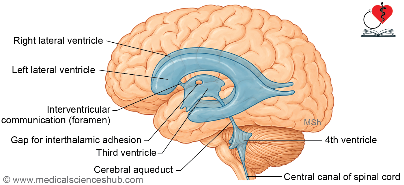
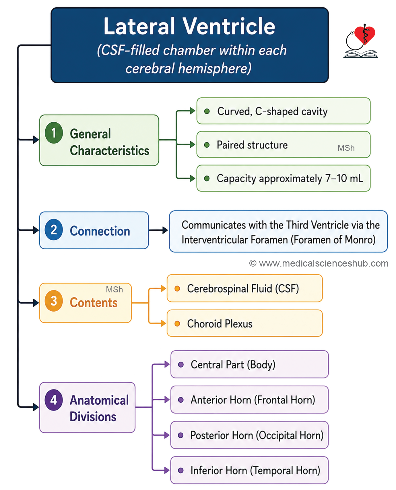
- The lateral ventricles are paired C-shaped cavities of the telencephalon, with one located within each cerebral hemisphere.
- They are the primary sites of CSF production in the brain.
- The two ventricles are separated from each other by the septum pellucidum.
- Their inner surfaces are lined by ependymal cells.
- Each ventricle has a capacity of approximately 7–10 mL.
Communications
- Each lateral ventricle communicates with the third ventricle through the interventricular foramen of Monro.
- This foramen serves as the primary pathway for CSF to exit the lateral ventricle and enter the broader ventricular system.
- Obstruction of the interventricular foramen of Monro blocks CSF outflow from the lateral ventricle, leading to progressive CSF accumulation and hydrocephalus with raised intracranial pressure.
Contents
The lateral ventricle contains two structures:
- Cerebrospinal fluid (CSF), which fills the ventricular cavity and is continuously circulated through the ventricular system.
- The choroid plexus, which is the primary source of CSF production within the lateral ventricle.
Parts of Lateral Ventricle
The lateral ventricle consists of a body and three horns:
- The body (central part) is the main portion of the ventricle, lying primarily within the parietal lobe.
- The anterior horn is a forward extension of the ventricle projecting into the frontal lobe.
- The posterior horn extends backward into the occipital lobe.
- The inferior horn is a downward and forward continuation of the body, extending into the temporal lobe.
Boundaries of the Parts of Lateral Ventricle
Central Part (body)
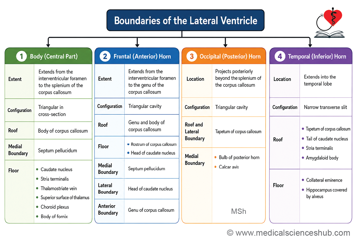
- The body lies primarily within the parietal lobe, extending from the interventricular foramen of Monro anteriorly to the splenium of the corpus callosum posteriorly.
- In coronal section, it has a triangular shape, with a roof, a medial wall, and a floor.
Boundaries
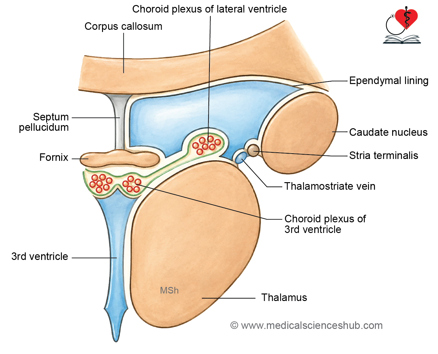
- The roof is formed by the undersurface of the corpus callosum.
- The medial wall is formed by the septum pellucidum.
- The floor slopes and is formed by the following structures, from lateral to medial:
- Caudate nucleus
- Stria terminalis
- Thalamostriate vein
- Lateral part of the upper surface of the thalamus
- Choroid plexus
- Upper surface of the body of the fornix
Anterior Horn
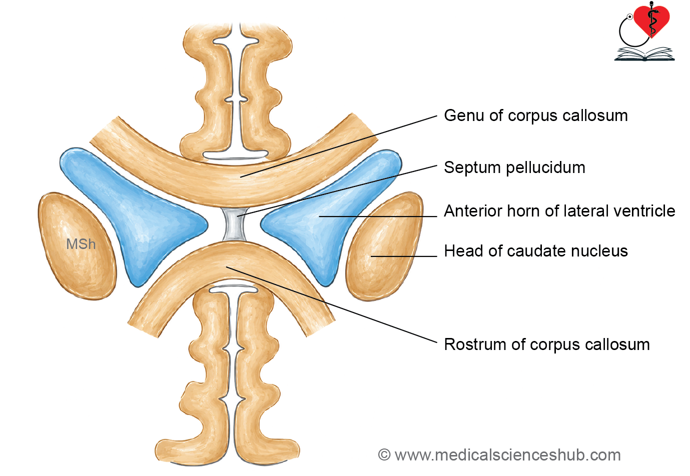
- The anterior horn extends forward from the interventricular foramen to the posterior surface of the genu of the corpus callosum, projecting into the frontal lobe.
- It is triangular in coronal section and has five walls:
- Roof: undersurface of the anterior body of the corpus callosum
- Floor: upper surface of the rostrum of the corpus callosum and the head of the caudate nucleus
- Medial wall: septum pellucidum
- Lateral wall: bulging head of the caudate nucleus
- Anterior wall: genu of the corpus callosum
Posterior Horn
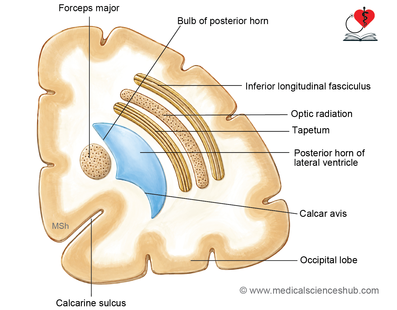
- The posterior horn extends backward behind the splenium of the corpus callosum into the occipital lobe.
- Its boundaries include:
- Roof and lateral wall: tapetum of the corpus callosum
- Medial wall: two elevations — the bulb of the posterior horn (produced by fibers of the forceps major) and the calcar avis (produced by the anterior part of the calcarine sulcus)
Inferior Horn
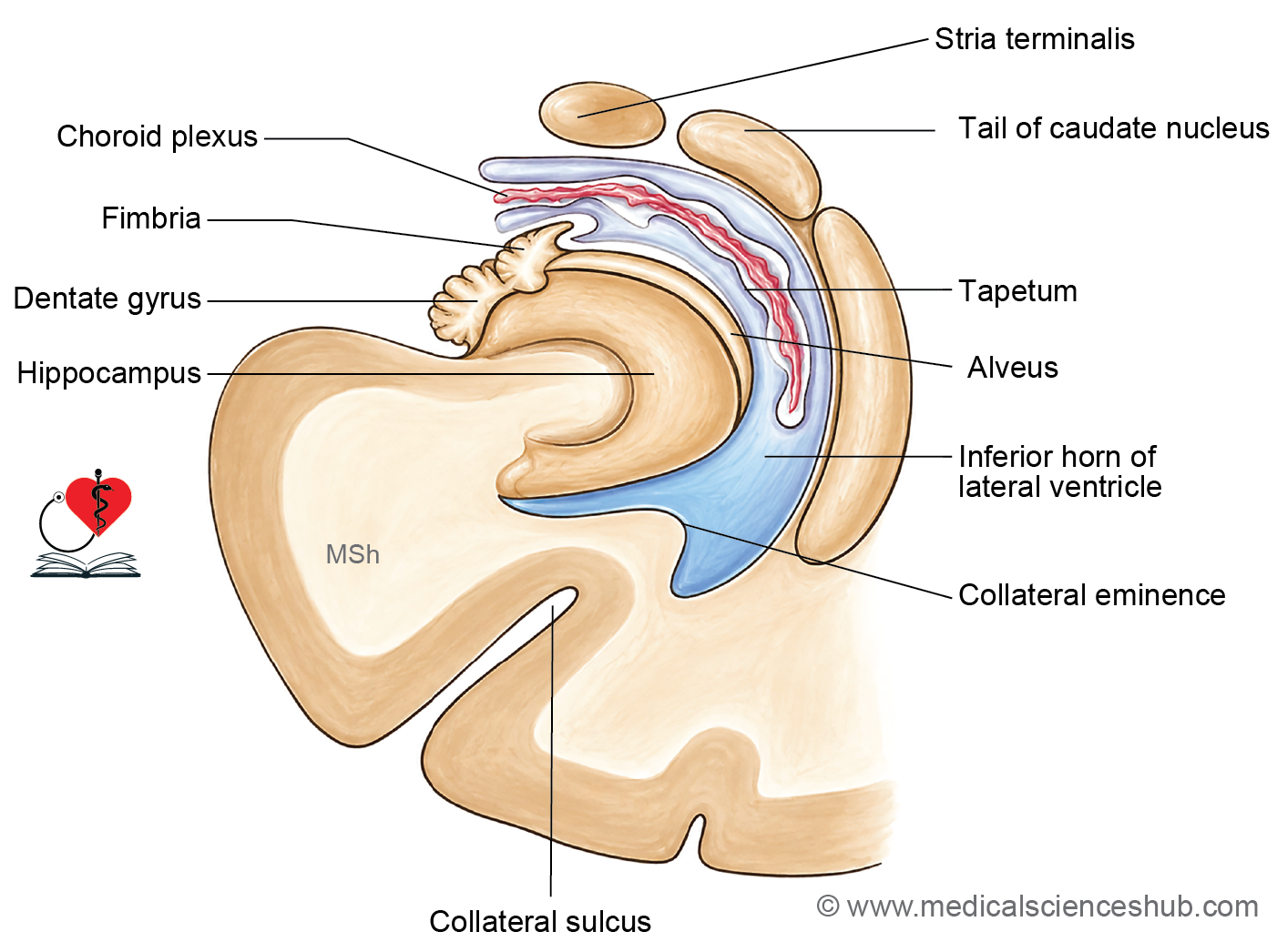
- The inferior horn is the largest of the three horns, extending downward and forward into the temporal lobe as a continuation of the body.
- The collateral trigone marks the junction where the posterior and inferior horns meet the body of the lateral ventricle.
- It appears as a transverse slit in cross-section, with a roof and floor:
- Roof: tapetum of the corpus callosum, tail of the caudate nucleus, stria terminalis, and amygdaloid body
- Floor: two elevations — the collateral eminence (produced by the collateral sulcus) and the hippocampus covered by the alveus
Blood Supply
- Anterior choroidal artery — This vessel arises from the internal carotid artery just proximal to its terminal bifurcation into the middle and anterior cerebral arteries. It courses posteriorly and laterally along the optic tract, enters the inferior horn of the lateral ventricle through the choroidal fissure, and supplies the choroid plexus in that region.
- Posterior choroidal arteries — These are branches of the posterior cerebral artery and are subdivided into two groups:
- Medial posterior choroidal artery — This vessel runs along the roof of the third ventricle and enters it through the choroidal fissure, supplying the choroid plexus of the third ventricle and contributing to that of the lateral ventricle.
- Lateral posterior choroidal artery — This vessel travels along the lateral surface of the thalamus and enters the lateral ventricle through the choroidal fissure of its posterior horn, supplying the choroid plexus in that territory.
CLINICAL NEUROANATOMY
Hypothalamic disorders
- Occlusion of the anterior choroidal artery is a recognized cause of a specific infarction syndrome, producing contralateral hemiplegia, hemianesthesia, and homonymous hemianopia, due to involvement of the posterior limb of the internal capsule, thalamus, and optic tract respectively.
- Visualization of the Lateral Ventricles
- Computed tomography (CT scan) — This is a widely used cross-sectional imaging modality that provides rapid visualization of ventricular size, shape, and any displacement, making it particularly useful in acute settings such as suspected hydrocephalus or intracranial hemorrhage.
- Magnetic resonance imaging (MRI) — This offers superior soft-tissue contrast compared to CT and allows detailed multiplanar visualization of the ventricular system and surrounding neural structures. It is the preferred modality for evaluating periventricular pathology, ventricular tumors, and subtle structural anomalies.
- Ventriculography (pneumoencephalography / air encephalography) — This was a historical radiological technique in which air or oxygen was introduced into the subarachnoid space via lumbar puncture. The gas displaced CSF within the subarachnoid space and ventricular system, producing negative contrast that enhanced radiological visualization of these spaces on plain radiographs.
- This procedure carried significant risks, including severe headache, herniation, and hemodynamic instability, and has been entirely replaced by CT and MRI in contemporary clinical practice.
- It was pioneered by Walter Dandy in 1919 and remained in use until the advent of modern cross-sectional imaging in the 1970s.
Important Questions
- Describe the lateral ventricle under the following headings: Situation and extent, parts (body and horns), boundaries of each part, communications and applied anatomy.
- Write a short note on the anterior (frontal) horn of the lateral ventricle.
- Discuss the boundaries of the body (central part) of the lateral ventricle.
- Write a short note on the posterior horn of the lateral ventricle.
- Write a short note on the inferior horn of the lateral ventricle.
Some Interesting Facts
- Choroid fissure is a “slit-like” gap between the fornix and thalamus through which choroid plexus enters the lateral ventricle.
- Choroid plexus is a highly vascular fold of pia mater. It produces CSF.
- Tela choroidea is a vascular fold of pia mater with covering of ependyma that protrudes into the ventricular cavity. The tela choroidea invaginates the cavity of lateral ventricle through C-shaped choroid fissure.
- Thalamostriate vein, also called the terminal vein, lies in the floor of the lateral ventricle. The vein begins near the anterior perforated substance, close to the junction of the caudate nucleus and the anterior limb of the internal capsule. From there, it runs backward in the groove between the thalamus and the caudate nucleus, called the sulcus terminalis. At the posterior end of the thalamus, it joins the choroidal vein to form the internal cerebral vein. This junction is located near the interventricular foramen (of Monro). Injury to this vein can lead to internal bleeding or complications related to deep brain drainage.
📝 Test Your Knowledge – Practice MCQs
Attempt the chapter MCQ quiz and assess your understanding of key concepts.
Click to see: Medical Disclaimer and Copyright Notice