
Competency
- AN71.1 Identify bone under the microscope; classify various types and describe the structure-function correlation of the same.
Introduction
- Bone is a specialized connective tissue characterized by an extracellular matrix rich in calcium salts.
- It consists of living cells embedded within a highly mineralized matrix.
- Matrix mineralization provides bones with strength, rigidity, and resistance to mechanical stress.
- Bone is a dynamic tissue that continuously undergoes remodeling in response to physical demands and hormonal influences.
- Bones protect vital organs, provide structural support, and serve as major reservoirs of calcium and phosphate.
- Bone also contains bone marrow, which is responsible for hematopoiesis (blood cell formation).
General Features of Bone
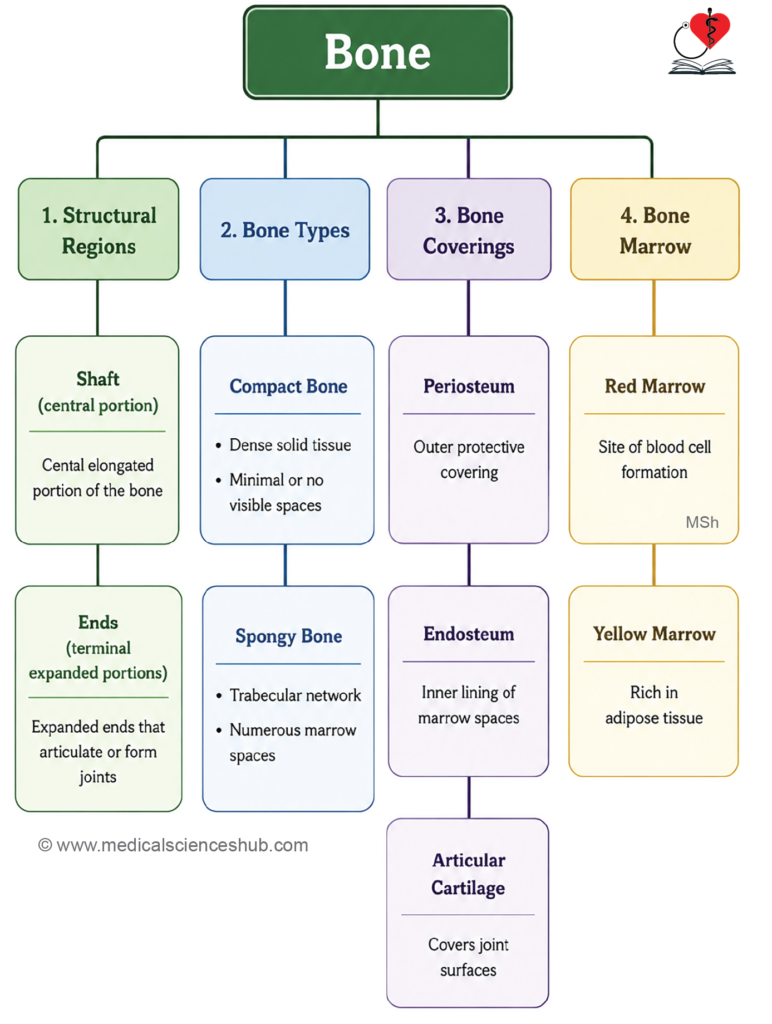
- A long bone consists of a central shaft (diaphysis) and two expanded ends (epiphyses).
- In cross-section, a long bone is composed of the diaphysis, epiphyses, and connective tissue coverings, namely the periosteum externally and endosteum internally.
- The diaphysis forms a strong tubular structure that encloses the medullary (marrow) cavity.
- Its wall is primarily made of compact (cortical) bone, which is dense and contains very few visible spaces.
- The epiphyses contain a thin outer layer of compact bone surrounding an internal network of bony trabeculae.
- This trabecular framework encloses numerous marrow-filled spaces, giving rise to spongy (cancellous or trabecular) bone.
- The articular surfaces of the epiphyses are covered by hyaline articular cartilage.
- This smooth, resilient cartilage reduces friction, absorbs mechanical stress, and facilitates efficient movement at synovial joints.
Covering of Bone
- Bone surfaces are covered by specialized connective tissue membranes that support growth, repair, and remodeling.
- The periosteum covers the external surface of bone, except at sites of tendon or ligament attachment and areas covered by articular cartilage.
- It consists of two layers: an outer fibrous layer and an inner osteogenic (cellular) layer containing osteoprogenitor cells.
- The endosteum is a thin membrane lining the internal surfaces of bone, including the medullary cavity and trabecular spaces of spongy bone.
- Unlike the periosteum, the endosteum is composed mainly of a single layer of osteoprogenitor cells.
- The endosteum is thinner than the periosteum and is the most metabolically active bone lining, playing a key role in bone remodeling and repair.
Composition of Bone Tissue
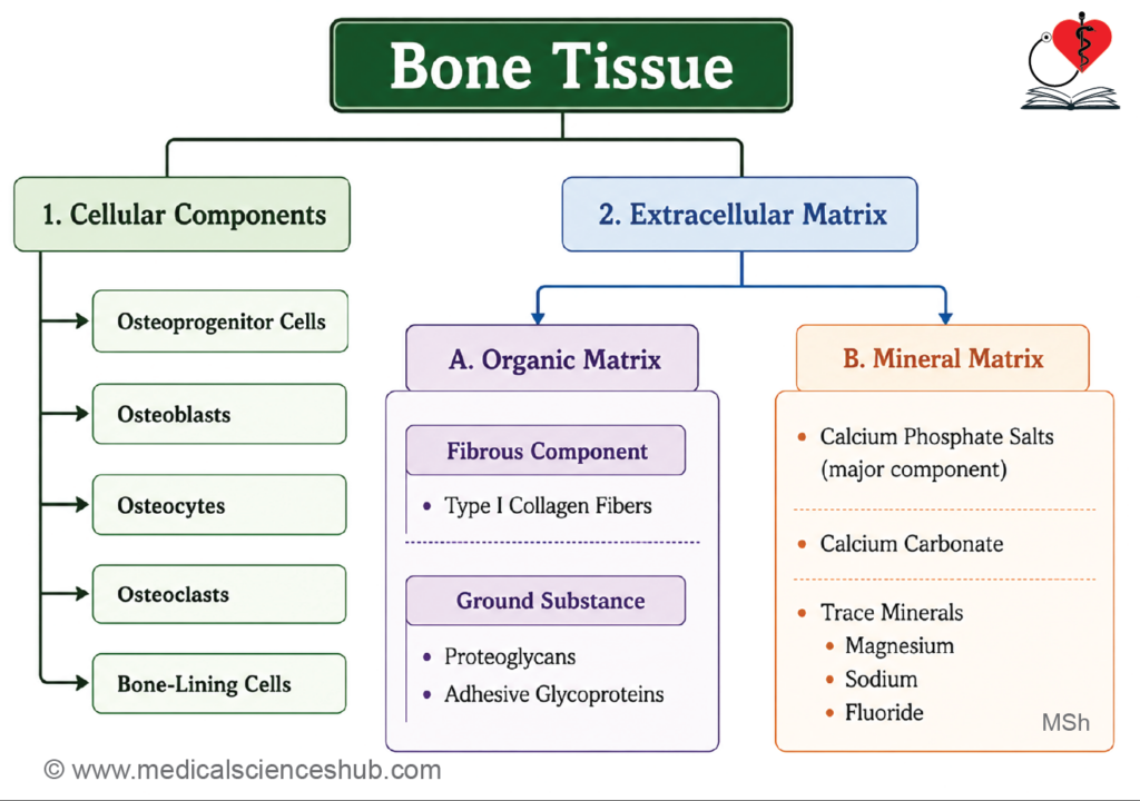
- Bone is a specialized type of connective tissue. It consists of three basic components as follows
- Cells
- Fibers
- Ground substance.
Cells of Bone Tissue
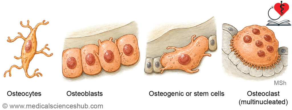
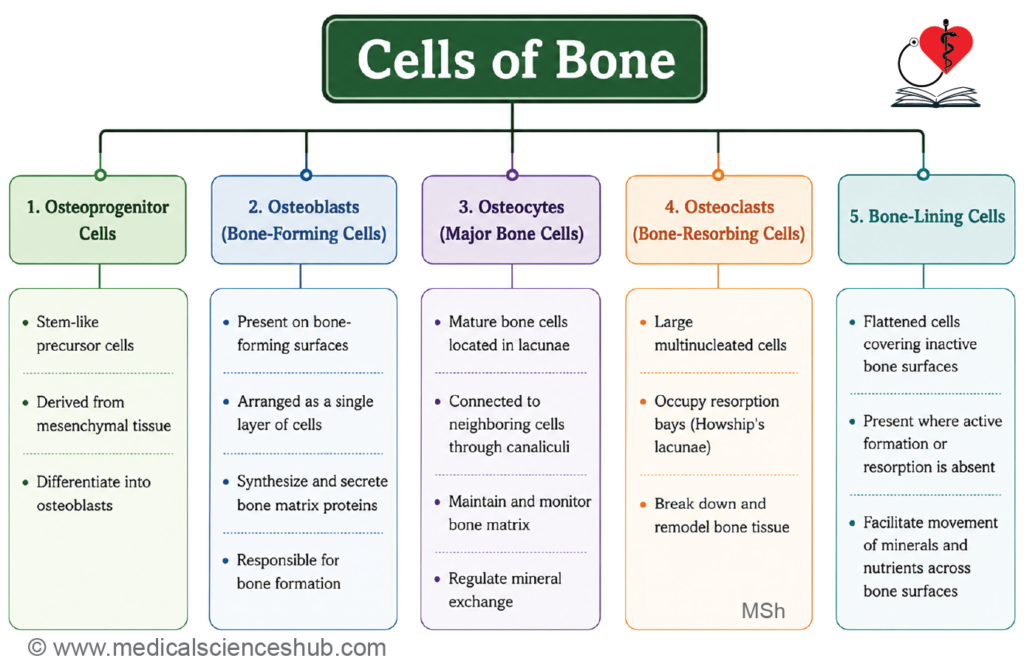
- Bone tissue consists of five types of cells: Osteoprogenitor cells, osteoblasts, osteocytes, bone lining cells, and osteoclasts.
Osteoprogenitor Cells
- Osteoprogenitor cells are mesenchymal stem cells that serve as precursors of bone-forming cells.
- They are located in the inner osteogenic layer of the periosteum, endosteum, and along the walls of Haversian and Volkmann’s canals.
- These cells differentiate into osteoblasts, which synthesize and form new bone tissue.
- Osteoprogenitor cells are abundant during fetal development and childhood, but their number gradually decreases with age.
- Bone morphogenetic proteins (BMPs) and insulin-like growth factors (IGFs) promote their differentiation into osteoblasts.
- Exposure to controlled electromagnetic fields may enhance osteogenic differentiation and support fracture healing.
- Histologically, they appear as flattened, spindle-shaped cells with elongated or oval nuclei and closely resemble fibroblasts.
Function: They give rise to osteoblasts.
Osteoblasts
- Osteoblasts are specialized bone-forming cells derived from osteoprogenitor cells.
- They are typically cuboidal to polygonal in shape and are arranged along surfaces where new bone is being deposited.
- Histologically, osteoblasts possess an eccentrically placed oval, euchromatic nucleus and a basophilic cytoplasm rich in protein-synthesizing organelles.
- Ultrastructurally, they contain abundant rough endoplasmic reticulum, Golgi apparatus, and secretory vesicles, reflecting their high synthetic activity.
- Cytoplasmic processes of adjacent osteoblasts communicate through gap junctions.
Functions of Osteoblasts
- Osteoblasts synthesize type I collagen, the principal organic component of bone matrix.
- They also produce matrix proteins such as osteocalcin, osteonectin, bone sialoprotein, and proteoglycans.
- These cells secrete alkaline phosphatase, an enzyme involved in bone mineralization.
- Elevated serum alkaline phosphatase may indicate increased bone formation, although it can also occur in conditions such as pregnancy and liver disease.
Osteocytes
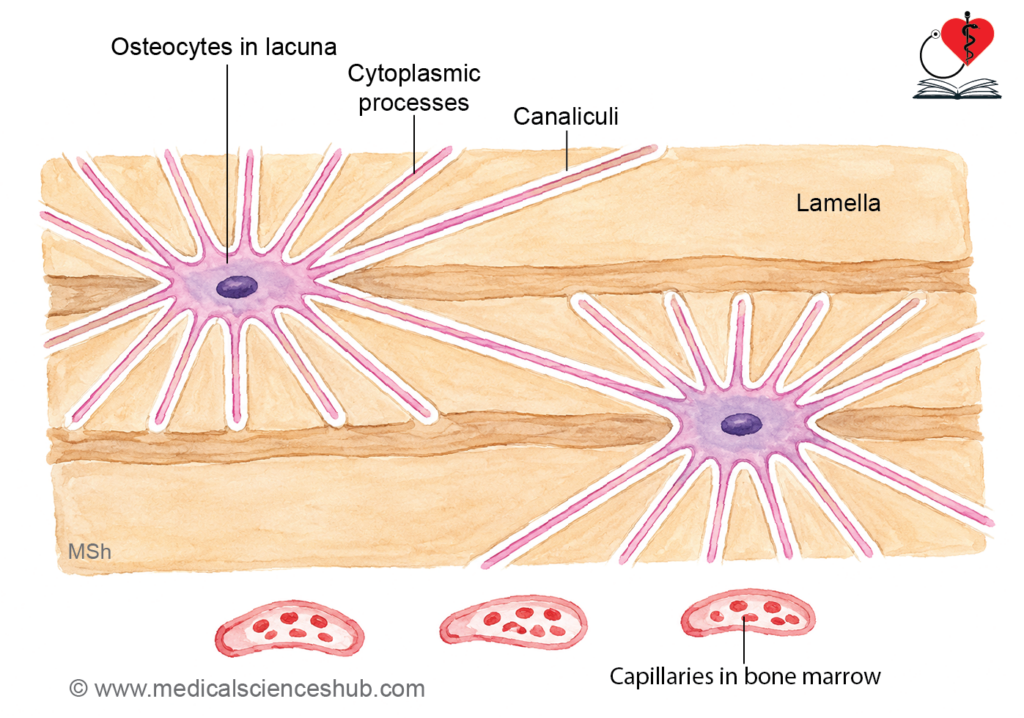
- Osteocytes are mature bone cells located within small cavities called lacunae in the mineralized bone matrix.
- They are derived from osteoblasts that become enclosed within the matrix they secrete.
- During transformation into osteocytes, the cells decrease in size and show reduced synthetic activity with fewer organelles.
- Osteocytes extend cytoplasmic processes through tiny channels called canaliculi, forming connections with neighboring osteocytes and surface bone cells.
- This interconnected network facilitates the exchange of nutrients, waste products, and signaling molecules within bone tissue.
- In routine H&E-stained sections, osteocytes exhibit lightly basophilic or eosinophilic cytoplasm due to limited rough endoplasmic reticulum.
- During histological preparation, decalcification may cause osteocytes to shrink or disappear from lacunae.
- The average lifespan of an osteocyte is approximately 10–20 years.
Functions of osteocytes
- Osteocytes maintain the structural integrity of bone and play a central role in bone remodeling and mechanosensation.
Osteoclasts
- Osteoclasts are large, multinucleated cells specialized for bone resorption.
- They are located on bone surfaces undergoing active remodeling and breakdown.
- During resorption, osteoclasts create shallow depressions known as Howship’s lacunae (resorption bays).
- Histologically, they are large cells (approximately 50–150 μm in diameter) with multiple nuclei and abundant eosinophilic cytoplasm.
- Osteoclasts secrete tartrate-resistant acid phosphatase (TRAP), an important marker of osteoclastic activity.
Functions of Osteoclasts
- Their primary function is the degradation and removal of bone matrix.
- They release lysosomal enzymes, including cathepsin K and matrix metalloproteinases, which digest the organic matrix.
- Osteoclasts also generate an acidic microenvironment that dissolves bone minerals and facilitates resorption.
Bone-lining Cells
- Bone-lining cells are flattened cells that cover bone surfaces where neither active bone formation nor bone resorption is occurring.
- They form a continuous, epithelium-like layer on the periosteal and endosteal surfaces and line internal bone channels.
- These cells are derived from inactive osteoblasts.
- Bone-lining cells help regulate the exchange of calcium, phosphate, and nutrients between bone tissue and the surrounding environment.
Bone Matrix
- Bone matrix is the extracellular material of bone and consists of organic and inorganic components.
- The organic component is composed of collagen fibers and ground substance, which together provide flexibility and tensile strength.
- Approximately 90% of the organic matrix is formed by collagen fibers, while the remaining 10% consists of ground substance containing proteoglycans and glycoproteins.
- The inorganic component is primarily made up of mineral salts, especially hydroxyapatite crystals, which impart hardness and resistance to compression.
- The combined action of organic and inorganic components gives bone its unique strength and durability.
Classification of Bones
- Bones can be classified using gross anatomical and histological characteristics.
- Based on gross (macroscopic) appearance:
- Compact bone is dense and lacks visible cavities.
- Spongy (cancellous or trabecular) bone contains numerous interconnected spaces, giving it a porous appearance.
- Based on histological structure:
- Woven (immature) bone contains irregularly arranged collagen fibers and is found during fetal development and early fracture repair.
- Lamellar (mature) bone contains organized layers (lamellae) of collagen fibers and forms the normal adult skeleton.
- Bones may also be classified according to shape into long, short, flat, irregular, and other categories in gross anatomy.
Compact Bones
- Compact bone is a dense type of bone that lacks visible cavities on gross examination.
- It forms the diaphysis of long bones, the thin outer layer of epiphyses, the outer and inner tables of flat bones, and the external layer of most other bones.
- In flat bones, the intervening spongy bone is known as the diploë.
- In living bone, the outer surface is covered by the periosteum, while the inner surface is lined by the endosteum.
- These coverings are absent in ground sections of dry bone prepared for microscopic study.
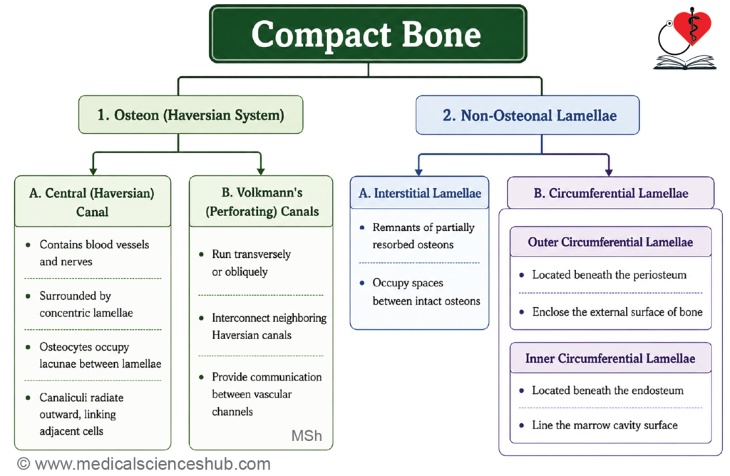
- Microscopically, compact bone is organized into three lamellar patterns:
- Haversian (osteonal) lamellae
- Interstitial lamellae
- Circumferential lamellae (outer and inner)
Haversian System of Lamellae
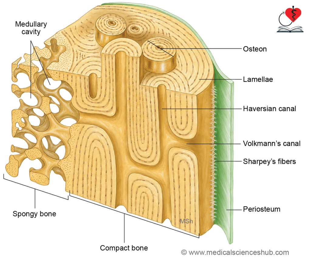
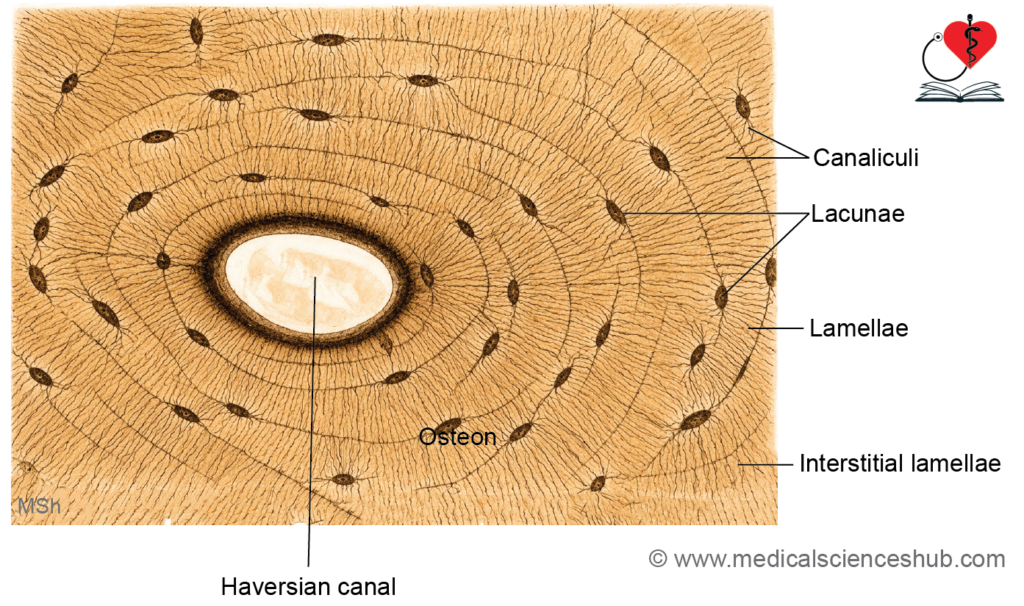
- The osteon (Haversian system) is the structural and functional unit of compact bone.
- Each osteon consists of a central Haversian canal surrounded by 4–15 concentric lamellae of mineralized bone matrix.
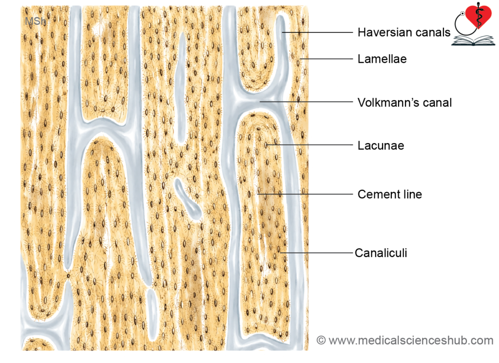
- Haversian canals run parallel to the long axis of the bone and, in living tissue, contain blood vessels, nerves, loose connective tissue, and lymphatics.
- In ground sections of dry bone, these canals may appear empty or contain debris.
- Small spaces called lacunae are located between adjacent lamellae and house osteocytes.
- Fine channels known as canaliculi radiate from lacunae and contain osteocyte processes, allowing communication and nutrient transport between cells.
- Within each lamella, collagen fibers are arranged parallel to one another, while fibers in adjacent lamellae are oriented in different directions, increasing bone strength.
- Volkmann’s (perforating) canals connect neighboring Haversian canals and transmit blood vessels and nerves from the periosteum into the bone.
- Unlike Haversian canals, Volkmann’s canals run obliquely or perpendicular to the bone axis and are not surrounded by concentric lamellae.
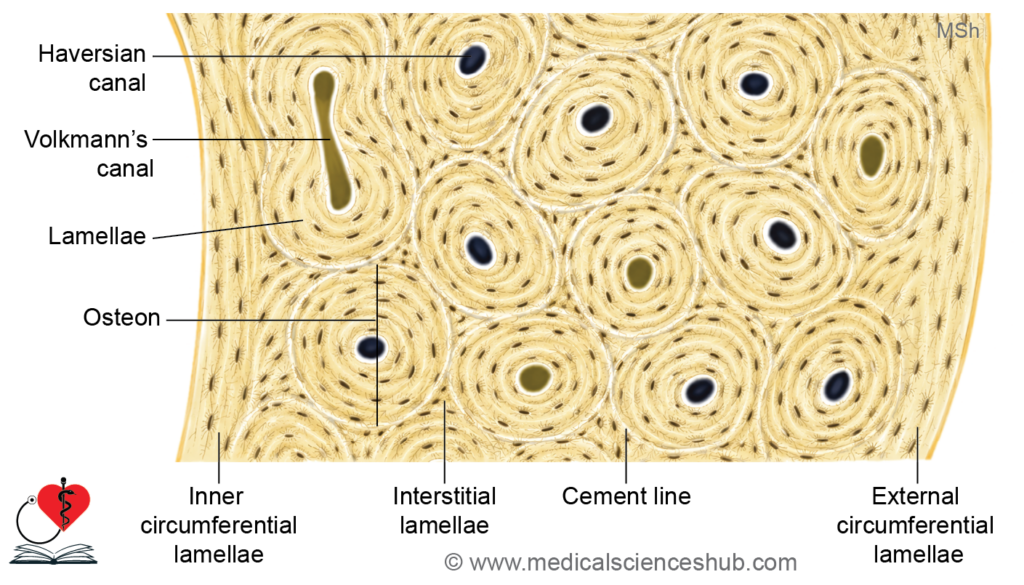
- In transverse sections, a Haversian canal appears as a circular or oval central opening encircled by concentric lamellae, whereas Volkmann’s canals appear as transverse or oblique channels connecting adjacent osteons.
- In longitudinal sections, Haversian canals run parallel to one another, while Volkmann’s canals cross between them.
- Each osteon is bounded by a distinct cement line, a highly mineralized layer that contains little or no collagen and clearly demarcates the osteon from surrounding bone.
Interstitial Lamellae
- Bone undergoes continuous remodeling, during which old osteons are resorbed and new osteons are formed.
- Newly formed osteons contain a central Haversian canal surrounded by concentric lamellae.
- Remnants of partially resorbed older osteons persist between adjacent osteons and are known as interstitial lamellae.
- Interstitial lamellae contain lacunae with osteocytes and interconnected canaliculi.
Circumferential Lamellae
- Circumferential lamellae are broad layers of bone matrix that encircle the entire circumference of compact bone.
- They are of two types:
- Outer circumferential lamellae, located immediately beneath the periosteum.
- Inner circumferential lamellae, situated adjacent to the endosteum and surrounding the medullary cavity.
Periosteum
- The periosteum is a connective tissue membrane covering the external surface of bone, except at articular surfaces.
- It consists of:
- An outer fibrous layer rich in collagen fibers.
- An inner osteogenic layer containing osteoprogenitor cells, especially prominent in growing bone.
- The periosteum is highly vascular, and its blood vessels communicate with osteonal vessels through Volkmann’s canals.
Endosteum
- The endosteum is a thin cellular lining covering the internal surfaces of bone, including the medullary cavity, trabecular spaces of spongy bone, and Haversian canals.
- It is typically a single-cell-thick layer composed of osteoblasts, osteoclasts, bone-lining cells, and osteoprogenitor cells, and plays an important role in bone growth, repair, and remodeling.
Spongy Bone
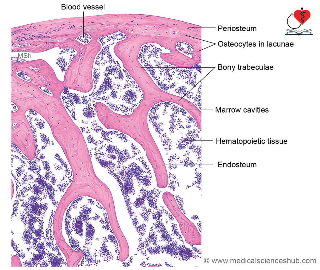
- Spongy bone, also known as cancellous or trabecular bone, forms the interior of epiphyses of long bones and the core of short, flat, and irregular bones.
- It is covered externally by a thin layer of compact bone.
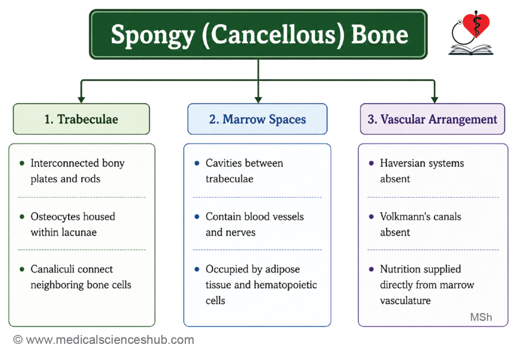
- Grossly, it has a porous appearance due to numerous spaces between interconnected bony plates or rods called trabeculae.
- Unlike compact bone, trabeculae do not contain Haversian systems (osteons).
- Osteocytes are located within lacunae in the trabeculae and communicate through fine canaliculi containing their cellular processes.
- Because trabeculae are thin, nutrients can diffuse directly from adjacent marrow spaces; therefore, a Haversian system is unnecessary.
- Trabecular surfaces are lined by endosteum and are associated with osteoblasts, osteoclasts, and osteoprogenitor cells, which participate in bone remodeling and repair.
Formation and Growth of Bone
- Most bones develop from mesenchymal tissue derived from mesoderm, although much of the facial skeleton receives contributions from neural crest cells.
- The process of bone formation is called ossification (osteogenesis).
- Ossification occurs by two principal mechanisms:
- Intramembranous ossification involves the direct differentiation of mesenchymal cells into bone-forming cells without a cartilage precursor.
- Bones formed by this process are called membranous bones.
- Examples include the bones of the cranial vault, much of the mandible, and a major part of the clavicle.
- Endochondral ossification involves the formation of a hyaline cartilage model that is subsequently replaced by bone.
- Bones formed through this mechanism are termed cartilaginous bones.
- Examples include most long bones (except much of the clavicle), the base of the skull, vertebrae, and ribs.
- Both processes contribute to the development, growth, and remodeling of the skeletal system.
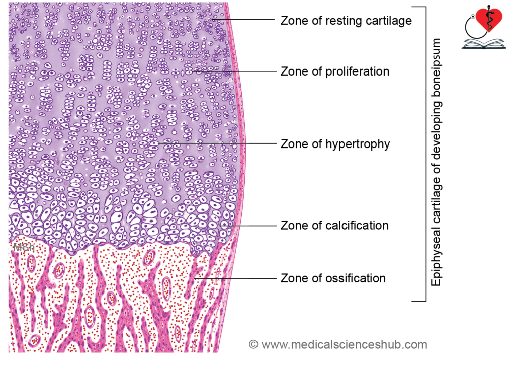
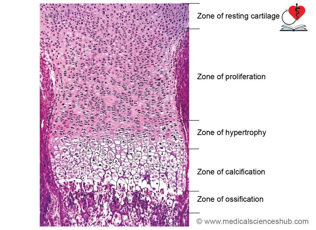
Zones of epiphyseal cartilage
- The epiphyseal (growth) plate is responsible for longitudinal growth of long bones. It appears during fetal development and remains active until skeletal maturity.
- During endochondral ossification, vascular endothelial growth factor (VEGF) promotes vascular invasion and replacement of cartilage by bone.
- The growth plate is divided into distinct zones:
- Zone of Reserve Cartilage
- Contains relatively inactive chondrocytes scattered singly within lacunae.
- Serves as a reservoir of cartilage cells.
- Zone of Proliferation
- Chondrocytes undergo rapid mitotic division.
- Cells become more numerous and are arranged in longitudinal columns parallel to the long axis of the bone.
- Zone of Hypertrophy
- Chondrocytes enlarge considerably and develop clear cytoplasm.
- The columnar arrangement becomes more prominent.
- Zone of Calcified Cartilage
- The cartilage matrix becomes calcified.
- Chondrocytes undergo apoptosis, leaving a calcified cartilage framework that temporarily supports new bone formation.
- Zone of Ossification (Resorption)
- Blood vessels, osteoprogenitor cells, and osteoblasts invade the calcified cartilage.
- Osteoblasts deposit bone matrix on remnants of calcified cartilage, forming mixed spicules.
- These spicules fuse and remodel to form bony trabeculae, contributing to bone elongation and growth.
- Zone of Reserve Cartilage
CLINICAL CORRELATION
- Osteogenesis imperfecta (brittle bone disease) is a genetic disorder caused primarily by defects in type I collagen synthesis. It results in fragile bones, recurrent fractures, blue sclerae, hearing impairment, joint laxity, and short stature.
- Scurvy, caused by vitamin C deficiency, impairs collagen formation and osteoid production. Spongy bone shows reduced trabeculae, while compact bone develops a thinner cortex.
- Rickets occurs in children due to vitamin D deficiency or impaired vitamin D metabolism, leading to defective mineralization of growing bones and deformities such as bowing of the legs.
- Osteomalacia is the adult counterpart of rickets and is characterized by inadequate mineralization of bone matrix, resulting in soft and weak bones.
- Osteoporosis involves decreased bone mass and deterioration of bone microarchitecture. It is common in older adults, especially postmenopausal women, due to increased bone resorption.
- Osteoma is a benign bone-forming tumor, whereas osteosarcoma is a malignant tumor of osteoblastic origin with the potential to metastasize.
Important Questions
- List the cells of bone and their functional correlation.
- List the cells of bones and their functions.
- Write a short note on transverse section of compact bone/ground bone.
- Write a short note on longitudinal section of compact bone or ground bone.
- Write a short note on zones of epiphyseal cartilage.
- List the zones of epiphyseal cartilage.
📝 Test Your Knowledge – Practice MCQs
Attempt the chapter MCQ quiz and assess your understanding of key concepts.
Click to see: Medical Disclaimer and Copyright Notice