
Competency
- AN62.4: Enumerate parts and major connections of basal ganglia
Introduction
- The basal nuclei (also called basal ganglia) are large clusters of grey matter embedded within the white matter of the cerebral hemispheres. They play a central role in regulating voluntary movement, maintaining posture, and supporting motor learning by modulating the output of the motor cortex.
- Damage to these structures produces characteristic movement disorders. Parkinson’s disease results from the loss of dopaminergic neurons and presents with resting tremor, rigidity, and slowed movement (bradykinesia). Chorea is marked by brief, irregular, involuntary movements, while dystonia involves sustained or repetitive muscle contractions that cause abnormal postures.
Components
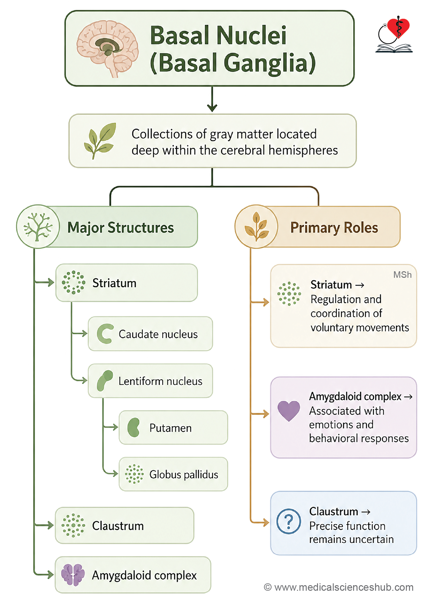
- The basal nuclei consist of three main components: the corpus striatum, the claustrum, and the amygdaloid body.
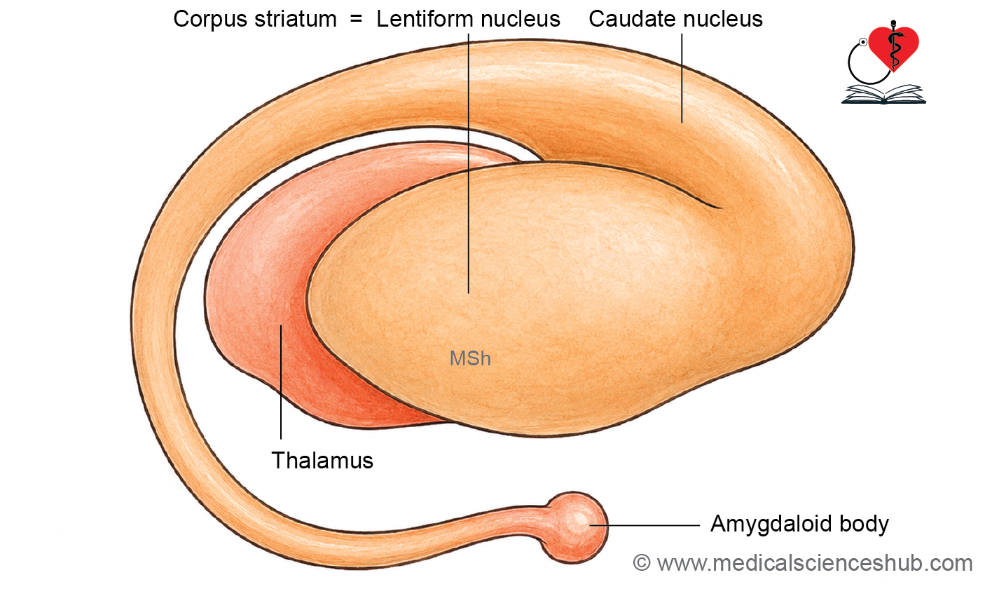
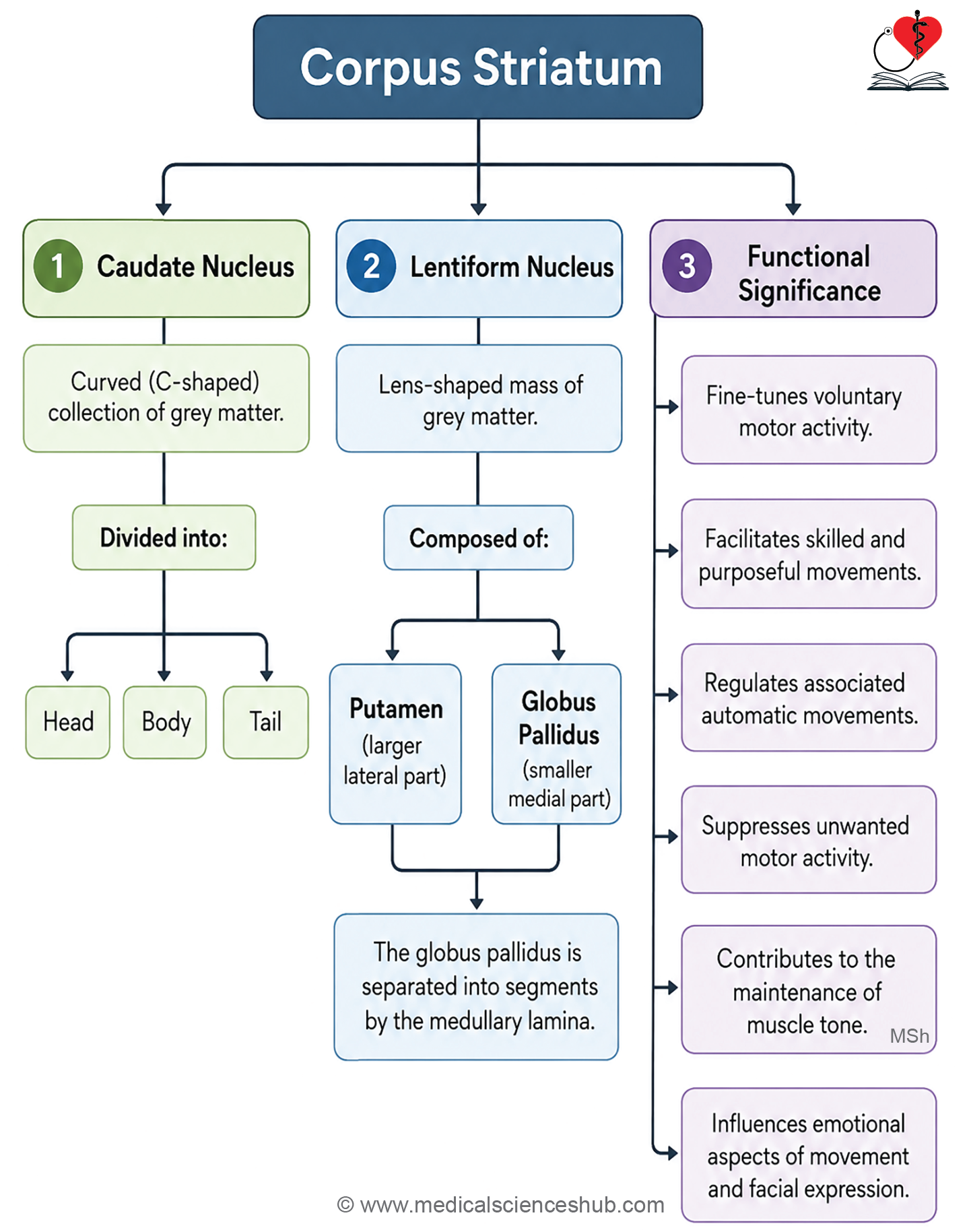
- The corpus striatum is further divided into the caudate nucleus and the lentiform nucleus; the lentiform nucleus itself comprises the putamen and the globus pallidus.
Functions
- The corpus striatum is a key component of the extrapyramidal system and is responsible for regulating muscle tone, maintaining posture, and coordinating the quality and smoothness of voluntary movements.
- The amygdaloid body is part of the limbic system and is involved in the emotional and motivational aspects that influence movement initiation and behavior.
- The function of the claustrum remains poorly understood and has not been definitively established.
Corpus Striatum
Location and divisions
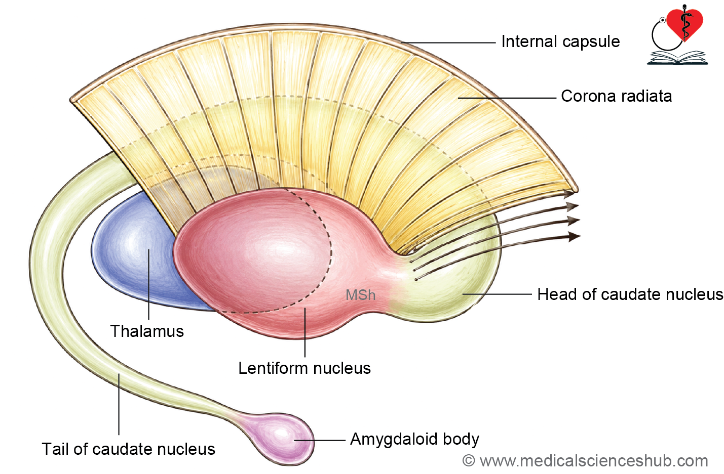
- The corpus striatum is located anterior and lateral to the thalamus.
- The fibers of the internal capsule divide it into two structures: the caudate nucleus and the lentiform nucleus.
- The lentiform nucleus is further subdivided into the putamen and the globus pallidus.
Caudate nucleus
- The caudate nucleus is the smaller, anterior component of the corpus striatum and lies medial to the internal capsule.
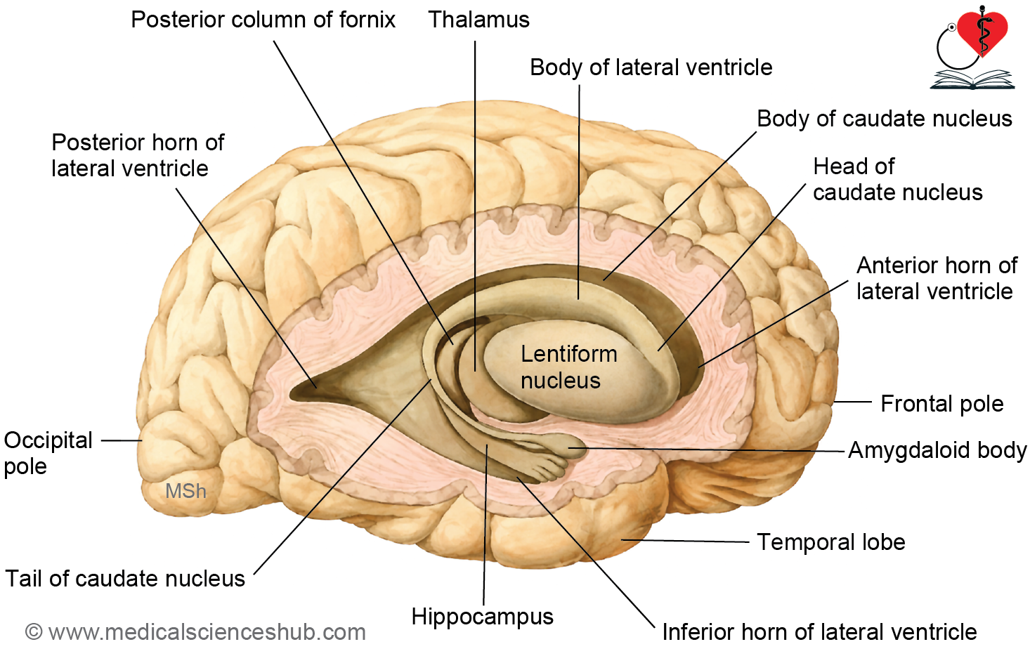
- It is a C-shaped mass of grey matter consisting of three continuous parts: a head, a body, and a tail.
- The head is the large, rounded anterior portion that forms the floor of the anterior horn of the lateral ventricle and is closely related to the lentiform nucleus.
- The body is a narrow, elongated segment that contributes to the floor of the body of the lateral ventricle, positioned between the thalamus and the ventricle.
- The tail is slender, curves downward and forward into the temporal lobe, forms the roof of the inferior horn of the lateral ventricle, and continues anteriorly to merge with the amygdaloid body.
Lentiform nucleus
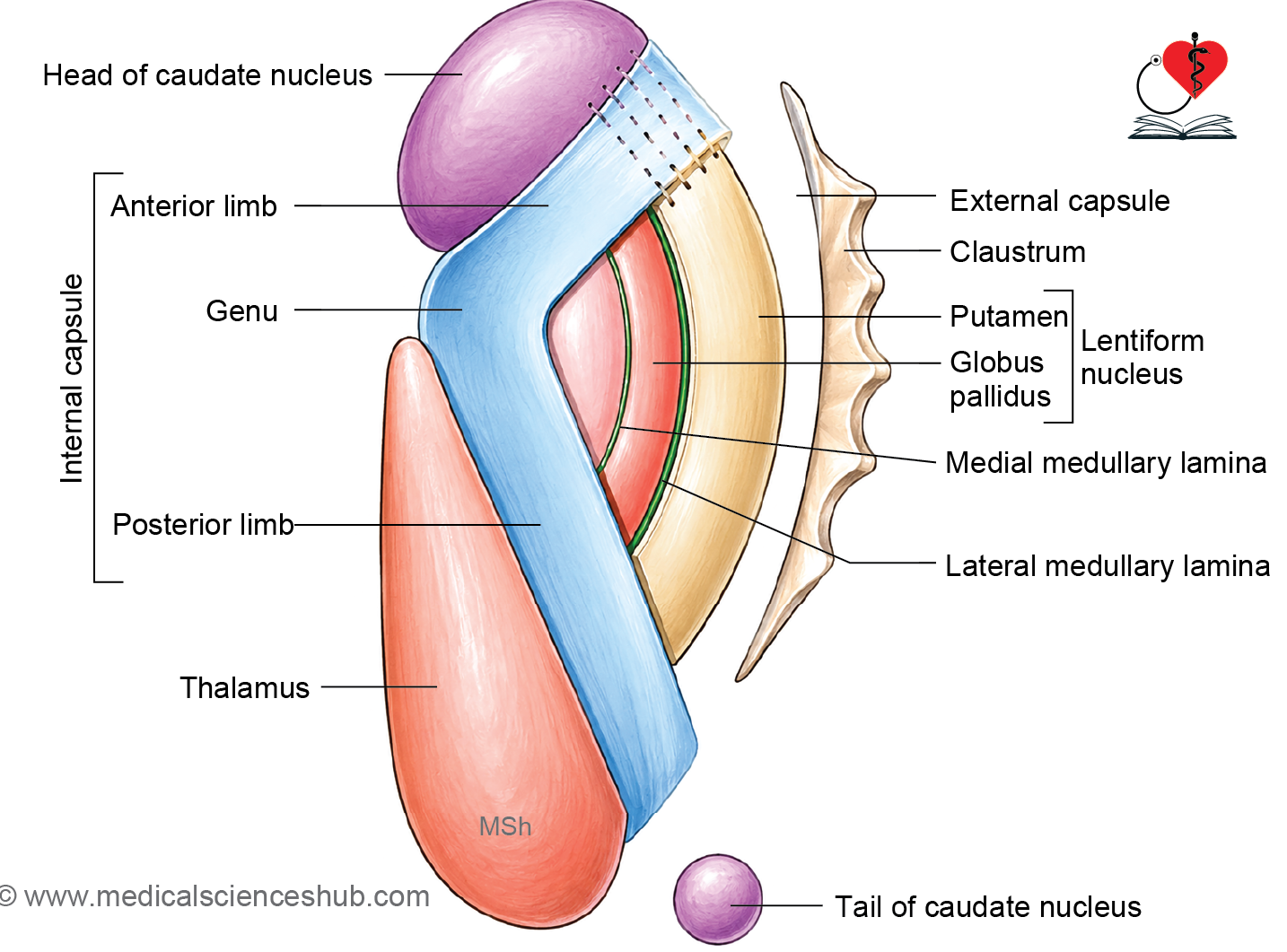
- The lentiform nucleus is a biconvex lens-shaped mass of grey matter located deep to the insular cortex and lateral to the internal capsule.
- Medially, it is separated from both the caudate nucleus and the thalamus by the internal capsule.
- A vertical sheet of white matter called the lateral medullary lamina divides the lentiform nucleus into two parts: the putamen and the globus pallidus.
- Putamen:
- It is the larger and more lateral of the two parts and appears darker due to its higher density of grey matter.
- Functionally, it works together with the caudate nucleus to form the striatum, which plays a central role in initiating and regulating voluntary movement.
- Globus pallidus:
- It is the smaller and more medial part; its pale appearance is due to the large number of myelinated fibers it contains (the term pallidus means pale).
- It is further divided by the medial medullary lamina into an inner (medial) segment and an outer (lateral) segment, both of which are important in suppressing unwanted movements.
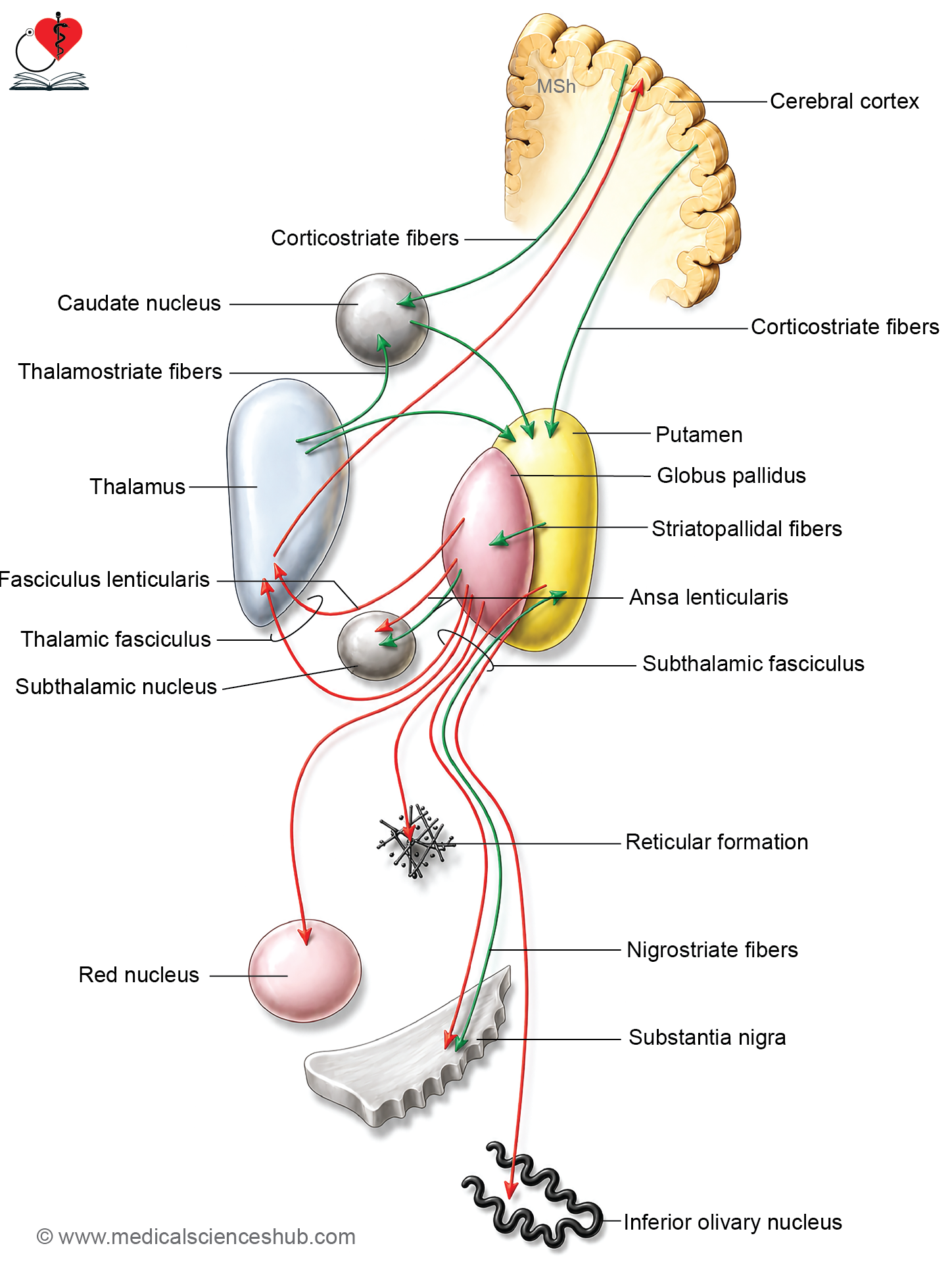
Table 13.1: Connection of corpus striatum
| Component | Afferent Connections (Inputs) | Efferent Connections (Outputs) |
|---|---|---|
| Neostriatum (caudate nucleus and putamen) | • Corticostriate fibres arising from the cerebral cortex • Thalamostriate fibres from the thalamus • Nigrostriate fibres originating in the substantia nigra | • Striatopallidal fibres projecting to the globus pallidus • Additional projections to the thalamus and substantia nigra |
| Paleostriatum (globus pallidus) | • Receives the majority of fibres from the neostriatum (caudate nucleus and putamen) • Smaller inputs from the substantia nigra, thalamus, and subthalamic region | • Pallidothalamic fibres to the thalamus • Connections to the hypothalamus, subthalamus, red nucleus, inferior olivary nucleus, and reticular formation through: 1. Ansa lenticularis 2. Fasciculus lenticularis 3. Subthalamic fasciculus |
Functions of Corpus Striatum
The corpus striatum is a key component of the extrapyramidal system and contributes to motor control in the following ways:
- It refines voluntary and reflex movements by ensuring that actions are smooth, accurate, and well-coordinated rather than jerky or imprecise.
- It supports fine motor control, enabling delicate and skilled tasks such as writing or threading a needle, through precise coordination of small muscle groups.
- It regulates automatic associated movements — the background motor activities that accompany voluntary actions, such as arm swinging during walking.
- It suppresses unwanted muscle activity by inhibiting unnecessary contractions, allowing purposeful movement to occur without interference.
- It helps maintain muscle tone and postural stability, providing a steady baseline of muscle activity during both rest and active movement.
- Through its connections with the limbic system, it contributes to the motor expression of emotions, including facial expressions, gestures, and changes in body posture during emotional states.
Claustrum
- The claustrum is a thin, saucer-shaped sheet of grey matter located deep to the insular cortex and is considered a detached extension of the insula.
- Medially, it is separated from the putamen by the external capsule; laterally, it is separated from the insular cortex by the extreme capsule.
- Its connections and functions have not yet been clearly established.
Amygdaloid Body
- The amygdaloid body is a small, almond-shaped mass of grey matter located in the temporal lobe, deep to the uncus, and positioned anterosuperior to the tip of the inferior horn of the lateral ventricle.
- Functionally, it is considered part of the limbic system.
Connections
- Afferent (incoming) fibers arrive primarily from the olfactory tract, linking the amygdaloid body to the sense of smell.
- Efferent (outgoing) fibers travel via a discrete bundle called the stria terminalis, which carries signals to the anterior commissure, the anterior perforated substance, and the hypothalamic nuclei.
Function
- As a component of the limbic system, the amygdaloid body plays a central role in processing and regulating emotional responses, including fear, aggression, and motivation.
Efferent Pathways of the Basal Nuclei
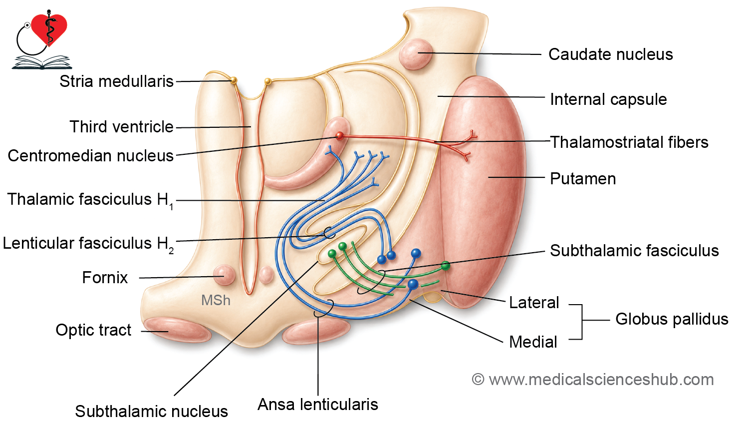
- The lenticular fasciculus carries efferent fibers from the globus pallidus, passes through the posterior limb of the internal capsule, travels superior to the subthalamic nucleus, and terminates in the thalamus.
- The ansa lenticularis is formed by efferent fibers from the globus pallidus that curve around the inferior aspect of the posterior limb of the internal capsule, forming a loop. These fibers terminate in the thalamus, substantia nigra, red nucleus, reticular nuclei, and inferior olivary nucleus.
- The thalamic fasciculus is formed by the combination of the lenticular fasciculus and the thalamic fibers of the ansa lenticularis; together they relay basal nuclei output to the thalamus.
- The subthalamic fasciculus consists of fibers that reciprocally connect the globus pallidus with the subthalamic nucleus, forming an important regulatory loop within the basal nuclei circuit.
CLINICAL NEUROANATOMY
Chorea
- Chorea is characterized by sudden, brief, irregular involuntary movements that shift unpredictably across muscle groups, commonly affecting the face, limbs, and trunk, and disrupting activities such as speaking and walking.
- There are two main types:
- Sydenham’s chorea occurs as a complication of rheumatic fever, most commonly in children and adolescents. It arises from autoimmune inflammation targeting the basal nuclei and is typically reversible with appropriate treatment.
- Huntington’s chorea is an autosomal dominant neurodegenerative disorder caused by progressive loss of neurons in the basal nuclei and cerebral cortex. It presents with worsening involuntary movements, personality changes, and cognitive decline, and currently has no cure, though symptoms can be managed.
- Athetosis: Athetosis is characterized by slow, continuous, writhing involuntary movements, most prominently affecting the fingers, hands, toes, and feet.
- Ballismus: It results from a lesion of the subthalamic nucleus and presents as violent, flinging involuntary movements of the trunk and proximal limbs. When the lesion is unilateral, it affects the contralateral side of the body and is termed hemiballismus.
- Wilson’s disease: It is an inherited disorder of copper metabolism that typically manifests in young adults. It leads to degeneration of the lenticular nucleus and liver cirrhosis, producing muscular rigidity, impaired voluntary movement, and episodes of uncontrolled laughing or crying. A characteristic feature is wing-beating tremor (asterixis), seen when the upper limbs are held in extension.
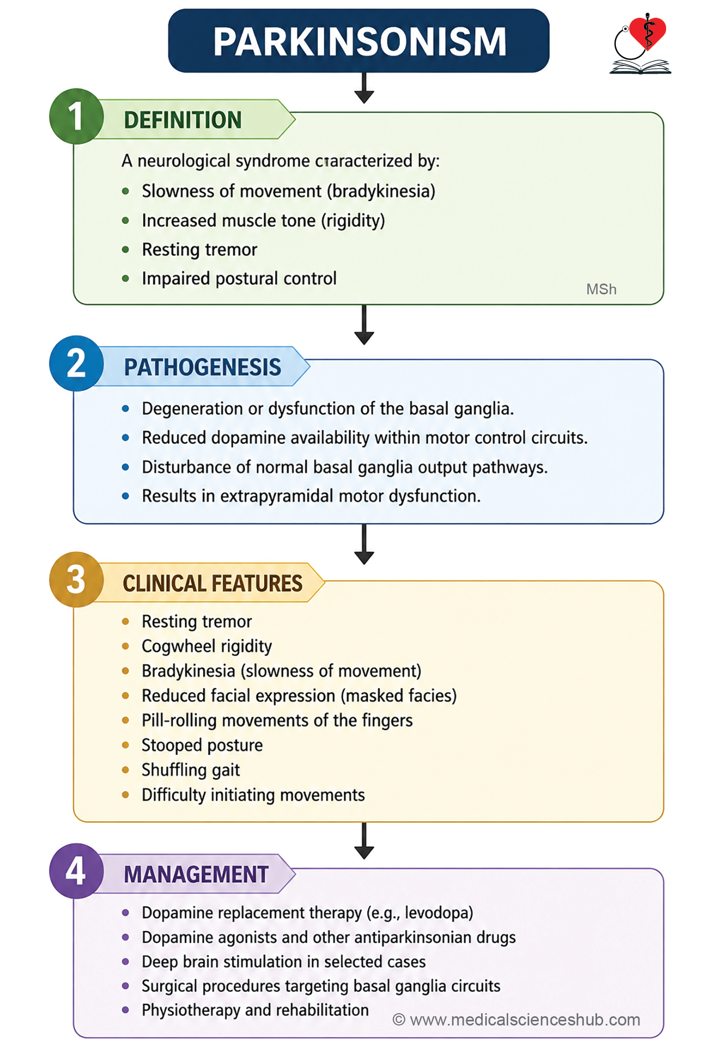
- Parkinsonism
- Parkinsonism is a clinical syndrome defined by the combination of bradykinesia, muscular rigidity, and tremor. It can result from Parkinson’s disease, drug side effects, infections, or vascular disorders affecting the brain.
- Anatomical Basis of Parkinson’s Disease:
- Parkinson’s disease (paralysis agitans) is a neurodegenerative disorder caused by deficiency of dopamine in the corpus striatum, affecting approximately 1% of individuals over the age of 50. Under normal conditions, melanin-containing neurons of the substantia nigra produce dopamine, which is transported to the corpus striatum via nigrostriatal fibers, where it inhibits neuronal activity and allows smooth motor control. Degeneration of the substantia nigra or its fibers leads to dopamine deficiency in the corpus striatum, loss of inhibition of striatal neurons, and the emergence of extrapyramidal motor symptoms.
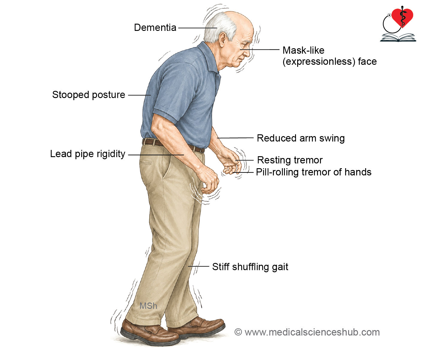
- Signs and Symptoms
- Resting tremor — rhythmic, involuntary shaking of the hands or limbs that is present at rest, diminishes during voluntary movement, and disappears during sleep.
- Muscular rigidity — increased resistance to passive movement, described as cogwheel rigidity (alternating resistance and relaxation) or lead pipe rigidity (uniform resistance throughout).
- Mask-like facies — reduced facial expression, diminished blinking, and an overall expressionless appearance due to facial muscle rigidity.
- Bradykinesia — slowness in initiating and executing movements, resulting in a characteristic shuffling gait with difficulty starting and stopping.
- Pill-rolling tremor — a repetitive circular movement of the thumb against the fingers, resembling the manual rolling of a small pill.
- Stooped posture — a flexed trunk, slightly bent knees, and adducted arms reflecting impaired postural reflexes and generalized rigidity.
- Loss of associated movements — automatic movements such as arm swinging during walking are reduced or absent, giving the gait a stiff, mechanical quality.
- Treatment
- Levodopa therapy — levodopa is a dopamine precursor that crosses the blood–brain barrier and is converted to dopamine within the brain, reducing tremor, rigidity, and bradykinesia. It is combined with carbidopa to prevent peripheral breakdown and reduce side effects.
Important Questions
- Describe the components of the basal ganglia and their anatomical relations.
- Explain the connections of the basal ganglia.
- Explain the role of the basal ganglia in the control of voluntary movements. Mention the clinical manifestations of basal ganglia lesions.
- Write a short note on Parkinsonism.
📝 Test Your Knowledge – Practice MCQs
Attempt the chapter MCQ quiz and assess your understanding of key concepts.
Click to see: Medical Disclaimer and Copyright Notice