
Competencies
- AN63.2: Describe anatomical basis of congenital hydrocephalus.
- AN64.2: Describe the development of neural tube, spinal cord, medulla oblongata, pons, midbrain, cerebral hemisphere, and cerebellum.
- AN64.3: Describe various types of open neural tube defects with its embryological basis.
- AN79.3: Describe the process of neurulation.
- AN79.5: Explain embryological basis of neural tube defects.
- AN79.6: Describe the diagnosis of pregnancy in first trimester and role of teratogens, alpha-fetoproteins. (This chapter includes alpha-fetoproteins).
Introduction
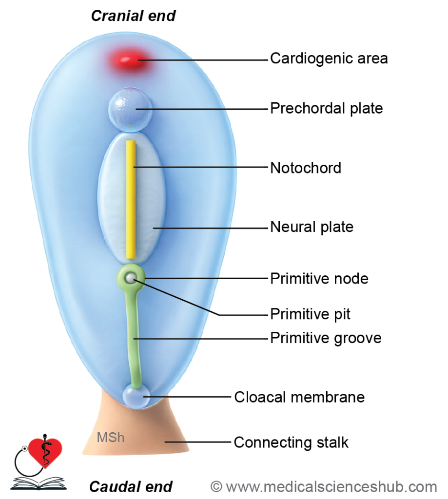
- The nervous system comprises the brain, spinal cord, peripheral nerves, and ganglia. It develops primarily from the neuroectoderm, a specialized region of surface ectoderm located between the primitive node and the prechordal plate.
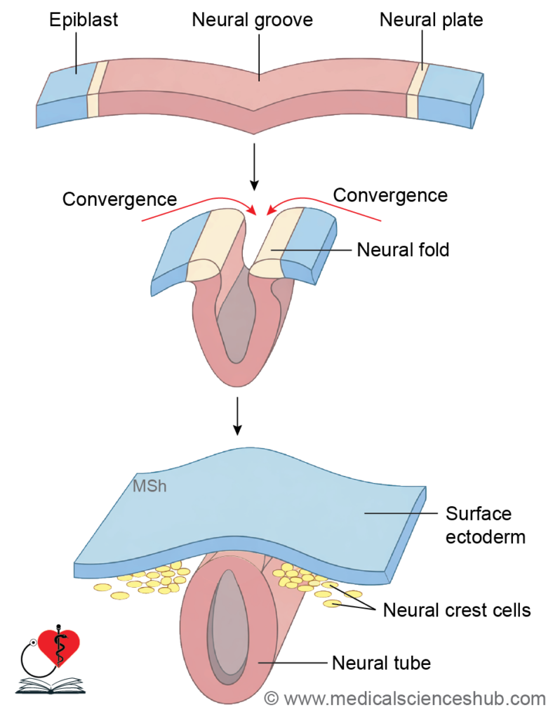
- The neuroectoderm forms the neural plate, which undergoes folding to produce the neural tube, the precursor of the central nervous system (CNS).
Neurulation
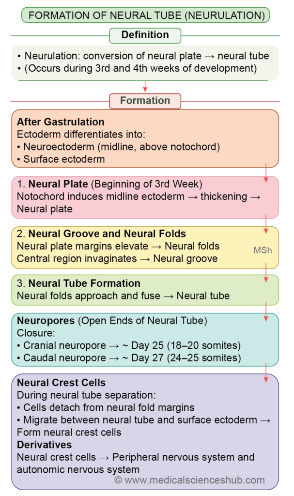
Definition: Neurulation is the process by which the neural tube forms from the neural plate.
Stages of Neurulation
1. Neural plate stage
During the presomitic period (days 16–19), the surface ectoderm thickens in the midline to form the neural plate. The underlying notochord acts as the primary inducer for this differentiation. The neural plate elongates in a craniocaudal direction.
2. Neural folding stage
Ongoing growth causes the neural plate to invaginate centrally, forming the neural groove with raised neural folds on either side. At the junction of neural plate and surface ectoderm, cells differentiate into the neural crest, which later gives rise to major components of the peripheral nervous system (PNS), including autonomic structures.
3. Neural tube formation
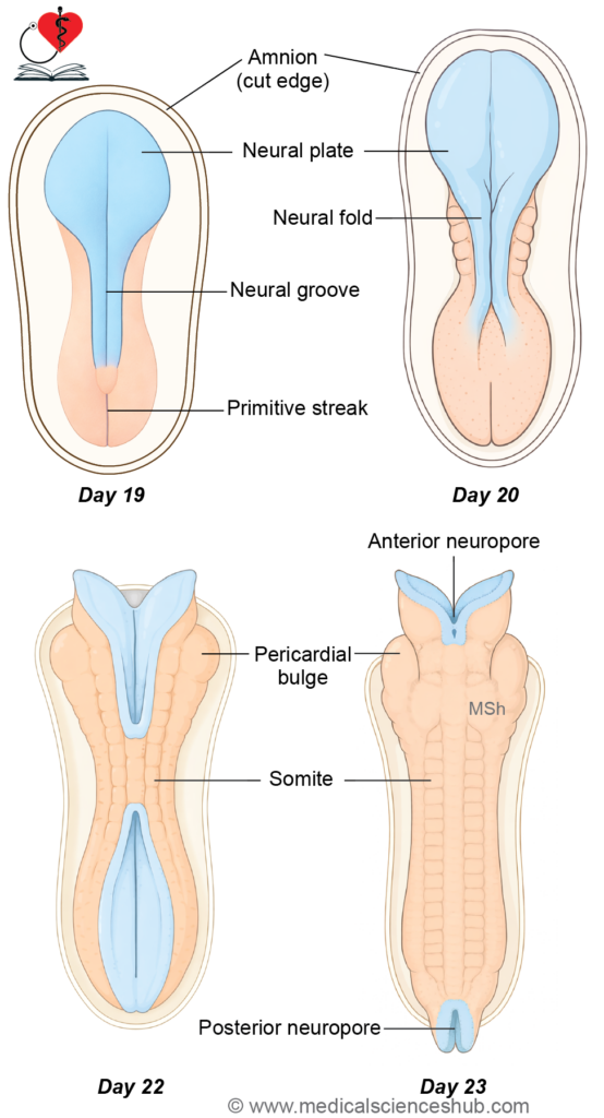
The neural folds gradually approach and fuse in the midline to form the neural tube. Fusion begins in the cervical region and proceeds both cranially and caudally. The neural tube subsequently develops into the CNS.
4. Neural tube closure
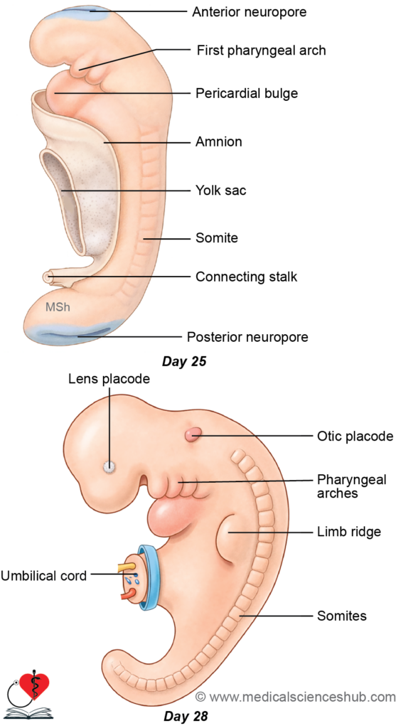
Initially, the neural tube remains open at both ends as the cranial (anterior) neuropore and caudal (posterior) neuropore. These openings allow temporary exchange of amniotic fluid, supporting early neuroectodermal development.
- The cranial neuropore closes around day 25 (~20 somites).
- The caudal neuropore closes around day 27 (~29 somites).
Failure of closure results in neural tube defects (NTDs). In later development, the cranial neuropore corresponds to the lamina terminalis, while the caudal neuropore is related to the terminal ventricle at the distal spinal cord.
Differentiation Of Neural Tube
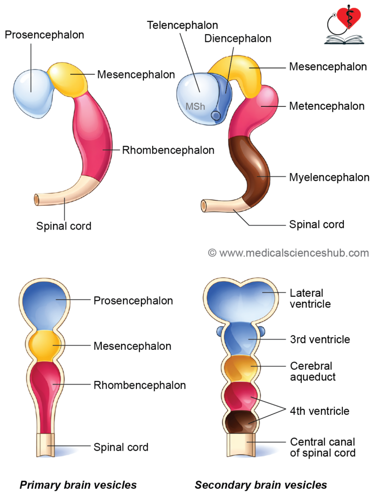
Formation of Primary Brain Vesicles
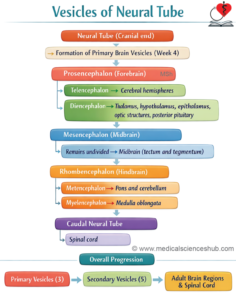
The expanded cranial end of the neural tube gives rise to three primary brain vesicles:
- Prosencephalon (forebrain) – the most cranial part
- Mesencephalon (midbrain) – the middle segment
- Rhombencephalon (hindbrain) – the caudal part
Formation of Secondary Brain Vesicles
With further development, these three vesicles differentiate into five secondary brain vesicles:
1. Prosencephalon divides into:
- Telencephalon – forms the cerebral hemispheres
- Diencephalon – forms the optic vesicles, thalamus, hypothalamus, pineal gland, and posterior pituitary (neurohypophysis)
2. Mesencephalon:
- Remains undivided and develops into the midbrain
3. Rhombencephalon divides into:
- Metencephalon – forms the pons and cerebellum
- Myelencephalon – forms the medulla oblongata
This orderly transformation of the neural tube establishes the structural basis of the central nervous system, with distinct regions specialized for higher functions, coordination, and vital autonomic control.
Flexures of Neural Tube
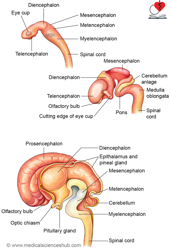
Unequal growth of different regions of the neural tube produces characteristic bends, along with localized thickenings and expansions. These structural changes are essential for proper brain organization.
Major Flexures of Neural Tube
The combined effects of embryonic folding and differential growth lead to four principal flexures:
- Cervical flexure
Located at the junction of the hindbrain and spinal cord, it forms an approximate 90° angle. - Cephalic (mesencephalic) flexure
Occurs in the midbrain region and is ventrally concave. It is a prominent early bend in brain development. - Pontine flexure
Found between the metencephalon and myelencephalon, this flexure is ventrally convex and contributes to the expansion of the fourth ventricle. - Telencephalic flexure
Develops between the telencephalon and diencephalon, reflecting rapid growth of the cerebral hemispheres.
Cavity of Neural Tube
- Initially, the cavity of the neural tube allows circulation of amniotic fluid, which supports early growth of the brain and spinal cord before full vascular development.
- As brain vesicles form, this cavity differentiates into the ventricular system:
- The telencephalon forms the lateral ventricles.
- The connection between telencephalon and diencephalon becomes the interventricular foramina (of Monro).
- The diencephalon forms the third ventricle.
- The mesencephalon forms the cerebral aqueduct (of Sylvius), linking the third and fourth ventricles.
- The rhombencephalon forms the fourth ventricle.
- The cavity of the spinal cord becomes the central canal, with a small terminal dilation known as the terminal ventricle in the conus medullaris.
- The fourth ventricle communicates with the subarachnoid space through one median aperture (foramen of Magendie) and two lateral apertures (foramina of Luschka), enabling cerebrospinal fluid circulation.
CLINICAL EMBRYOLOGY
Hydrocephalus
Hydrocephalus refers to an abnormal accumulation of cerebrospinal fluid (CSF) within the ventricular system of the brain.
Causes
- Obstruction of CSF flow within the ventricular pathways
- Excess production of CSF (rare; e.g., choroid plexus abnormalities)
- Impaired communication between the ventricles and the subarachnoid space
Pathology
Progressive accumulation of CSF leads to dilatation of the ventricles. In infants, this causes separation of cranial sutures, resulting in an enlarged head. Prolonged pressure may lead to compression and degeneration of neural tissue.
- Abnormal dilatation of the central canal of the spinal cord is termed syringomyelia, which is a distinct condition but may be associated with CSF flow abnormalities.
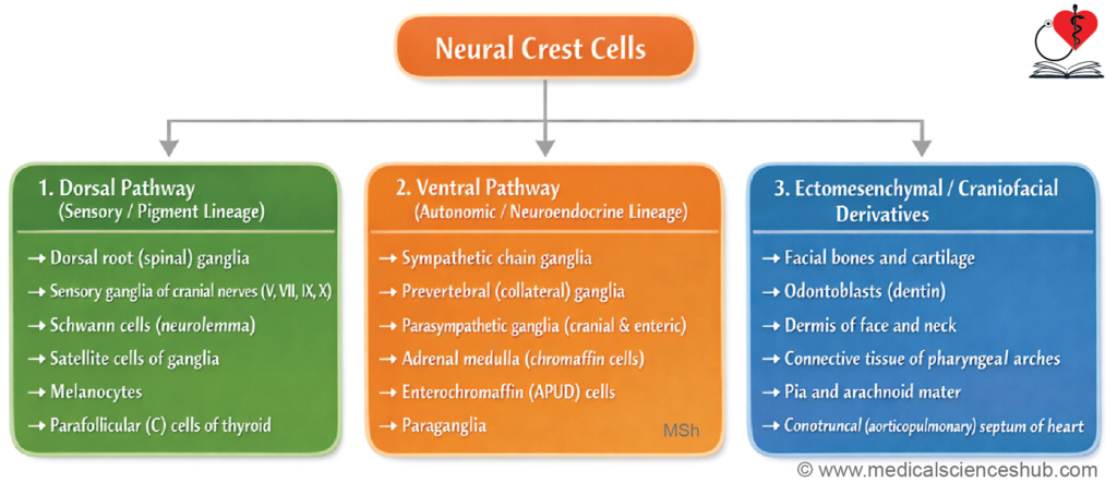
Neural Crest Cells
- During invagination of the neural plate, a specialized population of ectodermal cells appears at the margins of the neural groove. These cells are known as neural crest cells.
- After formation of the neural tube, neural crest cells occupy a transient position between the neural tube and the surface ectoderm.
- These cells then undergo extensive migration and differentiation. They are broadly organized into dorsal and ventral cell populations, which migrate along distinct pathways and give rise to a wide range of derivatives, particularly components of the peripheral nervous system (PNS) and other tissues.
Spinal Cord
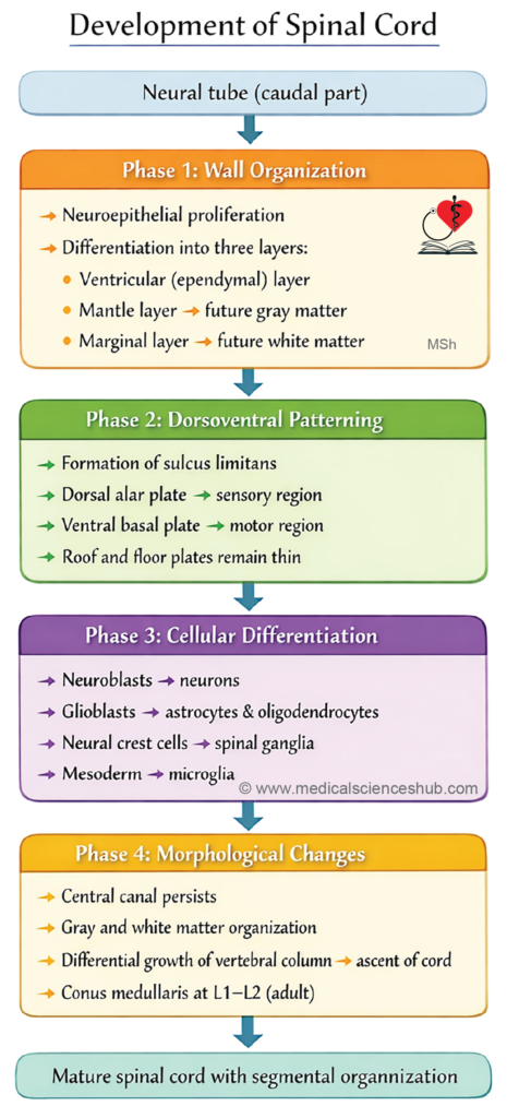
The portion of the neural tube caudal to the rhombencephalon develops into the spinal cord. Its formation occurs through a series of coordinated phases:
- Formation of mantle and marginal layers
- Formation of basal and alar plates
- Histogenesis of neural elements
- Positional changes of the spinal cord
Phases of Development
Phase 1: Formation of mantle and marginal layers
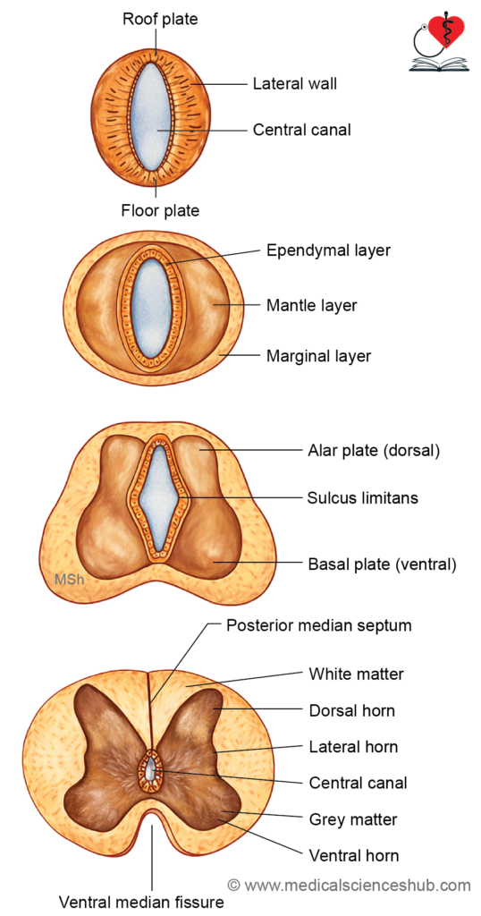
The early neural tube is lined by neuroepithelial cells, which proliferate and differentiate into three distinct layers:
- Ependymal layer (inner) – forms the lining of the central canal
- Mantle layer (middle) – gives rise to neurons and most neuroglial cells (future gray matter)
- Marginal layer (outer) – contains nerve fibers (axons) and develops into white matter, supported by neuroglia
Phase 2: Formation of basal and alar laminae
- Rapid proliferation within the mantle layer leads to regional specialization:
- The ventral region forms the basal plate, associated with motor (efferent) functions
- The dorsal region forms the alar plate, associated with sensory (afferent) functions
- The roof plate (dorsal midline) and floor plate (ventral midline) remain relatively thin.
- A longitudinal groove, the sulcus limitans, appears within the central canal and separates the alar and basal plates, establishing functional organization.
- Continued growth causes:
- Formation of the dorsal median septum due to expansion of the alar plate
- Development of the ventral median fissure due to expansion of the basal plate
- The remaining lumen persists as the central canal of the spinal cord.
- These early structural changes establish the fundamental sensory and motor organization of the spinal cord.
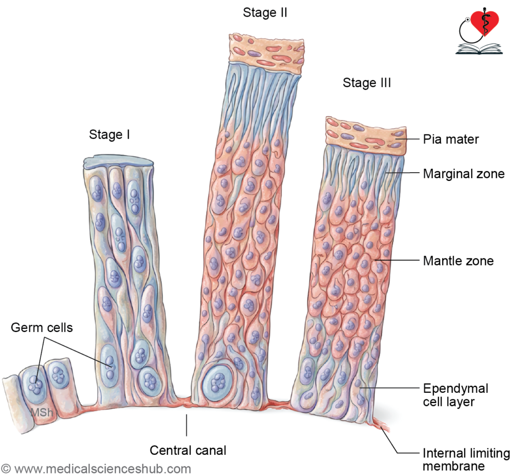
Phase 3: Histogenesis of cells in neural tube (Figs 22.5, 22.6)
- Cells of the mantle and marginal layers proliferate and differentiate into:
- Neuroblasts → form neurons
- Spongioblasts (glioblasts) → form supporting neuroglial cells
Development of Neurons
- Neuroblasts initially become apolar, then bipolar, and finally unipolar cells.
- The single process elongates to form the axon, while additional processes develop into dendrites, producing multipolar neurons (mature form).
- Neuroblasts lose their ability to divide after differentiation.
- Motor neurons differentiate earlier than sensory neurons.
Formation of Gray and White Matter
- The mantle layer forms gray matter, while the marginal layer forms white matter.
- From the basal plate:
- Neurons form the anterior (ventral) gray horn
- Their axons emerge as motor roots of spinal nerves
- From the alar plate:
- Neurons form the posterior (dorsal) gray horn
- Their axons contribute to ascending tracts
- Descending fibers from the brain also occupy the marginal layer.
- Some mantle cells differentiate into interneurons.
- Neural crest cells give rise to pseudounipolar neurons of the dorsal root ganglia.
- Central processes enter the spinal cord as sensory roots
- Peripheral processes form sensory fibers
- Sensory and motor roots unite to form a spinal nerve
- Cells at the junction of alar and basal plates form the lateral horn.
Development of Glial Cells
- Spongioblasts differentiate into:
- Astrocytes (protoplasmic and fibrous)
- Oligodendrocytes
- Microglia originate from mesenchyme and act as phagocytes.
The lining of the neural tube forms ependymal cells and contributes to the choroid plexus epithelium
Phase 4: Positional changes in spinal cord
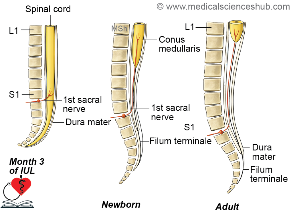
- During early development, the spinal cord occupies the entire length of the vertebral canal up to the third month of intrauterine life (IUL). At this stage, spinal nerves pass horizontally to exit through their corresponding intervertebral foramina.
- With growth, the vertebral column elongates more rapidly than the spinal cord. As a result, the terminal end of the spinal cord appears to ascend to a higher vertebral level.
- At birth, the spinal cord ends at the level of the third lumbar vertebra (L3). In adults, it typically terminates near the lower border of L1 or upper border of L2 vertebra.
Result of spinal cord recession
- Spinal nerves no longer originate at the same level as their exit foramina.
- They descend obliquely within the vertebral canal to reach their respective exit points.
- The lower extension of pia mater forms the filum terminale, which anchors the cord to the coccyx.
- The dura mater extends inferiorly up to the level of the second sacral vertebra (S2).
- The cauda equina consists of elongated lumbar, sacral, and coccygeal nerve roots formed due to differential growth.
- The pia mater is derived from neural crest cells, whereas the arachnoid mater and dura mater develop from surrounding mesenchyme.
Myelination
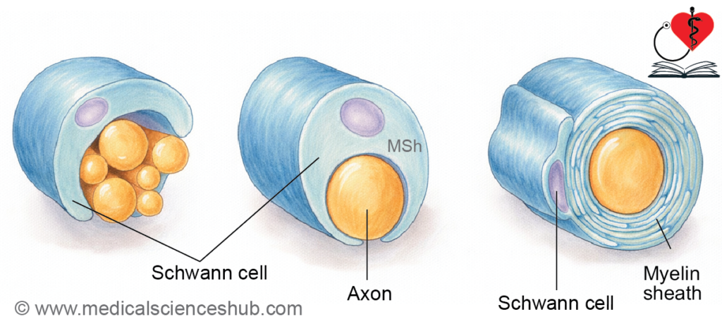
- Myelination starts with the onset of functional activity in nerve fibers. It begins around the fourth month of intrauterine life and continues after birth until approximately 2–3 years of age.
- Motor fibers undergo myelination earlier than sensory fibers. In the corticospinal tract, myelination commences near the ninth month of intrauterine life and extends up to about 2 years postnatally.
Functional Columns of Spinal Cord
Neurons derived from the basal plate (ventral) and alar plate (dorsal) are organized into longitudinal functional columns based on their roles.
Columns in basal lamina
- General somatic efferent (GSE) column
Supplies skeletal muscles, controlling voluntary movements. - General visceral efferent (GVE) column
Present mainly in the thoracolumbar and sacral regions as the lateral horn. It gives rise to preganglionic autonomic fibers (sympathetic and parasympathetic).
Columns in alar lamina
- General visceral afferent (GVA) column
Found predominantly in the thoracolumbar and sacral segments. It receives sensory input from viscera. - General somatic afferent (GSA) column
Receives somatic sensory information, including exteroceptive (e.g., touch, pain) and proprioceptive inputs.
CLINICAL EMBRYOLOGY
Neural Tube Defects
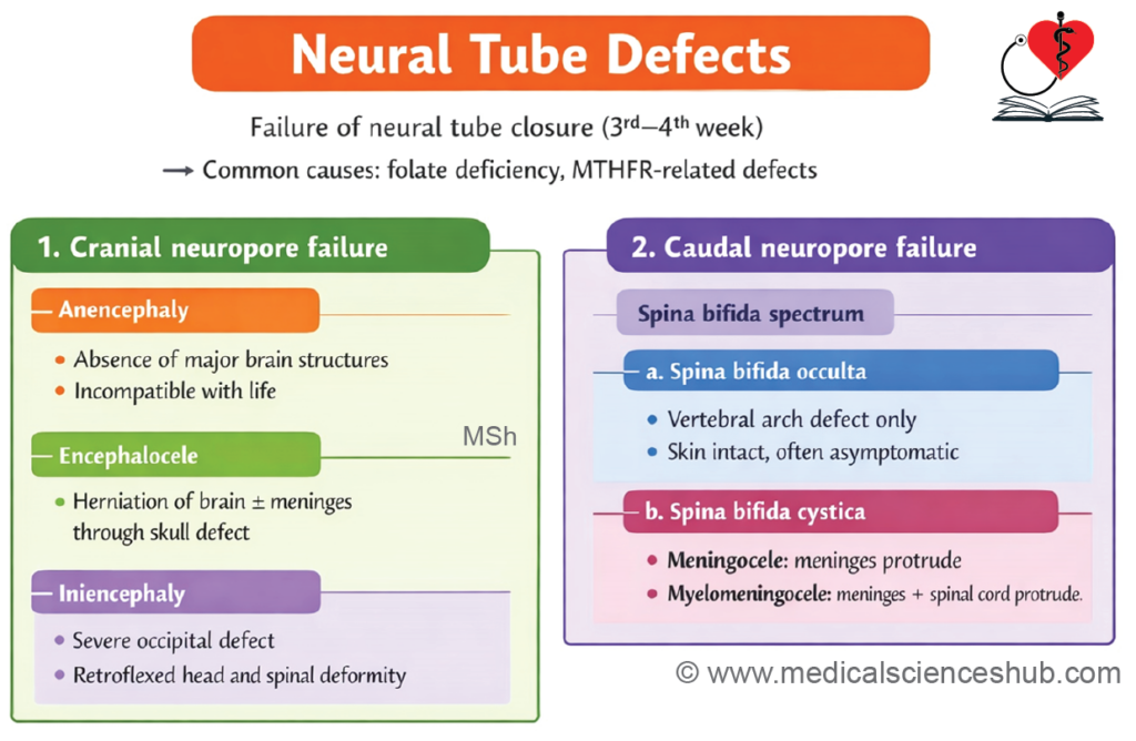
- Definition: Neural tube defects (NTDs) are congenital anomalies resulting from failure of neural tube closure.
- Closure begins in the third week of intrauterine life (IUL). The cranial neuropore closes by day 25, and the caudal neuropore by day 27.
- Etiology: The most common cause is folic acid (vitamin B9) deficiency, which impairs DNA synthesis, cell division, and myelination. Mutations in the MTHFR gene may also contribute. Maternal folic acid supplementation significantly reduces risk.
Spina Bifida
- Definition: A common NTD caused by incomplete closure of the neural tube during the first 4 weeks of development.
- Pathogenesis: Folate deficiency or MTHFR defect → defective neural tube closure → incomplete vertebral formation → bifid vertebrae.
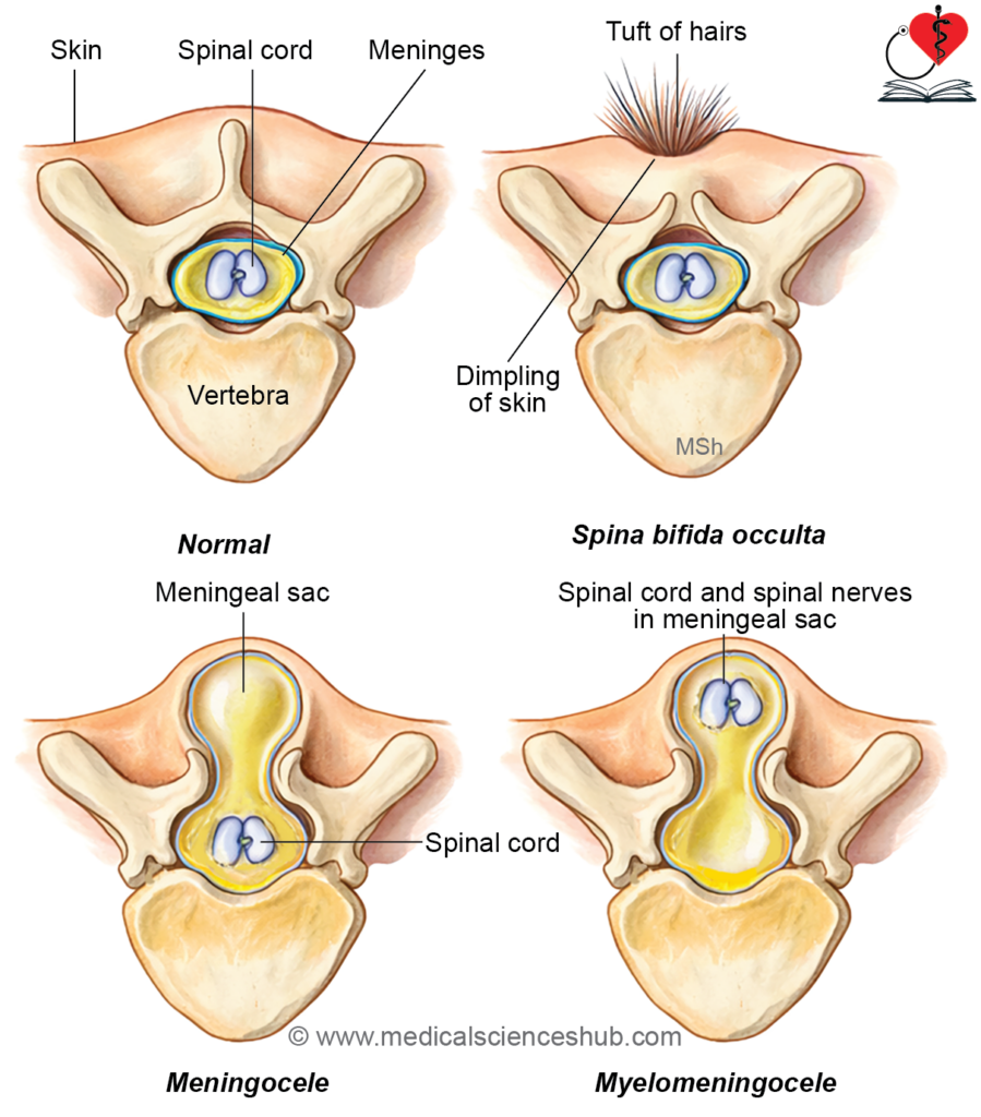
Types
1. Spina bifida occulta
- A mild form where the defect is not externally visible.
- Common in the lumbosacral region and may present as a skin dimple or tuft of hair.
- The spinal cord and meninges remain normal.
2. Spina bifida cystica
- Characterized by a visible cystic swelling:
- Meningocele: Herniation of meninges only through the defect
- Myelomeningocele: Herniation of spinal cord and meninges, often associated with neurological deficits
Prevention
Daily intake of 0.4 mg folic acid, starting before conception and continuing through early pregnancy, markedly lowers the risk of NTDs.
Screening
- Ultrasonography
- Elevated maternal serum α-fetoprotein (AFP)
Treatment
Surgical correction is typically performed after birth, depending on severity.
Other Neural Tube Defects
- Anterior spina bifida: Failure of fusion of vertebral body halves, allowing ventral meningeal protrusion.
- Anencephaly: Caused by failure of anterior neuropore closure, leading to severe absence of brain tissue.
- Encephalocele: Herniation of brain tissue and meninges through a skull defect.
- Iniencephaly: Characterized by defective occipital bone, cervical spina bifida, and marked retroflexion of the head.
- Rachischisis (myeloschisis): Severe defect due to failure of neural fold fusion, resulting in exposed neural tissue.
Functional Columns Of Brainstem
- The brainstem—comprising the medulla oblongata, pons, and midbrain—contains organized groups of neurons that form the cranial nerve nuclei.
- The sulcus limitans divides the brainstem into:
- Basal plate (ventral) → efferent (motor) columns
- Alar plate (dorsal) → afferent (sensory) columns
- These columns represent functionally distinct neuronal groups associated with cranial nerves.
Division of Functional Columns
Based on function, the columns are arranged as follows:
- General somatic efferent (GSE)
Supplies skeletal muscles of the head not derived from pharyngeal arches (e.g., extraocular muscles). - Special visceral efferent (SVE) / branchial efferent
Innervates muscles derived from pharyngeal arches, including facial expression, mastication, and muscles such as sternocleidomastoid and trapezius. - General visceral efferent (GVE)
Provides preganglionic parasympathetic fibers to glands and smooth muscles. - General visceral afferent (GVA)
Conveys visceral sensory information, mainly via the vagus nerve. - Special visceral afferent (SVA)
Responsible for taste sensation. - General somatic afferent (GSA)
Carries general sensory input (touch, pain, proprioception) from the face and mucosa of oral, nasal, and pharyngeal regions, primarily via the trigeminal nerve. - Special somatic afferent (SSA)
Transmits hearing and balance information (auditory and vestibular functions).- This functional organization reflects the embryological division of the brainstem into motor (basal) and sensory (alar) regions, ensuring precise integration of cranial nerve functions.
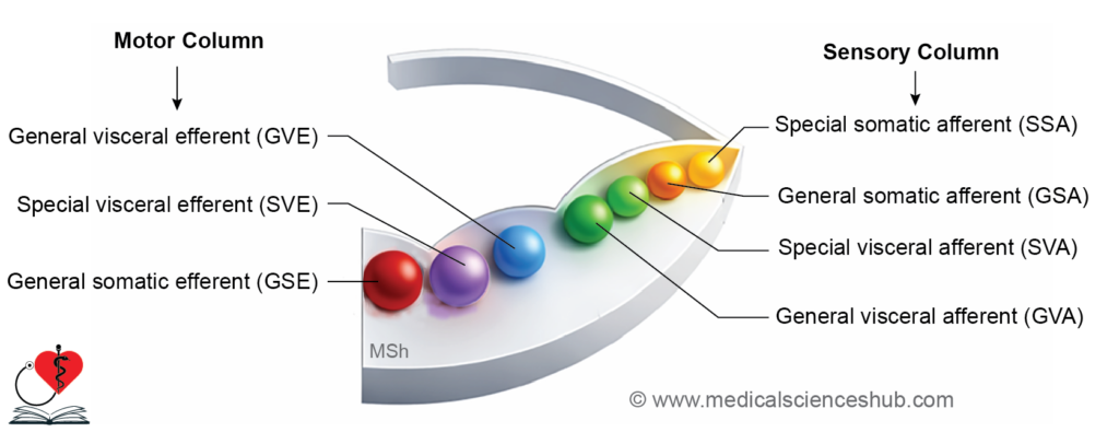
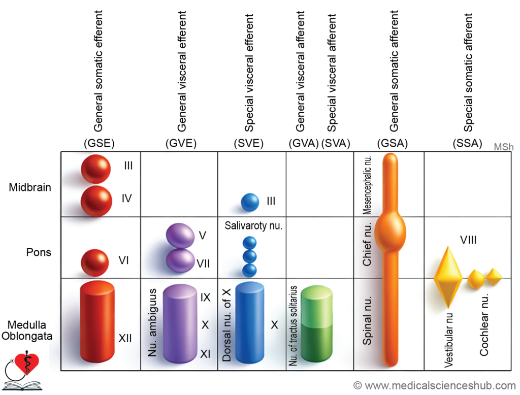
Table 22.1: Functional columns of brainstem: Efferent columns
| Component | Function | Nucleus (Location) | Cranial Nerve | Key Note |
|---|---|---|---|---|
| GSE (General somatic efferent) | Motor to non–pharyngeal arch striated muscles | Oculomotor (midbrain) | 3rd | All extraocular muscles except lateral rectus & superior oblique |
| Trochlear (midbrain) | 4th | Supplies superior oblique | ||
| Abducens (pons) | 6th | Supplies lateral rectus | ||
| Hypoglossal (medulla) | 12th | Supplies tongue muscles except palatoglossus | ||
| SVE (branchial efferent) | Motor to pharyngeal arch–derived muscles (incl. SCM, trapezius) | Trigeminal motor (pons) | 5th | 1st arch muscles (via mandibular nerve) |
| Facial motor (pons) | 7th | 2nd arch muscles | ||
| Nucleus ambiguus (medulla) | 9th | 3rd arch—stylopharyngeus | ||
| Nucleus ambiguus (medulla) | 10th | 4th arch (via superior laryngeal) | ||
| Nucleus ambiguus (medulla) | 11th | 6th arch (via recurrent laryngeal of vagus) | ||
| GVE (General visceral efferent) | Preganglionic parasympathetic fibers | Edinger–Westphal (midbrain) | 3rd | To sphincter pupillae & ciliary muscle |
| Superior salivatory & lacrimal (pons) | 7th | To lacrimal, submandibular, sublingual glands | ||
| Inferior salivatory (medulla) | 9th | To parotid gland | ||
| Dorsal motor nucleus (medulla) | 10th | To thoracoabdominal viscera |
Table 22.2: Functional columns of brainstem: Afferent columns
| Component | Function | Nucleus (Location) | Cranial Nerve(s) |
|---|---|---|---|
| GVA (General visceral afferent) | Visceral sensory input | Dorsal nucleus of vagus (medulla) | 10th |
| SVA(nutech printer) | Taste sensation | Nucleus tractus solitarius (pons–medulla) | 7th, 9th, 10th |
| GSA(General somatic afferent) | Proprioception & touch from face | Mesencephalic nucleus (midbrain) | 5th |
| Pain, temperature & crude touch (face, mucosa) | Spinal trigeminal nucleus (pons–medulla) | 5th | |
| SSA(Special somatic afferent) | Hearing and balance (inner ear) | Cochlear & vestibular nuclei (pons–medulla) | 8th |
Development Of Medulla Oblongata
- The rhombencephalon (hindbrain) differentiates into the myelencephalon, which forms the medulla oblongata, and the metencephalon, which gives rise to the pons and cerebellum. The junction between the midbrain and hindbrain is defined by the rhombencephalic isthmus. With further development of brain vesicles, the myelencephalon undergoes structural changes, and its cavity expands to form the inferior part of the fourth ventricle.
Developmental Stages
- The sulcus limitans separates the alar (sensory) plate from the basal (motor) plate. Expansion of the roof plate in the cranial region causes widening of the ventricular cavity. As a result, the alar plate shifts dorsolaterally, and both alar and basal plates contribute to the floor of the fourth ventricle.
Differentiation of Basal Lamina
The basal plate of the myelencephalon differentiates into organized motor columns:
- General somatic efferent (GSE): Forms the hypoglossal nucleus (CN XII), supplying intrinsic and extrinsic muscles of the tongue.
- Special visceral efferent (SVE): Develops into the nucleus ambiguus, innervating muscles derived from the pharyngeal arches via the glossopharyngeal (CN IX) and vagus (CN X) nerves.
- General visceral efferent (GVE): Gives rise to the dorsal motor nucleus of vagus and inferior salivatory nucleus, which regulate autonomic functions, including parotid gland secretion.
Differentiation of Alar Lamina
- Cells from each alar plate migrate ventrally into the marginal layer, forming the bulbopontine eminence, which later differentiates into the olivary nuclear complex. As white matter increases, these nuclei shift from their original positions to their definitive anatomical locations.
- The remaining alar plate cells organize into sensory columns:
- General visceral afferent (GVA): Associated with visceral sensory input, including components linked to the vagus nerve.
- Special visceral afferent (SVA): Forms the nucleus tractus solitarius, which receives taste fibers via cranial nerves IX and X.
- General somatic afferent (GSA): Gives rise to the spinal trigeminal nucleus and the gracile and cuneate nuclei, which process somatic sensory information.
- Special somatic afferent (SSA): Develops into the vestibular and cochlear nuclei for balance and hearing (cranial nerve VIII).
- The white matter of the medulla is primarily derived from ascending and descending fiber tracts.
Formation of Tela Choroidea and Choroid Plexus
- Expansion of the roof plate of the myelencephalon produces a thin ependymal layer forming the roof of the fourth ventricle. This layer is closely associated with vascular pia mater, together forming the tela choroidea.
- Invagination of vascular pia into the ependyma results in the formation of the choroid plexus, which is responsible for cerebrospinal fluid (CSF) production.
- Subsequently, openings develop in this region, forming the foramina of Luschka, which allow communication between the fourth ventricle and the subarachnoid space.
Development Of Pons
- The rhombencephalon (hindbrain) divides into the metencephalon (cranial) and myelencephalon (caudal). The metencephalon further differentiates into a ventromedial region, forming the pons, and a dorsolateral region (rhombic lip), which develops into the cerebellum. The roof plate becomes thin and expanded, contributing to the roof of the fourth ventricle, while the alar plate shifts dorsolaterally relative to the basal plate.
External Features
- The pontine flexure separates the pons from the medulla at the pontomedullary junction. The ventral surface shows a bulging basilar part with a basilar sulcus. Pontocerebellar fibers form the middle cerebellar peduncles. On the dorsal aspect, the facial colliculus appears in the floor of the fourth ventricle.
Formation of Pontine Nuclei
- Cells from the alar plate of the myelencephalon migrate ventrally to form the bulbopontine eminence. Its caudal part contributes to the olivary nuclei, while cranial migration gives rise to the pontine nuclei in the ventral pons. Axons from these nuclei form the middle cerebellar peduncles, connecting the pons to the cerebellum.
Differentiation of Basal Lamina
The basal plate of the pons forms motor columns:
- General somatic efferent (GSE): Motor nucleus of abducens nerve (CN VI) for the lateral rectus muscle.
- Special visceral efferent (SVE): Motor nucleus of trigeminal nerve (CN V) and facial nucleus (CN VII) supplying muscles of the first and second pharyngeal arches.
- General visceral efferent (GVE): Superior salivatory and lacrimal nuclei, controlling secretion of salivary and lacrimal glands via facial nerve (CN VII).
Differentiation of Alar Lamina
The alar plate gives rise to sensory columns:
- Special visceral afferent (SVA): Nucleus tractus solitarius receiving taste fibers via facial nerve (CN VII).
- General somatic afferent (GSA): Principal sensory nucleus and spinal trigeminal nucleus of CN V.
- Special somatic afferent (SSA): Vestibular and cochlear nuclei for balance and hearing.
The rhombic lip, a dorsolateral extension of the alar plate, expands to form the cerebellum.
Development Of Midbrain
The midbrain develops from the mesencephalon and undergoes organized differentiation into motor and sensory regions.
Stages of Development
The sulcus limitans divides the neural tube into a dorsal alar plate (sensory) and a ventral basal plate (motor).
Differentiation of Basal Lamina
The basal plate, together with the floor plate, forms the tegmentum of the midbrain. It differentiates into motor nuclei:
- General somatic efferent (GSE): Includes the oculomotor (CN III) and trochlear (CN IV) nuclei, which innervate extraocular muscles.
- General visceral efferent (GVE): Forms the Edinger–Westphal nucleus, responsible for parasympathetic control of the eye.
Differentiation of Alar Lamina
- The alar plate gives rise to important sensory and integrative structures. It forms the superior and inferior colliculi and pretectal nuclei. Some cells migrate ventrally to develop into the red nucleus and substantia nigra, while others contribute, along with basal plate derivatives, to the periaqueductal gray matter.
- Sensory columns derived from the alar plate include:
- General somatic afferent (GSA): Mesencephalic nucleus of trigeminal nerve.
- Special somatic afferent (SSA): Superior colliculus (visual reflexes) and inferior colliculus (auditory reflexes).
White Matter of Midbrain
- Descending tracts, including corticospinal, corticobulbar, and corticopontine fibers, pass through the ventral midbrain to form the crus cerebri (cerebral peduncles).
Cerebral Aqueduct
- The cavity of the mesencephalon becomes the cerebral aqueduct (of Sylvius), which connects the third ventricle to the fourth ventricle.
Development Of Cerebellum
- The cerebellum develops from the rhombic lips, which are bilateral expansions of the alar plate of the metencephalon. These lips enlarge, grow medially, and fuse to form the cerebellar plate, the primordium of the cerebellum.
- The cerebellar plate differentiates into a central vermis and paired cerebellar hemispheres. Cells from the mantle layer migrate outward to form the external granular layer, while deeper cells give rise to the cerebellar nuclei. The external granular layer later develops into the cerebellar cortex.
- Axonal connections establish major pathways: fibers from the dentate nucleus form the superior cerebellar peduncles, those from pontine nuclei form the middle peduncles, and inputs from olivary nuclei contribute to the inferior peduncles.
Stages of Development (Fig. 22.13)
- Cerebellar development begins around 40–45 days of intrauterine life. Initially, the right and left rhombic lips are separated by the roof plate but subsequently fuse in the midline. By approximately 12 weeks, the cerebellum shows a midline vermis and lateral enlargements forming the hemispheres.
- A transverse groove separates the flocculus and nodule, forming the flocculonodular lobe. Progressive formation of transverse fissures gives the cerebellum its characteristic folded appearance. The primary fissure divides the anterior lobe from the posterior lobe.
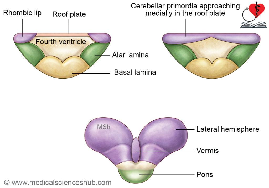
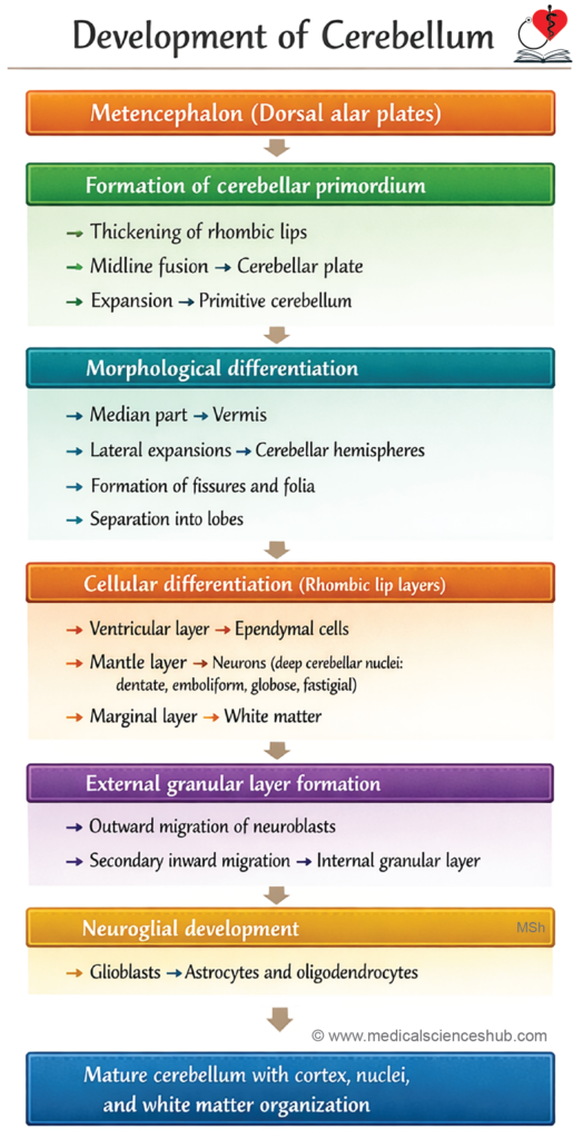
Table 22.3: Parts of cerebellum
| Lobe | Principal Components |
|---|---|
| Anterior lobe | Lingula Central lobule Culmen Ala Quadrangular lobule |
| Posterior lobe | Declive Folium Tuber Pyramid Uvula Simplex Biventral Semilunar Tonsil |
| Flocculonodular lobe | Nodule Flocculus |
Histogenesis of Cerebellum
- Initially, the cerebellar primordium consists of an outer marginal zone and an inner mantle zone of neuroepithelial cells. Cells from the mantle zone migrate outward across the marginal zone to form the external granular layer. These cells then proliferate and migrate inward, giving rise to the cerebellar cortex.
- The mature cortex is organized into three layers:
- Molecular layer: Contains stellate and basket cells.
- Purkinje cell layer: A single layer of Purkinje neurons.
- Granular layer: Composed of granule cells and Golgi cells.
- Development of the cerebellar cortex begins around the sixth month of intrauterine life and continues until approximately 18 months after birth, reflecting prolonged maturation.
- Non-migrating cells of the mantle zone form the deep cerebellar nuclei, including the dentate, emboliform, globose, and fastigial nuclei.
- The portion of the roof plate of the fourth ventricle, along with pia mater, that does not contribute to cerebellar formation develops into the superior and inferior medullary vela.
- Exposure to DNA synthesis inhibitors (e.g., certain antiviral drugs) during early childhood may impair cerebellar development.
Cerebellar Peduncles
The cerebellar peduncles are major fiber bundles formed within the white matter (marginal layer) of the cerebellum, connecting it with different parts of the brainstem.
- Superior cerebellar peduncle: Primarily contains efferent fibers arising from the dentate nucleus, transmitting output from the cerebellum to higher centers.
- Middle cerebellar peduncle: Composed mainly of afferent fibers from the pontine nuclei, conveying cortical input to the cerebellum.
- Inferior cerebellar peduncle: Contains predominantly afferent fibers from the inferior olivary nucleus, along with additional inputs from the spinal cord and vestibular system.
Together, these peduncles ensure efficient communication between the cerebellum and the brainstem, facilitating coordination and motor control.
Development Of Diencephalon
- The prosencephalon (forebrain) divides into the telencephalon (forming cerebral hemispheres) and the diencephalon. The diencephalon consists mainly of a roof plate and alar plates, and its cavity becomes the third ventricle. This ventricle communicates with the lateral ventricles via the interventricular foramen (of Monro) and with the fourth ventricle through the cerebral aqueduct.
Differentiation of Roof Plate
- The roof plate is formed by ependymal cells covered by vascular pia mater. Invagination of this vascular layer into the ventricular cavity produces the choroid plexus of the third ventricle, responsible for CSF secretion. The caudal part of the roof plate develops into the pineal gland.
Development of epithalamus
- The epithalamus arises from the roof plate and includes the habenular nuclei and habenular commissure, which are functionally related to olfactory pathways. A posterior commissure also forms caudal to the pineal gland. The pineal gland is often considered part of the epithalamus.
Differentiation of Alar Plates
- The alar plates form the lateral walls and floor of the diencephalon. They are subdivided by the hypothalamic sulcus and epithalamic sulcus into three regions:
- Epithalamus (dorsal)
- Thalamus (intermediate)
- Hypothalamus (ventral)
- The thalamus enlarges and projects into the third ventricle, forming the thalamic nuclei. As it expands, the hypothalamus is displaced caudoventrally. The floor of the diencephalon gives rise to the mammillary bodies.
- Around the 22nd day, the lateral wall of the hypothalamic region forms the optic sulcus, which evaginates to form the optic vesicle and subsequently the optic cup, giving rise to the retina and related structures.
- A downward extension from the floor, the infundibular process, develops into the posterior pituitary (neurohypophysis).
Development Of Cerebrum
- The prosencephalon (forebrain) differentiates into the telencephalon and diencephalon. The telencephalon forms two lateral outgrowths known as the cerebral hemispheres, along with a midline structure called the lamina terminalis.
- The cavity of the telencephalon develops into the paired lateral ventricles, which communicate with the third ventricle through the interventricular foramina (of Monro).
- Development of the cerebral hemispheres is typically described in two phases: early development (first two months), characterized by initial formation and expansion of the hemispheric vesicles, and later development (after the second month), during which rapid growth, cortical differentiation, and structural specialization occur.
Development during First 2 Months
- Each cerebral hemisphere initially consists of a thin pallium (future cortex) and a thick basal region. Cells from the basal region migrate into the pallium to form the cerebral cortex, while the remaining cells develop into the corpus striatum.
- The junction between expanding pallial regions becomes thin and is invaginated by the choroid plexus. The pallium differentiates into allocortex (including archipallium and paleopallium) and neocortex (neopallium), which constitutes the majority of the human cortex.
- Axonal pathways connecting cortical regions form the internal capsule, carrying both afferent and efferent fibers.
Development after 2nd Month
- The neocortex expands rapidly and overgrows the allocortex, leading to a relative reduction in ventricular size. In the floor of the hemisphere, neuronal masses condense to form the basal nuclei (corpus striatum).
- These nuclei differentiate into:
- Neostriatum: Includes caudate nucleus and putamen.
- Paleostriatum: Forms the globus pallidus.
- Archistriatum: Develops into the amygdaloid body.
- The putamen and globus pallidus fuse to form the lentiform nucleus. These structures are traversed by fibers of the internal and external capsules, establishing major neural pathways within the cerebrum.
Development of Lobes of Cerebral Hemispheres
- The cerebral cortex expands in specific directions during development, leading to the formation of distinct lobes of the cerebral hemispheres.
- Ventral expansion of the cortex gives rise to the frontal lobe.
- Dorsal growth contributes to the formation of the occipital lobe.
- Lateral (parietal) expansion results in the parietal lobe.
- The occipital pole extends ventrally and laterally to form the temporal lobe.
- These directional growth patterns establish the fundamental lobar organization of the cerebrum, which later becomes further refined by the development of sulci and gyri.
Effects of development of cerebral hemispheres
Expansion of the cerebral hemispheres produces several structural changes:
- Lateral ventricle: Growth of the temporal and occipital lobes leads to elongation of the lateral ventricle, forming the inferior (temporal) horn and posterior (occipital) horn.
- Caudate nucleus: The enlarging caudate nucleus follows the curvature of the ventricle and acquires a characteristic C-shaped configuration.
- Sulci and gyri formation: Continued cortical expansion results in folding of the surface. The lateral sulcus (Sylvian fissure) appears around the 4th month, while the central sulcus (of Rolando) becomes evident later in fetal life, contributing to surface complexity.
- Insula: A cortical region overlying the corpus striatum grows relatively slowly and forms the insula. It becomes deeply embedded within the lateral sulcus as surrounding cortical areas expand.
- Olfactory structures: An evagination from the frontal lobe develops into the olfactory bulb, while its connecting stalk forms the olfactory tract.
These changes establish the characteristic shape, internal organization, and surface pattern of the mature cerebrum.
Differentiation of Allocortex
- The allocortex develops into key components of the limbic system, which are involved in emotion, memory, and olfaction.
- It consists of two primitive regions: the archipallium, located on the medial surface of the cerebral hemisphere, and the paleopallium, situated on the ventral surface lateral to the corpus striatum.
- The allocortex gives rise to the limbic lobe, which differentiates into:
- Hippocampal formation: The dorsal part forms a thin layer of gray matter, the indusium griseum, over the corpus callosum. The ventral part develops into the hippocampus and dentate gyrus, which project into the inferior horn of the lateral ventricle.
- Hippocampal efferent pathways: Axons from the hippocampus form the fimbria, fornix, and associated commissural fibers, establishing connections with other limbic structures.
- In addition, the allocortex contributes to the formation of the olfactory bulb and tract, linking it functionally to the olfactory system.
Commissures of Telencephalon
The lamina terminalis represents the most anterior part of the telencephalon and serves as the pathway through which major commissural fibers connect the two cerebral hemispheres.
In the developing brain, this region gives rise to several important fiber bundles:
- Anterior commissure: Connects the temporal lobes, particularly regions related to olfaction.
- Corpus callosum: The largest commissure, linking corresponding areas of the right and left cerebral hemispheres. With hemispheric growth, it enlarges and becomes separated from the fornix by the septum pellucidum.
- Hippocampal (fornical) commissure: Connects the hippocampi of both sides, supporting limbic integration.
- Posterior commissure: Involved in pupillary light reflex pathways, though its connections are not fully defined.
- Habenular commissure: Connects the habenular nuclei of the epithalamus.
- Optic chiasma: Represents partial crossing of optic nerve fibers, essential for visual processing.
- At birth, the brain contains approximately 9–14 billion neurons. Postnatally, neuronal numbers remain largely constant, while neuroglial cells continue to proliferate. The adult cerebral cortex has an approximate surface area of 285,000 mm².
CLINICAL EMBRYOLOGY
Developmental Anomalies
- Anencephaly: A severe neural tube defect characterized by absence of major parts of the brain and skull. It shows higher incidence in females. In late pregnancy, it is often associated with polyhydramnios due to impaired fetal swallowing.
- Encephalocele: Results from defective closure of the cranial neural tube (commonly anterior neuropore), leading to herniation of brain tissue through a skull defect.
- Dandy–Walker malformation: Caused by failure of opening of the foramina of Magendie and Luschka, resulting in cystic dilation of the fourth ventricle and hypoplasia or agenesis of the cerebellar vermis.
- Microcephaly: Characterized by a markedly reduced brain size and head circumference. It may result from genetic factors or prenatal insults such as viral infections (e.g., cytomegalovirus) or radiation exposure.
- Arnold–Chiari malformation: Involves downward displacement of cerebellar tonsils through the foramen magnum, which may obstruct cerebrospinal fluid flow and cause neurological symptoms.
Important Questions
- Write a concise note on hydrocephalus, including its causes and clinical significance.
- Describe the process of neurulation, highlighting its key stages and outcomes.
- Enumerate the major derivatives of neural crest cells.
- Write a brief note on the neural crest, including its origin, migration, and significance.
- Explain the positional changes of the spinal cord during development.
- Write a short note on neural tube defects, with emphasis on spina bifida.
- Describe spina bifida, including its types and developmental basis.
- Outline the development of the cerebellum, mentioning its embryological origin and differentiation.
📝 Test Your Knowledge – Practice MCQs
Attempt the chapter MCQ quiz and assess your understanding of key concepts.
Click to see: Medical Disclaimer and Copyright Notice