
Competencies
- AN58.1: Identify external features of medulla oblongata.
- AN58.2: Describe transverse section of medulla oblongata at the level of (1) pyramidal decussation, (2) sensory decussation, (3) inferior olivary nucleus (ION).
- AN58.3: Enumerate cranial nerve nuclei in medulla oblongata with their functional groups.
- AN58.4: Describe the anatomical basis and effects of medial and lateral medullary syndromes.
Introduction
- The medulla oblongata is the vital lower part of the brainstem, linking the brain with the spinal cord and coordinating essential functions such as respiration, cardiovascular control, swallowing, and protective reflexes.
Brainstem
- The brainstem serves as the critical connection between the spinal cord and the cerebrum. It is situated in the posterior cranial fossa, resting on the sloping clivus, and is positioned between the diencephalon superiorly and the spinal cord inferiorly.
- The brainstem is divided into three continuous parts, arranged from inferior to superior: the medulla oblongata, the pons, and the midbrain. The medulla oblongata represents the upward continuation of the spinal cord, beginning at the level of the upper border of the posterior arch of the atlas vertebra. The midbrain, at the opposite end, connects the brainstem to the cerebral hemispheres.
- The brainstem contains four principal components.
- First, it carries ascending and descending fiber tracts that relay information between the spinal cord and higher brain centers.
- Second, it houses vital centers that regulate heart rate, blood pressure, and respiration — functions that are immediately life-threatening if disrupted.
- Third, the reticular formation, a diffuse network of neurons distributed throughout the brainstem, plays a central role in maintaining consciousness through the reticular activating system.
- Fourth, the brainstem contains the nuclei of cranial nerves III through XII, accounting for ten of the twelve pairs of cranial nerves.
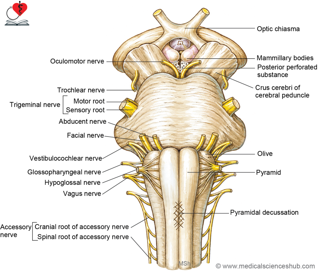
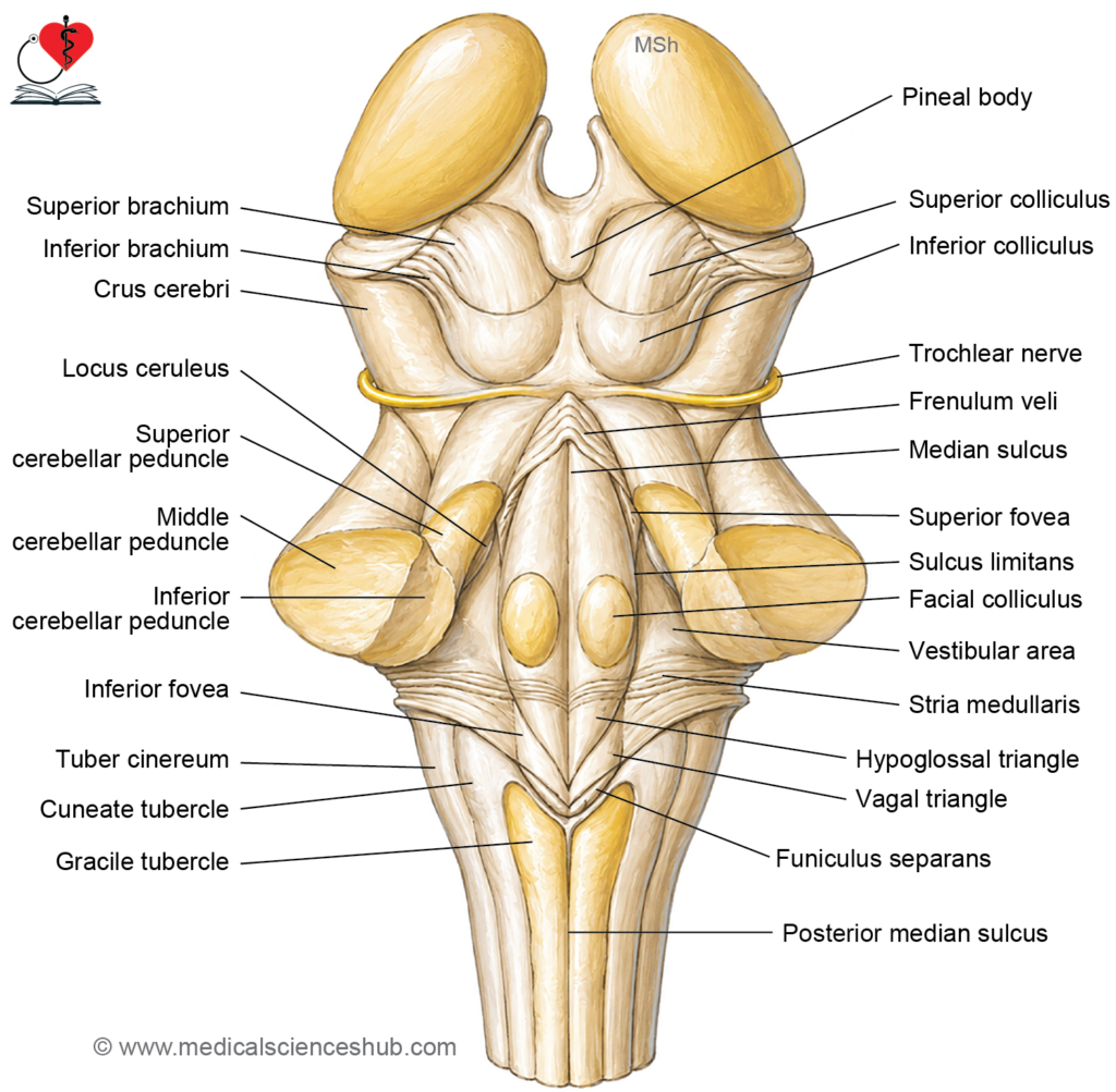
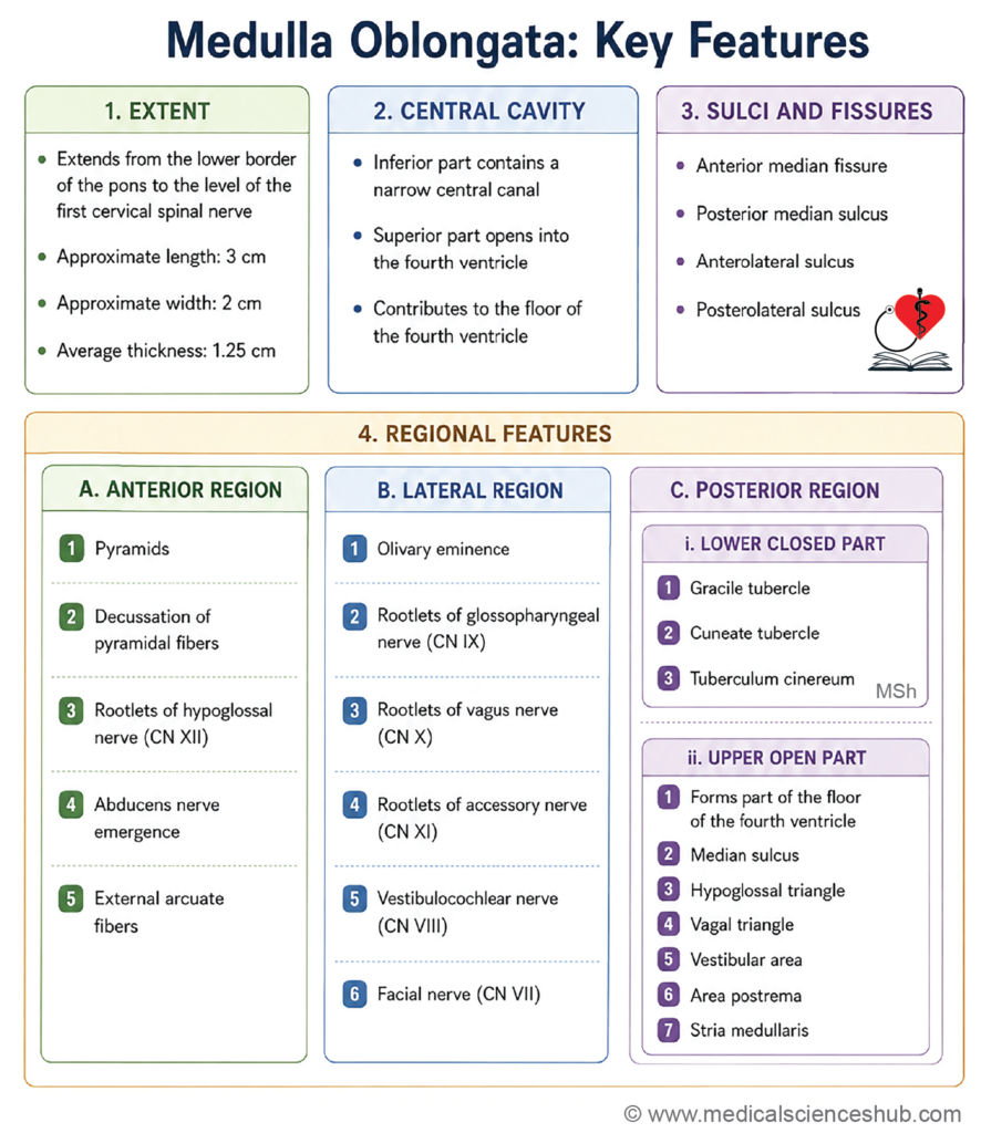
Medulla Oblongata
- The medulla oblongata is the most inferior component of the brainstem and represents the direct upward continuation of the spinal cord. It forms the lower and anterior portion of the hindbrain.
- In terms of extent, the medulla oblongata spans from the pontomedullary junction — marked by the lower border of the pons — down to the level of the first cervical spinal nerve, which corresponds to a transverse plane passing along the upper border of the posterior arch of the atlas (C1 vertebra).
- A tumor arising within the medulla oblongata or at the pontomedullary junction can exert pressure on surrounding neural and vascular structures. Depending on which structures are compressed, this may produce a range of neurological symptoms and clinical signs in the affected individual.
Shape and Measurements
- The medulla oblongata has a piriform (pear-shaped) configuration, being broader superiorly and tapering toward its inferior end.
- It measures approximately 3 cm in length, 2 cm in width at its widest point, and 1.25 cm in thickness.
Cavity of Medulla Oblongata
- The cavity medulla oblongata has two parts: Lower closed and upper open parts.
External Features
- The medulla oblongata has distinct anterior (ventral) and posterior (dorsal) surfaces, each marked by a series of longitudinal sulci and fissures that divide the structure into recognizable regions.
- The medulla is divided into right and left halves by two midline landmarks: the anterior median fissure on its ventral surface and the posterior median sulcus on its dorsal surface.
- The anterior median fissure is a deep vertical cleft running along the midline of the anterior surface, continuous inferiorly with the anterior median fissure of the spinal cord. In its lower portion, this fissure becomes relatively shallow due to two overlying structures: the crossing of pyramidal (corticospinal) fibers at the decussation of the pyramids, and the emergence of anterior external arcuate fibers.
- The posterior median sulcus is a faint longitudinal groove along the midline of the posterior surface, present in the closed (lower) part of the medulla. It is continuous inferiorly with the posterior median sulcus of the spinal cord.
- The foramen cecum is a small triangular recess located at the upper end of the anterior median fissure, at the level of the pontomedullary junction.
- The anterolateral and posterolateral sulci represent upward continuations of the corresponding sulci of the spinal cord. Together with the median fissure and sulcus, they divide the medulla oblongata into three longitudinal regions: anterior, lateral, and posterior.
Regions of Medulla Oblongata
- For descriptive purposes, the medulla oblongata is divided into three longitudinal regions by the anterolateral and posterolateral sulci:
- The anterior region lies between the anterior median fissure and the anterolateral sulcus. Its most prominent feature is the pyramid, a raised column of tissue containing descending corticospinal fibers.
- The lateral region lies between the anterolateral and posterolateral sulci. It contains the olive, an oval surface elevation produced by the underlying inferior olivary nucleus.
- The posterior region lies medial to the posterolateral sulcus. It contains the upward continuations of the posterior funiculi of the spinal cord, which expand in the upper medulla to form the gracile and cuneate tubercles.
Anterior Region
- The anterior region of the medulla oblongata contains three notable structures: the pyramids, the pyramidal decussation, and the emergence of the abducent nerve (cranial nerve VI).
- The pyramids are a pair of vertically elongated surface elevations situated on either side of the anterior median fissure. They are produced primarily by the descending fibers of the corticospinal (pyramidal) tract, and also contain corticonuclear and a small number of corticopontine fibers. These fiber bundles are covered superficially by the arcuate nucleus. The pyramids taper and flatten gradually toward the lower end of the medulla.
- At the inferior end of the anterior region, the fibers of the corticospinal tract cross the midline at the pyramidal decussation (motor decussation). A portion of these crossing fibers is visible on the surface, where they partially obliterate and interrupt the continuity of the anterior median fissure.
- The abducent nerve (CN VI) exits the brainstem at the pontomedullary junction, emerging just superior to the pyramid on the anterior surface of the medulla.
Lateral Region
- The lateral region of the medulla oblongata displays several important surface features, including the olives, multiple cranial nerve rootlets, and the anterior external arcuate fibers.
- The olives are paired oval surface elevations, one on each side, located just lateral to the pyramids. Each olive measures approximately 1.25 cm in its longitudinal axis. The elevation is produced by the underlying inferior olivary nucleus.
- The rootlets of the hypoglossal nerve (CN XII) emerge from the anterolateral sulcus, in the groove between the olive and the pyramid.
- The rootlets of the glossopharyngeal (CN IX), vagus (CN X), and the cranial part of the accessory nerve (CN XI) emerge along the posterolateral sulcus, dorsal to the olive.
- The facial nerve (CN VII) and vestibulocochlear nerve (CN VIII) emerge at the pontomedullary junction, superior to the olive.
- The anterior external arcuate fibers arise from the arcuate nucleus, exit through the anterior median fissure, and course horizontally across the surface of the pyramids and olives to reach the cerebellum via the inferior cerebellar peduncles.
Posterior Region
The posterior region of the medulla oblongata is subdivided into two parts based on the presence or absence of a central canal: a lower closed part and an upper open part.
Lower Closed Part
- The lower closed part lies between the posterolateral sulcus and the posterior median sulcus. It represents the upward continuation of the fasciculus gracilis and fasciculus cuneatus of the spinal cord.
- The gracile tubercle (clava) is a rounded surface elevation at the upper end of the fasciculus gracilis, produced by the underlying nucleus gracilis. Lateral to it, the cuneate tubercle is a similar elevation at the upper end of the fasciculus cuneatus, produced by the nucleus cuneatus. Both nuclei serve as relay stations for fine touch, vibration, and proprioceptive information ascending in the dorsal column–medial lemniscus pathway.
- The tuber cinereum is a small rounded elevation situated lateral to the cuneate tubercle. It is produced by the spinal tract of the trigeminal nerve and its associated nucleus. Inferiorly, it is continuous with the dorsolateral tract of Lissauer and the substantia gelatinosa of the spinal cord.
Upper Open Part
- In its upper portion, the posterior region of the medulla contributes to the floor of the fourth ventricle, as the central canal opens into this cavity.
- A median sulcus runs longitudinally along the floor of the fourth ventricle, dividing it into symmetrical right and left halves.
- Two triangular surface areas are visible in this region, each overlying an important underlying nucleus:
- The hypoglossal triangle overlies the hypoglossal nucleus (CN XII).
- The vagal triangle overlies the dorsal nucleus of the vagus nerve (CN X).
- Two functionally significant areas are also present:
- The vestibular area overlies the vestibular nuclei, which are involved in processing balance and spatial orientation signals transmitted via the vestibulocochlear nerve (CN VIII).
- The area postrema is located in the lower part of the vagal triangle. It is a circumventricular organ containing chemoreceptors that detect emetic agents in the blood and cerebrospinal fluid, playing a critical role in triggering the vomiting reflex.
Internal Structure Of Medulla Oblongata
- As the medulla oblongata is the direct upward continuation of the spinal cord, its lowermost portion retains the basic spinal cord organization, with a central core of grey matter surrounded by white matter. As one ascends through the medulla, this arrangement progressively breaks down — the central grey matter disperses and becomes reorganized into discrete nuclei scattered throughout the substance of the medulla.
- The internal structure of the medulla oblongata is most effectively studied by examining transverse sections taken at four representative levels, each corresponding to a distinct anatomical region and set of structural features:
- At the level of the pyramidal (motor) decussation, in the lower closed part of the medulla.
- At the level of the sensory (lemniscal) decussation, in the upper closed part of the medulla.
- At the level of the olives, corresponding to the open part of the medulla where the fourth ventricle begins to form.
- At the uppermost part of the medulla, immediately inferior to the pontomedullary junction.
TS of Medulla Oblongata at the Level of Pyramidal Decussation
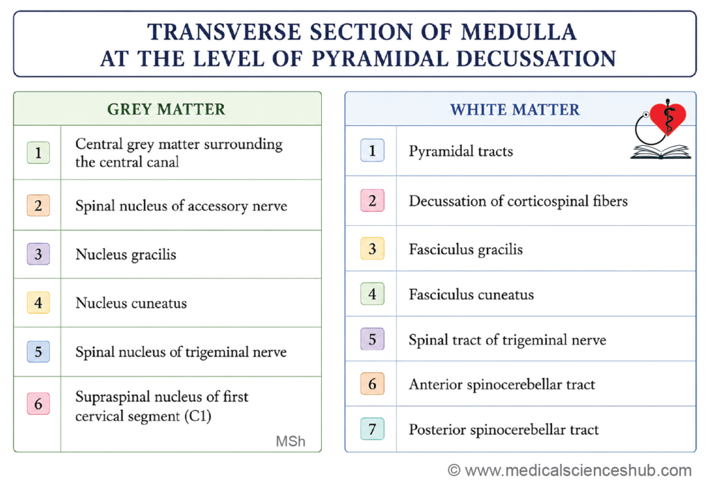
At the level of the pyramidal decussation — corresponding to the lowest part of the closed medulla, just superior to the spinal cord — a transverse section reveals the following features:
Grey Matter
- The central canal is present and surrounded by its central grey matter, retaining the basic organization seen in the spinal cord.
- The nucleus gracilis and nucleus cuneatus are present posteriorly, serving as relay nuclei for ascending dorsal column sensory pathways.
- The nucleus of the spinal tract of the trigeminal nerve is present, relaying pain and temperature information from the face and head.
- The spinal nucleus of the accessory nerve (CN XI) is identifiable at this level.
- The supraspinal nucleus of C1 is also present, representing the uppermost segment of the first cervical spinal nerve.
White Matter
- The pyramids are visible in the anterior part of the section, carrying descending corticospinal fibers.
- The pyramidal (motor) decussation is the defining feature of this level, where the majority of corticospinal fibers cross the midline to continue as the lateral corticospinal tract in the contralateral spinal cord.
- The spinal tract of the trigeminal nerve is present in the lateral region, carrying uncrossed pain and temperature impulses from the ipsilateral face.
- The fasciculus gracilis and fasciculus cuneatus occupy the posterior white matter, conveying fine touch, vibration, and proprioception from the lower and upper limbs respectively.
- The anterior and posterior spinocerebellar tracts are present laterally, transmitting unconscious proprioceptive information to the cerebellum.
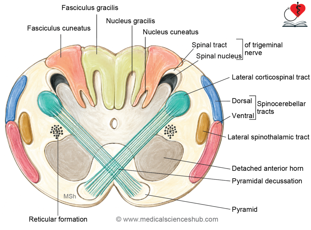
Key Description
Grey Matter
- The grey matter of the medulla oblongata at this level retains a spinal cord-like organization, containing a central canal flanked by ventral (anterior) and dorsal (posterior) grey columns, though these become progressively fragmented as one ascends through the medulla.
Pyramidal Decussation
- The pyramidal (corticospinal) tract mediates voluntary motor control of the contralateral half of the body. To achieve this crossed control, the majority of fibers decussate at the pyramidal decussation.
- Approximately 75–95% of corticospinal fibers cross the midline at this level, passing posteriorly and laterally to enter the lateral white column, where they descend as the lateral corticospinal tract.
- The bulk of the crossing fibers, together with the pyramids themselves, displaces the central canal posteriorly within the lower medulla.
Spinal Nucleus of the Accessory Nerve
- The spinal nucleus of the accessory nerve (CN XI) is a column of motor neurons that gives rise to the spinal part of the accessory nerve, which supplies the sternocleidomastoid and trapezius muscles.
- The decussating corticospinal fibers pass through the anterior grey column of the medulla, partially detaching a segment of it. This detached portion forms the spinal segment of the accessory nucleus, whose rootlets ascend within the vertebral canal, enter the skull through the foramen magnum, and briefly unite with the cranial root of the accessory nerve before separating again to reach their target muscles.
- The supraspinal nucleus is a further detached portion of the anterior grey column, giving rise to the motor fibers of the first cervical (C1) spinal nerve.
Nucleus Gracilis and Nucleus Cuneatus
- The nucleus gracilis and nucleus cuneatus are the relay nuclei of the dorsal column–medial lemniscus pathway, functioning as second-order neurons that transmit proprioception, vibration sense, and fine touch to the thalamus.
- The nucleus gracilis receives afferent input from the lower half of the body (carried by the fasciculus gracilis, also known as the tract of Goll), while the nucleus cuneatus receives input from the upper half of the body (carried by the fasciculus cuneatus, also known as the tract of Burdach).
- Both nuclei appear as narrow strip-like elevations of grey matter on the dorsal aspect of the medulla, with the nucleus gracilis positioned medial to the nucleus cuneatus.
Spinal Nucleus of the Trigeminal Nerve
- The spinal nucleus of the trigeminal nerve receives pain and temperature sensations from the face, scalp, and oral cavity, and is functionally analogous to the dorsal horn of the spinal cord.
- It forms part of the posterior grey matter of the medulla, situated lateral to the nucleus cuneatus. It extends from the pontomedullary junction inferiorly to the level of the second cervical spinal segment (C2).
- Collectively, the nucleus gracilis, nucleus cuneatus, and spinal nucleus of the trigeminal nerve constitute the functional equivalent of the posterior grey horn of the medulla oblongata.
Spinal Tract of the Trigeminal Nerve
- The spinal tract of the trigeminal nerve consists of descending primary afferent fibers of the trigeminal nerve that convey pain and temperature information from the face, scalp, and oral cavity to the spinal nucleus below.
- It runs between the spinal nucleus of the trigeminal nerve and the lateral surface of the medulla, and is functionally analogous to the dorsolateral tract of Lissauer in the spinal cord.
Reticular Formation
- The reticular formation at this level consists of a diffuse network of scattered neurons and interconnecting nerve fibers located within the lateral white column of the medulla, forming part of the larger reticular system that extends throughout the brainstem.
TS of Medulla Oblongata at the Level of Sensory Decussation
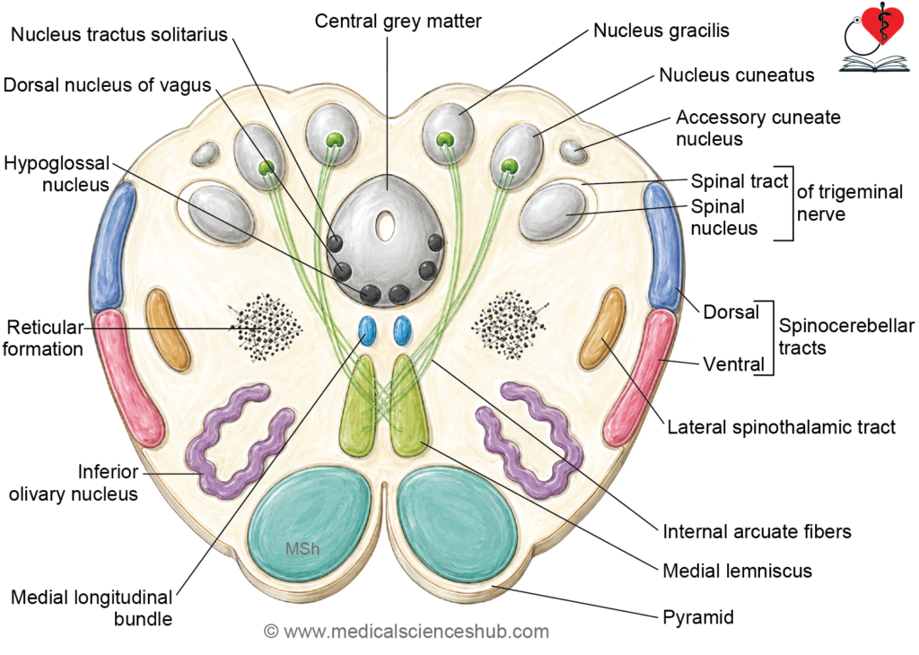
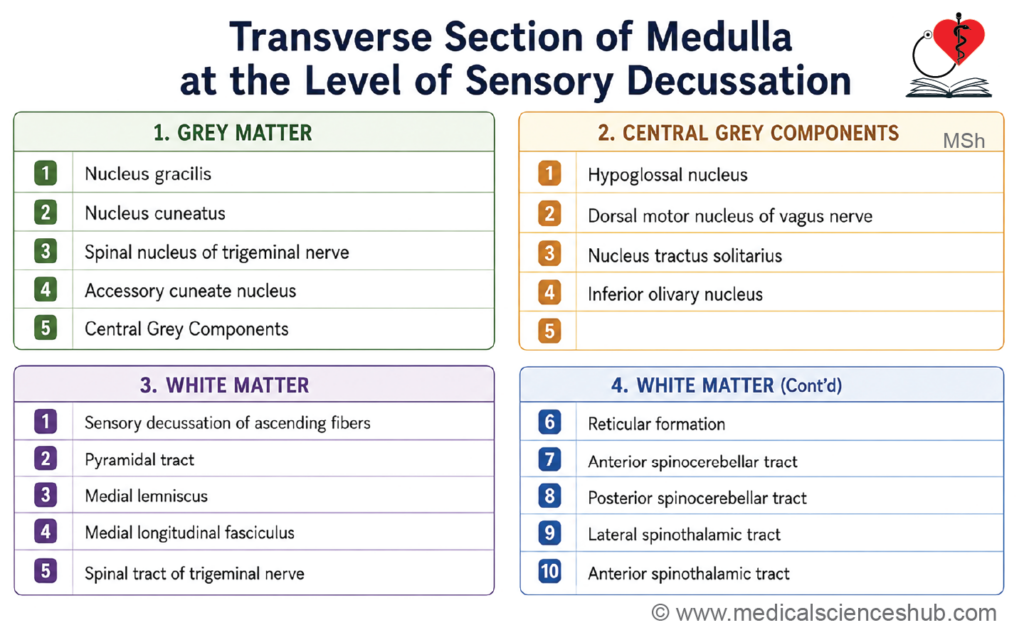
At the level of the sensory decussation — corresponding to the upper closed part of the medulla and also referred to as the level of the internal arcuate fibers — a transverse section reveals the following features:
Grey Matter
- The central grey matter surrounds the central canal and contains several important nuclei at this level:
- The hypoglossal nucleus (CN XII) provides motor innervation to the muscles of the tongue.
- The dorsal nucleus of the vagus nerve (CN X) is the primary parasympathetic nucleus of the vagus, supplying thoracic and abdominal viscera.
- The nucleus tractus solitarius receives visceral afferent and gustatory (taste) inputs from cranial nerves VII, IX, and X.
- The inferior olivary nucleus is a prominent folded grey matter structure that relays information to the cerebellum via the inferior cerebellar peduncle, playing a key role in motor coordination and learning.
- The nucleus gracilis and nucleus cuneatus are still present at this level and are in the process of giving rise to internal arcuate fibers, which form the sensory decussation.
- The nucleus of the spinal tract of the trigeminal nerve continues at this level, processing pain and temperature information from the face and oral cavity.
- The accessory cuneate nucleus lies lateral to the nucleus cuneatus. It receives proprioceptive input from the upper limb and transmits it to the cerebellum via the posterior spinocerebellar tract, functioning as the upper limb equivalent of Clarke’s column in the spinal cord.
White Matter
- The sensory decussation, formed by internal arcuate fibers, is the defining feature of this level. These are axons of second-order neurons arising from the nucleus gracilis and nucleus cuneatus that cross the midline and ascend as the medial lemniscus, carrying fine touch, vibration, and proprioception to the contralateral thalamus.
- The pyramids remain visible in the anterior part of the section, continuing to carry descending corticospinal fibers.
- The medial longitudinal fasciculus (MLF) is a paired white matter tract situated near the midline, dorsal to the pyramids. It coordinates conjugate eye movements and head-neck reflexes by interconnecting the nuclei of cranial nerves III, IV, and VI with vestibular and cervical motor neurons.
- The spinal tract of the trigeminal nerve continues laterally, carrying descending pain and temperature fibers toward the spinal nucleus below.
- The reticular formation occupies the lateral white matter, contributing to autonomic regulation, consciousness, and other integrative functions.
- The anterior and posterior spinocerebellar tracts are present in the lateral region, transmitting unconscious proprioceptive signals to the cerebellum.
- The lateral spinothalamic tract and anterior spinothalamic tract are present, carrying pain and temperature and crude touch and pressure sensations respectively from the contralateral body to the thalamus.
Key Description
Grey Matter at the Level of Sensory Decussation
- At this level, the previously compact central grey matter begins to disperse. It separates into the central grey matter proper — containing the major cranial nerve nuclei — and a number of peripheral nuclei, including the nucleus gracilis, nucleus cuneatus, accessory cuneate nucleus, and the spinal nucleus of the trigeminal nerve.
Hypoglossal Nucleus
- The hypoglossal nucleus is the general somatic efferent (GSE) motor nucleus of the hypoglossal nerve (CN XII), supplying all intrinsic and extrinsic muscles of the tongue, with the exception of the palatoglossus.
- It is approximately 2 cm in length and occupies a paramedian position within the central grey matter, lying in the floor of the fourth ventricle deep to the hypoglossal triangle.
- It receives descending input from corticonuclear (corticobulbar) fibers. Damage to the hypoglossal nucleus or its nerve results in ipsilateral paralysis and wasting of the tongue musculature.
Dorsal Nucleus of the Vagus
- The dorsal nucleus of the vagus is the largest parasympathetic (general visceral efferent, GVE) nucleus in the brainstem. It lies dorsolateral to the hypoglossal nucleus in the floor of the fourth ventricle, deep to the vagal triangle.
- It gives rise to preganglionic parasympathetic fibers of the vagus nerve (CN X), which supply the heart, the smooth muscle and glands of the respiratory tract, and the gastrointestinal tract as far as the splenic flexure of the colon.
Nucleus Tractus Solitarius
- The nucleus tractus solitarius (NTS) is an elongated nucleus situated dorsolateral to the dorsal nucleus of the vagus. It represents the special visceral afferent (SVA) column of the brainstem, primarily receiving gustatory (taste) afferents from the facial (CN VII), glossopharyngeal (CN IX), and vagus (CN X) nerves.
- Its efferent fibers cross the midline and ascend as the solitariothalamic tract to reach the ventral posteromedial (VPM) nucleus of the thalamus, and subsequently project to the primary somatosensory cortex and insula.
- The caudal portion of the NTS is functionally designated the cardiorespiratory nucleus. It receives visceral afferents from the lungs, trachea, larynx, and gastrointestinal tract, contributing to the regulation of respiratory rhythm. It also receives input from the carotid sinus and aortic body (chemoreceptors), through which it modulates cardiovascular function via the reticular formation.
Sensory Decussation (Internal Arcuate Fibers)
- The internal arcuate fibers are the axons of second-order neurons arising from the nucleus gracilis (tract of Goll) and nucleus cuneatus (tract of Burdach). They course ventromedially, cross the midline at the sensory decussation, and ascend as the medial lemniscus to reach the ventral posterolateral (VPL) nucleus of the thalamus, conveying fine touch, vibration, and conscious proprioception from the contralateral body.
- Within the medial lemniscus, the fibers maintain a precise somatotopic organization: fibers from the nucleus gracilis (representing the lower limb and lower trunk) are positioned ventral to those from the nucleus cuneatus (representing the upper trunk and upper limb). From ventral to dorsal, the arrangement follows the sequence: lower limb → lower trunk → upper trunk → upper limb.
Pyramid
- The pyramids remain prominent at this level, containing the descending fibers of the corticospinal and corticonuclear tracts. In the lower medulla, the majority of corticospinal fibers cross the midline at the pyramidal decussation to form the lateral corticospinal tract in the contralateral spinal cord.
Medial Longitudinal Fasciculus
- The medial longitudinal fasciculus (MLF) is a heavily myelinated, paired fiber bundle running in a paramedian position throughout the brainstem. It serves as the principal pathway coordinating conjugate eye movements and gaze stability in response to vestibular, auditory, and visual stimuli.
- It receives afferent input from three main sources:
- The vestibular nuclei, conveying signals related to balance and head position.
- The lateral lemniscus, conveying signals related to auditory processing.
- The superior colliculus, conveying signals related to visual reflexes.
- It projects efferent fibers to:
- The nuclei of cranial nerves III (oculomotor), IV (trochlear), and VI (abducens), coordinating extraocular muscle movements.
- The spinal nucleus of the accessory nerve (CN XI) and the anterior horn cells of the upper cervical spinal cord, coordinating head and neck movements.
- In summary, the MLF integrates vestibular, auditory, and visual inputs to produce coordinated movements of the eyes, face, and neck.
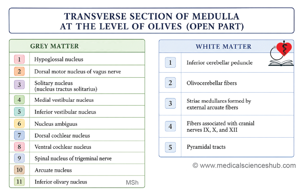
TS of Medulla Oblongata at the Level of Inferior Olivary Nucleus
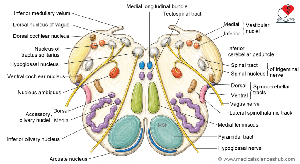
At the level of the inferior olivary nucleus, the transverse section passes through the floor of the fourth ventricle and represents the open part of the medulla. It reveals the following features:
Grey Matter
Cranial Nerve Nuclei
- The hypoglossal nucleus (CN XII), dorsal nucleus of the vagus (CN X), and nucleus tractus solitarius remain present at this level, as described at the level of the sensory decussation.
- The inferior and medial vestibular nuclei are present, receiving equilibrium and spatial orientation signals transmitted via the vestibulocochlear nerve (CN VIII), and projecting to the MLF, cerebellum, and spinal cord.
- The nucleus ambiguus is a motor nucleus located within the reticular formation of the lateral medulla. It contributes motor fibers to the glossopharyngeal (CN IX), vagus (CN X), and cranial part of the accessory nerve (CN XI), supplying the striated muscles of the pharynx, larynx, and soft palate. It represents the special visceral efferent (SVE) column of the medulla.
- The dorsal and ventral cochlear nuclei receive auditory afferents from the cochlear division of the vestibulocochlear nerve (CN VIII) and relay signals to higher auditory centers via the lateral lemniscus.
- The nucleus of the spinal tract of the trigeminal nerve continues at this level, processing pain and temperature information from the face and oral cavity.
Other Grey Matter Structures
- The inferior olivary nucleus is the most prominent grey matter structure at this level. It is a large, folded laminar nucleus that generates climbing fiber projections to the contralateral cerebellar cortex via the inferior cerebellar peduncle. It plays a crucial role in motor learning, coordination, and timing of movements.
- The arcuate nucleus is a small collection of neurons situated on the anterior surface of the pyramid. It is considered a displaced pontine nucleus and gives rise to anterior external arcuate fibers, which travel to the cerebellum via the inferior cerebellar peduncle.
White Matter
- The inferior cerebellar peduncle (restiform body) is a large fiber bundle forming on the posterolateral aspect of the medulla at this level. It serves as the principal pathway connecting the medulla and spinal cord to the cerebellum, transmitting olivocerebellar, spinocerebellar, and arcuatocerebellar fibers.
- The pyramidal tract continues in the anterior part of the section, carrying descending corticospinal and corticonuclear fibers.
- Olivocerebellar fibers arise from the inferior olivary nucleus, cross the midline, and pass through the inferior cerebellar peduncle to reach the contralateral cerebellar cortex as climbing fibers.
- The striae medullares (external arcuate fibers) are superficial fibers that arise from the arcuate nucleus, course across the dorsal surface of the medulla, and enter the cerebellum via the inferior cerebellar peduncle.
- The fibers of cranial nerves IX (glossopharyngeal), X (vagus), and XI (accessory) are present at this level, emerging from or converging toward their respective nuclei and exiting along the posterolateral sulcus of the medulla.
Key Description
Hypoglossal Nucleus
- The hypoglossal nucleus is an elongated nucleus approximately 2 cm in length, situated in the upper medulla in the floor of the fourth ventricle, deep to the hypoglossal triangle.
- It gives rise to the motor fibers of the hypoglossal nerve (CN XII), supplying all intrinsic and extrinsic muscles of the tongue, with the exception of the palatoglossus.
Olivary Nuclear Complex
- The olivary nuclear complex is located in the medulla oblongata, posterolateral to the pyramid. It consists of three nuclei: the inferior olivary nucleus, the medial accessory olivary nucleus, and the dorsal accessory olivary nucleus. Together, they form part of the spino-olivo-cerebellar pathway, contributing to motor coordination and learning.
- The inferior olivary nucleus is the largest of the three. It has a characteristic folded, crumpled-bag appearance with its hilus facing medially, and produces the surface elevation known as the olive.
- The medial accessory olivary nucleus lies between the medial lemniscus and the inferior olivary nucleus, while the dorsal accessory olivary nucleus lies dorsal to it. Both accessory nuclei are phylogenetically older than the inferior olivary nucleus and are connected primarily with the paleocerebellum.
- Afferent connections include spino-olivary fibers from the contralateral body and projections from the ipsilateral red nucleus.
- Efferent fibers form the olivocerebellar tract, which exits through the hilus of the inferior olivary nucleus, crosses the midline, and enters the contralateral inferior cerebellar peduncle. These fibers reach the cerebellar cortex as climbing fibers, terminating on Purkinje cells.
Arcuate Nucleus
- The arcuate nucleus is a displaced component of the pontine nuclei, located on the ventromedial surface of the pyramids on either side of the anterior median fissure.
- It receives afferent input from corticopontine fibers originating in the cerebral cortex, and relays this information to the cerebellum, contributing to the precision and coordination of voluntary movements.
- Its efferent fibers follow two routes to reach the cerebellum via the inferior cerebellar peduncle:
- Anterior external arcuate fibers course over the external surface of the pyramids and olive to enter the contralateral inferior cerebellar peduncle.
- A second group of fibers pass posteriorly, exit through the posterior median sulcus, decussate with corresponding fibers from the opposite side, and run laterally beneath the floor ependyma of the fourth ventricle as the striae medullares, ultimately reaching the cerebellum through the inferior cerebellar peduncle.
Nucleus Ambiguus
- The nucleus ambiguus is a motor nucleus situated deep within the reticular formation of the upper medulla. It represents the special visceral efferent (SVE) column and gives rise to branchiomotor fibers that join the glossopharyngeal (CN IX), vagus (CN X), and the cranial part of the accessory nerve (CN XI).
- These fibers supply the pharyngeal constrictor muscles, the intrinsic muscles of the larynx, and the striated muscles of the palate and upper esophagus — all structures derived from the pharyngeal arches — and are essential for phonation and swallowing.
Vestibular Nuclear Complex
- The vestibular nuclear complex lies beneath the floor of the fourth ventricle, spanning the upper medulla and lower pons. It comprises four nuclei: the superior, inferior, medial, and lateral vestibular nuclei. The superior and lateral nuclei lie in the pons, while the inferior and medial nuclei lie in the medulla. The medial vestibular nucleus is the largest of the four.
- Afferent inputs arrive from the vestibular division of the vestibulocochlear nerve (CN VIII), spinovestibular fibers, cerebellovestibular fibers, and brainstem reticular fibers.
- Efferent projections serve multiple functions:
- Vestibulocerebellar fibers project to the cerebellum to coordinate balance-related motor adjustments.
- The vestibulospinal tract descends to the spinal cord, modulating spinal reflex activity and regulating extensor muscle tone for postural control.
- Projections to the medial longitudinal fasciculus (MLF) connect with the nuclei of CN III, IV, and VI and the spinal nucleus of CN XI, coordinating conjugate eye movements and head movements in response to vestibular stimuli.
- Ascending projections reach the thalamus and ultimately the primary vestibular cortex in the parietal lobe, enabling conscious perception of head position and movement.
- Dysfunction of the vestibular nuclei commonly manifests as vertigo, nausea, and motion sickness.
Cochlear Nuclei
- The cochlear nuclei are the second-order neurons of the auditory pathway, responsible for the initial central processing of sound. They are divided into the ventral (anterior) cochlear nucleus and the dorsal (posterior) cochlear nucleus, both located along the lateral surface of the inferior cerebellar peduncle.
- The dorsal cochlear nucleus forms a surface prominence called the auditory tubercle (acoustic eminence) on the posterior surface of the inferior cerebellar peduncle, visible in the floor of the fourth ventricle. The ventral cochlear nucleus lies on the ventrolateral surface of the peduncle, at the junction between the vestibular and cochlear divisions of CN VIII.
- Both nuclei receive afferent input from the spiral ganglion of the cochlea via the cochlear division of the vestibulocochlear nerve.
- Their efferent fibers ascend to the contralateral inferior colliculus via the lateral lemniscus, following two principal routes:
- Fibers from the dorsal cochlear nucleus form the dorsal acoustic stria, cross the midline, and ascend in the contralateral lateral lemniscus.
- Fibers from the ventral cochlear nucleus travel via the intermediate acoustic stria and the trapezoid body, cross the midline, and ascend through the contralateral lateral lemniscus.
- The trapezoid body is a prominent transversely oriented fiber bundle at the pontomedullary junction, containing decussating auditory fibers from the intermediate acoustic stria and projections from the superior olivary nuclei. It also contains relay nuclei that synapse with ascending auditory fibers. A proportion of auditory fibers do not decussate and ascend ipsilaterally.
Superior Olivary Complex
- The superior olivary complex consists of the medial and lateral superior olivary nuclei together with the periolivary nuclei, located rostral and anterior to the facial nerve nucleus in the pons.
- It plays a central role in auditory processing and binaural sound localization by comparing differences in sound frequency and intensity between the two ears.
Inferior Cerebellar Peduncle
- The inferior cerebellar peduncle extends from the dorsolateral aspect of the medulla oblongata to the cerebellum and is the principal route for afferent input to the cerebellum from the medulla and spinal cord.
- Its intramedullary portion is divided into two components:
- The restiform body (lateral, larger component) carries the majority of afferent fibers to the cerebellum, including the cuneocerebellar tract (posterior external arcuate fibers), dorsal spinocerebellar tract, olivocerebellar tract (crossed fibers from the inferior olivary nucleus), anterior external arcuate fibers (from the arcuate nucleus), and reticulocerebellar fibers.
- The juxtarestiform body (medial, smaller component) carries both afferent and efferent fibers connecting the cerebellum with the vestibular nuclei, forming part of the cerebellar circuitry for balance and postural regulation.
Blood Supply of Medulla Oblongata
- The blood supply of the medulla oblongata is of considerable clinical significance, as occlusion of specific arteries produces distinct and recognizable neurological syndromes.
- The medulla oblongata is supplied by three main arterial sources:
- The vertebral arteries (fourth part) run along the anterior surface of the medulla and give rise to multiple perforating branches that supply the medial and paramedian regions.
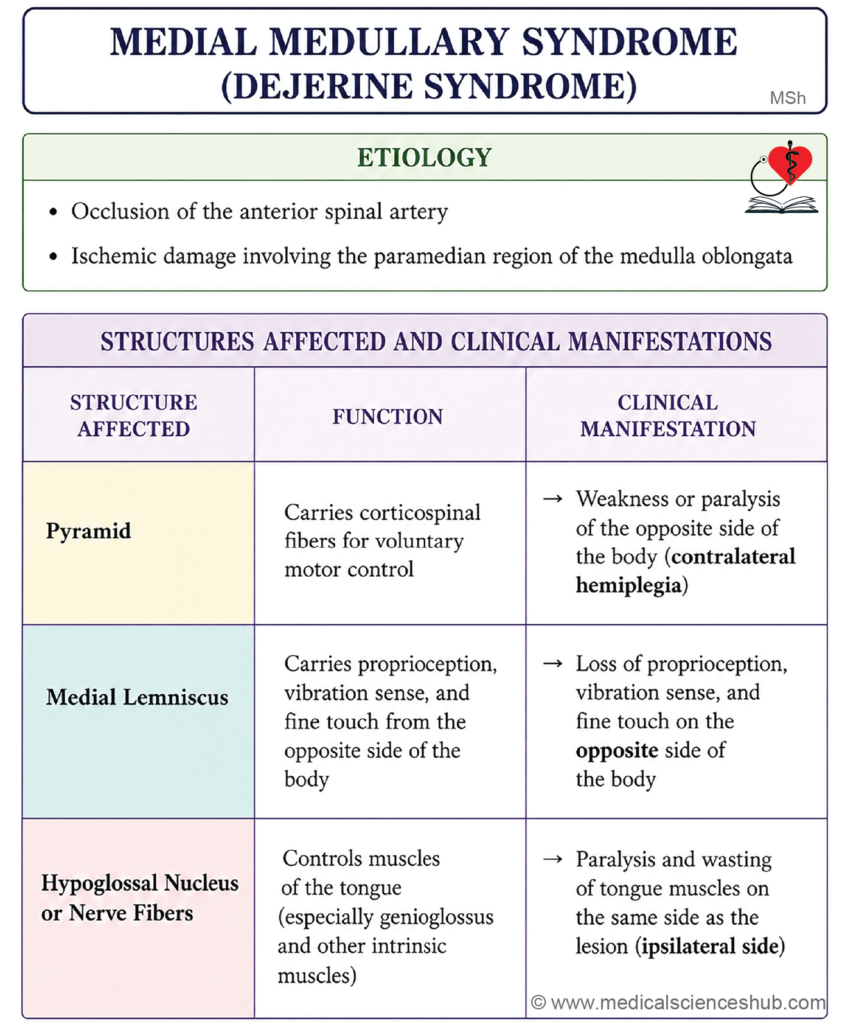
- The anterior spinal artery is formed by the union of two small branches, one arising from each vertebral artery, which fuse to form a single midline vessel. It descends through the anterior median fissure of the medulla, supplying the medial medullary structures, including the pyramids, medial lemniscus, and hypoglossal nerve fibers. Occlusion of this artery results in medial medullary syndrome (Dejerine syndrome), characterized by contralateral hemiplegia, contralateral loss of fine touch and proprioception, and ipsilateral hypoglossal nerve palsy.
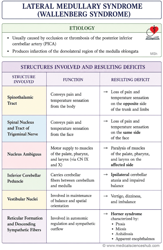
- The posterior inferior cerebellar artery (PICA) supplies the dorsolateral medulla and the inferior cerebellum. Thrombosis or occlusion of this artery produces lateral medullary syndrome (Wallenberg syndrome), one of the most clinically recognized brainstem stroke syndromes, presenting with ipsilateral facial sensory loss, contralateral body sensory loss, dysphagia, hoarseness, ipsilateral Horner syndrome, and cerebellar ataxia.
CLINICAL NEUROANATOMY
Vital Centers
- The vital centers controlling respiration, heart rate, and blood pressure are located in the medulla oblongata, in the region of the floor of the fourth ventricle. Injury to these centers is potentially fatal, as they regulate functions essential for survival.
Medial Medullary Syndrome (Dejerine Syndrome)
- Named after the French neurologist Joseph Jules Dejerine (1849–1917), this condition is also referred to as anterior bulbar syndrome or inferior alternating syndrome.
- It results from a vascular lesion involving the anterior spinal artery, leading to infarction of the paramedian region of the medulla oblongata.
- The structures damaged in this syndrome include the pyramid (corticospinal tract), the medial lemniscus, and the hypoglossal nucleus or nerve fibers (CN XII).
- The clinical presentation includes three characteristic features:
- Contralateral hemiplegia (weakness of the trunk and limbs on the opposite side of the lesion), due to damage to the corticospinal tract in the pyramid.
- Contralateral loss of fine touch, vibration sense, and conscious proprioception, due to damage to the medial lemniscus.
- Ipsilateral lower motor neuron paralysis of the tongue, due to damage to the hypoglossal nucleus or its emerging fibers, causing the tongue to deviate toward the side of the lesion on protrusion.
Lateral Medullary Syndrome (Wallenberg Syndrome)
- Named after the German neurologist Adolf Wallenberg (1862–1949), this condition is also known as posterior inferior cerebellar artery (PICA) syndrome.
- It results from occlusion of the posterior inferior cerebellar artery, a branch of the vertebral artery, leading to infarction of the dorsolateral medulla oblongata.
- The structures damaged include the spinothalamic tract, the spinal nucleus and tract of the trigeminal nerve, the nucleus ambiguus, the inferior cerebellar peduncle, the vestibular nuclei, the reticular formation, and the descending hypothalamospinal sympathetic fibers.
- The clinical presentation includes the following features:
- Contralateral loss of pain and temperature sensation in the trunk and limbs, due to damage to the spinothalamic tract.
- Ipsilateral loss of pain and temperature sensation over the face, due to damage to the spinal nucleus and tract of the trigeminal nerve.
- Ipsilateral paralysis of the palate, pharynx, and larynx, due to damage to the nucleus ambiguus, resulting in dysphagia (difficulty swallowing) and dysphonia (hoarseness of voice).
- Ipsilateral cerebellar ataxia, due to damage to the inferior cerebellar peduncle, manifesting as incoordination of limb movements on the same side as the lesion.
- Vertigo, nausea, and vomiting, due to involvement of the vestibular nuclei.
- Ipsilateral Horner syndrome — comprising ptosis (drooping of the upper eyelid), miosis (constriction of the pupil), and anhidrosis (loss of facial sweating) — due to interruption of the descending sympathetic fibers from the hypothalamus.
Important Questions
- Draw a well-labeled diagram showing TS of medulla at the level of pyramidal decussation.
- Draw a well-labeled diagram of TS of medulla oblongata at the level of sensory decussation.
- Draw a well-labeled diagram of TS of medulla oblongata at the level of olives or open part of medulla.
- Write a short note on lateral medullary syndrome (Wallenberg syndrome)
- Write a short note on medial medullary syndrome (Dejerine syndrome).
📝 Test Your Knowledge – Practice MCQs
Attempt the chapter MCQ quiz and assess your understanding of key concepts.
Click to see: Medical Disclaimer and Copyright Notice