
Competencies
- AN43.2 Identify, describe and draw the microanatomy of tonsil.
- AN70.2 Identify the lymphoid tissue under the microscope and describe microanatomy of lymph node, spleen, thymus, tonsil and correlate the structure with function
Introduction
- The lymphatic system plays a vital role in body defense by contributing to immunity against infectious agents and harmful substances.
- Immunity is the ability of the body to resist infections and toxins through the action of antibodies, immune cells, and activated white blood cells (leukocytes).
- Immunity is broadly classified into two types:
- Innate (natural) immunity
- Acquired (adaptive) immunity
- Innate immunity is present from birth and provides immediate, non-specific protection against a wide range of pathogens.
- Acquired immunity develops during life following exposure to specific antigens and generates targeted immune responses.
- Acquired immunity is of two types:
- Active immunity: Develops when exposure to an antigen, microorganism, or toxin stimulates the body’s own immune system to produce a protective response and immunological memory.
- Passive immunity: Results from the transfer of preformed antibodies from another source, such as maternal antibodies or administered immunoglobulins, providing immediate but temporary protection.
Lymphatic Tissue
- Lymphoid tissue is a specialized connective tissue composed of lymphocytes, plasma cells, reticular cells, and a supporting network of reticular fibers.
- It plays a crucial role in immune defense by recognizing foreign antigens, removing aged or damaged cells, and generating antibody-mediated immune responses.
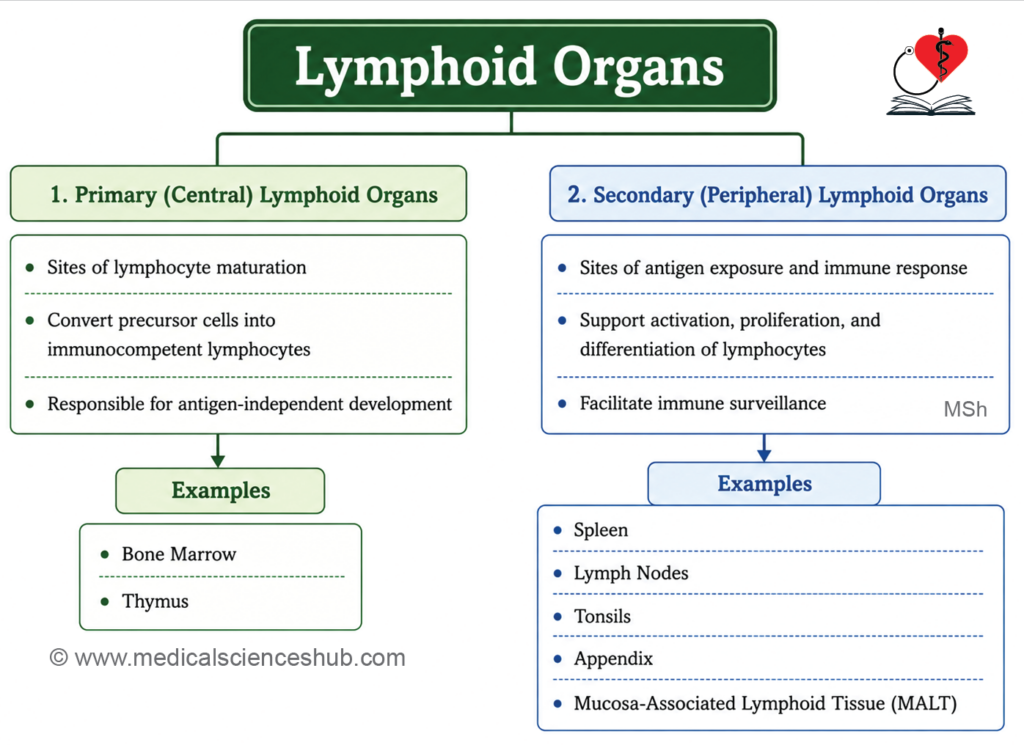
- Lymphoid organs are classified into primary and secondary lymphoid organs based on their functions.
Primary Lymphoid Organs
- Also known as central lymphoid organs.
- They are responsible for the production, maturation, and differentiation of lymphocytes from stem cells.
- Examples include the bone marrow, where B lymphocytes mature, and the thymus, where T lymphocytes undergo maturation.
Secondary Lymphoid Organs
- These organs provide sites where mature lymphocytes encounter foreign antigens and initiate immune responses.
- They facilitate activation, proliferation, and differentiation of lymphocytes.
- Examples include the spleen, lymph nodes, tonsils, appendix, and Peyer’s patches of the ileum.
Diffuse and Dense Lymphoid Tissues
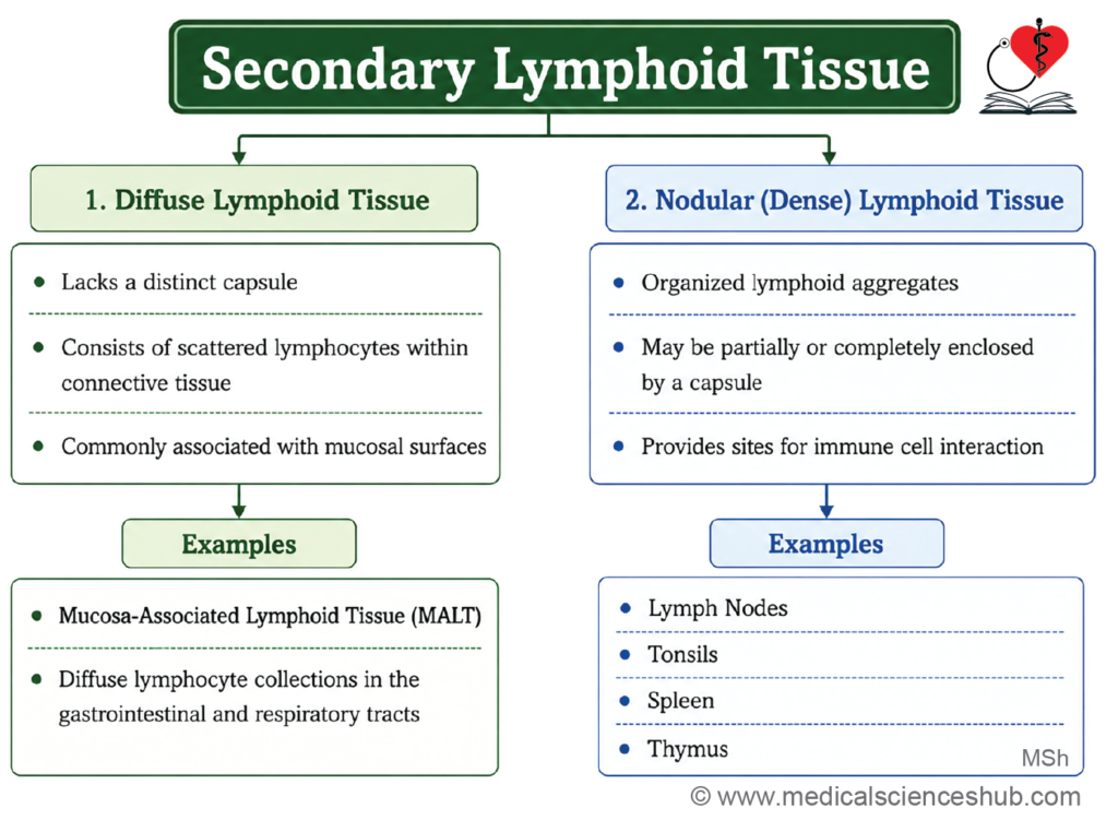
- Lymphoid tissues are grouped as diffuse and dense lymphoid tissues.
Diffuse Lymphoid Tissue
- Consists of scattered lymphocytes within the mucosa of the gastrointestinal, respiratory, urinary, and reproductive tracts.
- It is commonly referred to as mucosa-associated lymphoid tissue (MALT).
- The ileum is a prominent site containing MALT due to the presence of Peyer’s patches.
- MALT provides immune protection against microorganisms entering through mucosal surfaces.
- It is generally non-encapsulated and lacks a distinct outer capsule.
Dense Lymphoid Tissue
- Composed of concentrated aggregates of lymphoid cells.
- It is typically enclosed within a well-defined connective tissue capsule.
- Examples include the lymph nodes, spleen, thymus, and tonsils.
- These structures serve as organized sites for immune surveillance and lymphocyte activation.
Cells of Lymphoid System
- Lymphoid tissue contains lymphocytes along with supporting cells such as reticular cells and epithelioreticular cells.
- Several immune cells are associated with the lymphoid system, including monocytes, macrophages, neutrophils, basophils, and dendritic cells.
- Identification and differentiation of lymphoid cells depend on specific surface molecules known as clusters of differentiation (CD) markers, which are widely used in immunological classification and diagnosis.
Lymphocytes
- Lymphocytes are a subtype of white blood cells and form the principal cellular component of lymphoid tissue.
- They typically possess a large, densely stained (heterochromatic) round nucleus surrounded by a thin rim of cytoplasm.
- Small lymphocytes measure approximately 5–8 μm, while large lymphocytes measure 9–15 μm in diameter.
- Routine histological examination cannot reliably distinguish lymphocyte subtypes.
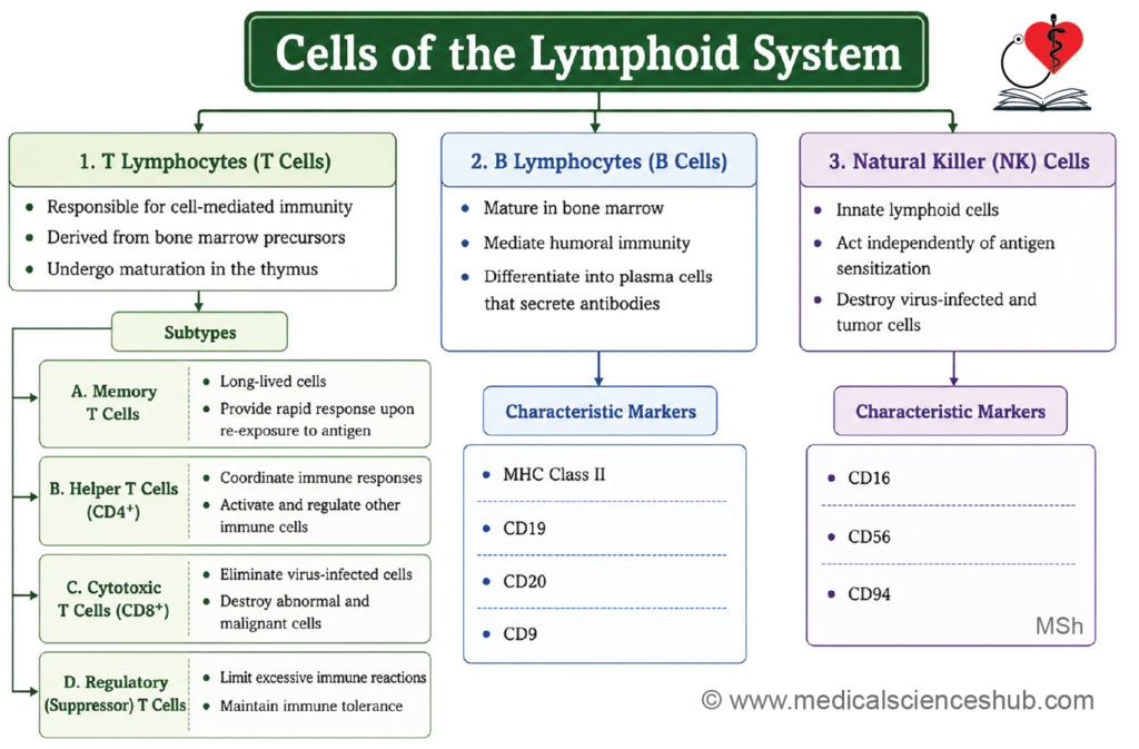
- Based on the expression of specific cluster of differentiation (CD) markers, lymphocytes are classified into T lymphocytes, B lymphocytes, and natural killer (NK) cells.
- All lymphocytes originate from hematopoietic stem cells in the bone marrow.
T Lymphocytes (T Cells)
- Immature lymphoid precursors migrate from the bone marrow to the thymus, where they mature into functional T cells.
- T cells are responsible for cell-mediated immunity.
- They express a T-cell receptor (TCR) that recognizes antigens presented on major histocompatibility complex (MHC) molecules.
- Following antigen exposure, T cells differentiate into effector T cells or memory T cells.
- Memory T cells persist for long periods and respond rapidly upon re-exposure to the same antigen.
Types of Effector T Cells
- Helper T cells (CD4+ T cells):
- Express CD4 surface glycoproteins.
- Regulate immune responses by activating B cells, plasma cells, macrophages, and cytotoxic T cells.
- Secrete cytokines, including interleukins, that coordinate immune activity.
- Cytotoxic T cells (CD8+ T cells):
- Express CD8 surface glycoproteins.
- Destroy virus-infected cells, tumor cells, intracellular pathogen-infected cells, and transplanted foreign cells.
- Regulatory T cells (Treg cells):
- Suppress excessive immune responses.
- Help maintain immune tolerance and prevent autoimmune reactions.
B Lymphocytes (B Cells) /Plasma Cells
- B cells were first identified in the bursa of Fabricius of birds, from which their name is derived.
- In humans, they develop and mature in the bone marrow.
- B cells are responsible for humoral immunity, which is mediated by antibodies.
- They possess membrane-bound B-cell receptors (BCRs) that recognize specific antigens.
- Upon activation, B cells differentiate into:
- Plasma cells, which secrete large amounts of immunoglobulins (antibodies).
- Memory B cells, which provide long-term immunological memory.
- Common B-cell markers include CD19, CD20, and CD79a.
- B cells express MHC class II (MHC II) molecules and can function as antigen-presenting cells.
Natural Killer Cells
- NK cells are lymphocytes that are neither T cells nor B cells.
- They originate from the bone marrow and constitute approximately 5–10% of circulating lymphocytes.
- NK cells provide rapid innate immune defense against virus-infected and malignant cells.
- Characteristic NK-cell markers include CD16 and CD56.
- They can destroy antibody-coated target cells through antibody-dependent cellular cytotoxicity (ADCC).
- Unlike T cells, NK cells do not require prior antigen sensitization for their cytotoxic activity.
Antigen-Presenting Cells
- Antigen-presenting cells (APCs) capture, process, and present antigens to CD4+ helper T lymphocytes through MHC class II (MHC II) molecules.
- During antigen processing, foreign material is degraded within lysosomes, and antigenic peptides are displayed on the cell surface bound to MHC II molecules.
- Major APCs include macrophages, Kupffer cells of the liver, Langerhans cells of the epidermis, dendritic cells of lymphoid organs, B lymphocytes, and certain epithelioreticular cells of the thymus.
Reticular Cells
- Reticular cells are specialized connective tissue cells that form the supporting framework of lymphoid organs such as the spleen, lymph nodes, and tonsils.
- They synthesize type III collagen (reticular fibers) and ground substance.
- These cells are elongated and possess slender cytoplasmic processes that contribute to the reticular network.
Lymph
- Lymph is a clear to pale-yellow fluid that circulates through lymphatic vessels and lymph nodes.
- It is formed from excess interstitial (tissue) fluid that enters lymphatic capillaries and contains water, plasma proteins, and dissolved substances.
- Lymph contains white blood cells, predominantly lymphocytes, which contribute to immune defense.
- Intestinal lymph may contain fat-rich chylomicrons absorbed from the digestive tract; this milky lymph is known as chyle.
- Lymph can also transport microorganisms, foreign particles, and occasionally malignant cells from tissues.
Lymphatic Vessels
- Lymphatic vessels collect excess interstitial fluid, plasma proteins, lipids, and other large molecules from tissues and return them to the bloodstream.
- The lymphatic vascular system consists of lymphatic capillaries, larger lymphatic vessels, lymphatic trunks, and the thoracic duct.
Lymph Capillaries
- Lymphatic capillaries begin as blind-ended channels within connective tissues.
- They are lined by a thin layer of endothelial cells and are more permeable than blood capillaries.
- Their high permeability allows absorption of large molecules, including dietary lipids in the intestine.
- Lymphatic capillaries are present in most tissues but are absent in the epidermis, cornea, cartilage, hair, nails, bone marrow, splenic red pulp, choroid of the eye, internal ear, central nervous system, and most meninges.
- Lymph reaches lymph nodes through afferent lymphatic vessels and leaves through efferent lymphatic vessels.
Larger Lymph Vessels
- Lymphatic capillaries unite to form progressively larger vessels and trunks.
- Major lymphatic channels include the thoracic duct, right lymphatic duct, jugular trunks, subclavian trunks, bronchomediastinal trunks, intestinal trunks, and lumbar trunks.
- Similar to veins, larger lymphatic vessels possess tunica intima, tunica media, and tunica adventitia.
- Distinguishing features include:
- Elastic fibers are present throughout all vessel layers.
- Smooth muscle cells occur in both the tunica media and tunica adventitia.
- In the thoracic duct, smooth muscle fibers are predominantly arranged in a longitudinal orientation, facilitating lymph propulsion.
Lymphatic Nodule/Follicle
- A lymphatic nodule (lymphoid follicle) is a localized, well-defined aggregation of lymphocytes within lymphoid tissue.
- It is typically spherical or ovoid in shape and lacks a surrounding capsule.
- Lymphatic nodules are found in most secondary lymphoid tissues but are absent in the thymus.
- They are classified into primary and secondary nodules.
Primary Nodules
- Consist mainly of small, mature lymphocytes with a relatively uniform appearance.
- They develop before birth and may transform into secondary nodules after antigenic stimulation.
Secondary Nodules
- Form following exposure to an antigen and contain a prominent germinal center.
- The germinal center contains actively proliferating lymphoblasts, plasmablasts, and follicular dendritic cells (FDCs) that support lymphocyte activation and proliferation.
- Surrounding the germinal center is the corona (mantle zone), which contains densely packed mature lymphocytes.
- Histologically, germinal centers appear lighter than the surrounding mantle zone because of their larger, less condensed nuclei.
Lymph Node
- A lymph node is a small, kidney-shaped lymphoid organ that filters lymph and plays an important role in immune surveillance.
- It has a convex outer surface and a concave hilum, which serves as the entry and exit point for blood vessels and lymphatic structures.
- Multiple afferent lymphatic vessels enter the lymph node through its convex surface and deliver lymph for filtration.
- After passing through the node, filtered lymph exits via one or more efferent lymphatic vessels at the hilum.
- Blood vessels also enter and leave the lymph node through the hilum, supplying nutrients and supporting immune cell activity.
- Lymph nodes act as sites for antigen recognition, lymphocyte activation, and removal of foreign particles and microorganisms.
Histology of Lymph Node
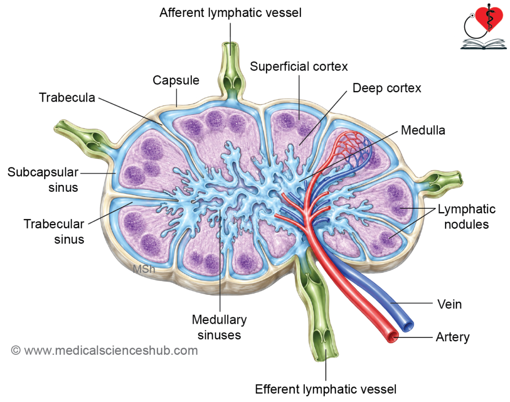
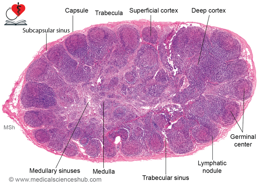
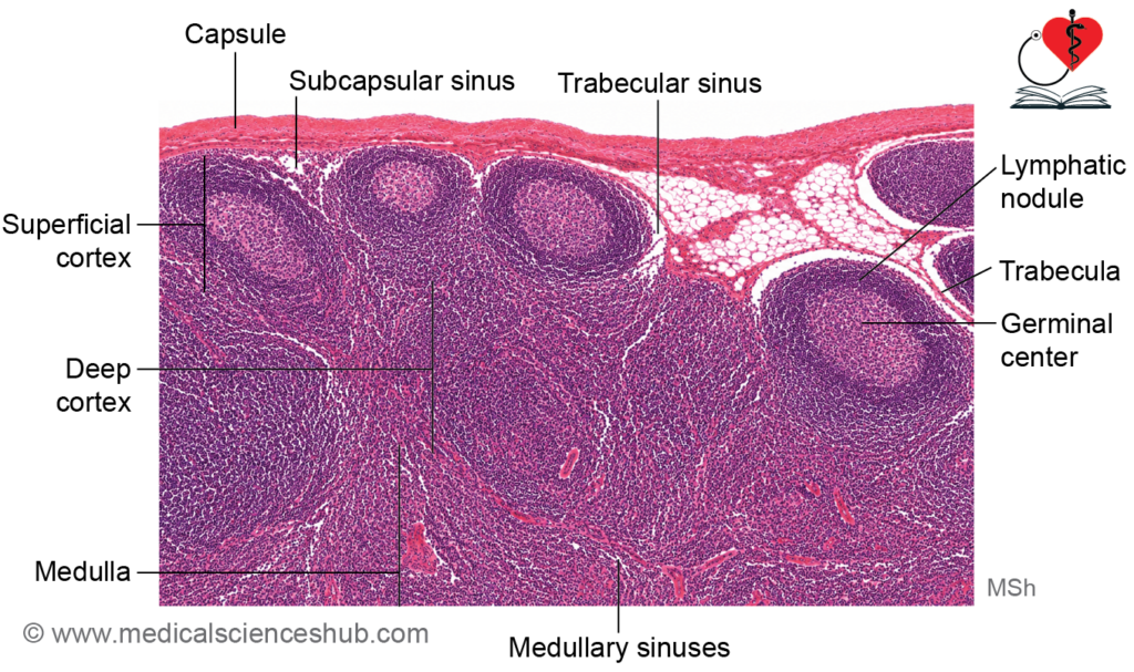
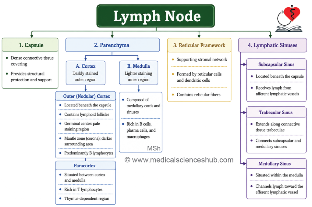
- A lymph node is enclosed by a dense connective tissue capsule, from which connective tissue trabeculae extend into the interior of the organ.
- The supporting framework consists of reticular tissue, composed of reticular cells, reticular fibers (type III collagen), macrophages, and dendritic cells.
- Reticular cells are elongated cells that synthesize the reticular fiber network, while dendritic cells function as important antigen-presenting cells.
The lymph node parenchyma is divided into an outer cortex and an inner medulla.
Parenchyma of Lymph Node
- The cortex is the darkly stained outer region of the lymph node.
- It consists of:
- Superficial (nodular) cortex, which contains numerous lymphatic follicles (nodules).
- Paracortex (deep cortex), located between the superficial cortex and medulla.
- The nodular cortex is rich in B lymphocytes.
- Secondary lymphoid follicles contain a pale germinal center, where B-cell proliferation and differentiation occur, surrounded by a darker mantle (corona) zone of mature lymphocytes.
- The paracortex is predominantly populated by T lymphocytes and is therefore termed the thymus-dependent zone.
Sinuses of Lymph NodeViva
- Lymph flows through a system of interconnected sinuses:
- Subcapsular sinus: Located immediately beneath the capsule and receives lymph from afferent lymphatic vessels.
- Trabecular (intermediate) sinuses: Run alongside trabeculae and connect the subcapsular sinus to the medullary sinuses.
- Medullary sinuses: Receive lymph from trabecular sinuses and drain it toward efferent lymphatic vessels.
- These sinuses contain a reticular meshwork that facilitates lymph filtration, trapping of microorganisms and cellular debris, and presentation of antigens to immune cells.
Medulla
- The medulla forms the central, lighter-staining region of the lymph node.
- It contains:
- Medullary cords, composed mainly of B cells, plasma cells, and macrophages.
- Medullary sinuses, which separate adjacent cords and converge at the hilum to drain into efferent lymphatic vessels.
- The medulla plays an important role in antibody production and final filtration of lymph before it leaves the node.
Functions of Lymph Node
- Lymph nodes filter lymph and remove microorganisms, foreign particles, and cellular debris.
- They serve as sites for antigen presentation to T lymphocytes, initiating immune responses.
- Lymph nodes promote activation and proliferation of B lymphocytes.
- Activated B cells differentiate into plasma cells, which produce antibodies.
- Thus, lymph nodes support both humoral immunity and cell-mediated immunity while contributing to host defense.
CLINICAL CORRELATION
- Lymphadenitis is inflammation of a lymph node, resulting in enlargement due to increased immune cell activity and lymphocyte proliferation. Common causes include infection and involvement of lymph nodes by metastatic cancer. Clinical features include swollen, tender lymph nodes and fever.
- In lymph node metastasis, malignant cells from a primary tumor become trapped within lymph nodes, proliferate, and form secondary tumor deposits.
Thymus (Thymic Gland)
- Thymus and bone marrow are the primary lymphoid organs responsible for lymphocyte development and maturation.
- The thymus is a bilobed organ located in the anterior part of the superior mediastinum.
- Embryologically, it develops mainly from the third pharyngeal pouch endoderm, while mesenchymal tissue contributes connective tissue components.
- The thymus enlarges from birth until puberty and subsequently undergoes gradual age-related shrinkage, known as thymic involution.
Histology of Thymus
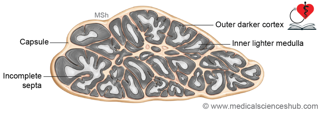
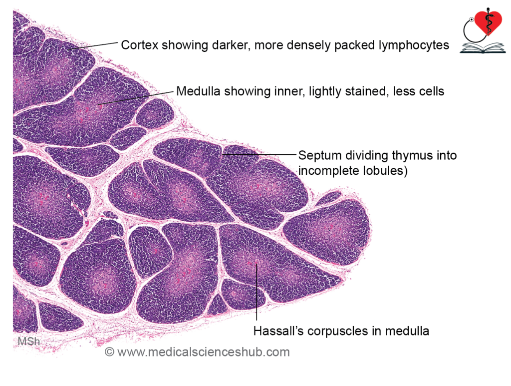
- The thymus is enclosed by a thin connective tissue capsule that sends septa (trabeculae) into the organ, dividing it into incomplete thymic lobules.
- Each lobule consists of an outer darkly stained cortex and an inner lightly stained medulla.
- Because the septa do not completely separate the lobules, the medullae of adjacent lobules are continuous.
- Unlike lymph nodes, the thymus does not contain lymphoid nodules or germinal centers.
Cortex
- The cortex is the peripheral, deeply basophilic region due to its high density of thymocytes (immature T lymphocytes).
- Thymocytes are supported by a network of epithelioreticular cells, which are derived from the endoderm of the third pharyngeal pouch.
- Macrophages are also present and remove apoptotic or degenerating thymocytes.
Medulla
- The medulla is the central, lightly stained region because it contains fewer thymocytes.
- It contains both thymocytes and epithelioreticular cells.
- The medulla of neighboring lobules is continuous, a characteristic feature of the thymus.
- The most distinctive feature of the medulla is the presence of Hassall’s corpuscles.
Hassall’s/Thymic CorpusclesViva, Neet
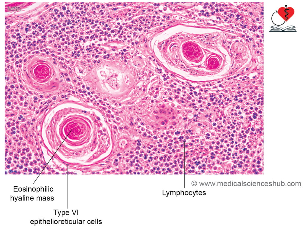
- Hassall’s corpuscles are rounded or ovoid structures found exclusively in the thymic medulla.
- They consist of concentrically arranged type VI epithelioreticular cells surrounding a central eosinophilic, hyalinized core.
- Their number increases with age and they are involved in thymocyte maturation and immune regulation.
- Epithelioreticular cells within these corpuscles produce cytokines such as IL-4 and IL-6, which support T-cell development.
- The thymus also secretes hormones including thymosin and thymopoietin, which contribute to T-lymphocyte maturation.
Epithelioreticular cells
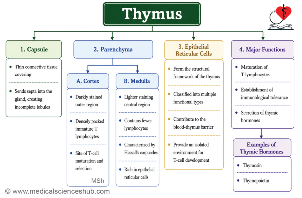
- Epithelioreticular cells form the supporting framework of the thymus and provide the specialized microenvironment required for T-lymphocyte maturation.
- These cells are derived from the endoderm of the third pharyngeal pouch and are often referred to as nurse cells because they support developing thymocytes.
- Six types of epithelioreticular cells are recognized. Types I–III are located in the cortex, whereas Types IV–VI are found in the medulla.
Functions of Different Types
- Type I cells: Form a barrier between the thymic cortex and surrounding connective tissue and blood vessels through tight junctions.
- Type II cells: Support developing thymocytes and participate in T-cell selection and maturation.
- Type III cells: Contribute to the boundary between the cortex and medulla.
- Type IV cells: Work with Type III cells to form the corticomedullary junction.
- Type V cells: Create the cellular framework of the medulla.
- Type VI cells: Form the concentric layers of Hassall’s corpuscles and secrete cytokines such as IL-4 and IL-6, which aid thymocyte maturation.
- The thymus also produces hormones, including thymosin and thymopoietin, which contribute to T-cell development and differentiation.
Functions of Thymus
- The thymus is the primary site for the maturation, differentiation, and selection of T lymphocytes.
- It provides a specialized and protected microenvironment necessary for T-cell development.
- Epithelioreticular cells secrete hormones such as thymopoietin and thymosin, which support T-cell maturation.
- The thymus is essential for the establishment of effective cell-mediated immunity during early life.
- After puberty, it undergoes gradual age-related shrinkage, known as thymic involution.
Blood–Thymic Barrier
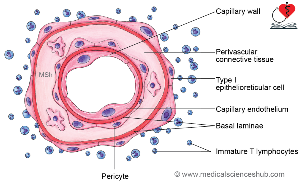
- The blood-thymic barrier protects developing T lymphocytes from exposure to circulating antigens during maturation.
- It creates an isolated microenvironment essential for proper T-cell development.
- The barrier is formed by:
- Continuous capillary endothelium with a basal lamina and pericytes,
- Perivascular connective tissue containing macrophages,
- Type I epithelioreticular cells.
- Tight (occluding) junctions between endothelial and epithelioreticular cells restrict antigen entry and maintain immune isolation of immature thymocytes.
CLINICAL CORRELATION
Myasthenia gravis
- Myasthenia gravis is an autoimmune disorder characterized by antibodies directed against acetylcholine receptors at the neuromuscular junction.
- It is frequently associated with thymic abnormalities, including thymic hyperplasia and, in some cases, thymoma.
- Loss of functional receptors impairs neuromuscular transmission, leading to muscle weakness and fatigue.
- Common manifestations include ptosis (drooping eyelids), diplopia (double vision), dysarthria, and difficulty swallowing.
DiGeorge syndrome
- This genetic disorder results from deletion of a segment of chromosome 22q11.2.
- It is characterized by underdevelopment or absence of the thymus and parathyroid glands.
- Defective thymic development causes severe deficiency of T lymphocytes and impaired cell-mediated immunity, leading to recurrent infections.
- Hypoparathyroidism may result in hypocalcemic tetany.
- Associated features include congenital heart defects, cleft palate, and developmental delay.
Spleen
- The spleen is the largest secondary lymphoid organ in the body.
- It functions as a blood-filtering organ, removing aged or damaged red blood cells (RBCs) and contributing to immune surveillance.
- The spleen is located in the left upper quadrant of the abdomen, beneath the diaphragm.
- It possesses a diaphragmatic surface facing laterally and a visceral surface facing medially.
- The visceral surface contains the hilum, through which the splenic artery, splenic vein, lymphatic vessels, and nerves enter or leave the organ.
- The spleen plays important roles in blood filtration, immune responses, and recycling of erythrocyte components.
Gross Section of Spleen
- A fresh cut section of the spleen reveals two distinct components: white pulp and red pulp.
- White pulp appears as scattered grayish-white areas distributed throughout the splenic tissue.
- Red pulp surrounds the white pulp and constitutes the majority of the spleen.
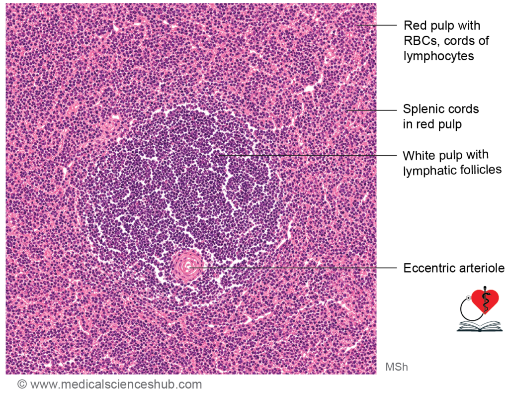
- Histologically, white pulp consists of dense aggregates of lymphocytes surrounding branches of the splenic artery and appears basophilic on hematoxylin and eosin (H&E) staining.
- Red pulp is composed of numerous blood-filled splenic sinusoids and splenic cords, giving it a reddish appearance.
- The white pulp is primarily involved in immune responses, whereas the red pulp functions in blood filtration and removal of aged or damaged erythrocytes.
Histology of Spleen
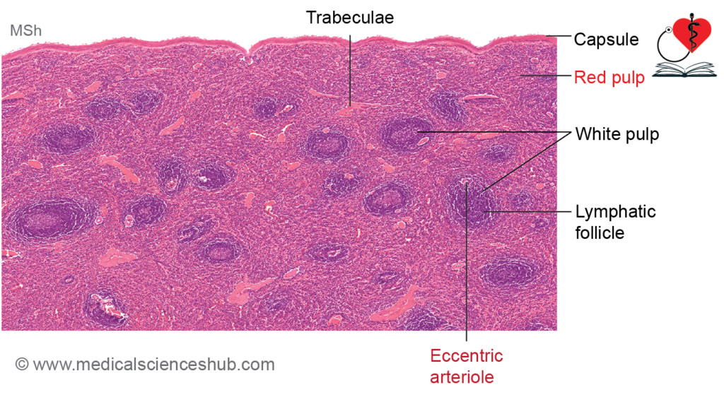
- Histologically, spleen shows capsule, white pulp and red pulp. Spleen does not have afferent lymphatics.
Capsule and Trabeculae
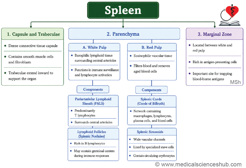
- The spleen is enclosed by a dense connective tissue capsule from which trabeculae extend into the parenchyma and branch repeatedly.
- A supporting framework is formed by reticular cells and reticular fibers.
- The capsule and trabeculae contain myofibroblasts, whose contraction helps mobilize and release stored blood from the spleen when required.
White Pulp
- The white pulp of the spleen consists of lymphoid tissue and appears as pale areas in a fresh section.
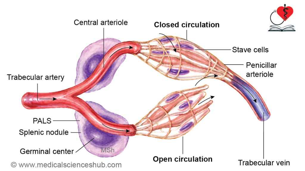
- Branches of the splenic artery form central arterioles, which are surrounded by cylindrical aggregates of lymphocytes called periarteriolar lymphoid sheaths (PALS).
- PALS are composed predominantly of T lymphocytes and therefore represent the thymus-dependent zone of the spleen.
- In histological cross-sections, PALS appears as a lymphoid sheath surrounding a central arteriole.
- Following antigenic stimulation, B lymphocytes proliferate within the white pulp to form splenic nodules (Malpighian corpuscles) containing a germinal center.
- Expansion of the germinal center displaces the central arteriole toward the periphery, producing the characteristic eccentric arteriole seen in splenic nodules.
Red Pulp
- Red pulp surrounds the white pulp and constitutes the major portion of the splenic parenchyma.
- It is composed of two principal components:
- Splenic cords (Cords of Billroth)
- Splenic sinusoids
Splenic Cords (Cords of Billroth)
- These are irregular, interconnected cords of tissue forming a supportive network within the red pulp.
- They contain reticular cells, reticular fibers, lymphocytes, plasma cells, macrophages, and numerous red blood cells (RBCs).
- Macrophages within the cords play an important role in the removal of aged or damaged erythrocytes.
Splenic Sinusoids
- Splenic cords are separated by wide vascular channels known as splenic sinusoids.
- Sinusoids possess a large lumen and are lined by elongated endothelial cells arranged parallel to the vessel axis.
- Their basal lamina is discontinuous, and adjacent endothelial cells are separated by slit-like gaps.
- These structural features allow blood cells to move between the splenic cords and sinusoids, facilitating blood filtration.
- Unlike most capillaries, splenic sinusoids lack pericytes and smooth muscle cells.
- Because RBCs are present in both the splenic cords and sinusoids, distinguishing the two structures histologically may be challenging.
Marginal Zone
- The marginal zone is a specialized region located at the interface between the white pulp and red pulp of the spleen.
- It contains a network of reticular tissue, blood sinusoids, lymphocytes, macrophages, and numerous antigen-presenting cells (APCs).
- This region serves as an important site for immune surveillance of blood-borne antigens.
- Antigen-presenting cells capture and process circulating antigens and present them to lymphocytes, thereby initiating appropriate immune responses.
- The marginal zone plays a key role in linking blood filtration with activation of both innate and adaptive immunity.
Functions of Spleen
- The spleen performs important immune, hematological, and blood-filtering functions.
- It activates lymphocytes through the action of antigen-presenting cells (APCs) and initiates immune responses against blood-borne antigens.
- The spleen serves as a site for the proliferation and activation of T lymphocytes and B lymphocytes.
- It removes and destroys aged, damaged, or abnormal red blood cells (RBCs) and platelets from the circulation.
- During fetal life, the spleen functions as a significant hematopoietic organ, contributing to blood cell formation.
- The spleen also acts as a reservoir, storing a limited volume of blood and releasing it when needed.
CLINICAL CORRELATION
Splenomegaly
- Splenomegaly refers to abnormal enlargement of the spleen.
- Common causes include hematological disorders such as thalassemia and sickle cell disease, infections (e.g., malaria and typhoid), autoimmune diseases, leukemia, and portal hypertension associated with liver disease.
Splenectomy
- Splenectomy is the surgical removal of the spleen.
- It may be performed for splenic trauma, certain hematological disorders, malignancies, or severe hemolytic anemia.
- Following splenectomy, susceptibility to infections increases because of reduced splenic immune function.
Palatine Tonsil
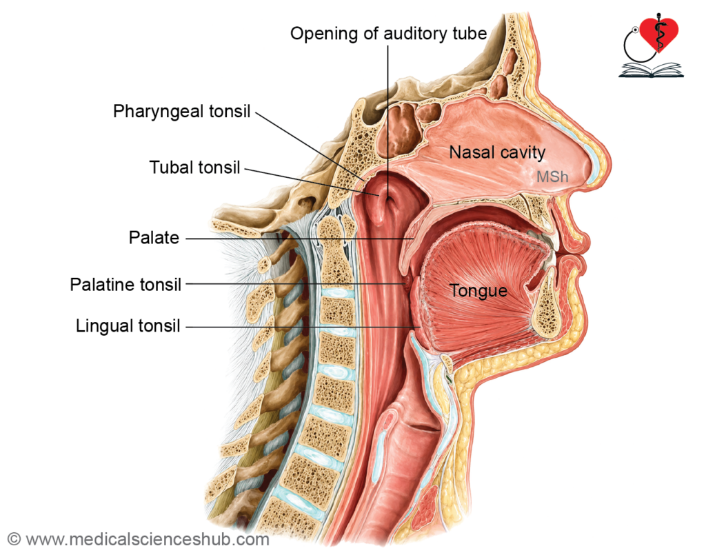
- Tonsils are aggregates of lymphoid tissue located at the junction of the nasal and oral cavities with the pharynx.
- Together, they form Waldeyer’s tonsillar ring, which includes the pharyngeal, tubal, palatine, and lingual tonsils.
- The palatine tonsils are the largest and are situated on either side of the oropharynx, between the palatoglossal and palatopharyngeal arches.
- Tonsils constitute an important component of mucosa-associated lymphoid tissue (MALT).
- They provide immune surveillance and help initiate immune responses against inhaled and ingested pathogens.
Histology of Palatine Tonsil
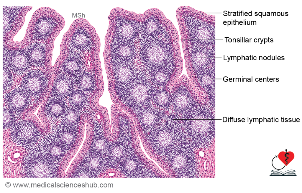
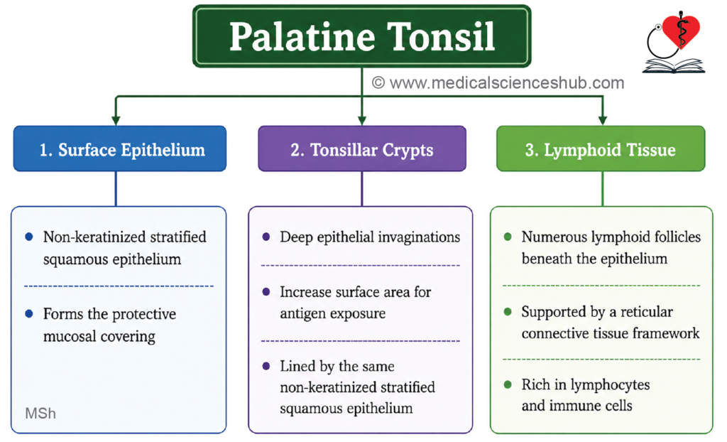
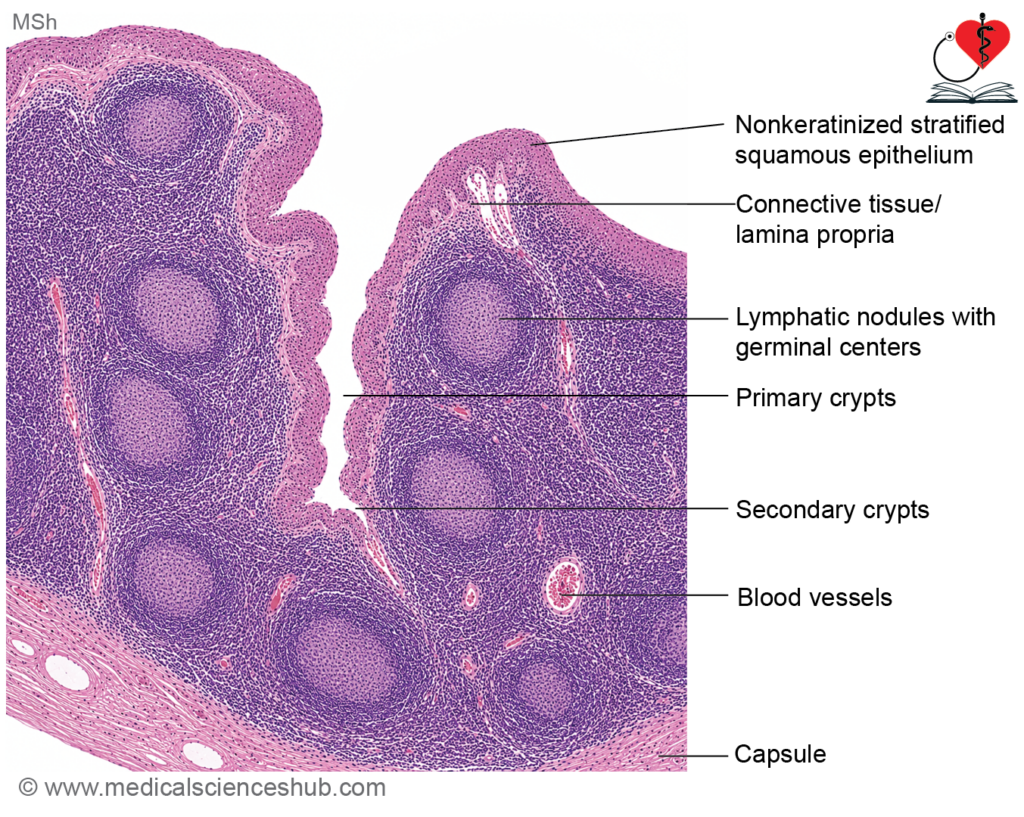
- Histologically, the palatine tonsil consists of a lining epithelium, tonsillar crypts, and abundant lymphoid follicles.
- The oral surface is covered by non-keratinized stratified squamous epithelium.
- This epithelium invaginates deeply into the tonsil to form tonsillar crypts, which increase the surface area exposed to antigens.
- Crypts are lined by the same non-keratinized stratified squamous epithelium and often contain infiltrating lymphocytes, making the boundary between epithelium and lymphoid tissue indistinct.
- The lateral surface of the tonsil is enclosed by a connective tissue capsule, from which efferent lymphatic vessels emerge.
- Beneath the epithelium lies dense lymphoid tissue containing numerous lymphoid follicles, supported by a network of reticular fibers.
Functions of Tonsil
- Produces and supports the proliferation of lymphocytes.
- Participates in antigen presentation and initiates immune responses against inhaled and ingested pathogens.
CLINICAL CORRELATION
- Tonsillitis is inflammation of the tonsils, most commonly caused by viral or bacterial infections.
- Tonsillectomy is the surgical removal of the tonsils and is often performed in cases of recurrent or chronic tonsillitis.
- Salivary corpuscles are formed when shed epithelial cells and lymphocytes from the palatine tonsils mix with saliva in the oral cavity.
Important Questions
- List the primary and secondary lymphoid organs.
- List the cells of lymphoid tissue.
- What is lymph.
- What is lymphatic follicle.
- Write a short note on blood–thymic barrier.
- Write a short note on histology of thymus.
- Write a short note on histology of spleen.
- Write a short note on histology of tonsil.
📝 Test Your Knowledge – Practice MCQs
Attempt the chapter MCQ quiz and assess your understanding of key concepts.
Click to see: Medical Disclaimer and Copyright Notice