
Competencies
- AN25.2: Describe development of pleura, lungs, and heart.
- AN25.4: Describe embryological basis of (1) atrial septal defect, (2) ventricular septal defect, (3) Fallot’s tetralogy, (4) tracheooesophageal fistula.
Introduction
- The increasing metabolic and nutritional demands of the developing embryo lead to the early formation of the cardiovascular system, which begins to develop during the third week of intrauterine life. All structural components of this system arise from the mesoderm. This section focuses on the early stages of heart development.
Establishment of the Cardiogenic Area (Heart Field)
- During the third week, cardiac progenitor cells appear in the epiblast, located lateral to the primitive streak. These cells migrate through the primitive streak in a cranial direction and accumulate within the splanchnopleuric mesoderm, where they form a horseshoe-shaped primary heart field by the end of the third week.
- With the formation of the head fold, this cardiogenic region shifts ventrally and comes to lie anterior to the foregut within the developing pericardial cavity.
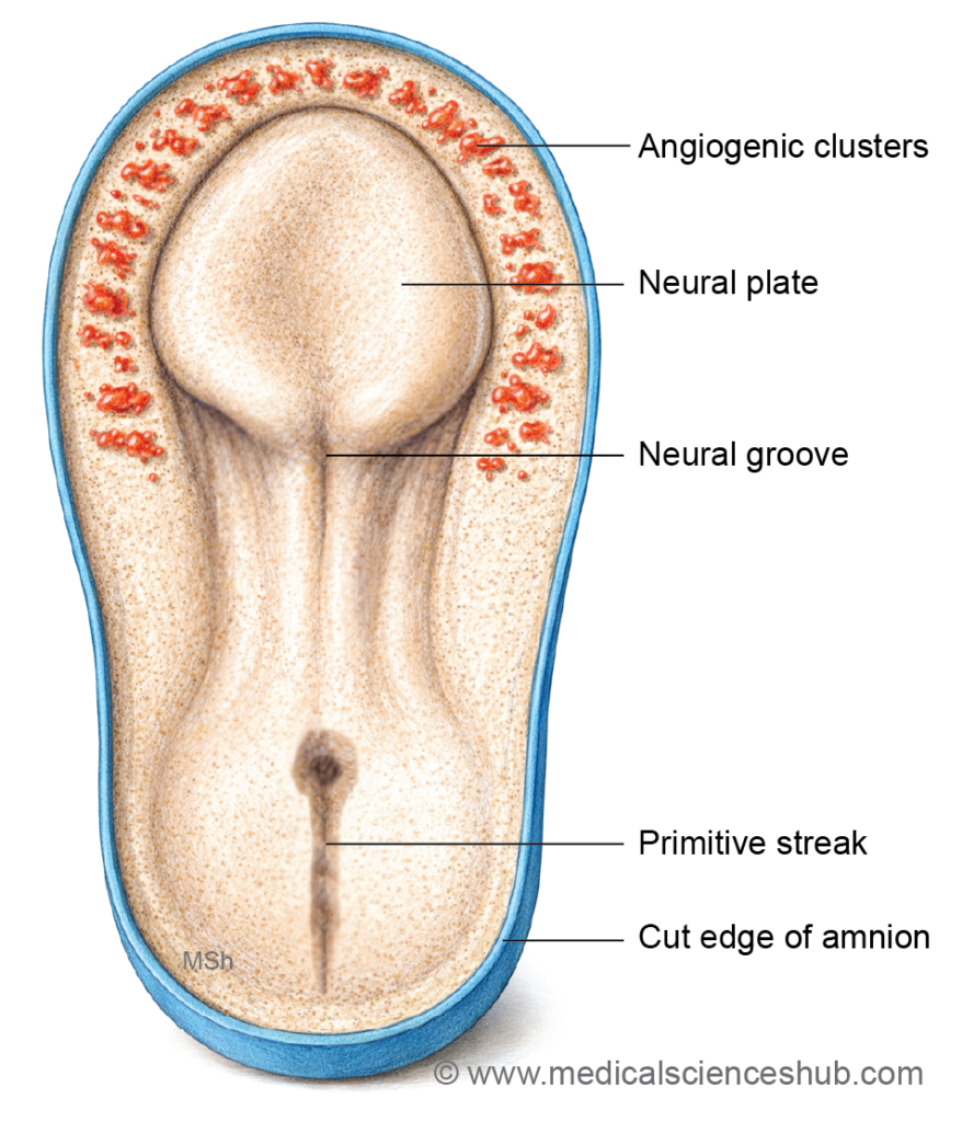
Primary and Secondary Heart Fields
- Most of the heart develops from the primary heart field, which contributes mainly to the left ventricle and parts of the atrial region. Around day 20–21, an additional group of progenitor cells forms the secondary heart field in the splanchnopleuric mesoderm located ventral to the pharynx. Cells from this region contribute to the right ventricle and the outflow tract of both ventricles.
- Notably, the circulatory system is the first functional system to develop in the embryo.
Formation of the Primitive Heart Tube
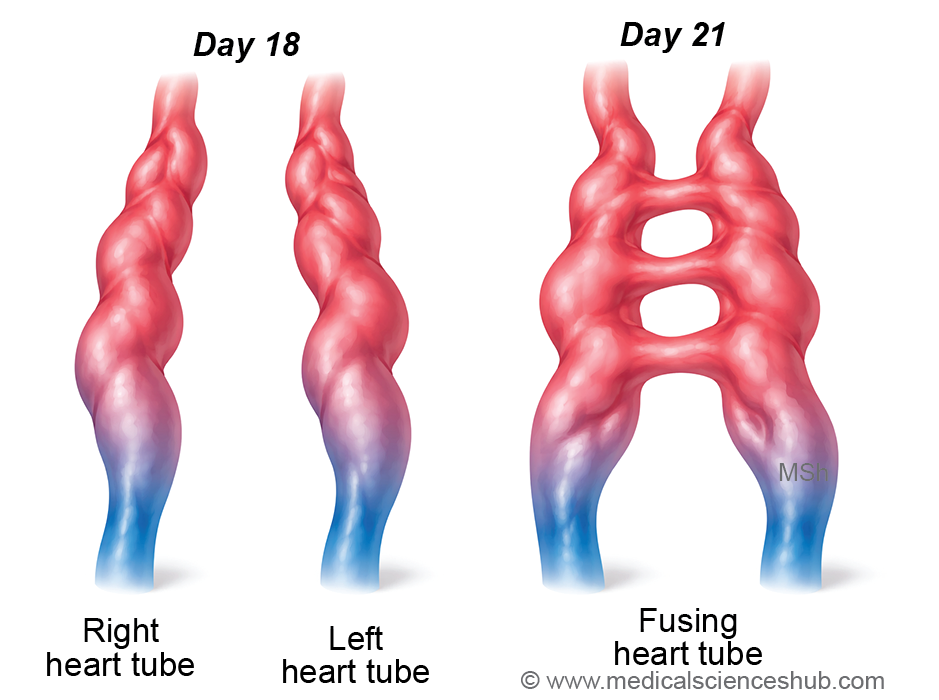
- The endoderm of the primitive pharynx induces vasculogenesis within the primary heart field, leading to the formation of small vascular channels. These channels fuse to produce two endothelial heart tubes (right and left), which later merge to form the primitive heart tube. The inner lining of this tube differentiates into the endocardium.
- The surrounding splanchnopleuric mesoderm forms a myoepicardial mantle, which subsequently differentiates into:
- Myocardium – the muscular layer of the heart (derived from splanchnopleuric mesoderm)
- Epicardium – the visceral layer of the pericardium
- The somatopleuric mesoderm surrounding the pericardial cavity forms the parietal layer of the pericardium.
- The primitive heart begins to beat around day 22, and by approximately day 24, blood circulation within the embryo is established.
Heart Tubes
Formation of the Primitive Heart Tube
- During the third week of development, two endothelial heart tubes form within the cardiogenic region. These tubes gradually move toward the midline and fuse to form a single primitive heart tube. However, the extreme ends of the tube initially remain bifurcated.
- The cranial end of the tube is known as the arterial end, while the caudal end is called the venous end.
Dilatations of the Heart Tube
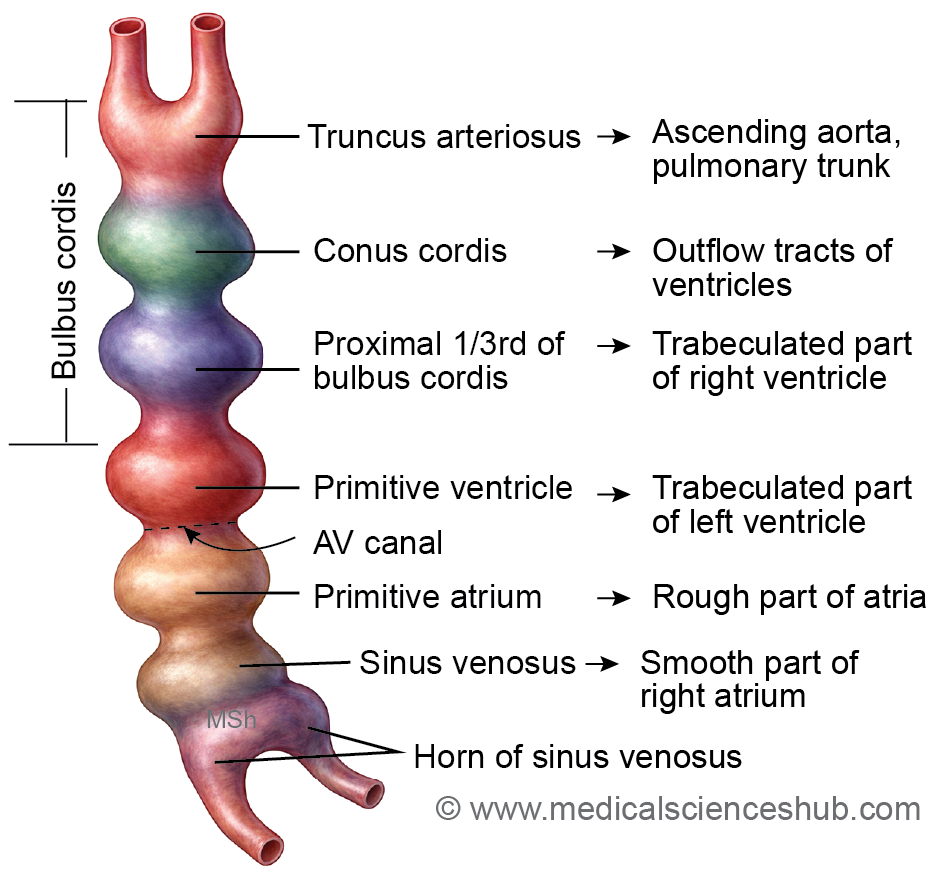
- Soon after its formation, the primitive heart tube develops a series of segmental dilatations arranged from cranial to caudal direction:
- Bulbus cordis, which later differentiates into:
- Truncus arteriosus
- Conus cordis
- Proximal bulbar part
- Primitive ventricle
- Primitive atrium
- Sinus venosus
- Bulbus cordis, which later differentiates into:
- These dilated regions represent the early structural components from which the chambers and outflow tracts of the adult heart develop.
Arterial End of the Heart Tube
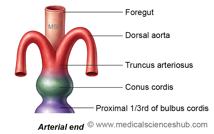
- The arterial end, represented by the truncus arteriosus, initially divides into right and left limbs. These limbs connect with the dorsal aortae through the first pair of pharyngeal arch arteries.
- Subsequently, six pairs of pharyngeal arch arteries develop and link the truncus arteriosus with the dorsal aortae. These arteries run along either side of the foregut (primitive pharynx).
Venous End of the Heart Tube
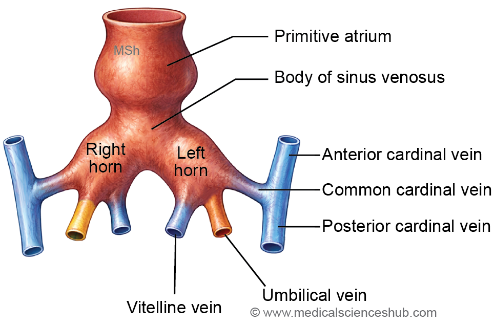
- The caudal portion of the heart tube forms the sinus venosus, which consists of right and left horns.
- Each horn receives three major embryonic veins (arranged from lateral to medial):
- Common cardinal vein – drains blood from the body wall
- Umbilical vein – carries oxygenated blood from the placenta
- Vitelline vein – returns blood from the yolk sac
- These venous channels play a key role in establishing the early embryonic venous circulation.
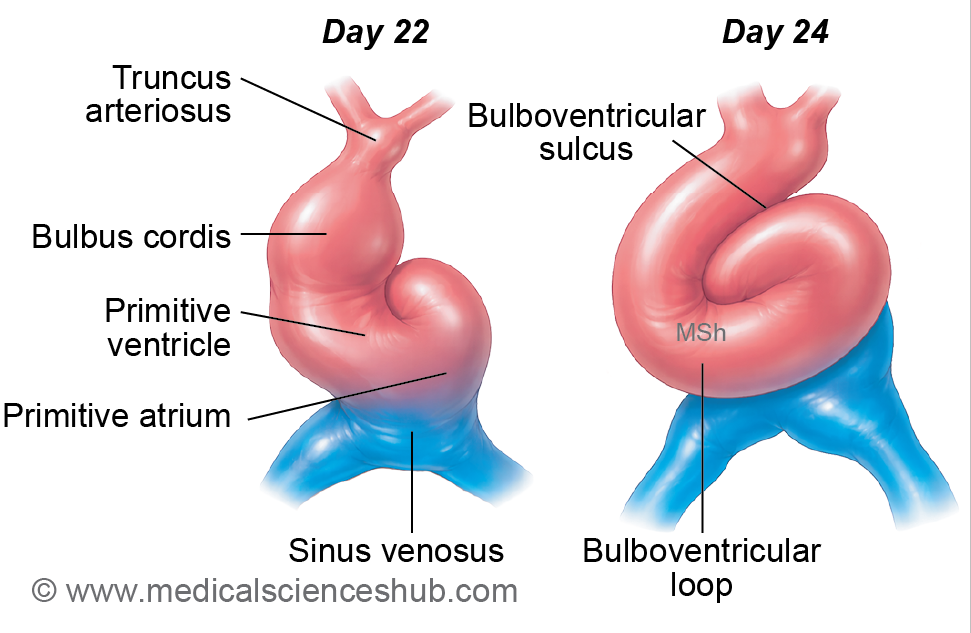
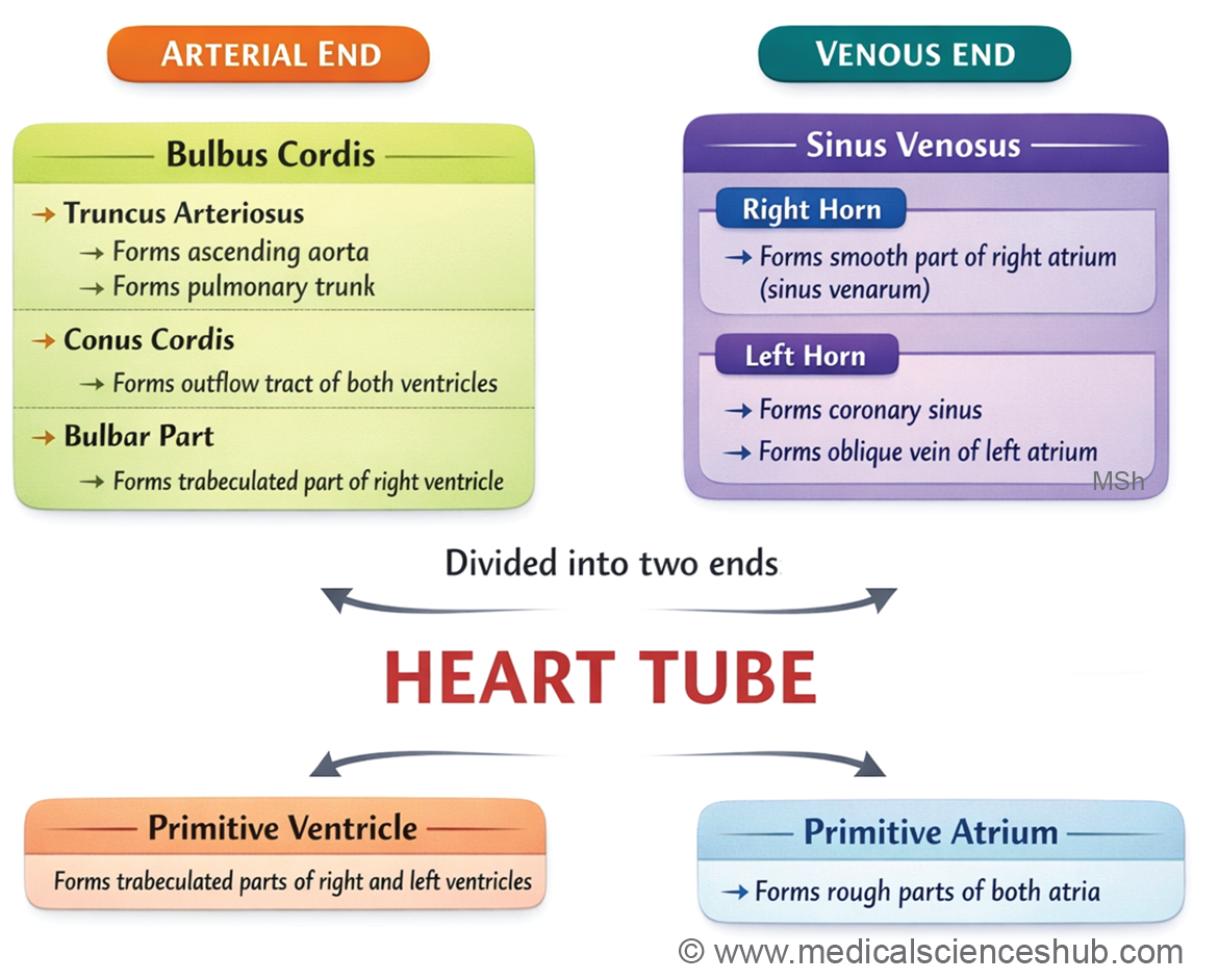
Table 17.1: Fate of the Components of the Primitive Heart Tube
| Component of Primitive Heart Tube | Major Adult Derivatives |
|---|---|
| Truncus arteriosus | Divides to form the ascending aorta and pulmonary trunk, which constitute the main arterial outflow tracts of the heart. |
| Bulbus cordis | Develops into the smooth outflow regions of the ventricles: the conus arteriosus of the right ventricle and the aortic vestibule of the left ventricle. |
| Primitive ventricle | Forms the trabeculated (muscular) portions of both the right and left ventricles. |
| Primitive atrium | Gives rise to the trabeculated parts of the right and left atria, including the auricular appendages. |
| Sinus venosus | Differentiates asymmetrically: the right horn becomes the sinus venarum (smooth posterior wall of the right atrium), while the left horn forms the coronary sinus and the oblique vein of the left atrium. |
Acquisition Of External Features Of The Adult Heart
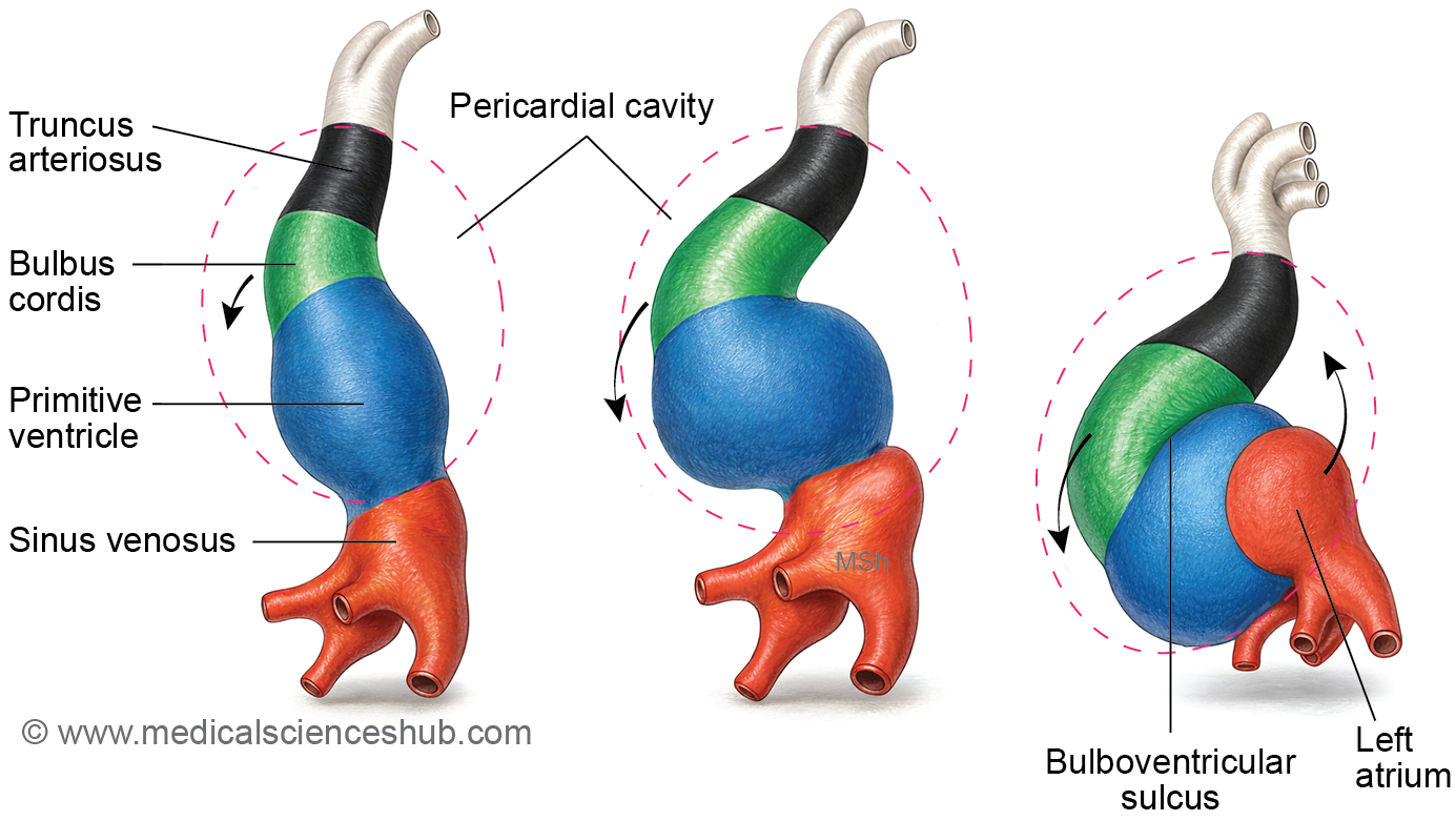
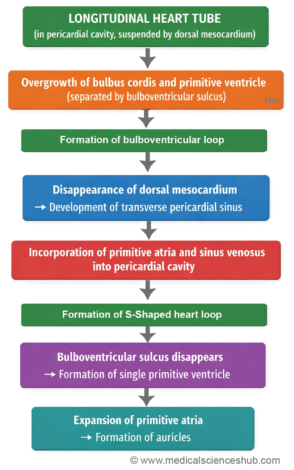
- The primitive heart tube undergoes growth, bending, and rearrangement to develop the external morphology of the adult heart.
- Initially, the heart tube lies longitudinally within the pericardial cavity. It is attached to the dorsal wall of the cavity by a fold of pericardium called the dorsal mesocardium.
- Formation of the bulboventricular loop: Rapid growth of the bulbus cordis and primitive ventricle causes the heart tube to bend ventrally, producing a characteristic U-shaped bulboventricular loop.
- Formation of the transverse sinus: The portion of the dorsal mesocardium connecting the bulboventricular loop subsequently degenerates. This creates a passage within the pericardial cavity known as the transverse pericardial sinus.
- Formation of the S-shaped cardiac loop: As the primitive atrium and sinus venosus detach from the septum transversum, they shift dorsocranially within the pericardial cavity relative to the primitive ventricle. This rearrangement produces an S-shaped cardiac loop. The bulbus cordis and primitive ventricle are initially separated by the bulboventricular sulcus, which later disappears as both regions merge to form a continuous ventricular chamber.
- Formation of auricles: The primitive atrium lies posterior to the truncus arteriosus. With expansion, it extends forward on both sides of the truncus arteriosus to form the right and left auricles, which contribute to the external appearance of the adult atria.
Development Of Atria
- The right and left atria arise from multiple embryonic structures that gradually remodel to form the definitive atrial chambers.
Right Atrium
- The rough, trabeculated part of the right atrium and the right auricle develop from the right half of the primitive atrium.
- The smooth posterior wall, known as the sinus venarum, originates from incorporation of the right horn of the sinus venosus into the atrial wall.
- The right venous valve contributes to several important structures, including the crista terminalis, the valve of the inferior vena cava, and the valve of the coronary sinus.
- A small ventral smooth region of the right atrium is derived from the right portion of the atrioventricular canal.
Left Atrium
- The anterior rough part of the left atrium and the left auricle develop from the left half of the primitive atrium.
- The posterior smooth-walled portion, located between the openings of the pulmonary veins, forms through incorporation of the pulmonary veins into the atrial wall.
- The ventral smooth region of the left atrium develops from the left portion of the atrioventricular canal.
- Further details of atrial development involve septation and remodeling processes described in subsequent sections.
Sinus Venosus
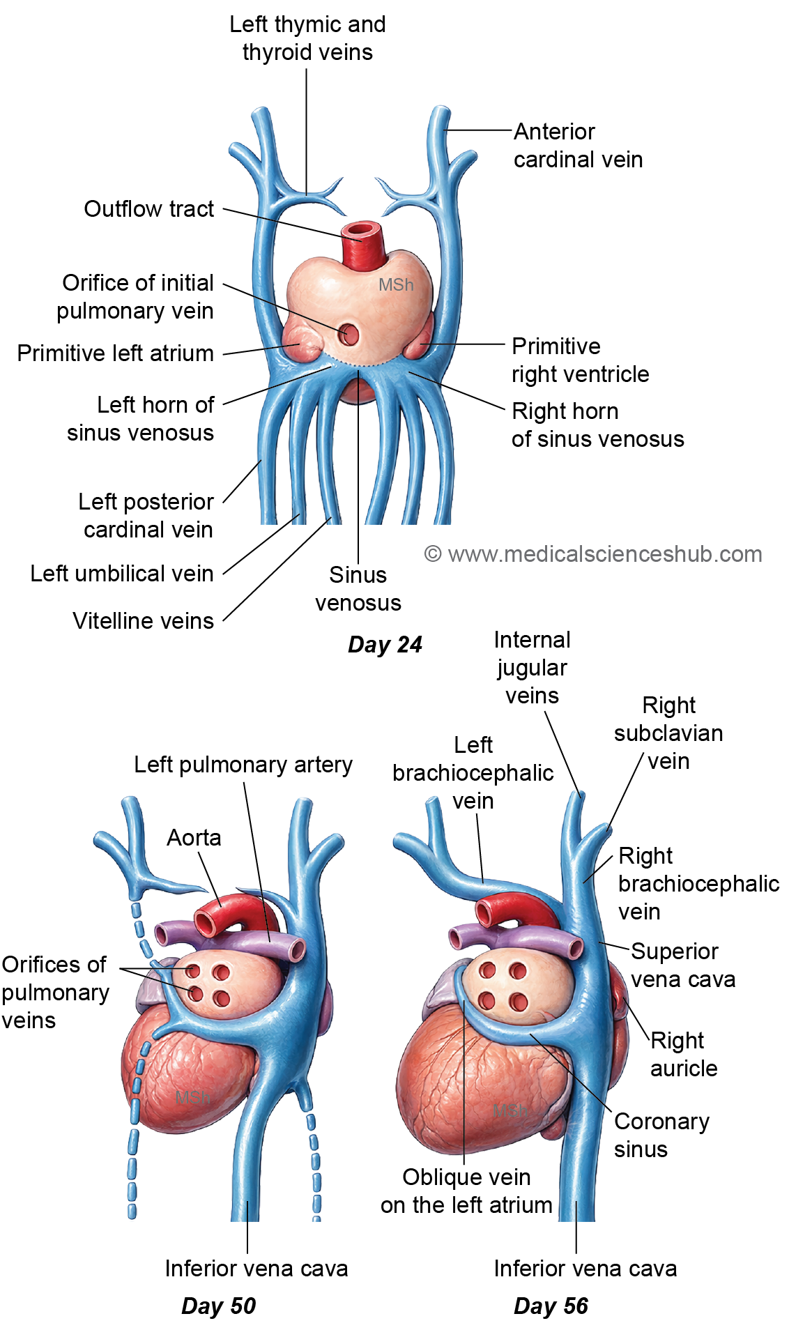
- The sinus venosus forms the caudal end of the primitive heart tube and represents the venous inflow region of the developing heart. Its unfused portion consists of right and left horns, each receiving venous blood from several embryonic vessels.
- Each horn receives blood through three main veins:
- Vitelline veins from the yolk sac
- Umbilical veins from the placenta
- Common cardinal veins (ducts of Cuvier) from the embryonic body wall
- The sinoatrial orifice is the opening that connects the sinus venosus with the primitive atrium, allowing venous blood to enter the atrial chamber.
Changes in the Left Horn
- Near the sinoatrial orifice, a crescent-shaped sinoatrial fold develops. This fold separates the left horn from the primitive atrium.
- Consequently, the left horn gradually loses its direct atrial connection and becomes a tributary of the right horn.
Fate of the Sinoatrial Orifice
- Initially, the sinoatrial orifice is a wide, transversely oriented opening located centrally.
- With development of the sinoatrial fold, the opening narrows and becomes a slit-like passage.
- Its margins form two important structures:
- Right venous valve
- Left venous valve
- Fusion of the cranial ends of these valves produces the septum spurium, whereas fusion of their caudal ends forms the sinus septum.
Changes in the Atrioventricular Canal
- The atrioventricular (AV) canal is the communication between the primitive atrium and the primitive ventricle.
- Thickened mesenchymal regions called atrioventricular cushions develop in the dorsal and ventral walls of the canal. These cushions arise from subendocardial mesenchyme within the cardiac jelly, which lies between the endocardium and myocardium.
- Growth and fusion of the AV cushions produce the septum intermedium, which divides the AV canal into right and left channels.
- Most major cardiac septa form between the 27th and 37th days of embryonic development.
Fate of Tributaries of Sinus Venous
- The vessels draining into the sinus venosus undergo significant remodeling during cardiac development. Their transformation contributes to several major systemic venous structures in the adult circulation.
- The right common cardinal vein forms a major portion of the superior vena cava.
- The right vitelline vein contributes to the terminal segment of the inferior vena cava.
- The left horn of the sinus venosus, together with the left common cardinal vein, develops into the coronary sinus.
- A remnant of the left common cardinal vein persists as the oblique vein of the left atrium.
- The cranial segment of the right posterior cardinal vein contributes to the arch of the azygos vein.
- The right umbilical vein and left vitelline vein disappear by approximately the fifth week of development.
- Most of the left common cardinal vein degenerates by the tenth week, leaving only small remnants.
- The left umbilical vein partially persists. Its proximal portion regresses, while the distal part carries oxygenated blood from the placenta to the inferior vena cava through the ductus venosus during fetal life. After birth, this vessel becomes the ligamentum teres of the liver.
- The right umbilical vein and left vitelline vein regress and become obliterated by the fifth week of embryonic development.
- Most of the left common cardinal vein subsequently degenerates and is obliterated by approximately the tenth week, leaving only small remnants that contribute to the coronary sinus system.
Development Of Interatrial Septum
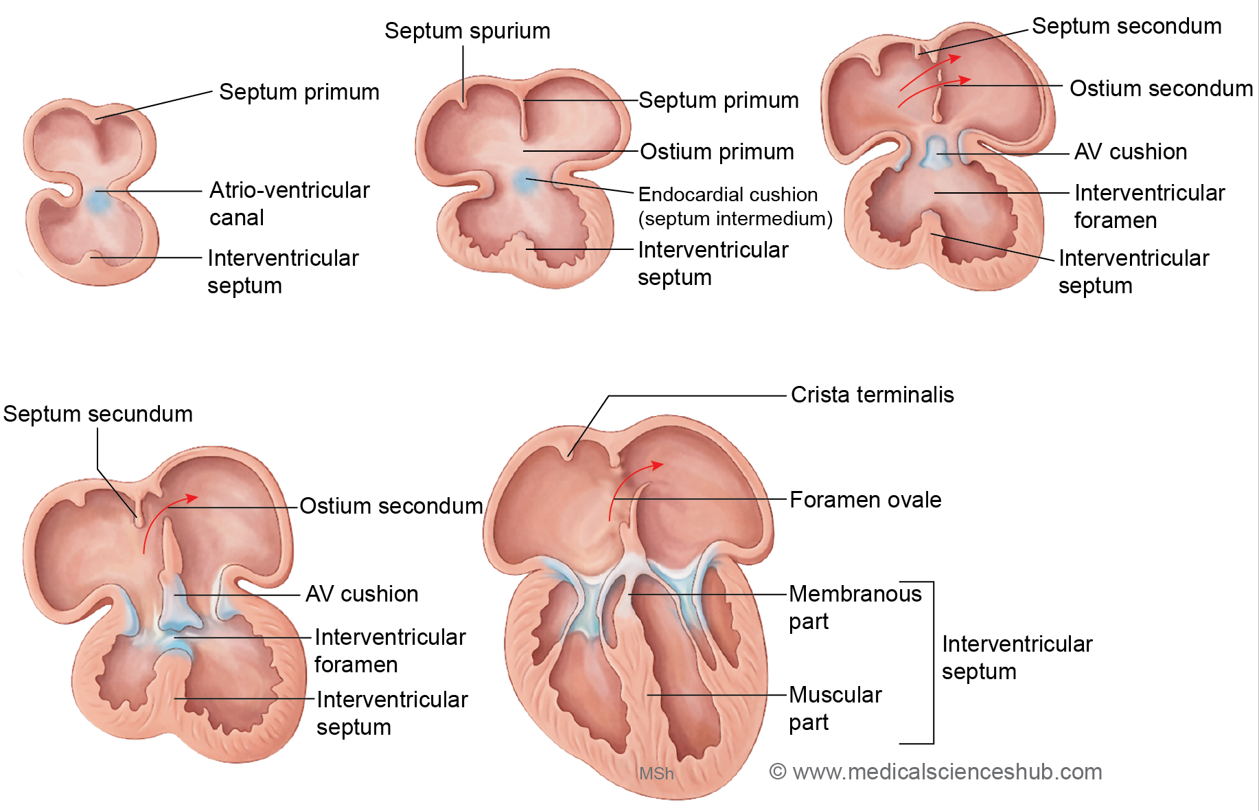
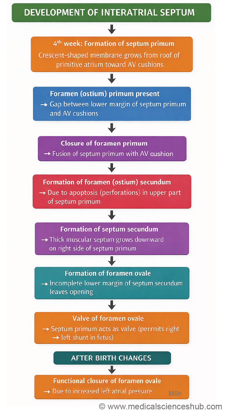
- The interatrial septum forms during the fifth week of intrauterine development through the coordinated growth of two septa: septum primum and septum secundum.
Stages of Development
- Septum primum: Toward the end of the fourth week, the septum primum begins to grow downward from the roof of the primitive atrium, slightly to the left of the septum spurium and the sinoatrial opening. It initially appears as a thin crescent-shaped membrane.
- Foramen (ostium) primum: As the septum primum grows toward the atrioventricular cushions (forming the septum intermedium), a temporary opening remains between its lower edge and the cushions. This gap is called the foramen primum, allowing blood flow between the two atria.
- Foramen (ostium) secundum: When the septum primum fuses with the atrioventricular cushions, the foramen primum closes. At the same time, programmed cell death creates a new opening in the upper part of the septum primum, known as the foramen secundum, which maintains interatrial blood flow.
- Septum secundum: A thicker, crescent-shaped septum secundum develops from the roof of the primitive atrium, positioned between the septum spurium and the septum primum.
- Foramen ovale: The septum secundum grows downward toward the septum intermedium, partially covering the foramen secundum. This arrangement creates an oblique channel called the foramen ovale, which allows blood to pass from the right atrium to the left atrium during fetal life.
- Changes after birth: After birth, increased left atrial pressure presses the septum primum against the septum secundum, functionally closing the foramen ovale. In the adult heart, the floor of the fossa ovalis represents the septum primum, while the free inferior margin of the septum secundum forms the annulus (limbus) of the fossa ovalis.
Function of Foramen Ovale
- The foramen ovale functions as a valve-like opening formed by the overlap of the thick septum secundum and the thin, mobile septum primum. The septum primum acts as a flexible flap valve.
- This arrangement permits unidirectional flow of blood from the right atrium to the left atrium, while preventing reverse flow. As a result, oxygenated blood entering the right atrium from the placenta can pass directly into the left atrium.
- The shunt created by the foramen ovale allows most blood to bypass the nonfunctional fetal lungs, directing it into the systemic circulation instead of passing through the right ventricle and pulmonary circulation.
- After birth, expansion of the lungs initiates pulmonary circulation, which increases venous return to the left atrium. The resulting rise in left atrial pressure presses the septum primum against the septum secundum, producing functional closure of the foramen ovale. Over time, permanent anatomical fusion of the septa usually occurs.
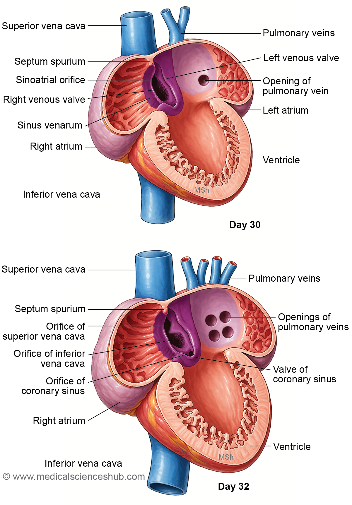
Formation of Sinus Venarum (Absorption of Sinus Venosus in the Right Atrium)
- During cardiac development, the sinus venosus becomes incorporated into the right atrium, producing structural remodeling of the atrial wall.
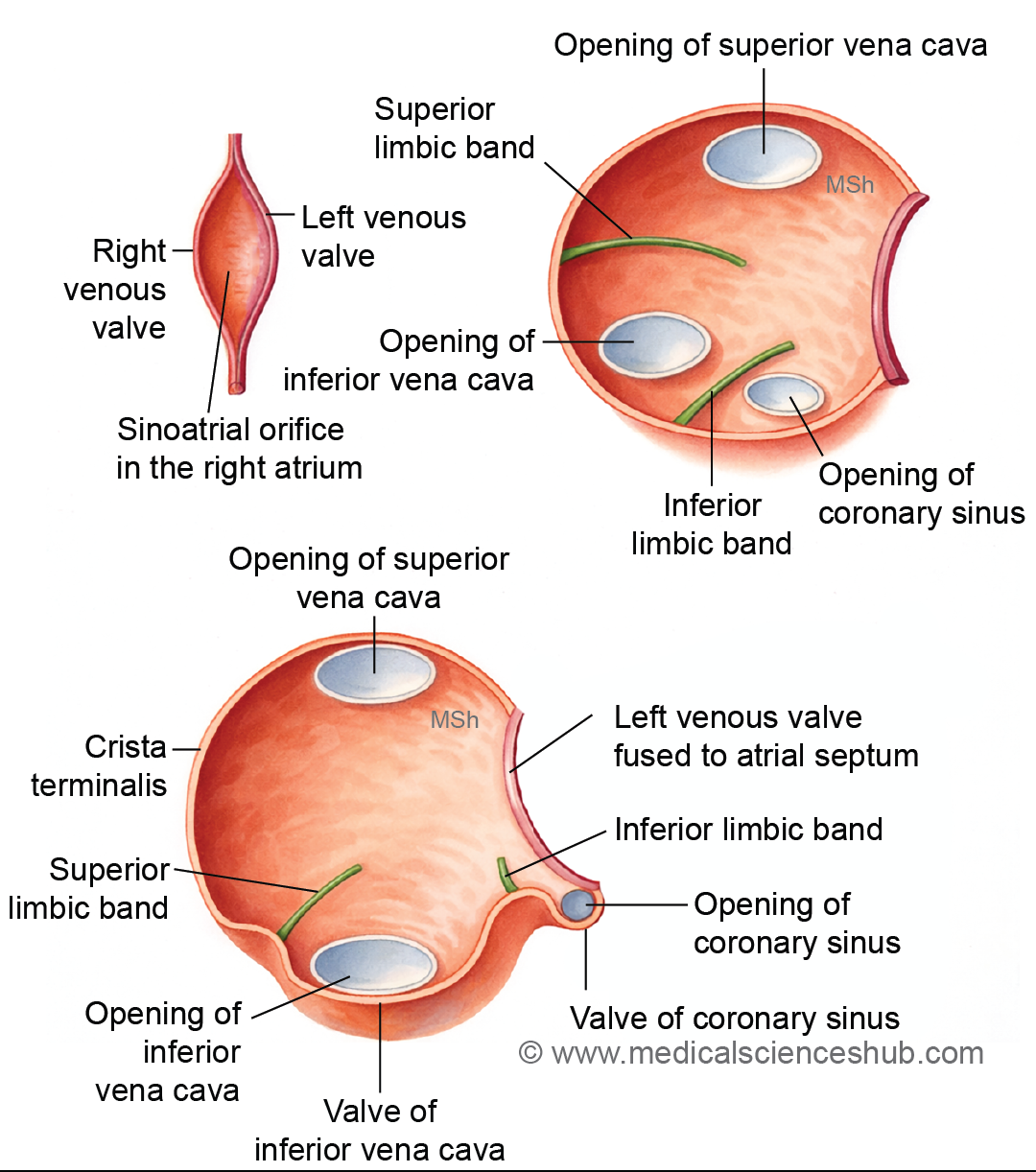
- The right and left horns of the sinus venosus are gradually absorbed into the right atrium. This process results in the separation of the openings of the superior vena cava, inferior vena cava, and coronary sinus within the atrial cavity.
- Two muscular ridges, the superior and inferior limbic bands, subdivide the right venous valve into three distinct regions.
- The right venous valve gives rise to several important structures:
- Crista terminalis
- Valve of the inferior vena cava
- Valve of the coronary sinus
- The left venous valve gradually merges with the interatrial septum during atrial septation.
- The portion of the sinus venosus incorporated into the atrial wall forms the sinus venarum, which constitutes the smooth posterior part of the right atrium.
Absorption of Pulmonary Veins into Left Atrium
- During early cardiac development, the dorsal wall of the left atrium forms an outgrowth known as the primordial pulmonary vein.
- This single pulmonary vein initially divides into two branches and subsequently into four pulmonary veins, each draining blood from the developing lungs.
- As the left atrium enlarges, the proximal portions of these pulmonary veins become incorporated into the atrial wall.
- This incorporation results in the separate openings of the four pulmonary veins into the left atrium, rather than a single common channel.
- The absorbed portions of the pulmonary veins contribute to the formation of the smooth posterior wall of the left atrium, whereas the rough anterior part of the atrium develops from the primitive atrium.
CLINICAL EMBRYOLOGY
Atrial septal defects (ASD)
- Definition: Atrial septal defect is a congenital heart defect characterized by an abnormal opening in the interatrial septum that allows communication between the right and left atria.
- Embryological basis: The defect results from abnormal development of the septum primum or septum secundum during formation of the interatrial septum.
- Types
- Ostium secundum ASD – Most common type; caused by excessive resorption of septum primum or inadequate development of septum secundum.
- Ostium primum ASD – Occurs in the lower part of the atrial septum due to failure of septum primum to fuse with endocardial cushions.
- Sinus venosus ASD – Located near the opening of the superior vena cava.
- Coronary sinus ASD – Rare defect near the coronary sinus.
- Physiological effect
- Blood usually flows from the left atrium to the right atrium (left-to-right shunt).
- This leads to increased pulmonary blood flow and right heart enlargement.
- Clinical features
- Many patients remain asymptomatic in childhood.
- Large defects may produce dyspnea, fatigue, and recurrent respiratory infections.
- A wide fixed splitting of the second heart sound is a typical clinical finding.
Probe patency of foramen ovale
- Definition: Probe patency of the foramen ovale refers to a condition where the septum primum and septum secundum do not fuse completely, leaving a small slit-like opening that can be crossed by a probe.
- Embryological explanation
- After birth, increased pressure in the left atrium normally presses the septum primum against the septum secundum, functionally closing the foramen ovale.
- In some individuals, the septa fail to fuse permanently.
- Features
- Occurs in about 20–25% of adults.
- Usually not a true atrial septal defect because the septal tissues are present but not fused.
- Clinical significance
- Normally asymptomatic.
- May permit paradoxical embolism, where a venous clot passes into systemic circulation.
Dextrocardia
- Definition: Dextrocardia is a congenital condition in which the heart is positioned on the right side of the thoracic cavity instead of the left.
- Embryological cause: It results from abnormal looping of the primitive heart tube during early embryonic development.
- Types
- Isolated dextrocardia – Only the heart is on the right side.
- Dextrocardia with situs inversus – Mirror-image arrangement of thoracic and abdominal organs.
- Dextrocardia with congenital heart defects – Often associated with septal defects or transposition anomalies.
- Clinical importance
- Individuals with situs inversus totalis may have normal cardiac function.
- Isolated dextrocardia is frequently associated with other congenital cardiac malformations.
Development Of Ventricles
- The ventricles of the heart arise from different embryonic regions of the primitive heart tube.
- The rough (trabeculated inflow parts) of both ventricles develop from the primitive ventricle.
- The smooth outflow regions—the infundibulum of the right ventricle and the aortic vestibule of the left ventricle—are derived from the conus cordis, which represents the middle third of the bulbus cordis.
Bulbus Cordis
- The bulbus cordis is a dilated segment at the arterial end of the developing heart tube. It differentiates into three distinct parts, each contributing to specific cardiac structures.
- Proximal one-third
- This portion merges with the primitive ventricle, forming the bulboventricular chamber.
- It later develops into the trabeculated part of the right ventricle.
- Middle one-third (Conus cordis)
- This conical region, called the conus cordis, gives rise to the outflow tracts of both ventricles.
- Distal one-third (Truncus arteriosus)
- The distal segment forms the truncus arteriosus.
- A spiral aorticopulmonary septum later divides it into the pulmonary trunk and the ascending aorta.
Formation Of Interventricular Septum
Formation of the Interventricular Septum
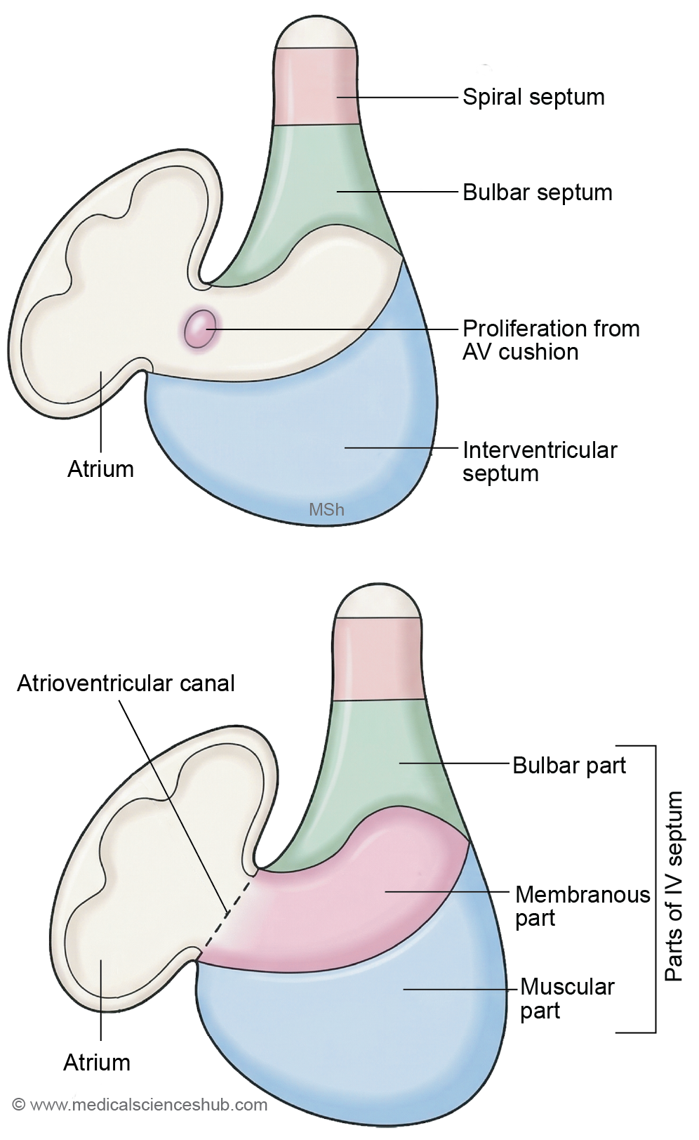
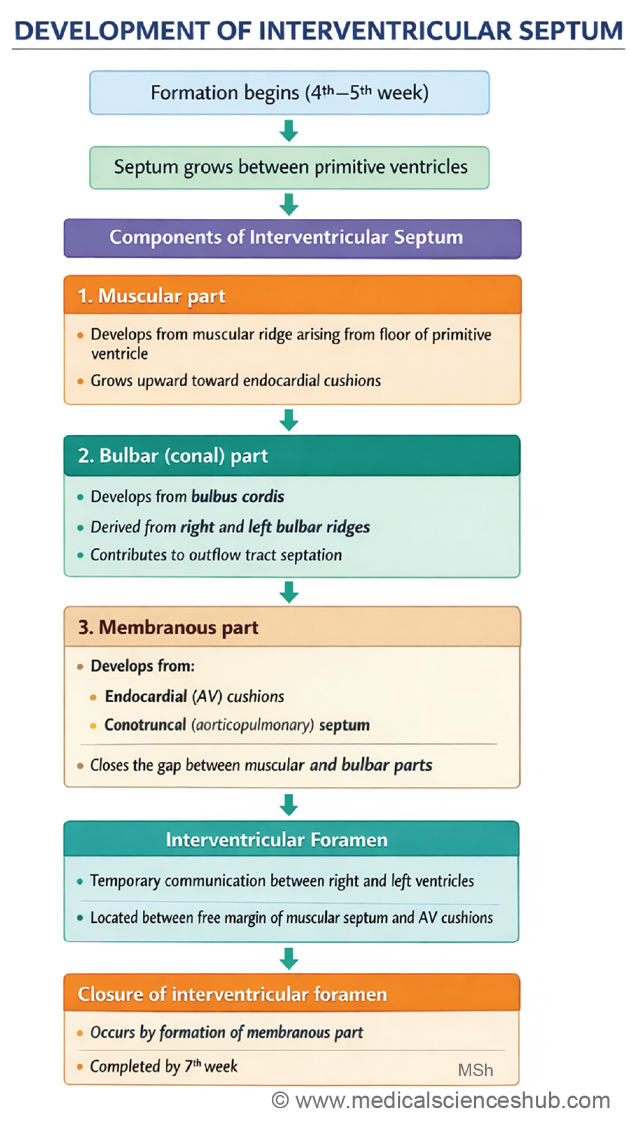
- The interventricular septum separates the right and left ventricles and develops from three embryonic components: the muscular part, bulbar part, and membranous part.
Components of the Interventricular Septum
- Muscular part: Develops from a muscular ridge arising from the floor of the bulboventricular cavity.
- Bulbar part: Forms from the right and left bulbar ridges originating in the conus cordis.
- Membranous part: Produced by proliferation of tissue from the atrioventricular (AV) cushions, which closes the gap between the muscular and bulbar components.
Stages of Development
1. Muscular part
- A muscular interventricular ridge grows upward from the floor of the bulboventricular cavity, partially dividing the primitive ventricle into right and left chambers.
- This ridge grows toward the septum intermedium (formed by the AV cushions) but does not completely fuse with it.
- Consequently, the ventricles initially remain connected through the interventricular foramen, located above the muscular septum.
2. Bulbar part
- In the conus cordis, the right and left bulbar ridges develop and fuse to form the bulbar (conal) septum.
- This septum grows downward toward the muscular interventricular septum.
- During this stage, the interventricular foramen persists between the muscular and bulbar components.
3. Membranous part
- By approximately the eighth week, proliferation of tissue from the AV cushions and bulbar ridges closes the interventricular foramen, forming the membranous part of the interventricular septum.
- The interventricular foramen is temporarily necessary until proper separation of the bulbus cordis and truncus arteriosus occurs.
- The anterior portion of the membranous septum separates the right and left ventricles, whereas the posterior portion separates the right atrium from the left ventricle.
Formation of Aorticopulmonary Septum
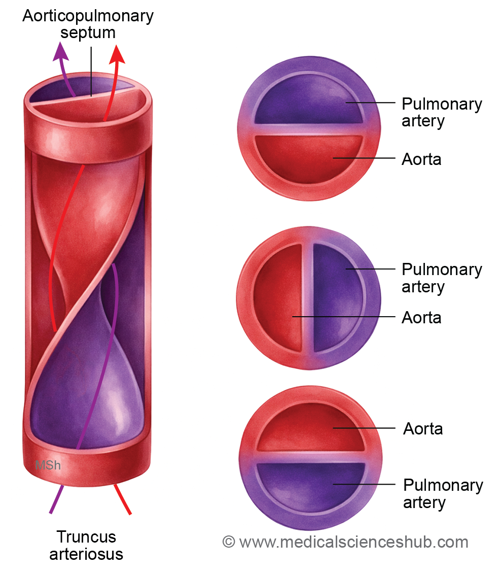
- During cardiac development, a spiral partition known as the aorticopulmonary septum forms within the truncus arteriosus. This septum divides the common arterial outflow tract into two major vessels: the ascending aorta and the pulmonary trunk.
Stages of Development
- Formation of truncal ridges: Two longitudinal swellings called truncal ridges develop within the wall of the truncus arteriosus. These ridges gradually enlarge and fuse in the midline, forming the aorticopulmonary (spiral) septum.
- Fusion with the bulbar septum: The developing aorticopulmonary septum aligns with the bulbar septum of the conus cordis and eventually fuses with it. At this stage, the aorta is positioned posterior to the pulmonary trunk.
- Spiral arrangement of the septum: The septum follows a spiral course as it partitions the outflow tract. Consequently, the aorta, initially located behind the pulmonary trunk, shifts toward the right side and finally lies anterior to the pulmonary trunk near its origin.
CLINICAL EMBRYOLOGY
Transposition of great vessels
- Transposition of the Great Vessels (TGV) is a congenital heart defect in which the aorta arises from the right ventricle and the pulmonary trunk arises from the left ventricle.
- It occurs due to failure of the aorticopulmonary septum to spiral normally during cardiac development.
- As a result, systemic and pulmonary circulations run in parallel rather than in series.
- Deoxygenated blood circulates through the body, while oxygenated blood repeatedly circulates through the lungs.
- Survival depends on mixing of blood through defects such as atrial septal defect, ventricular septal defect, or patent ductus arteriosus.
- Affected newborns usually present with severe cyanosis soon after birth.
Persistent (common) truncus arteriosus
- Persistent truncus arteriosus is a congenital anomaly in which a single arterial trunk arises from the heart instead of separate aorta and pulmonary trunk. It results from failure of the truncus arteriosus to divide because the aorticopulmonary septum does not form.
- The single vessel supplies systemic, pulmonary, and coronary circulations.
- A ventricular septal defect is usually present beneath the truncal valve.
- Oxygenated and deoxygenated blood mix, producing cyanosis and increased pulmonary blood flow.
- The condition is commonly associated with neural crest cell migration defects.
Ventricular Septal Defects (VSD)
Definition: Ventricular septal defect is a congenital defect characterized by an abnormal opening in the interventricular septum, allowing communication between the right and left ventricles.
Embryological Basis
- The interventricular septum develops from two components:
- Muscular interventricular septum
- Membranous septum formed by endocardial cushions and conotruncal ridges.
- VSD occurs when these structures fail to fuse completely.
Types
Ventricular septal defects are classified according to their location in the interventricular septum:
- Membranous VSD
- Located in the membranous part of the septum near the aortic and tricuspid valves.
- Most common type of VSD.
- Muscular VSD
- Occurs in the muscular portion of the interventricular septum.
- Often small and may close spontaneously during childhood.
- Inlet VSD
- Located in the posterior septum near the atrioventricular valves.
- Frequently associated with atrioventricular septal defects.
- Outlet (Conal/Infundibular) VSD
- Found in the outflow part of the septum near the aortic and pulmonary valves.
- Often linked with conotruncal anomalies.
Physiological Consequences
- Blood flows from the left ventricle to the right ventricle (left-to-right shunt) because left ventricular pressure is higher.
- This causes increased pulmonary blood flow and volume overload of the right ventricle and lungs.
Complications
- Pulmonary hypertension
- Cardiac enlargement
- Heart failure in severe cases
- Long-standing defects may lead to Eisenmenger syndrome (reversal of shunt with cyanosis).
Clinical Features
- Heart murmur
- Shortness of breath
- Poor growth in infants
- Some small defects may close spontaneously during childhood.
Development Of Valves Of Heart
Atrioventricular Valves
- The atrioventricular (AV) valves regulate blood flow between the atria and ventricles. The tricuspid valve lies between the right atrium and right ventricle, whereas the mitral (bicuspid) valve lies between the left atrium and left ventricle.
- In early development, the atria communicate with the ventricles through a single atrioventricular canal.
- Proliferation of subendocardial mesenchyme produces endocardial cushions around this canal.
- These cushions undergo remodeling and excavation, forming the cusps of the AV valves.
- The free margins of the cusps become attached to papillary muscles of the ventricular wall through thin fibrous cords called chordae tendineae.
- Valve structure differs on each side of the heart:
- On the right side, three cushions—anterior, posterior, and septal—give rise to the tricuspid valve.
- On the left side, two cushions—anterior and posterior—form the mitral (bicuspid) valve.
Pulmonary and Aortic Valves
The semilunar valves develop from endocardial cushions located in the truncus arteriosus near its junction with the conus cordis.
- Initially, two cushions (right and left) appear in this region.
- Shortly afterward, two additional cushions (ventral and dorsal) develop, forming four swellings.
- When the aorticopulmonary septum divides the truncus arteriosus into the ascending aorta and pulmonary trunk, the cushions are partitioned into separate sets.
- Subsequent excavation and remodeling of these cushions produce the cusps of the aortic and pulmonary valves.
Because of the spiral rotation of the outflow tract, the final arrangement of cusps is:
- Aortic valve: one anterior cusp and two posterior cusps.
- Pulmonary valve: one posterior cusp and two anterior cusps.
Development Of Conducting System Of Heart
- The cardiac conduction system, responsible for initiating and coordinating the heartbeat, begins to develop around the fifth week of intrauterine life. Its major components arise from specialized regions of the developing heart.
- Sinoatrial (SA) node
The SA node, the primary pacemaker of the heart, develops during the fifth week. After incorporation of the sinus venosus into the right atrium, the SA node becomes positioned near the opening of the superior vena cava. - Atrioventricular (AV) node and bundle of His
The AV node and the atrioventricular bundle (bundle of His) originate from tissue in the interatrial septal region, close to the opening of the coronary sinus. These structures develop from the dorsal endocardial cushion of the atrioventricular canal around the sixth week of development. - Purkinje fibers
The bundle of His divides into right and left bundle branches, which extend along the ventricular septum and spread within the ventricular walls as Purkinje fibers, forming the terminal part of the cardiac conduction network.
Development Of Pericardium
- The pericardium, which encloses and protects the heart, develops from different components of the embryonic mesoderm.
- The serous pericardium develops from mesodermal layers:
- Visceral layer (epicardium): derived from splanchnopleuric mesoderm
- Parietal layer: derived from somatopleuric mesoderm
- The fibrous pericardium develops mainly from the somatopleuric mesoderm with contribution from the septum transversum.
- The pericardial cavity originates from the intraembryonic coelom.
Stages of Development
- During head folding, the pericardial cavity comes to lie on the ventral side of the foregut. The developing heart tube, surrounded by the myoepicardial mantle, invaginates this cavity. The inner layer of this mantle forms the visceral pericardium (epicardium).
- Initially, the heart tube is suspended within the pericardial cavity by a double-layered fold called the dorsal mesocardium, which resembles a mesentery.
- As cardiac looping occurs, the arterial and venous ends of the heart tube move closer. The dorsal mesocardium then degenerates, creating the transverse pericardial sinus. At this stage, the visceral and fibrous pericardium become continuous around the arterial and venous poles of the heart.
- Subsequent reorientation of the superior vena cava (SVC) and inferior vena cava (IVC), together with incorporation of the pulmonary veins into the left atrium, leads to the formation of the oblique pericardial sinus.
- The somatopleuric mesoderm lining the pericardial cavity differentiates to form the parietal layer of the serous pericardium and the fibrous pericardium
CLINICAL EMBRYOLOGY
Tetralogy of Fallot (ToF)
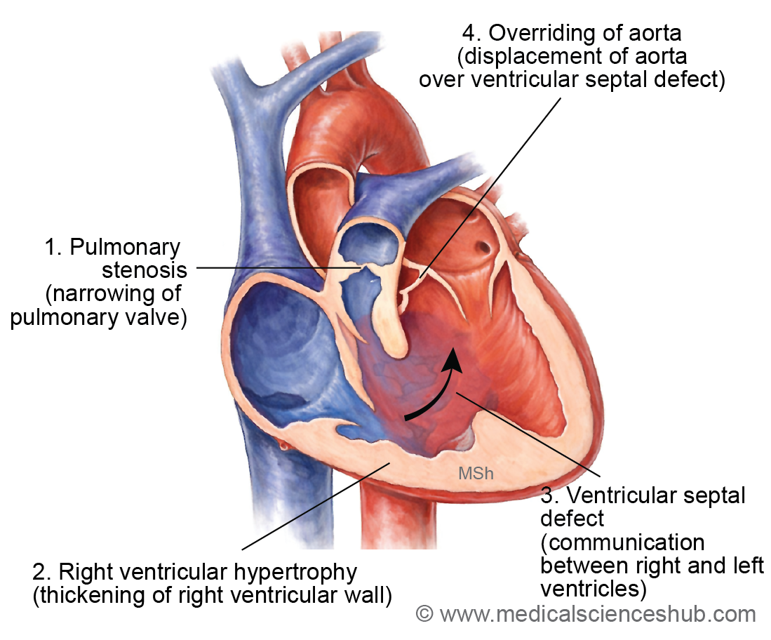
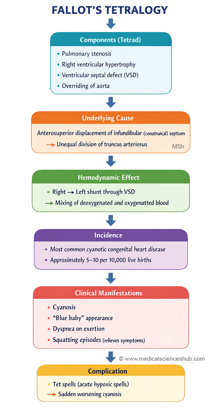
- Tetralogy of Fallot (ToF) is a congenital cardiac malformation and represents the most common cyanotic congenital heart disease. It results from unequal division of the conus cordis, leading to anterior displacement of the conotruncal (aorticopulmonary) septum during cardiac development.
Components
Tetralogy of Fallot is characterized by four major structural abnormalities:
- Pulmonary stenosis – narrowing of the right ventricular outflow tract.
- Ventricular septal defect (VSD) – an abnormal opening in the interventricular septum.
- Overriding aorta – the aorta is positioned directly above the VSD, receiving blood from both ventricles.
- Right ventricular hypertrophy – thickening of the right ventricular wall due to increased pressure.
Etiology
- In many cases, the exact cause is unknown.
- It may be associated with maternal phenylketonuria.
- Mutations in the JAG1 gene, which influences neural crest cell–mediated formation of the conotruncal septum, have also been implicated.
Diagnosis
- Echocardiography is the primary diagnostic method.
- Chest X-ray may show a characteristic “boot-shaped” (coeur en sabot) heart, caused by right ventricular hypertrophy.
Pathophysiology
- Pulmonary stenosis increases resistance to blood flow from the right ventricle.
- This leads to elevated right ventricular pressure and right ventricular hypertrophy.
- Blood is then shunted from the right ventricle to the left ventricle through the VSD.
- The overriding aorta receives mixed oxygenated and deoxygenated blood, resulting in systemic cyanosis.
Clinical Manifestations
- Cyanosis, producing the classic “blue-baby” appearance of the lips and nail beds.
- Easy fatigability and reduced exercise tolerance.
- Tet spells, which are sudden hypoxic episodes characterized by shortness of breath, deep cyanosis, agitation, and possible syncope.
Important Questions
- List the derivatives of the various segments of the primitive heart tube.
- Describe the anatomy and developmental importance of the bulbus cordis.
- Explain the development of the sinus venosus and its contributions to the adult heart.
- Describe the embryological development of the right atrium.
- Explain the formation of the interatrial septum.
- Discuss the development and functional role of the foramen ovale in fetal circulation.
- Describe the formation of the interventricular septum.
- Provide an overview of atrial septal defects (ASDs), including their types and significance.
- Explain the anatomical basis and clinical relevance of a patent foramen ovale.
- Describe ventricular septal defects (VSDs), including their developmental origin.
- Outline the features of Tetralogy of Fallot.
- Explain the embryological basis of Tetralogy of Fallot.
- Describe ectopia cordis, including its developmental origin and clinical implications.
📝 Test Your Knowledge – Practice MCQs
Attempt the chapter MCQ quiz and assess your understanding of key concepts.
Click to see: Medical Disclaimer and Copyright Notice