
Competencies
- AN69.1 Identify elastic and muscular blood vessels, capillaries under the microscope.
- AN69.2 Describe the various types and structure-function correlation of blood vessel.
- AN69.3 Describe the ultrastructure of blood vessels.
Introduction
- The cardiovascular system is responsible for transporting blood between the heart and body tissues, ensuring the delivery of oxygen, nutrients, and other essential substances.
- It consists of the heart and a network of blood vessels. The heart is a muscular pump that propels blood into the arterial system and receives blood returning through veins.
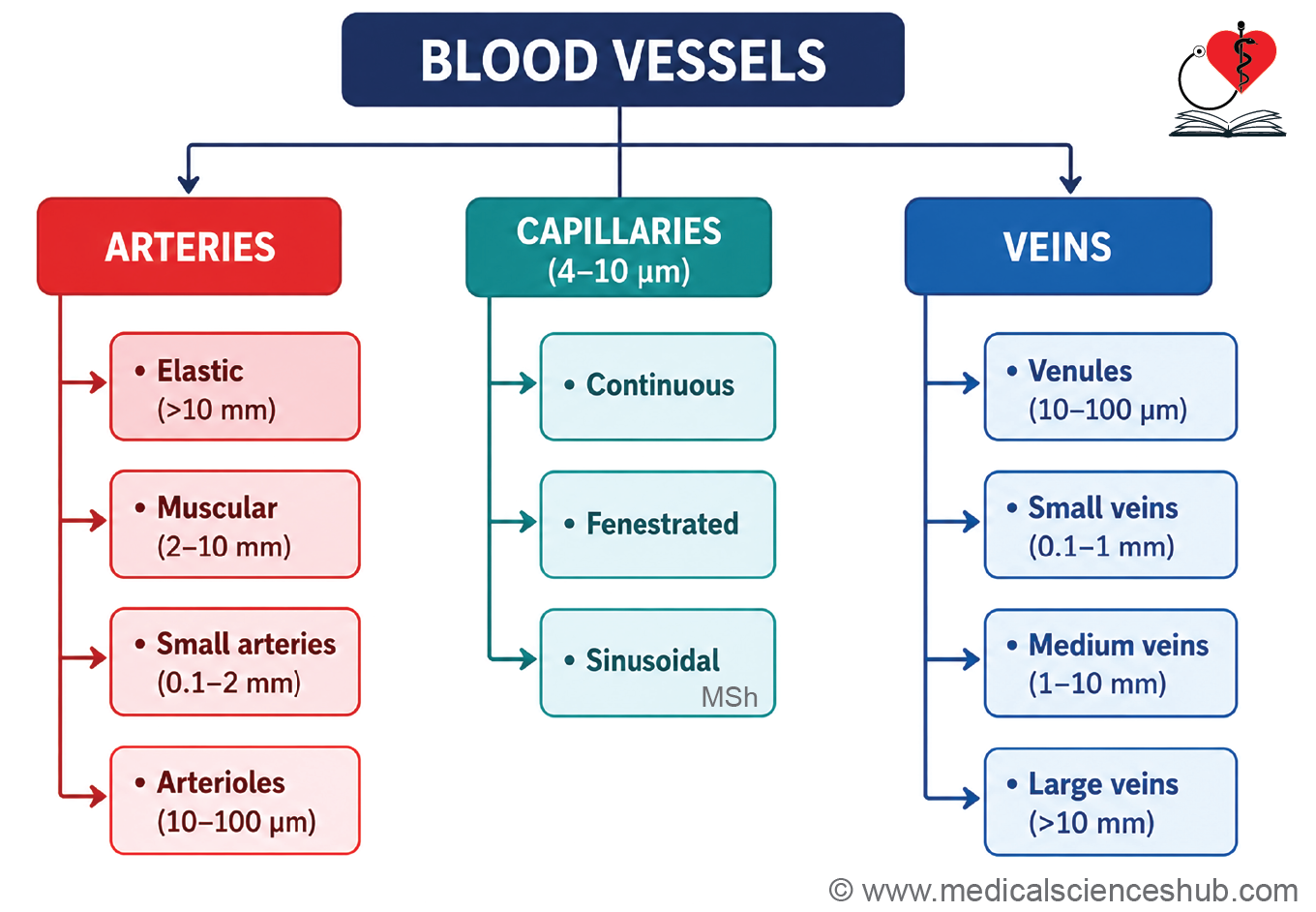
- Blood vessels are classified into arteries, arterioles, capillaries, venules, and veins.
- Arteries carry blood away from the heart, while veins return blood to the heart.
- Arterioles are small branches of arteries that regulate blood flow into capillaries.
- Capillaries are the smallest blood vessels and serve as the primary sites for exchange of gases, nutrients, and waste products between blood and tissues.
- Venules collect blood from capillaries and gradually merge to form veins.
- Major circulatory pathways include systemic circulation, pulmonary circulation, and portal circulation.
- Systemic and pulmonary circulations contain a single capillary network, whereas portal circulations contain two successive capillary beds connected by a portal vessel. Examples include the hepatic portal system and hypothalamo-hypophyseal portal system.
- All blood vessels are lined internally by endothelium, a specialized simple squamous epithelium.
Endothelium
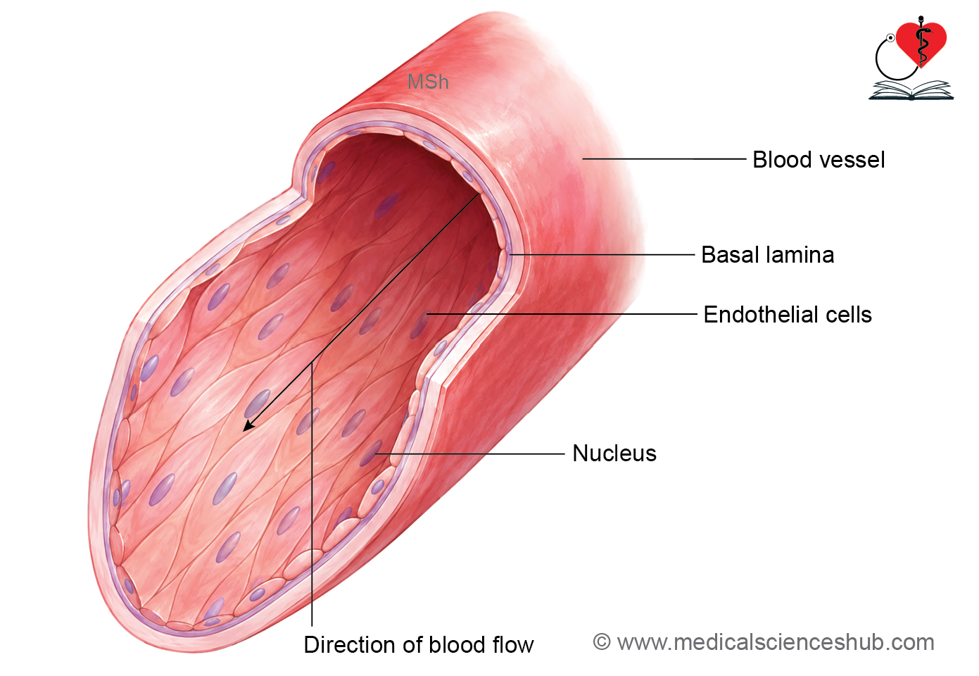
- Endothelium is a specialized simple squamous epithelium that lines the inner surface of the heart, blood vessels, and lymphatic vessels.
- It is composed of endothelial cells, which are flattened, elongated, and polygonal in shape. Their long axis is oriented parallel to the direction of blood flow.
- In histological sections stained with H&E, endothelial cells appear thin, with scant cytoplasm and elongated, dark-staining nuclei.
- Ultrastructurally, endothelial cells contain organelles such as mitochondria, endoplasmic reticulum, and cytoskeletal microfilaments. Adjacent cells are joined by tight junctions and rest on a basal lamina, which contains type IV collagen.
Functions of Endothelium
- Provides a smooth, non-thrombogenic surface that facilitates the uninterrupted flow of blood.
- Acts as a selective permeability barrier, regulating the movement of substances between blood and tissues through diffusion, vesicular transport, and receptor-mediated mechanisms.
- Synthesizes and releases von Willebrand factor (vWF), which promotes platelet adhesion and contributes to blood clotting following vascular injury.
- Produces nitric oxide (NO) and prostacyclin, which cause vasodilation and inhibit platelet aggregation.
- Releases endothelin and expresses angiotensin-converting enzyme (ACE), both of which contribute to vasoconstriction and regulation of vascular tone.
- Maintains local blood flow by balancing vasodilatory and vasoconstrictive influences.
- Secretes various growth factors, including fibroblast growth factors, platelet-derived growth factors, and colony-stimulating factors, which support tissue repair and cellular growth.
- Produces endothelium-derived relaxing factor (EDRF), now recognized primarily as nitric oxide, which promotes relaxation of vascular smooth muscle.
- Synthesizes components of the basal lamina, including type IV collagen.
Basic Structure of Blood Vessels
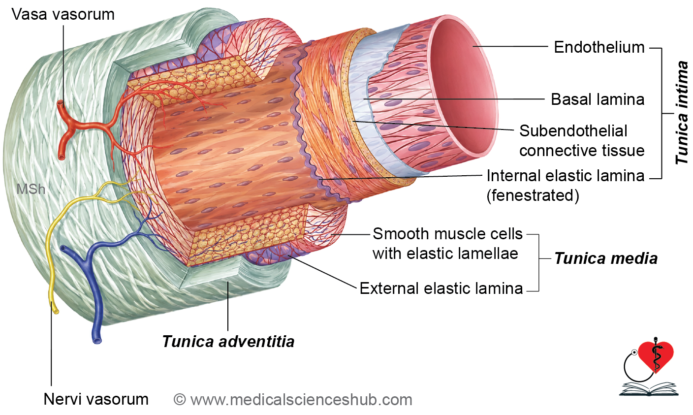
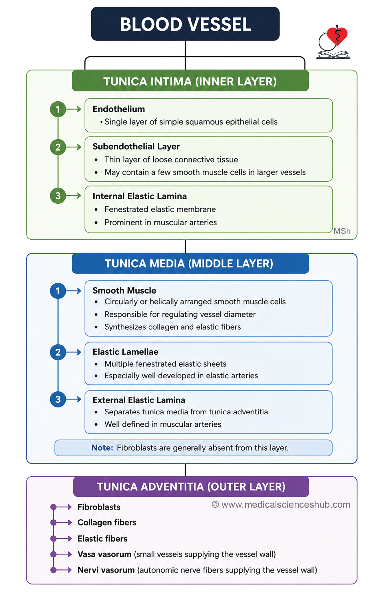
· Histologically, most blood vessels consist of three concentric layers: tunica intima, tunica media, and tunica adventitia.
· The structure and relative thickness of these layers vary according to the type, size, and functional requirements of the blood vessel.
Tunica Intima
- Tunica intima is the innermost layer of a blood vessel and is in direct contact with circulating blood.
- It consists of three components: endothelium, subendothelial connective tissue, and the internal elastic lamina.
- The endothelium is formed by a single layer of simple squamous endothelial cells resting on a basal lamina.
- Endothelial cells are flattened and aligned parallel to the direction of blood flow.
- The subendothelial layer contains loose connective tissue with collagen and elastic fibers.
- The internal elastic lamina is composed of fenestrated elastic sheets that provide flexibility and allow diffusion of nutrients to deeper layers of the vessel wall.
Tunica Media
- Tunica media is the middle layer of the blood vessel wall and is composed primarily of concentrically arranged smooth muscle cells.
- These smooth muscle cells have elongated nuclei and are embedded within elastic fibers, collagen fibers, and extracellular matrix components.
- In addition to regulating vessel diameter, smooth muscle cells synthesize collagen, elastic fibers, and proteoglycans.
- Elastic fibers are organized as fenestrated elastic lamellae.
- The tunica media is generally thicker in arteries than in veins due to a greater amount of smooth muscle.
- The external elastic lamina separates the tunica media from the tunica adventitia.
- Smooth muscle cells mediate vasoconstriction and vasodilation under autonomic control and can proliferate in response to endothelial growth factors.
Tunica Adventitia
- Tunica adventitia is the outermost layer of a blood vessel and is composed mainly of connective tissue containing collagen fibers, elastic fibers, fibroblasts, and occasional macrophages.
- It provides structural support and anchors the vessel to surrounding tissues.
- Vasa vasorum are small blood vessels that supply nutrients to the outer layers of large blood vessel walls.
- Nervi vasorum are autonomic nerve fibers, predominantly sympathetic, that regulate vascular tone by promoting vasoconstriction through the release of norepinephrine.
Classification of Blood Vessels
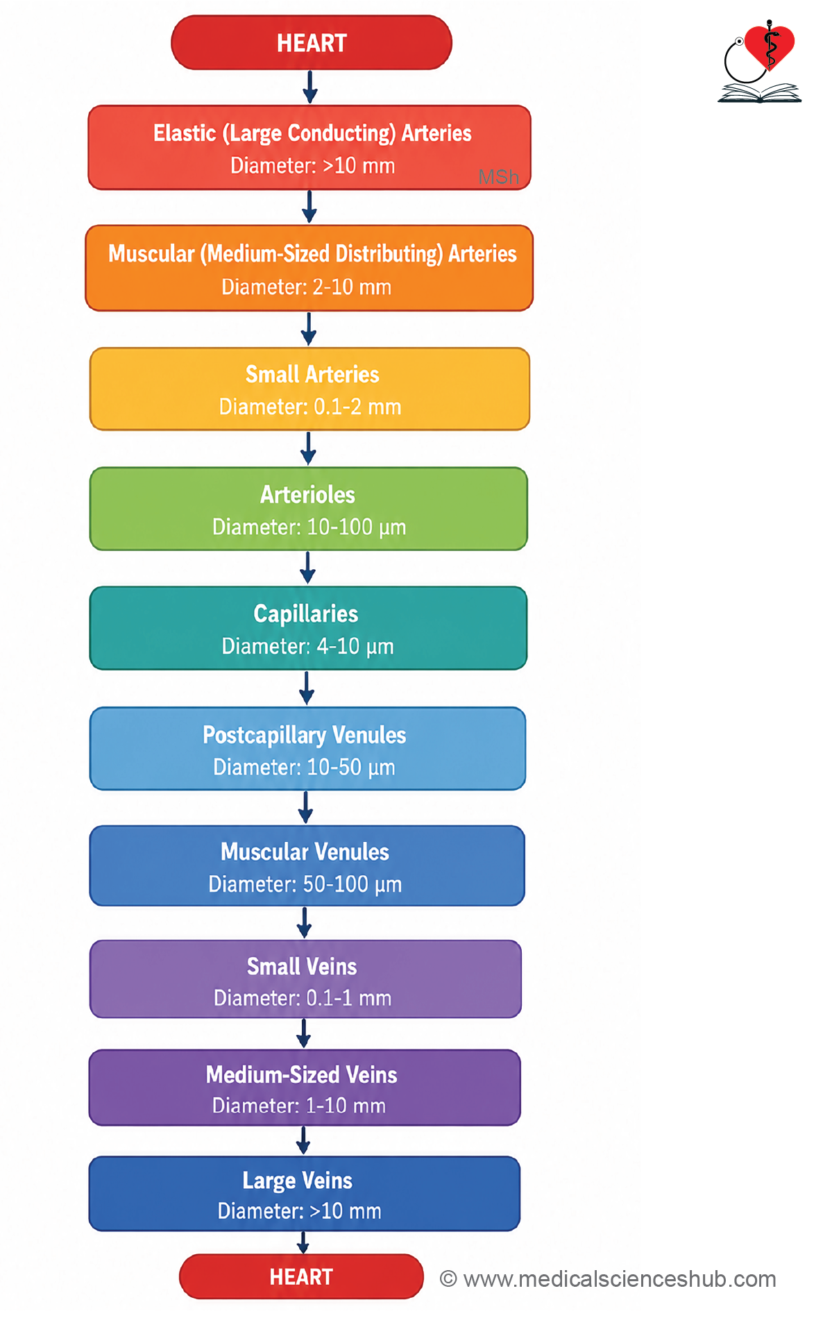
- Blood vessels are broadly classified into arteries, capillaries, and veins.
- Arteries transport blood away from the heart, capillaries facilitate exchange between blood and tissues, and veins return blood to the heart.
Arteries
Based on the composition of the tunica media, arteries are classified into:
- Elastic (large) arteries: Contain abundant elastic lamellae and smooth muscle, allowing expansion during cardiac systole.
- Muscular (medium-sized) arteries: Have a predominance of smooth muscle with fewer elastic fibers.
- Small arteries: Possess up to eight layers of smooth muscle cells in the tunica media.
- Arterioles: The smallest arteries, typically containing one to two layers of smooth muscle, which regulate blood flow into capillary beds.
Capillaries
- Capillaries form extensive vascular networks and are the primary sites for the exchange of gases, nutrients, and metabolic waste products.
- They are classified according to the structure of the endothelium and basal lamina:
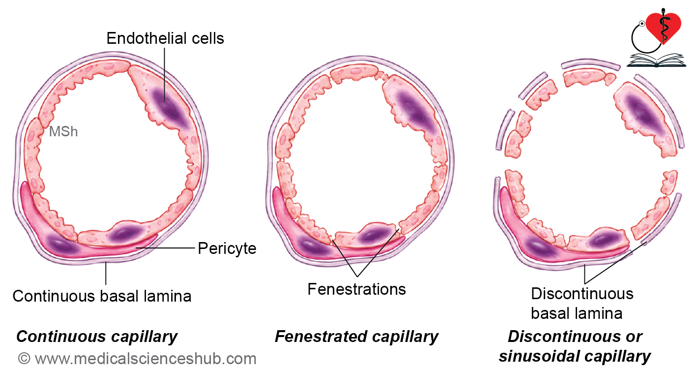
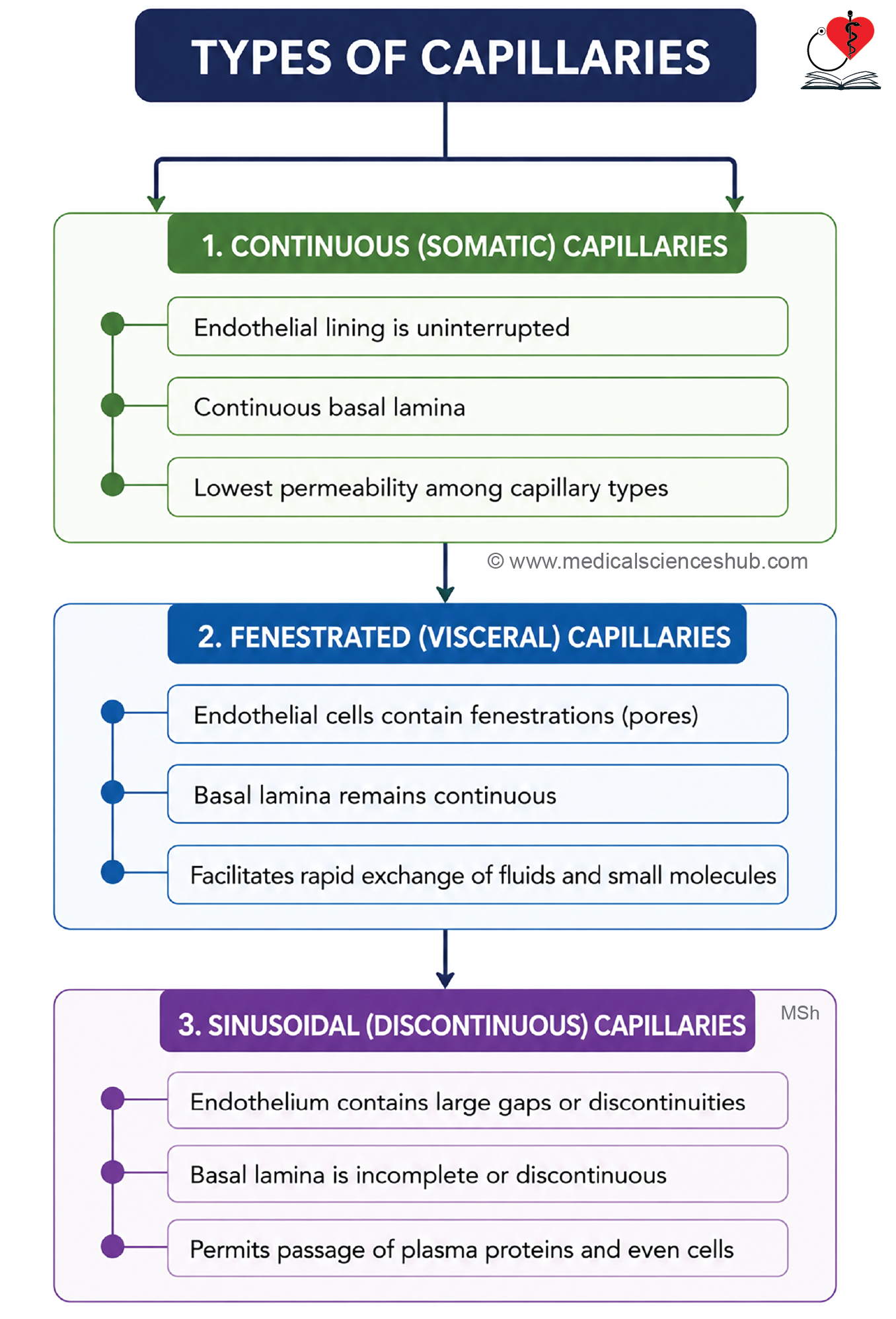
- Continuous capillaries: Have uninterrupted endothelial cells and a continuous basal lamina.
- Fenestrated capillaries: Contain pores (fenestrations) in endothelial cells, while the basal lamina remains continuous.
- Sinusoidal capillaries (sinusoids): Possess discontinuous endothelium and basal lamina, allowing the passage of larger molecules and cells.
Veins
Veins are categorized by size into:
- Venules, which receive blood from capillaries.
- Small veins.
- Medium-sized veins, which often accompany arteries.
- Large veins, which return large volumes of blood directly to the heart.
Elastic Arteries
- Elastic arteries, also known as large arteries or conducting arteries, are the largest arteries in the body, typically measuring more than 10 mm in diameter.
- Examples include the aorta, pulmonary trunk, common carotid artery, subclavian artery, brachiocephalic trunk, and common iliac artery.
- These vessels transport blood from the heart to muscular arteries and contain abundant elastic tissue in their walls.
- During ventricular systole, elastic arteries expand to accommodate the surge of blood ejected from the heart.
- During ventricular diastole, elastic recoil of the arterial wall helps propel blood forward, maintaining continuous blood flow and stable arterial pressure.
- This pressure-maintaining mechanism is known as the Windkessel effect.
Structure of Elastic Artery
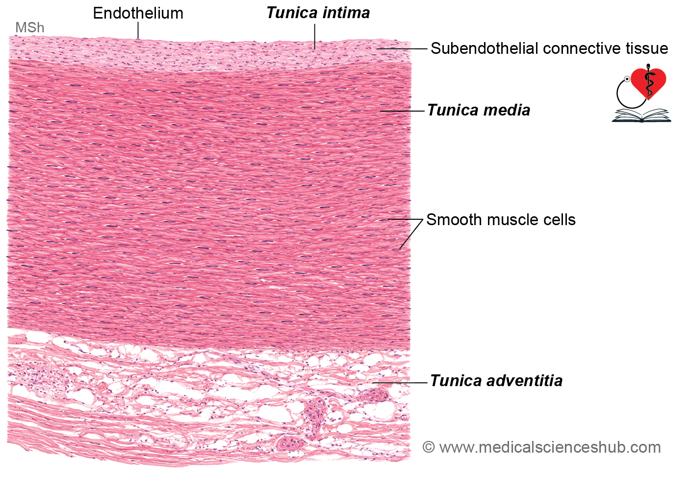
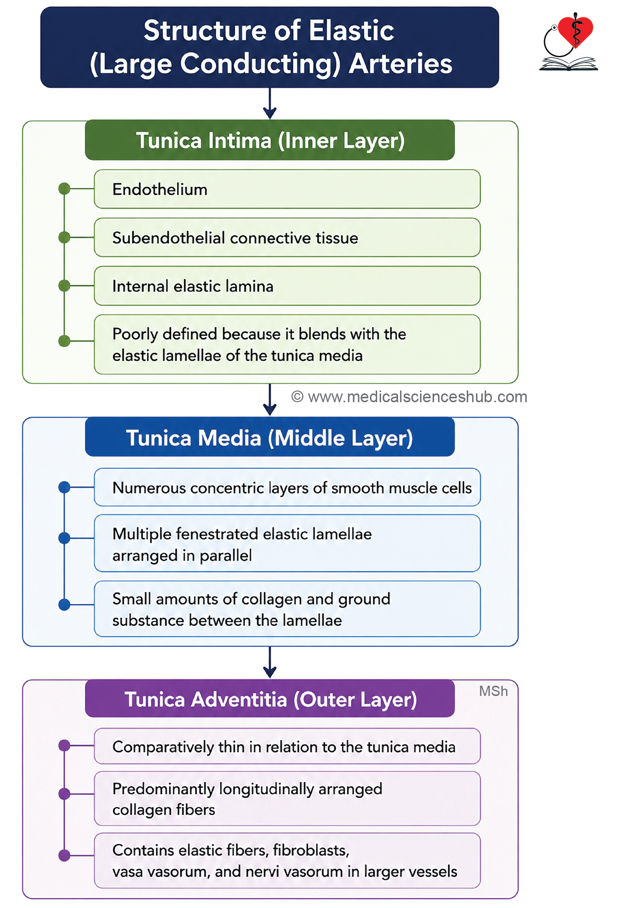
The wall of an elastic artery is composed of three layers: tunica intima, tunica media, and tunica adventitia.
Tunica Intima
- The tunica intima consists of endothelium, subendothelial connective tissue, and an internal elastic lamina.
- The endothelium is a simple squamous epithelium resting on a basal lamina, with cells aligned parallel to blood flow.
- The subendothelial layer contains connective tissue with collagen fibers, elastic fibers, and a few smooth muscle cells.
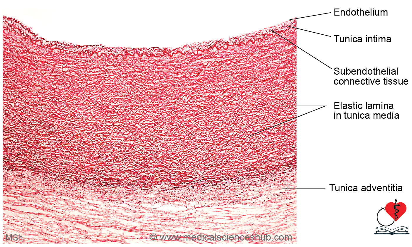
- The internal elastic lamina is formed by fenestrated elastic sheets. In elastic arteries, it is often difficult to distinguish because numerous elastic lamellae are present in the tunica media.
Tunica Media
- The tunica media is the thickest layer and the most prominent feature of elastic arteries.
- It contains multiple concentric layers of smooth muscle cells interspersed with numerous fenestrated elastic lamellae.
- Smooth muscle cells are spindle-shaped, possess elongated nuclei, and are arranged in a spiral pattern around the vessel.
- These cells synthesize elastin, collagen, and extracellular matrix components.
- In the adult aorta, the tunica media typically contains about 40–70 elastic lamellae, which provide elasticity and resilience to the vessel wall.
- The internal and external elastic laminae are not clearly distinguishable from the surrounding elastic lamellae.
Tunica Adventitia
- The tunica adventitia is relatively thin compared with the tunica media.
- It contains longitudinally arranged collagen fibers, fine elastic fibers, fibroblasts, and macrophages.
- The adventitia houses vasa vasorum, which supply the outer regions of the vessel wall, and nervi vasorum, autonomic nerve fibers that regulate vascular smooth muscle activity.
Muscular Arteries
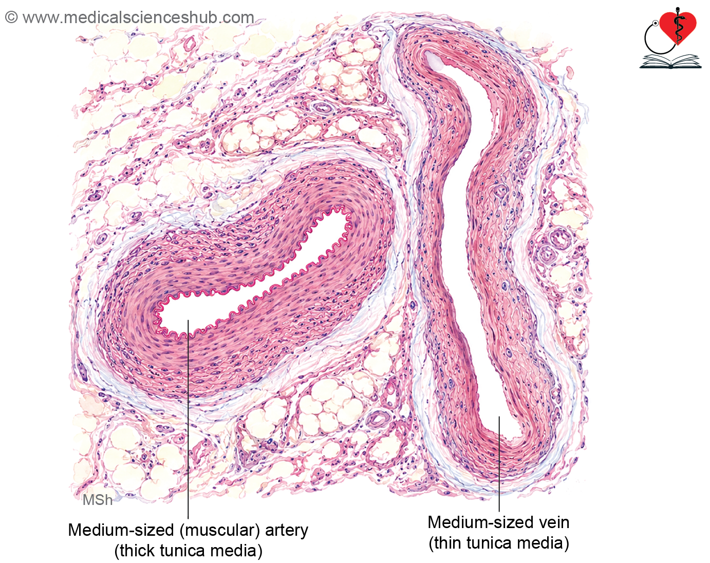
- Muscular arteries, also known as medium-sized arteries, typically have a diameter ranging from approximately 2–10 mm.
- These vessels are often referred to as distributing arteries because they deliver blood from large elastic arteries to specific organs and tissues.
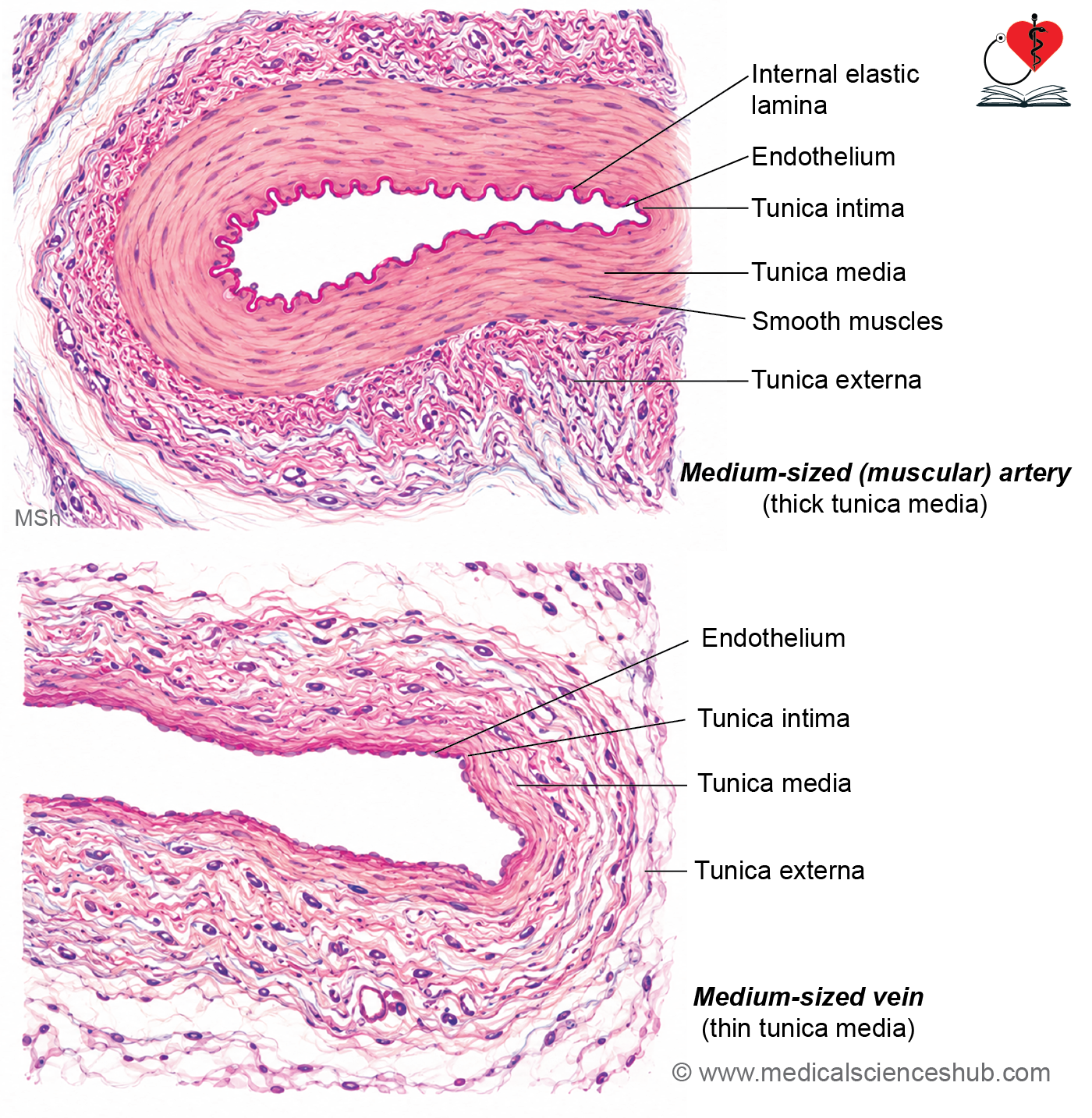
- The most characteristic feature of muscular arteries is a thick tunica media composed predominantly of multiple layers of smooth muscle cells, with relatively fewer elastic fibers than elastic arteries.
- Due to the reduced elastic content, the internal elastic lamina is usually prominent and can be readily identified in histological sections.
- Smooth muscle cells within the tunica media are arranged concentrically and play a key role in regulating vascular diameter.
- Contraction of these cells produces vasoconstriction, which reduces vessel lumen size and blood flow.
- Relaxation of smooth muscle cells causes vasodilation, increasing lumen diameter and enhancing blood flow to tissues.
- Through these mechanisms, muscular arteries contribute significantly to the regulation of tissue perfusion and systemic blood pressure.
Structure of Muscular Arteries
- Muscular arteries develop from elastic arteries through a gradual reduction in vessel diameter, a decrease in elastic tissue, and an increase in smooth muscle within the vessel wall.
- Their wall is composed of three layers: tunica intima, tunica media, and tunica adventitia.
Tunica Intima
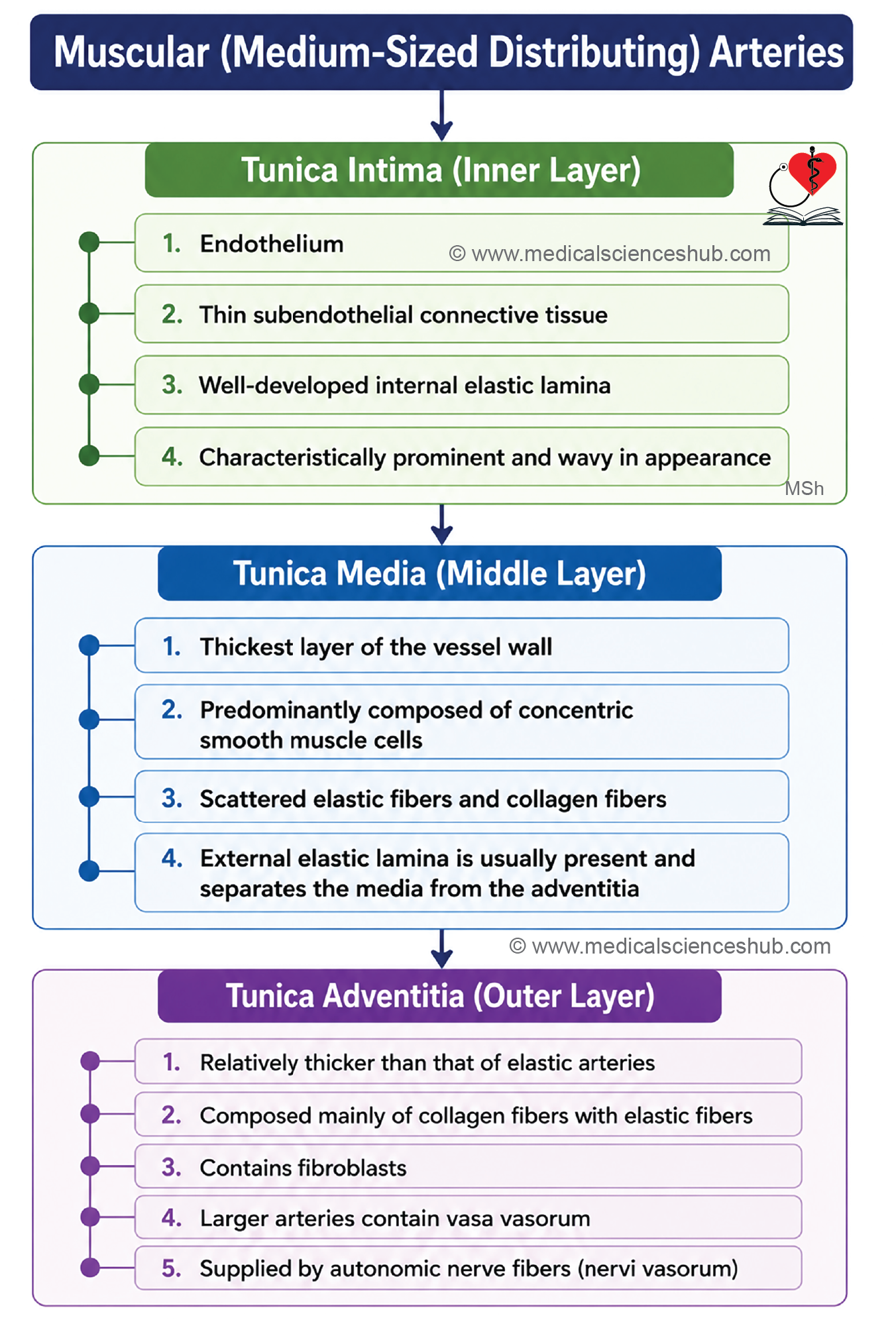
- The tunica intima consists of endothelium, a thin subendothelial connective tissue layer, and a prominent internal elastic lamina.
- The endothelium is a simple squamous epithelium resting on a basal lamina.
- Because the subendothelial layer is minimal, the internal elastic lamina appears as a distinct wavy structure in histological sections.
Tunica Media
- The tunica media is the most prominent layer and is composed mainly of concentric layers of smooth muscle cells.
- These spindle-shaped cells are arranged in a spiral pattern and play an important role in regulating vascular diameter and blood pressure.
- The layer contains relatively small amounts of elastic fibers and collagen fibers.
- Smooth muscle cells are interconnected by gap junctions, allowing coordinated contraction.
- Fibroblasts are generally absent from the tunica media.
- An external elastic lamina may be present at the boundary between the tunica media and tunica adventitia.
Tunica Adventitia
- The tunica adventitia is composed of connective tissue and is relatively thicker than that of elastic arteries.
- It contains vasa vasorum, which supply the vessel wall, and nervi vasorum, which provide autonomic innervation to vascular smooth muscle.
Arterioles
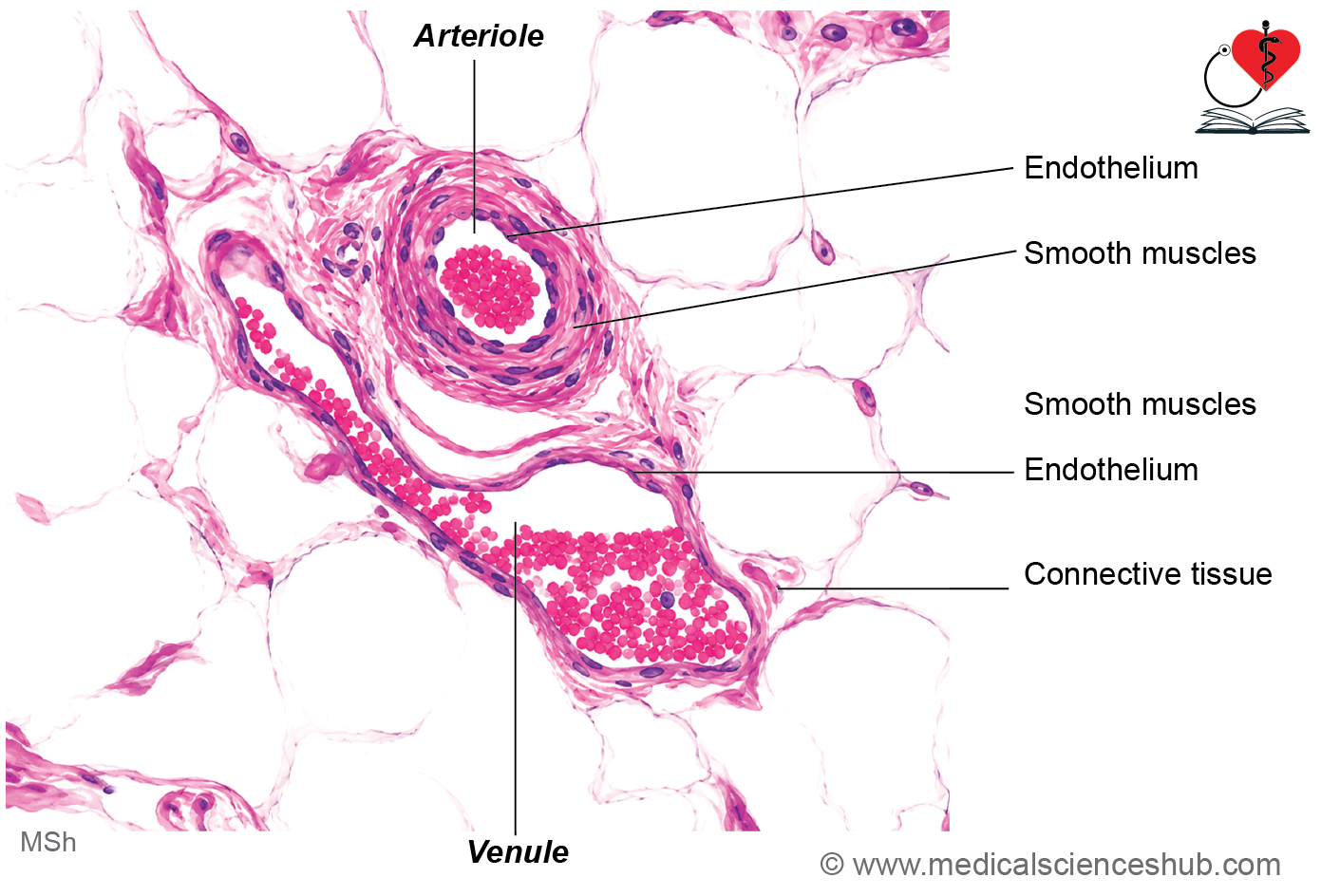
- Arterioles are small branches of muscular arteries, typically measuring 10–100 μm in diameter.
- They further divide to form capillary networks within tissues.
- Arterioles are classified into large (muscular) arterioles and terminal arterioles.
- By altering vascular resistance through smooth muscle contraction and relaxation, arterioles regulate capillary blood flow and contribute significantly to the maintenance of blood pressure.
Structure of Arteriole
Arterioles are small blood vessels composed of tunica intima, tunica media, and a thin tunica adventitia.
Tunica intima
- The tunica intima consists of a simple squamous endothelium resting on a thin layer of subendothelial connective tissue.
- The internal elastic lamina is usually absent or poorly developed in arterioles.
Tunica media
- The tunica media contains 1–3 concentric layers of smooth muscle cells.
- Large arterioles generally have 2–3 smooth muscle layers, whereas terminal arterioles possess only a single layer.
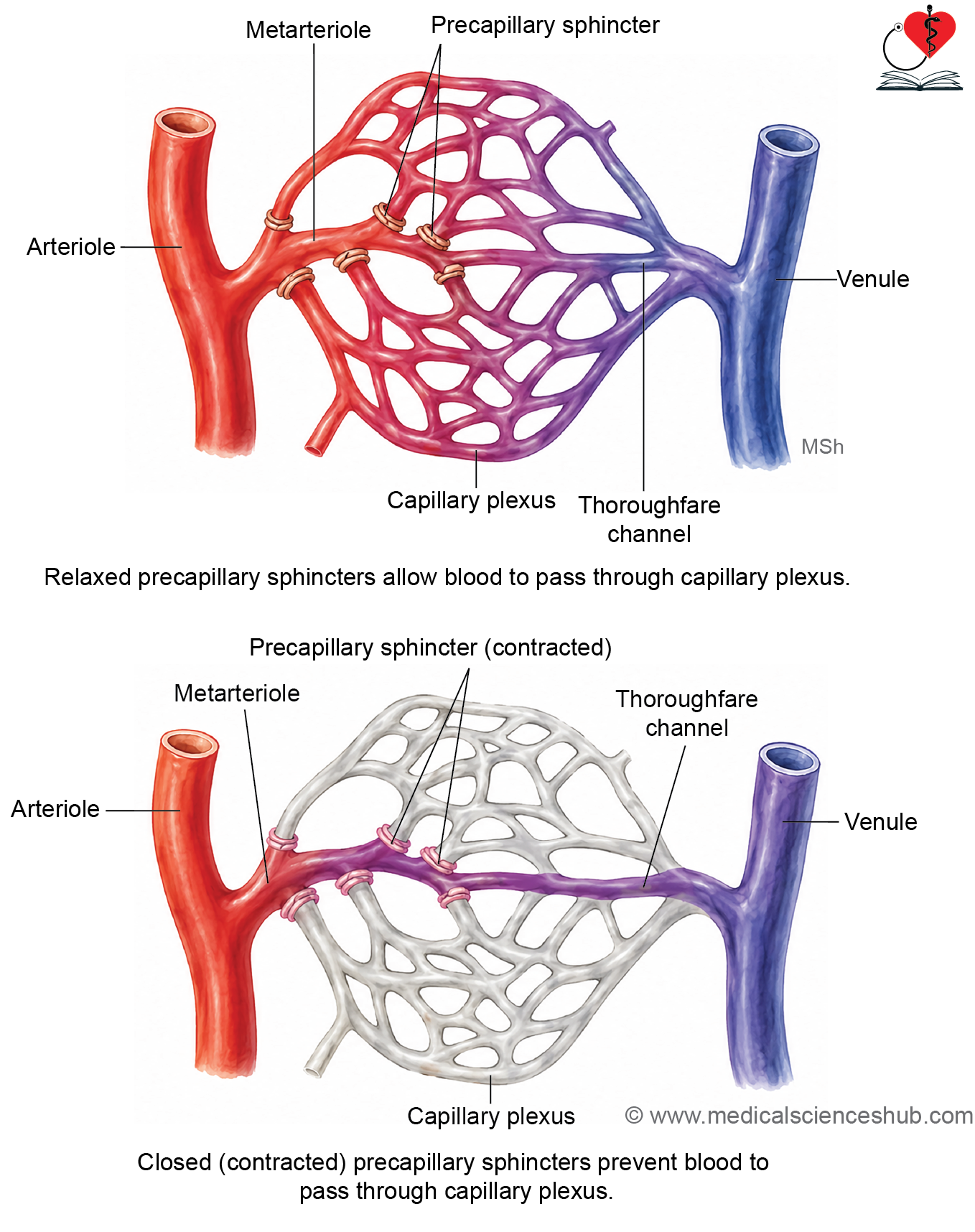
- At the junction of terminal arterioles and capillaries, smooth muscle forms precapillary sphincters, which regulate blood entry into capillary beds.
- Contraction and relaxation of arteriolar smooth muscle control vascular resistance and influence systemic blood pressure.
Tunica adventitia
The tunica adventitia is a thin connective tissue layer that separates the arteriole from surrounding tissues and may be difficult to distinguish microscopically.
Functions of Arterioles
- Arterioles regulate the distribution of blood to tissues by controlling flow through capillary networks.
- They are the principal resistance vessels of the circulatory system and play a major role in maintaining arterial blood pressure.
CLINICAL CORRELATION
- Atherosclerosis is a chronic arterial disease characterized by the accumulation of atheromatous plaques within the tunica intima. These plaques contain lipids, cholesterol, macrophages, foam cells, smooth muscle cells, and fibrous connective tissue. Progressive plaque formation narrows the arterial lumen, reducing blood flow and increasing the risk of myocardial infarction, stroke, and other ischemic disorders.
- Hypertension is defined as persistently elevated arterial blood pressure. Traditionally, it has been described as a systolic pressure above 140 mmHg or a diastolic pressure above 90 mmHg. Age-related vascular changes, including increased vascular resistance, reduced elasticity, increased collagen deposition, and smooth muscle hypertrophy, contribute to hypertension.
- An aneurysm is a localized abnormal dilation of an artery caused by weakening of the vessel wall, particularly the tunica media. Degeneration of elastic fibers and their replacement by collagen can predispose to aneurysm formation.
- Common risk factors include atherosclerosis, syphilis, Marfan syndrome, and Ehlers-Danlos syndrome.
- Marfan syndrome is an autosomal dominant connective tissue disorder, while Ehlers-Danlos syndrome comprises inherited disorders affecting collagen structure and function.
- Syphilis is a sexually transmitted infection caused by the bacterium Treponema pallidum and may involve the cardiovascular system in advanced stages.
- Thromboangiitis obliterans (Buerger disease) is a recurrent inflammatory disorder affecting small and medium-sized arteries and veins, primarily in the extremities. It is strongly associated with tobacco use and leads to vascular occlusion, reduced blood flow, and pain in the affected limbs. Severe cases may result in tissue ischemia and gangrene.
Table 10.1: Differences between elastic artery and muscular artery
| Feature | Elastic Artery | Muscular Artery | Arteriole |
|---|---|---|---|
| Tunica Intima | Contains a well-developed subendothelial connective tissue layer. Internal elastic lamina is usually indistinct due to abundant elastic lamellae. | Subendothelial connective tissue is minimal. Internal elastic lamina is prominent and clearly visible. | Subendothelial connective tissue is very thin or absent. Internal elastic lamina is absent or present only in larger arterioles. |
| Tunica Media | Thickest layer; contains numerous concentric elastic lamellae with smooth muscle cells interspersed between them. | Predominantly composed of multiple layers of smooth muscle cells with relatively few elastic fibers. | Consists of 1–3 layers of smooth muscle cells with little or no elastic tissue. |
| Tunica Adventitia | Relatively thin connective tissue layer. | Relatively thick fibroelastic connective tissue layer. | Thin connective tissue layer that may be poorly defined. |
| Principal Function | Conducts blood from the heart and dampens pressure fluctuations through elastic recoil. | Distributes blood to specific organs and regulates blood flow. | Controls blood flow into capillary beds and contributes significantly to vascular resistance. |
| Examples | Aorta, common carotid artery, subclavian artery, pulmonary trunk. | Brachial artery, femoral artery, radial artery. | Terminal and muscular arterioles within tissues. |
Capillaries
- Capillaries are the smallest blood vessels and form extensive networks connecting arterioles to venules.
- They are the primary sites for the exchange of gases, nutrients, waste products, and fluids between blood and tissues.
- Capillaries typically measure 4–10 μm in diameter, with an average diameter of about 8 μm.
- In narrow capillaries, red blood cells (RBCs) must deform to pass through the lumen.
- The capillary wall consists only of endothelial cells and their basal lamina; tunica media and tunica adventitia are absent.
- Some capillaries contain pericytes, which are embedded within the basal lamina and partially surround the endothelial cells.
- Pericytes possess elongated nuclei and cytoplasmic processes and can differentiate into smooth muscle cells or fibroblasts, contributing to vascular repair and stability.
Classification of Capillaries
- Based on their structure, capillaries are classified into continuous, fenestrated, and sinusoidal (discontinuous) types.
- These types differ in endothelial continuity and permeability.
- Some classifications consider sinusoids as a distinct vascular category rather than a capillary subtype.
Continuous Capillaries (Somatic)
- Continuous capillaries possess an uninterrupted layer of endothelial cells joined by tight junctions and surrounded by a continuous basal lamina.
- Exchange of substances occurs through the endothelial cytoplasm by diffusion and transcytosis using pinocytic vesicles.
- Tight junctions restrict the passage of large molecules, contributing to selective permeability.
- These capillaries are commonly found in connective tissue, skeletal and cardiac muscle, skin, lungs, and the central nervous system, where controlled exchange is essential.
Fenestrated Capillaries (Visceral )
- Fenestrated capillaries contain numerous small pores called fenestrations within their endothelial cells, typically measuring 50–80 nm in diameter.
- These pores increase capillary permeability and facilitate rapid exchange of fluids and dissolved substances.
- The basal lamina remains continuous and completely surrounds the capillary wall.
- In most fenestrated capillaries, the fenestrations are covered by thin diaphragms, which help regulate filtration; however, diaphragms are absent in renal glomerular capillaries.
- Fenestrated capillaries are commonly found in the intestinal villi, endocrine glands, pancreas, choroid plexus, ciliary body of the eye, gallbladder, and renal glomeruli.
Sinusoids (Discontinuous Capillaries)
- Sinusoids (discontinuous capillaries) are highly permeable vascular channels characterized by large gaps between endothelial cells and a discontinuous basal lamina.
- Their wide lumen and slow blood flow facilitate the exchange of large molecules, proteins, and even cells between blood and surrounding tissues.
- Sinusoids are found in the liver, spleen, bone marrow, lymph nodes, adrenal cortex, pituitary gland, parathyroid gland, and carotid body.
- In the liver, sinusoidal walls contain Kupffer cells, which are specialized macrophages, and hepatic stellate (Ito) cells, which store vitamin A.
- In the spleen, elongated endothelial cells create intercellular slits that permit selective passage of blood cells.
Arteriovenous (AV) shunts/anastomosis
- Arteriovenous (AV) shunts or anastomoses are direct vascular connections between an arteriole and a venule, bypassing the capillary bed.
- These channels provide an alternative pathway for blood flow and help regulate tissue perfusion.
- AV shunts are commonly found in the skin of the fingertips, toes, nose, lips, and in the erectile tissues of the penis and clitoris.
Structure
- AV shunts may be straight or coiled in configuration.
- Their walls are lined by endothelium and surrounded by several layers of smooth muscle cells.
- Contraction of these smooth muscle cells closes the shunt, directing blood through the capillary network.
Functions
- AV shunts play a key role in thermoregulation by controlling blood flow through superficial skin vessels. When open, they allow blood to bypass capillaries, reducing heat loss regulation demands.
- In erectile tissues, they help regulate blood distribution to cavernous spaces, contributing to erection.
- AV shunts are less developed in newborns and tend to decrease in number with aging, which may reduce the efficiency of temperature regulation.
Glomus Body
- A glomus body is a specialized AV shunt composed of a cluster of small vessels.
- It is commonly located in the skin of the fingers, toes, lips, nose, and tongue.
- The vessel walls contain modified smooth muscle cells called epithelioid cells, which participate in regulating local blood flow.
Veins
- Veins carry blood toward the heart.
- Based on diameter, veins are classified as: Venules, small veins, medium-sized veins, and large veins.
Venules and Small Veins
- Venules are the smallest veins and collect blood from capillary networks. They generally have a diameter of 0.1 mm or less.
- Postcapillary venules (10–50 μm) receive blood directly from capillaries and are composed of endothelium supported by pericytes.
- Muscular venules (50–100 μm) possess an endothelial lining, 1–2 layers of smooth muscle cells, and an outer connective tissue layer.
- Venules serve as the initial channels for returning blood from tissues to the venous circulation.
Medium-Sized Veins
- Medium-sized veins have a diameter of approximately 1–10 mm and possess three layers: tunica intima, tunica media, and tunica adventitia, although these layers are less distinct than in arteries.
- The tunica intima consists of endothelium, basal lamina, a thin subendothelial connective tissue layer, and an often discontinuous internal elastic lamina.
- The tunica media is relatively thin and contains only a few layers of smooth muscle cells.
- The tunica adventitia is the thickest layer and is composed of connective tissue rich in collagen and elastic fibers.
- Examples include the radial, tibial, and popliteal veins.
- These veins contain venous valves, which prevent backflow of blood. Valves are particularly numerous in the lower limbs, where they assist venous return against gravity.
Large Veins
- Large veins are vessels with a diameter greater than 10 mm and are composed of tunica intima, tunica media, and tunica adventitia.
- Examples: Superior vena cava, inferior vena cava.
Tunica intima
- The tunica intima consists of endothelium, basal lamina, and a thin subendothelial connective tissue layer containing a few smooth muscle cells.
Tunica media
- The tunica media is relatively thin and contains circularly arranged smooth muscle cells, collagen fibers, and fibroblasts.
- The boundary between the tunica intima and tunica media is often indistinct.
Tunica adventitia
- The tunica adventitia is the thickest and most prominent layer of large veins.
- It contains longitudinally arranged smooth muscle cells, along with collagen fibers, elastic fibers, and fibroblasts.
- Examples of large veins include the superior vena cava and inferior vena cava.
CLINICAL CORRELATION
- Deep vein thrombosis (DVT) is the formation of a blood clot within a deep vein, most commonly in the lower limbs. It is associated with prolonged immobilization, extended bed rest, and orthopedic conditions requiring limb casts. A detached clot may travel to the lungs and cause a pulmonary embolism, a potentially life-threatening condition.
- Varicose veins are abnormally dilated and tortuous superficial veins, frequently affecting the lower limbs. Predisposing factors include venous valve incompetence, reduced vascular smooth muscle tone, and prolonged standing. Similar venous dilatations occur as hemorrhoids in the anal canal and as varices in the lower esophagus.
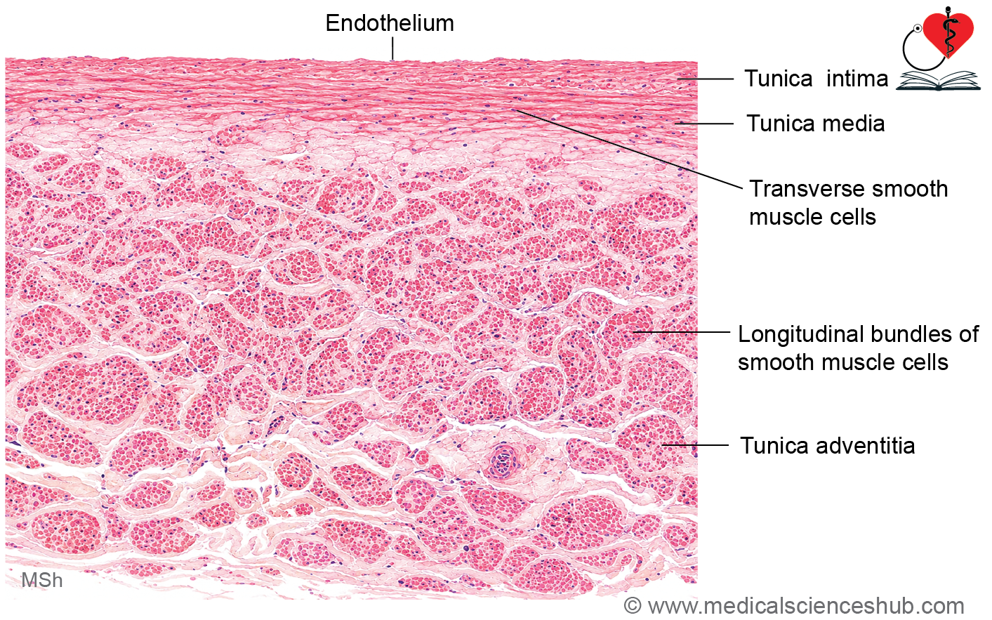
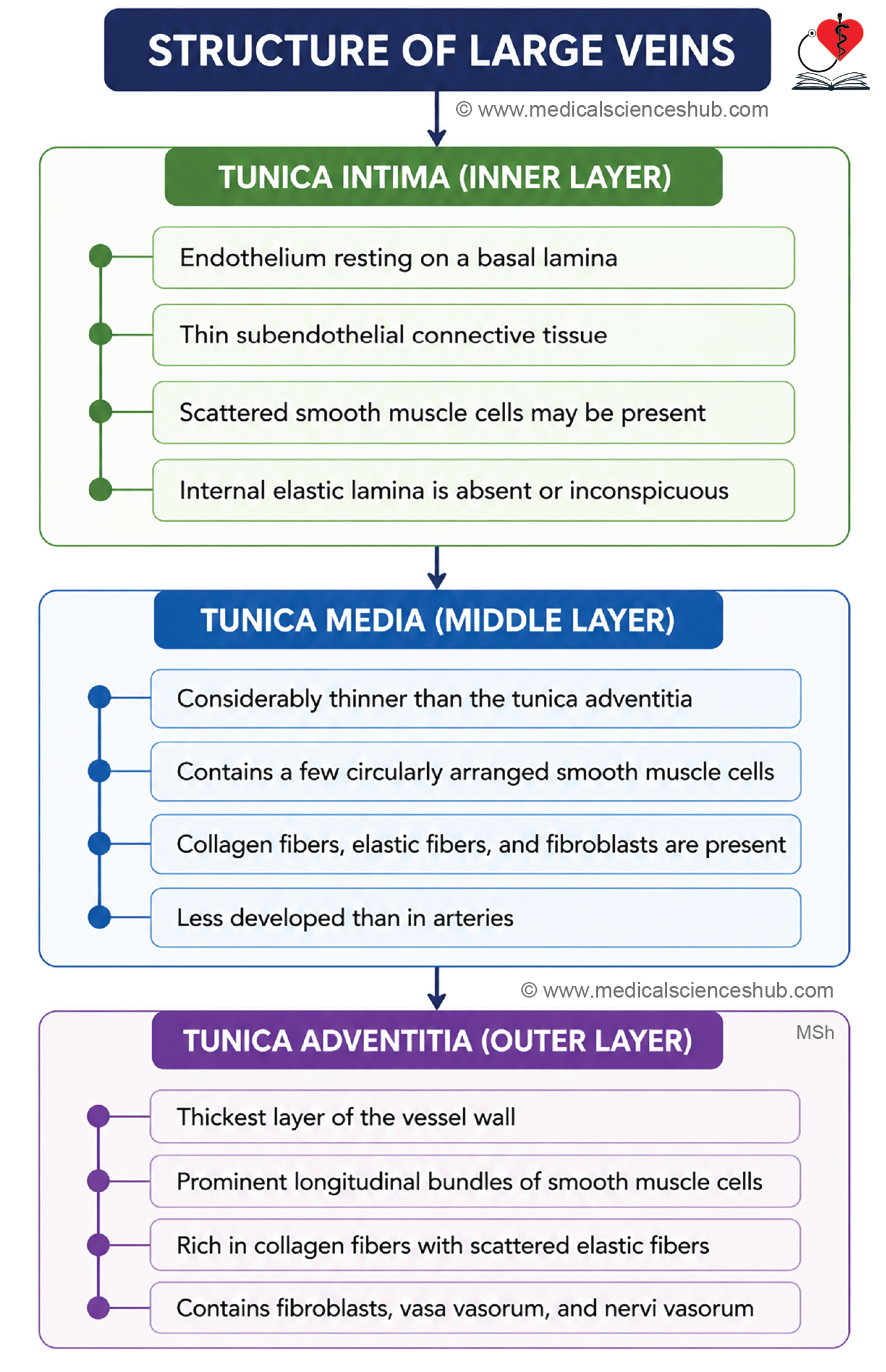
Table 10.2: Differences between artery and vein
| Feature | Artery | Vein |
|---|---|---|
| Wall Thickness | Wall is relatively thick and strong. | Wall is comparatively thin and less muscular. |
| Lumen | Lumen is usually round, oval, and remains patent. | Lumen is often irregular, collapsed, or flattened in histological sections. |
| Internal Elastic Lamina | Well-developed and easily identifiable in most medium-sized arteries. | Usually absent or poorly developed. |
| Tunica Media | Thickest layer of the vessel wall; contains numerous smooth muscle cells and elastic fibers. It is generally thicker than the tunica adventitia. | Relatively thin; contains fewer smooth muscle cells and more collagen fibers. It is usually thinner than the tunica adventitia. |
| Tunica Adventitia | Composed mainly of connective tissue containing collagen and elastic fibers. | Often the thickest layer, especially in large veins, and may contain longitudinal smooth muscle bundles in addition to connective tissue. |
| Layer Definition | Tunica intima, tunica media, and tunica adventitia are usually well defined. | Vessel layers are often less distinct, particularly in small and medium-sized veins. |
| Valves | Valves are absent under normal conditions. | Valves are commonly present, especially in veins of the lower limbs, to prevent backflow of blood. |
| Primary Function | Carries blood away from the heart under higher pressure. | Returns blood toward the heart under lower pressure. |
Table 10.3: Differences between large-sized vein, medium-sized vein and venule
| Feature | Large Vein | Medium-Sized Vein | Venule |
|---|---|---|---|
| Diameter | Greater than 10 mm | Approximately 1–10 mm | Approximately 10–100 μm |
| Tunica Intima | Contains endothelium with a thin subendothelial connective tissue layer. | Endothelium is supported by a very thin or inconspicuous subendothelial connective tissue layer. | Consists mainly of endothelium; subendothelial connective tissue is absent or minimal. |
| Tunica Media | Relatively thin; contains smooth muscle cells, collagen fibers, and fibroblasts. The boundary with the adventitia may be indistinct. | Thin layer containing a few smooth muscle cells, collagen fibers, and elastic fibers. | Contains 1–2 layers of smooth muscle cells in muscular venules; postcapillary venules lack a true tunica media. |
| Tunica Adventitia | Thickest layer; contains longitudinally arranged smooth muscle cells, collagen fibers, elastic fibers, and fibroblasts. | Prominent connective tissue layer rich in collagen fibers, elastic fibers, and fibroblasts. | Thin connective tissue layer containing a small amount of collagen and a few fibroblasts. |
| Valves | Usually absent in major central veins such as the venae cavae. | Commonly present, especially in veins of the limbs. | Absent. |
| Examples | Superior vena cava, inferior vena cava | Radial vein, tibial vein, popliteal vein | Postcapillary venules and muscular venules within tissues. |
Important Questions
- List the functions of endothelium.
- Write a short note on histology of elastic/large artery.
- Write a short note on histology of muscular arteries.
- List the differences between elastic artery, muscular artery, and arteriole.
- Write a short note on capillaries.
- Write a short note on sinusoids.
- Write a short note on AV shunts.
- Draw well-labeled diagram of histology of mediumsized vein.
- Write a short note on histology of large vein.
- List the differences between artery and vein.
- List the differences between large-sized vein, medium-sized vein and venule.
📝 Test Your Knowledge – Practice MCQs
Attempt the chapter MCQ quiz and assess your understanding of key concepts.
Click to see: Medical Disclaimer and Copyright Notice