
Competency
- AN52.1 Describe and identify the microanatomical features of gastrointestinal system: Liver, gallbladder, pancreas.
Introduction
- The liver, gallbladder, and pancreas are vital accessory organs of digestion. The liver produces bile and performs important metabolic functions, the gallbladder stores and concentrates bile, while the pancreas secretes digestive enzymes and hormones essential for digestion and glucose regulation.
Liver
- The liver is the largest gland in the human body, weighing approximately 1.5 kg in adults.
- It functions primarily as a modified exocrine gland that secretes bile and also performs numerous metabolic, detoxification, and storage functions.
Gross Organization of Liver
- The liver receives a dual blood supply:
- Hepatic artery supplies oxygenated blood.
- Portal vein carries nutrient-rich blood from the gastrointestinal tract.
- Venous blood leaves the liver through the hepatic veins, which drain into the inferior vena cava.
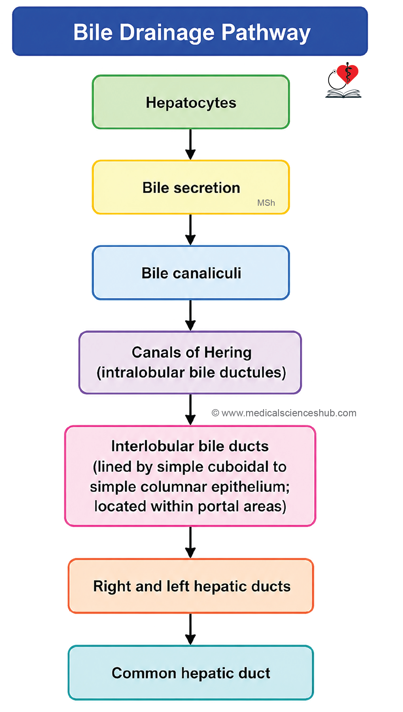
- Bile produced by hepatocytes is transported through bile canaliculi to the extrahepatic biliary system.
- Anatomically, the liver is divided into four lobes:
- Right lobe
- Left lobe
- Quadrate lobe
- Caudate lobe
- Clinically, functional segments based on vascular supply and biliary drainage are more important than anatomical lobes.
Structural Organization of Liver
The liver consists of the following major components:
- Connective tissue capsule and stroma
- Hepatocytes (functional parenchymal cells)
- Sinusoids and associated blood vessels
- Perisinusoidal space (Space of Disse)
- Bile canaliculi
Connective Tissue Capsule
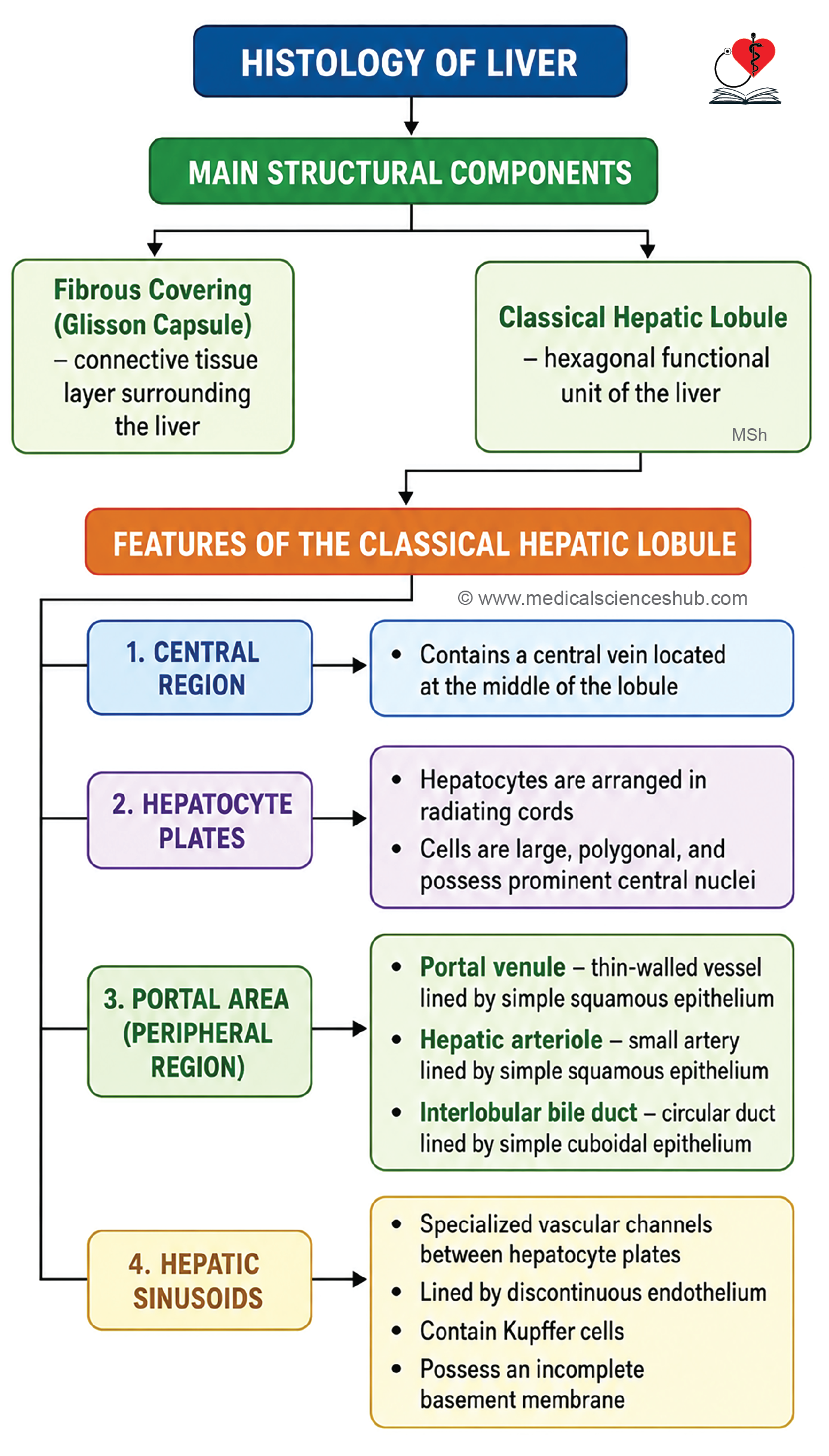
- The liver is enclosed by a thin connective tissue covering called Glisson’s capsule.
- Extensions of this connective tissue accompany branches of the portal vein, hepatic artery, and bile ducts into the liver parenchyma.
- A delicate framework of reticular fibers (type III collagen) supports the hepatic tissue.
- These reticular fibers are argentophilic, meaning they can be demonstrated by silver impregnation staining techniques.
Hepatocytes (Liver Cells)
- Hepatocytes are the principal functional cells of the liver and constitute the bulk of the hepatic parenchyma.
- They are large, polygonal cells measuring approximately 20–30 μm in diameter.
- The cells contain centrally placed, round nuclei with abundant euchromatin, reflecting their high metabolic and synthetic activity.
- One or two prominent nucleoli are usually present.
- Many hepatocytes are binucleated or polyploid, a feature associated with liver growth and regeneration.
- The liver possesses a remarkable capacity for regeneration following tissue injury or partial loss.
- The cytoplasm is typically mildly eosinophilic and contains abundant organelles involved in protein synthesis, metabolism, and detoxification.
Histochemical Features
- PAS stain demonstrates intracellular glycogen stores.
- Sudan stains identify lipid droplets within hepatocytes.
- Lipofuscin pigment may be seen in lysosomes, particularly in older cells.
- Numerous mitochondria reflect the high metabolic activity of hepatocytes.
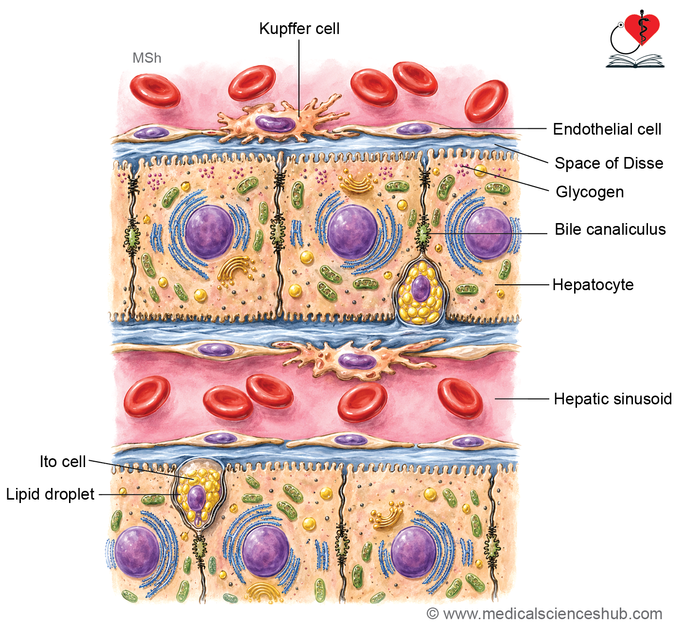
Surfaces of Liver Cells
- Sinusoidal surface: Faces the hepatic sinusoids and bears numerous microvilli projecting into the Space of Disse, increasing the surface area for exchange of nutrients and metabolites.
- Canalicular surface: Forms the walls of bile canaliculi together with adjacent hepatocytes. Tight junctions prevent leakage of bile into surrounding tissues.
- Intercellular surface: Contacts neighboring hepatocytes and contributes to structural organization and cell-to-cell communication within hepatic plates.
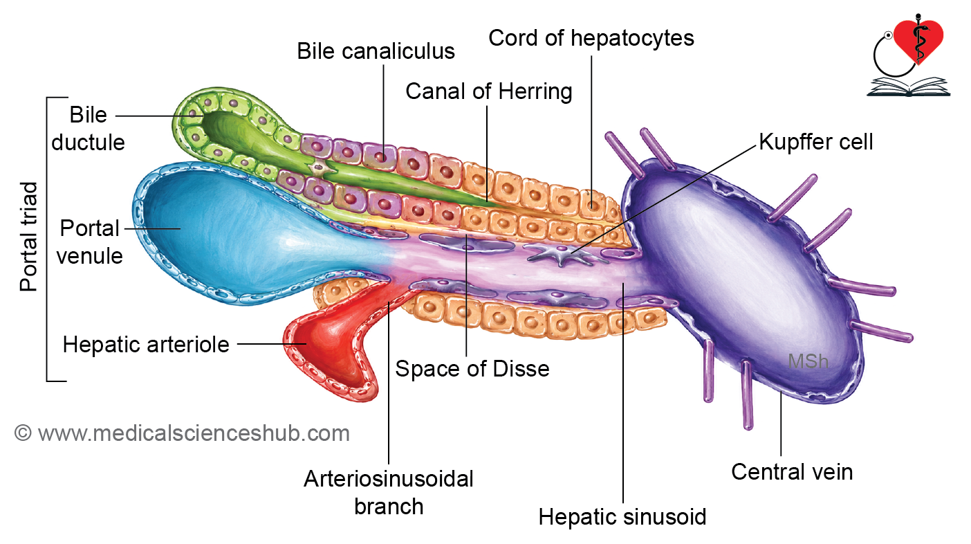
Blood Vessels of Liver
- The liver receives a dual blood supply:
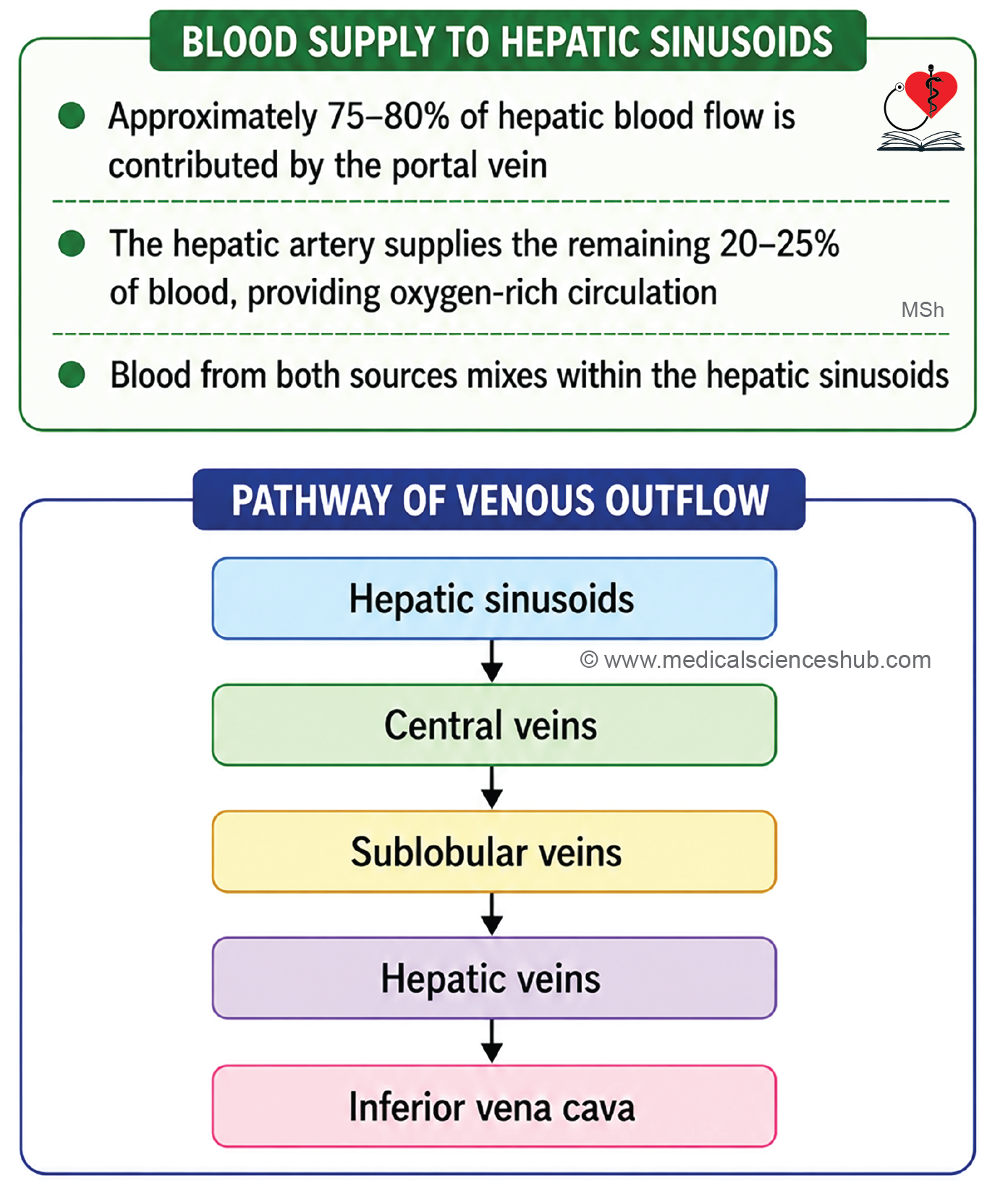
- Portal vein: Contributes approximately 75% of hepatic blood flow and carries nutrient-rich blood from the gastrointestinal tract, spleen, and pancreas.
- Hepatic artery: Supplies approximately 25% of blood flow and provides oxygenated blood.
- Bile produced by hepatocytes flows through bile canaliculi, which drain into bile ductules and subsequently into the extrahepatic biliary system.
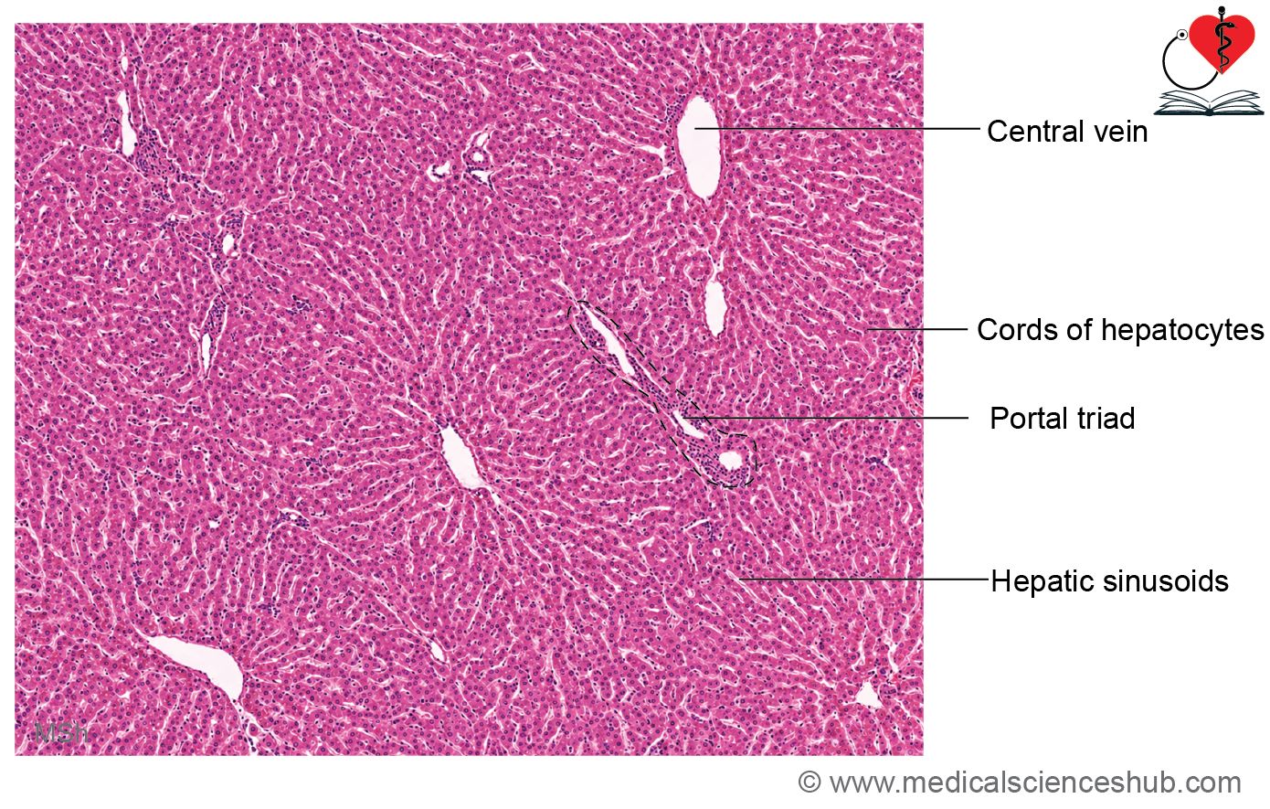
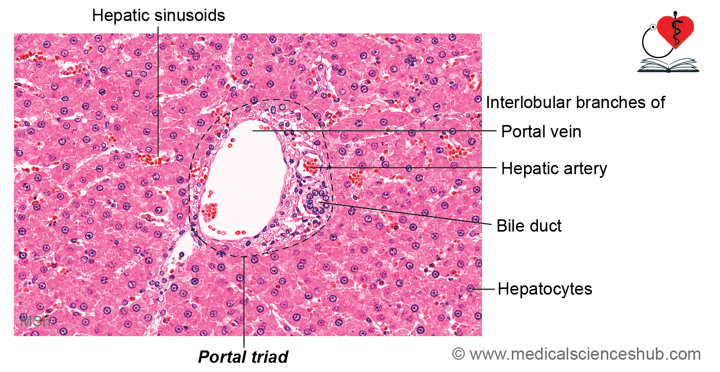
- At the periphery of hepatic lobules, connective tissue regions known as portal triads (portal canals) contain:
- A branch of the portal vein
- A branch of the hepatic artery
- A bile ductule
- The central vein is not a component of the portal triad.
- Blood from branches of the portal vein and hepatic artery mixes within the hepatic sinusoids, where exchange with hepatocytes occurs.
- Blood flow follows the sequence:
- Portal venule and hepatic arteriole → hepatic sinusoids → central vein → sublobular veins → hepatic veins → inferior vena cava.
- Hepatic veins are characteristically valveless, allowing unobstructed venous drainage from the liver.
Concept of liver lobules
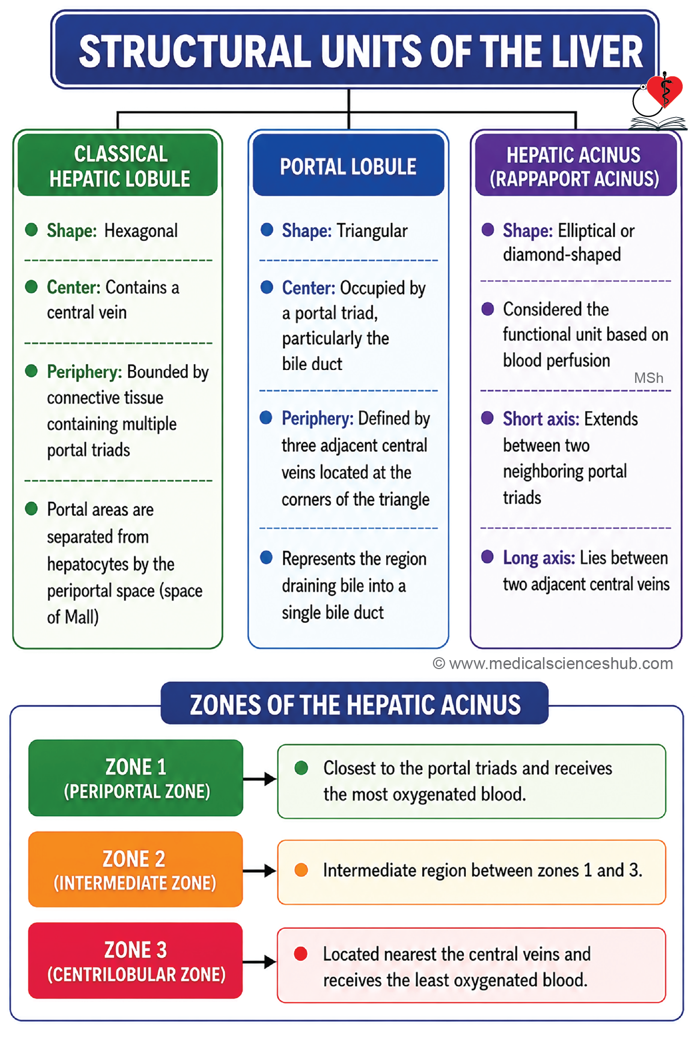
- The liver parenchyma is organized into structural units called liver lobules.
- Three complementary models are used to describe hepatic organization:
- Classical liver lobule
- Portal lobule
- Hepatic acinus (acinus of Rappaport)
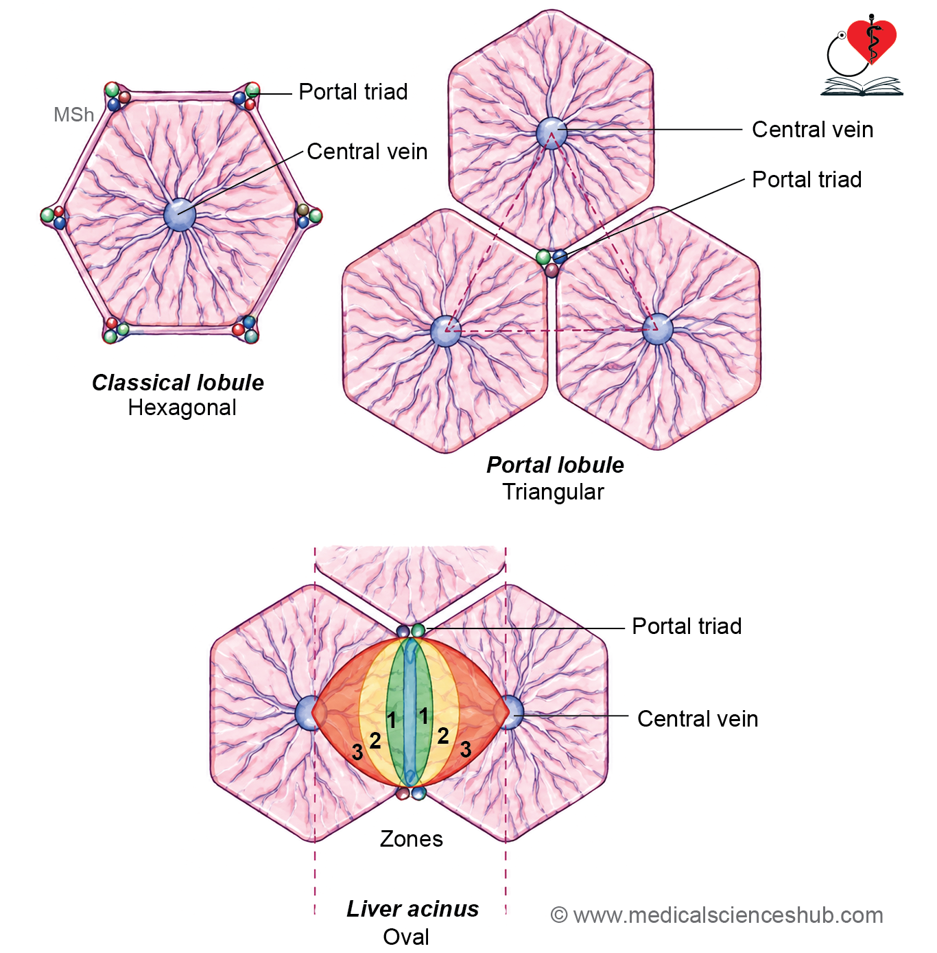
Classical liver lobule
- The classical lobule is a roughly hexagonal unit of liver tissue.
- A central vein occupies the center of the lobule.
- Plates or cords of hepatocytes radiate outward from the central vein and are separated by hepatic sinusoids.
- Portal triads are located at the corners of the hexagon within connective tissue regions known as portal canals.
- Adjacent lobules are separated by only a small amount of connective tissue in humans, making their boundaries indistinct.
- The Space of Mall (periportal space) lies between the portal tract connective tissue and peripheral hepatocytes and serves as an important site for lymph formation.
- Classical lobules are more clearly defined in some animals, such as pigs, because of a more prominent connective tissue framework.
Portal lobule
- The portal lobule emphasizes the exocrine function of the liver, namely bile secretion.
- It is a triangular region of hepatic tissue centered on a portal triad, particularly its bile duct.
- The three surrounding central veins form the corners of the triangle.
- It represents the area of liver tissue that drains bile into a single bile ductule.
Liver acinus (Portal acinus or hepatic acinus or acinus of Rappaport)
- The hepatic acinus is considered the most functionally relevant unit of the liver.
- It is diamond-shaped and is supplied by terminal branches of the hepatic artery and portal vein.
- The short axis lies between two adjacent portal triads, while the long axis extends between two neighboring central veins.
Zones of the Hepatic Acinus
- Zone 1 (periportal zone):
- Closest to the portal triad.
- Receives the most oxygenated and nutrient-rich blood.
- First region exposed to blood-borne toxins and nutrients.
- Zone 2 (intermediate zone):
- Lies between zones 1 and 3.
- Exhibits intermediate metabolic activity and blood supply.
- Zone 3 (centrilobular zone):
- Closest to the central vein.
- Receives the least oxygenated blood.
- Most susceptible to hypoxic injury and ischemic damage.
Liver sinusoids
- Hepatic sinusoids are specialized vascular channels located between the plates of hepatocytes and facilitate exchange between blood and liver cells.
- The sinusoidal wall is lined by:
- Discontinuous endothelial cells with large fenestrations.
- Kupffer cells, which are resident stellate macrophages responsible for phagocytosis and immune surveillance.
- The Space of Disse (perisinusoidal space) is a narrow extracellular compartment situated between the sinusoidal endothelium and hepatocytes.
- Numerous hepatocyte microvilli project into the Space of Disse, greatly increasing the surface area available for metabolic exchange.
- Efficient transfer of substances occurs because the hepatic sinusoid lacks a typical filtration barrier due to:
- Wide gaps between endothelial cells.
- An incomplete or discontinuous basal lamina.
- During fetal development, the Space of Disse can serve as a site of hematopoiesis.
- Hepatic stellate cells (Ito cells) are located within the Space of Disse. These mesenchymal cells store vitamin A and lipid droplets under normal conditions.
- In chronic liver injury, Ito cells become activated and transform into myofibroblast-like cells, producing excessive extracellular matrix. This process contributes to hepatic fibrosis and may progress to cirrhosis.
Table 16.1: Cells of liver
| Cell Type | Location | Principal Functions |
|---|---|---|
| Hepatocytes | Arranged in hepatic cords or plates between sinusoids | Synthesize and secrete bile, metabolize and detoxify drugs and toxins, store glycogen, and participate in protein and lipid metabolism |
| Kupffer Cells | Located along the luminal surface of hepatic sinusoids | Specialized liver macrophages that remove aged erythrocytes, microorganisms, cellular debris, and other particulate matter through phagocytosis |
| Hepatic Stellate (Ito) Cells | Situated in the perisinusoidal space (Space of Disse) | Store vitamin A and lipids, support tissue repair, and transform into myofibroblast-like cells during chronic liver injury, contributing to hepatic fibrosis |
| Sinusoidal Endothelial Cells | Form the lining of hepatic sinusoids | Possess fenestrations that facilitate exchange of nutrients, plasma proteins, and metabolites between sinusoidal blood and hepatocytes |
| Hematopoietic Cells (Fetal Liver) | Present within the Space of Disse during fetal development | Participate in the formation and maturation of blood cells before birth |
CLINICAL CORRELATION
- Hepatitis refers to inflammation of the liver and may result from viral infections (such as hepatitis viruses), bacterial infections, drugs, toxins, or autoimmune disorders.
- Liver cirrhosis is a chronic condition in which damaged hepatocytes are progressively replaced by fibrous tissue, leading to distortion of normal liver architecture and impaired function.
- Common causes include chronic alcohol consumption, viral hepatitis, drugs, toxins, and autoimmune liver diseases.
- Portal hypertension is an abnormal increase in pressure within the portal venous system, usually caused by increased resistance to blood flow through the liver.
- It commonly develops in cirrhosis and leads to dilation of portosystemic (portocaval) venous anastomoses.
- Wilson disease is an autosomal recessive disorder caused by mutations in the ATP7B gene, resulting in defective copper transport and excretion.
- Excess copper accumulates in the liver, brain, and cornea, producing liver damage, neurological manifestations, and cirrhosis.
- Liver failure occurs when extensive hepatocyte loss severely impairs hepatic function.
- Reduced detoxification capacity causes accumulation of toxic metabolites in the bloodstream.
- Severe liver failure may lead to hepatic encephalopathy, progressing to hepatic coma and potentially death if untreated.
Gallbladder
- The gallbladder is a pear-shaped, muscular reservoir that stores and concentrates bile produced by the liver.
- It has a storage capacity of approximately 30–50 mL and is located in a fossa on the inferior surface of the liver.
- Bile enters the gallbladder through the cystic duct.
Histology of Gallbladder
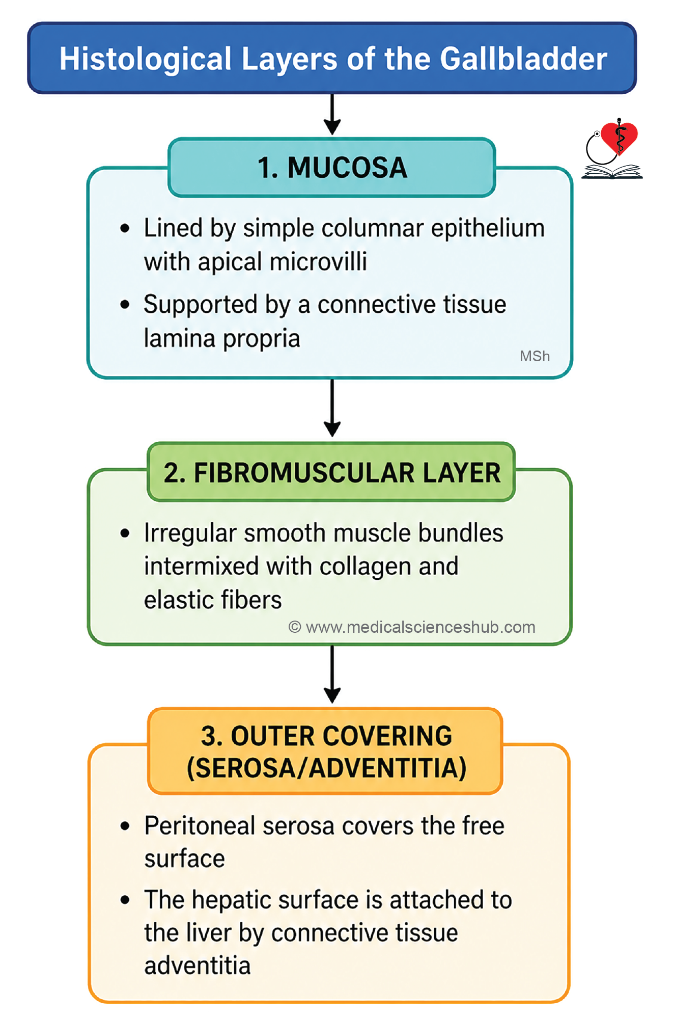
- The wall of the gallbladder consists of three layers:
- Mucosa
- Fibromuscular coat
- Serosa or adventitia
- Unlike most parts of the gastrointestinal tract, the gallbladder lacks both submucosa and muscularis mucosae.
Mucosa
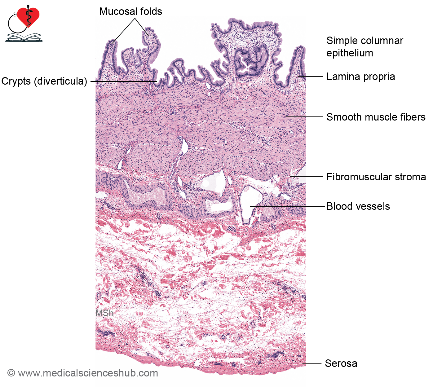
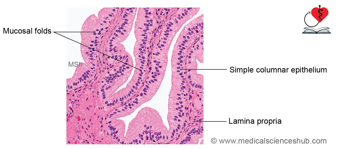
- The mucosa forms numerous temporary folds that flatten when the gallbladder becomes distended.
- It is lined by simple columnar epithelium with basally located nuclei.
- Epithelial cells possess abundant microvilli, forming an apical striated border that enhances absorption of water and electrolytes from bile.
- The lamina propria consists of connective tissue rich in fenestrated capillaries.
- Mucosal folds may create crypt-like recesses, and mucous glands may be present near the neck of the gallbladder.
- Lymphatic vessels are generally absent within the lamina propria.
Fibromuscular Coat/muscularis Externa
- This layer contains irregularly arranged bundles of smooth muscle interwoven with collagen and elastic fibers.
- Muscle fibers do not form distinct circular or longitudinal layers.
- Deep mucosal invaginations extending into the muscle layer are known as Rokitansky–Aschoff sinuses. These are mucosal outpouchings rather than true glands.
Serosa/Adventitia
- The outer layer contains connective tissue, blood vessels, lymphatics, nerves, and elastic fibers.
- Surfaces not attached to the liver are covered by serosa, consisting of visceral peritoneum lined by mesothelium.
CLINICAL CORRELATION
- Cholecystitis refers to inflammation of the gallbladder.
- Cholelithiasis is the formation of gallstones within the gallbladder.
- Chronic gallstone disease and longstanding cholecystitis increase the risk of adenocarcinoma of the gallbladder.
Pancreas
- The pancreas is a retroperitoneal gland extending from the C-shaped curve of the duodenum to the hilum of the spleen.
- It is anatomically divided into the head, neck, body, and tail.
Pancreatic Ducts
- The exocrine secretions of the pancreas are drained mainly by two ducts:
- Main pancreatic duct (duct of Wirsung):
- Traverses the entire length of the pancreas.
- Joins the common bile duct to form the hepatopancreatic ampulla (ampulla of Vater).
- Opens into the second part of the duodenum at the major duodenal papilla.
- The opening is regulated by the sphincter of Oddi, which controls the flow of bile and pancreatic juice and helps prevent reflux of intestinal contents.
- Accessory pancreatic duct (duct of Santorini):
- Drains a smaller portion of the pancreas.
- Opens separately into the duodenum through the minor duodenal papilla.
- Main pancreatic duct (duct of Wirsung):
- These ducts ensure efficient delivery of pancreatic enzymes and bicarbonate-rich secretions into the duodenum for digestion.
Histology of Pancreas
Capsule
- The pancreas is enclosed by a thin capsule of loose connective tissue.
- Connective tissue septa extend inward from the capsule, dividing the gland into poorly defined lobules.
Exocrine and Endocrine Pancreas
The pancreatic parenchyma consists of two distinct parts:
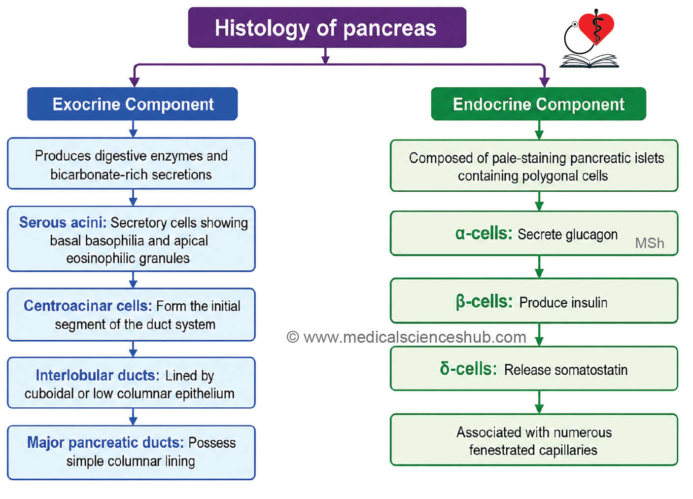
- Exocrine pancreas, which produces digestive enzymes and bicarbonate-rich secretions.
- Endocrine pancreas (Islets of Langerhans), which secretes hormones such as insulin and glucagon directly into the bloodstream.
Exocrine Pancreas (Serous Gland)
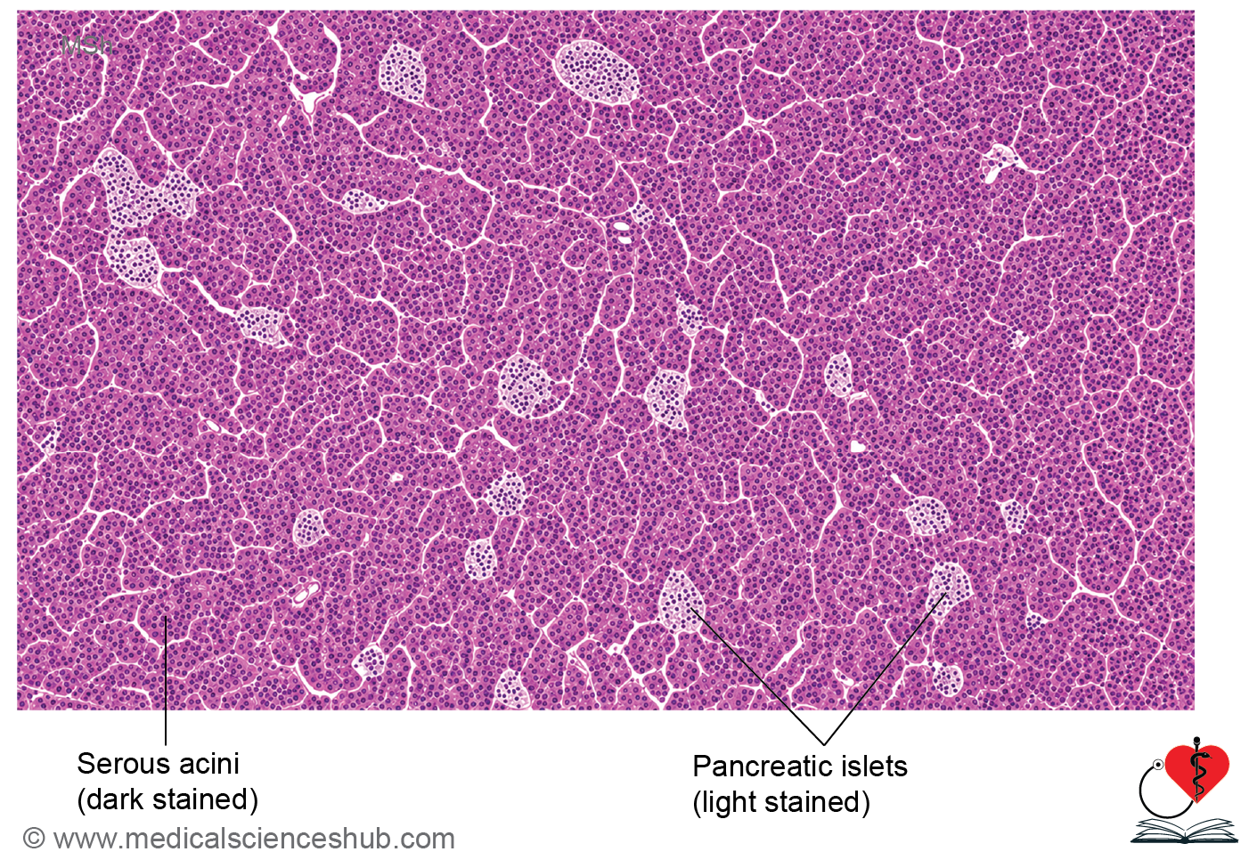
- The exocrine pancreas is a compound tubuloacinar serous gland.
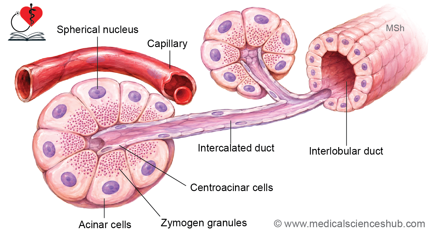
- It is composed of numerous pancreatic acini, which appear round or oval and possess small central lumina.
- Acinar cells are pyramidal with broad basal regions and narrow apical surfaces facing the lumen.
- Nuclei are round and located near the base of the cells.
Acinar Cells
- On H&E staining, the basal cytoplasm appears basophilic because of abundant rough endoplasmic reticulum.
- The apical cytoplasm is eosinophilic due to the presence of zymogen granules.
- Zymogen granules contain inactive digestive enzymes, including proteases, amylases, lipases, and nucleases.
- These granules are released by exocytosis and are often more prominent during fasting.
Centroacinar Cells
- Centroacinar cells are unique to the pancreas and represent the initial portion of the duct system within the acinus.
- They appear pale-staining with centrally placed flattened nuclei.
- These cells contribute bicarbonate-rich fluid to pancreatic secretions.
Pancreatic Duct System
- The ductal pathway is:
- Intercalated ducts → Interlobular ducts → Main and accessory pancreatic ducts
- Intercalated ducts begin as centroacinar cells and are lined by simple squamous to low cuboidal epithelium.
- Interlobular ducts are lined by cuboidal to low columnar epithelium.
- The main and accessory pancreatic ducts are lined by simple columnar epithelium.
- Unlike salivary glands, the pancreas lacks striated ducts.
Functions and Regulation
- Intercalated ducts add water and bicarbonate ions to pancreatic secretions.
- Exocrine pancreatic activity is regulated by the hormones secretin and cholecystokinin (CCK), as well as autonomic neural input.
Endocrine Pancreas
- The endocrine pancreas is composed of numerous small clusters of hormone-secreting cells known as Islets of Langerhans, which are scattered throughout the pancreatic tissue.
- These islets constitute approximately 1–2% of the pancreatic volume, and the human pancreas contains about one million islets.
- On H&E staining, the islets appear as pale-staining clusters of polygonal cells surrounded by richly vascularized capillaries.
- The surrounding exocrine pancreatic acini stain more deeply, making the islets readily distinguishable.
- Individual endocrine cell types cannot be reliably identified with routine H&E staining.
Cell Types of the Islets
- Special histological stains and immunohistochemical techniques demonstrate several endocrine cell populations:
- Alpha (A) cells – secrete glucagon.
- Beta (B) cells – secrete insulin and are the most abundant cell type.
- Delta (D) cells – secrete somatostatin.
- PP (F) cells – secrete pancreatic polypeptide.
- Smaller populations of other endocrine cells, including enterochromaffin cells, may also be present.
- The rich capillary network within the islets enables rapid release of hormones directly into the bloodstream, allowing effective regulation of glucose metabolism and digestive functions.
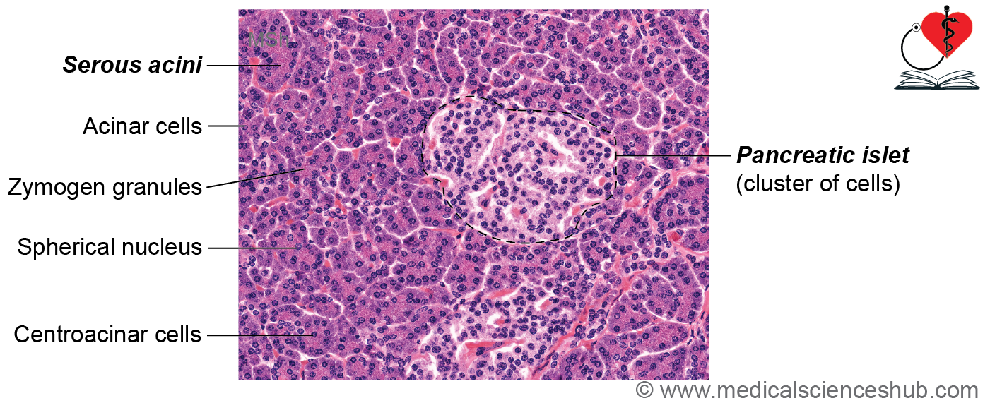
Table 16.2: Exocrine pancreas
| Component of Exocrine Pancreas | Histological Characteristics / Epithelium |
|---|---|
| Serous Acini | Composed of pyramidal secretory cells with basophilic basal cytoplasm due to abundant rough endoplasmic reticulum and eosinophilic apical cytoplasm containing zymogen granules. Myoepithelial cells are absent. |
| Centroacinar Cells | Pale-staining cells located at the center of acini; lined by simple squamous to low cuboidal epithelium. They represent the beginning of the duct system. |
| Intercalated Ducts | Lined by simple low cuboidal epithelium and receive secretions directly from centroacinar cells. |
| Interlobular Ducts | Lined by cuboidal to low columnar epithelium and located within connective tissue septa between lobules. |
| Striated Ducts | Absent in the pancreas, unlike in major salivary glands. |
| Main and Accessory Pancreatic Ducts | Lined predominantly by simple columnar epithelium that facilitates the transport of pancreatic secretions to the duodenum. |
Table 16.3: Cells of islets of Langerhans
| Cell Type | Hormone/Secretion | Histological Features and Distribution |
|---|---|---|
| Alpha (A or A₂) Cells | Glucagon | Constitute approximately 15–20% of islet cells. Predominantly located at the periphery of the islets. Show strong staining with acid fuchsin. |
| Beta (B) Cells | Insulin | Form the largest population, accounting for about 60–70% of islet cells. Typically concentrated in the central region of the islets. Stain characteristically with aldehyde fuchsin. |
| Delta (D, A₁, or Type III) Cells | Somatostatin | Represent approximately 5–10% of islet cells. Usually situated near the periphery. These argyrophilic cells demonstrate affinity for silver stains and may also stain with acid fuchsin. |
| PP (F) Cells | Pancreatic Polypeptide | Found mainly in the peripheral portions of the islets, particularly in the head of the pancreas. Regulate pancreatic exocrine secretion and gastrointestinal activity. |
| D₁ Cells | Vasoactive Intestinal Peptide (VIP) | Relatively few in number. Secrete VIP, which influences intestinal motility, blood flow, and glandular secretion. |
| Enterochromaffin (EC) Cells | Serotonin, Motilin, and Substance P | Scattered neuroendocrine cells that produce bioactive peptides involved in gastrointestinal motility and secretory functions. |
CLINICAL CORRELATION
- Acute pancreatitis is a sudden inflammation of the pancreas caused by premature activation of pancreatic digestive enzymes within the gland rather than in the intestine.
- Activated enzymes lead to autodigestion, resulting in inflammation, edema, tissue necrosis, and potentially life-threatening complications.
- Chronic pancreatitis is a long-standing inflammatory disorder characterized by recurrent episodes of pancreatic injury, progressive fibrosis, and gradual loss of pancreatic function.
- In both acute and chronic pancreatitis, blood levels of serum amylase and serum lipase are commonly elevated and serve as important diagnostic markers.
- Diabetes mellitus develops when insulin production is inadequate or when body tissues fail to respond appropriately to insulin.
- Type 1 diabetes mellitus results from destruction or failure of pancreatic beta cells, leading to absolute insulin deficiency.
- Type 2 diabetes mellitus is primarily caused by insulin resistance, in which target tissues respond poorly to insulin, often accompanied by a relative reduction in insulin secretion.
- Persistent pancreatic damage may impair both the endocrine and exocrine functions of the pancreas.
Important Questions
- Write a short note on liver lobules.
- Write a short note on classical hepatic lobule.
- Write a short note on histology of liver.
📝 Test Your Knowledge – Practice MCQs
Attempt the chapter MCQ quiz and assess your understanding of key concepts.
Click to see: Medical Disclaimer and Copyright Notice