
Competency
- AN43.2 Identify, describe and draw the microanatomy of cornea,retina.
Introduction
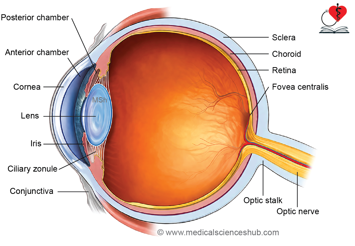
- The eye is the peripheral organ of vision located within the bony orbit.
- The eyeball is approximately spherical, with an average diameter of about 24–25 mm.
- Eye movements are controlled by six extraocular muscles, while surrounding adipose tissue provides cushioning and support.
- The wall of the eyeball is composed of three concentric layers:
- Fibrous coat: Consists of the sclera and cornea.
- Vascular coat (uvea): Includes the choroid, ciliary body, and iris.
- Neural coat: Formed by the retina.
- The eyeball contains three fluid-filled chambers:
- Anterior chamber: Located between the cornea and the anterior surfaces of the iris and lens.
- Posterior chamber: Situated between the posterior surface of the iris and the ciliary body, zonular fibers, and lens.
- Vitreous chamber: Extends between the lens and the retina.
- The anterior and posterior chambers contain aqueous humor and communicate through the pupil.
- The vitreous chamber contains vitreous humor, a transparent gel-like substance.
- The cornea, lens, aqueous humor, and vitreous humor together form the major refractive media of the eye, helping to focus light onto the retina.
Cornea
- The cornea is the transparent anterior portion of the fibrous coat of the eyeball and is continuous with the sclera at the corneoscleral limbus.
- It is avascular, highly innervated, convex anteriorly, and approximately 1 mm thick.
- Corneal transparency is essential for proper refraction of light entering the eye.
Histology of Cornea
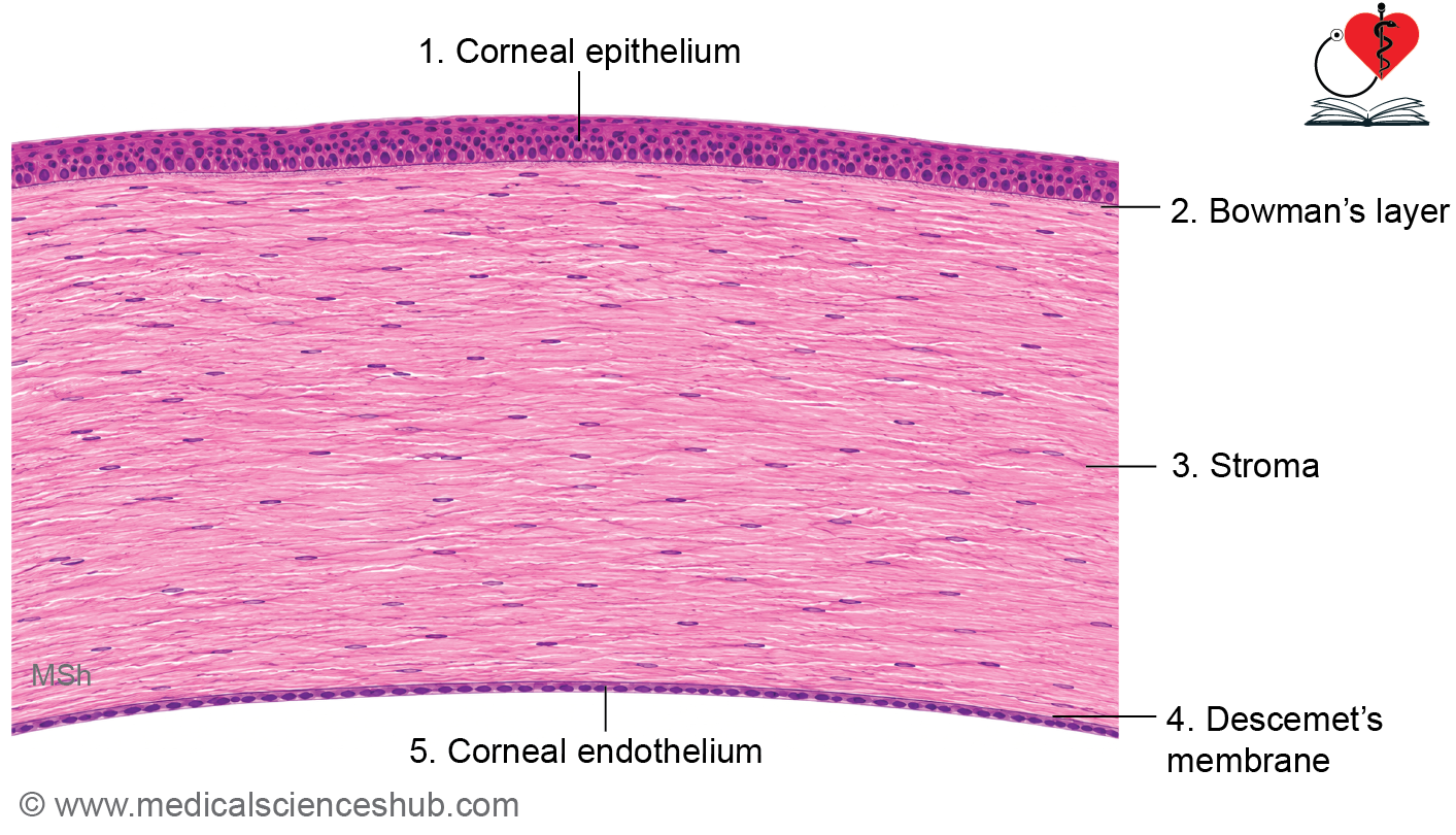
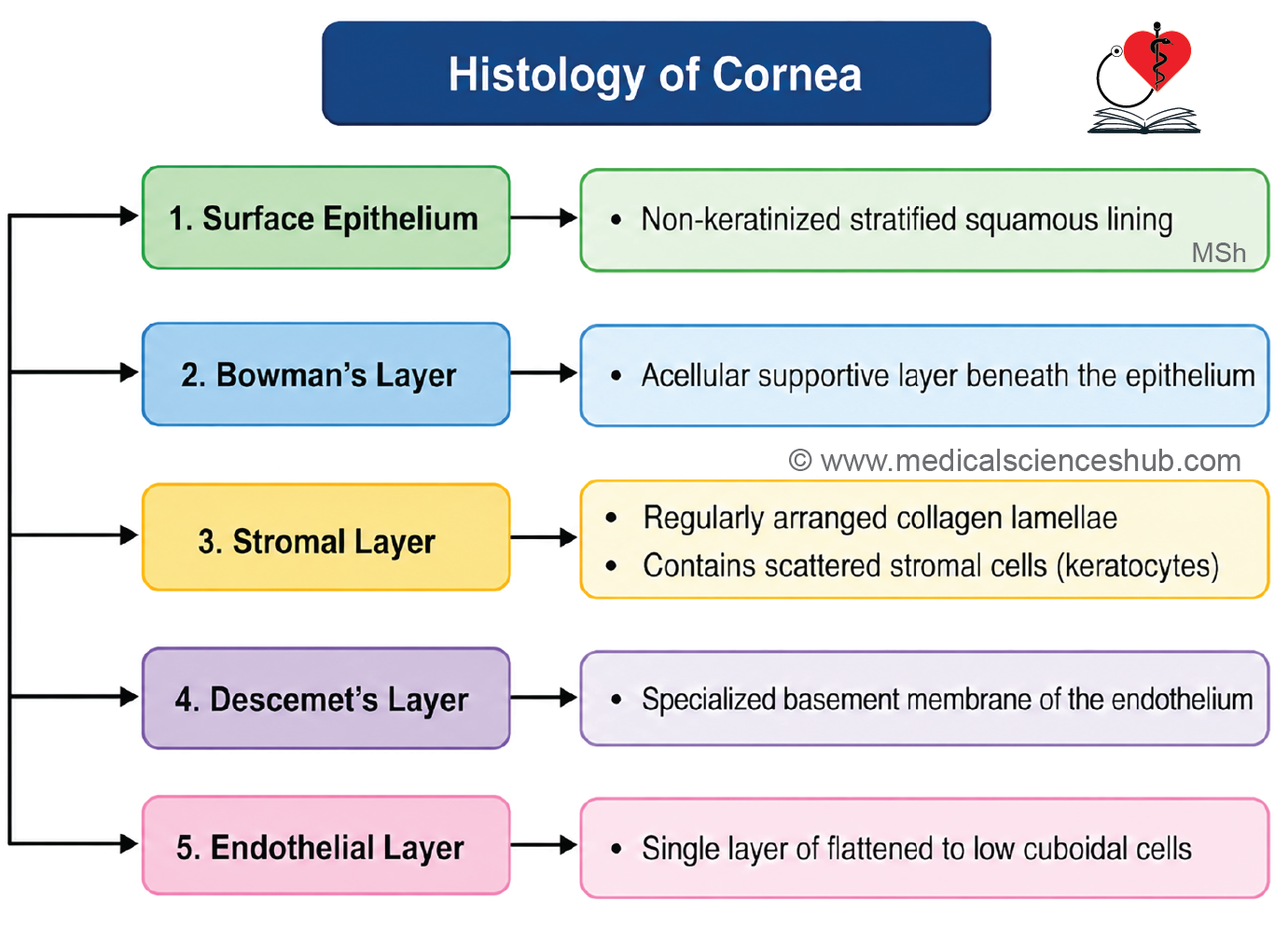
The cornea is composed of five distinct layers arranged from anterior to posterior: Corneal epithelium, Bowman’s membrane, corneal stroma, Descemet’s membrane, and corneal endothelium.
Corneal Epithelium
- The outer surface is lined by nonkeratinized stratified squamous epithelium.
- Basal cells are columnar, whereas superficial cells are flattened with condensed nuclei.
- The epithelium regenerates rapidly, usually within about one week.
- Limbal stem cells located at the corneoscleral junction replenish epithelial cells.
- Numerous free nerve endings make the cornea highly sensitive to touch.
- Surface microvilli help retain the tear film, maintaining corneal lubrication and optical clarity.
Bowman’s Membrane (Anterior Basement Membrane/Limiting Lamina)
- This acellular layer lies directly beneath the corneal epithelium.
- It is composed of randomly arranged collagen fibrils and measures approximately 8–10 μm in thickness.
- It provides structural support and acts as a protective barrier against infection and injury.
Corneal Stroma (Substantia Propria)
- The stroma forms about 90% of corneal thickness.
- It consists of regularly arranged lamellae of type I collagen, ground substance, and specialized fibroblasts called keratocytes.
- Collagen fibers run parallel within each lamella and at right angles to those in adjacent lamellae.
- This highly ordered arrangement is responsible for corneal transparency.
- The extracellular matrix contains proteoglycans rich in sulfated glycosaminoglycans.
Descemet’s Membrane
- This layer is the true basement membrane of the corneal endothelium.
- It appears as a homogeneous, PAS-positive membrane.
- At the corneal periphery, it contributes to the formation of the pectinate ligament, which participates in aqueous humor drainage.
- Peripheral fibers also help maintain corneal curvature.
Corneal Endothelium
- The posterior surface of the cornea is lined by a single layer of flattened to low cuboidal endothelial cells.
- These cells are in direct contact with the aqueous humor of the anterior chamber.
- Specialized intercellular junctions maintain tissue integrity while permitting controlled fluid movement.
- The endothelium actively removes excess water from the stroma, preserving corneal transparency and optimal optical function.
Sclera
- The sclera is the tough, white, opaque fibrous coat that forms the posterior five-sixths of the eyeball.
- It provides structural support, maintains the shape of the eye, and serves as the attachment site for extraocular muscles.
- The sclera is composed of:
- Dense bundles of collagen fibers
- A small amount of elastic fibers
- Ground substance
- Scattered fibroblasts
- Its white, opaque appearance results from the irregular arrangement of collagen fibers, which scatter light.
Layers of the Sclera
- Although not sharply demarcated, the sclera can be divided into three layers:
- Episclera (Episcleral Layer)
- The outermost layer composed of loose connective tissue.
- Sclera Proper (Tenon’s Capsule/Substantia Propria)
- The thick middle layer consisting of densely packed collagen fibers.
- It provides most of the sclera’s strength and rigidity.
- Suprachoroid Lamina (Lamina Fusca)
- The innermost layer adjacent to the choroid.
- It contains delicate connective tissue fibers and serves as a transition zone between the sclera and choroid.
Layers of Sclera (Ill-defined)
Layers of the Sclera
- Although not sharply demarcated, the sclera can be divided into three layers:
- Episclera (Episcleral Layer)
- The outermost layer composed of loose connective tissue.
- Sclera Proper (Tenon’s Capsule/Substantia Propria)
- The thick middle layer consisting of densely packed collagen fibers.
- It provides most of the sclera’s strength and rigidity.
- Suprachoroid Lamina (Lamina Fusca)
- The innermost layer adjacent to the choroid.
- It contains delicate connective tissue fibers and serves as a transition zone between the sclera and choroid.
Uvea/Vascular Coat
- Deep to sclera, there is a vascular coat of eyeball.It consists of choroid, ciliary body, and iris.
Choroid
- The choroid is the highly vascular, pigmented layer situated between the sclera and retina.
- Its dark coloration is due to abundant melanocytes, which absorb scattered light and improve visual clarity.
- The choroid extends from the optic nerve posteriorly to the ora serrata anteriorly.
1. Choriocapillaris (Choriocapillary Layer)
- This layer lies immediately external to Bruch’s membrane.
- It contains a dense network of blood vessels embedded within connective tissue.
- The outer region contains larger blood vessels, while the inner choriocapillaris consists of numerous capillaries that supply the outer retina.
- Numerous pigmented cells contribute to the dark appearance of the choroid.
- Externally, the vascular layer is supported by the suprachoroid lamina (lamina fusca), a thin nonvascular connective tissue layer adjacent to the sclera.
2. Bruch’s Membrane (Lamina Vitrea)
- Bruch’s membrane is a thin, transparent layer located between the choriocapillaris and the retinal pigment epithelium.
- It is composed of collagen and elastic fibers arranged in specialized layers. This membrane forms part of the barrier and exchange interface between the choroid and retina.
Ciliary Body
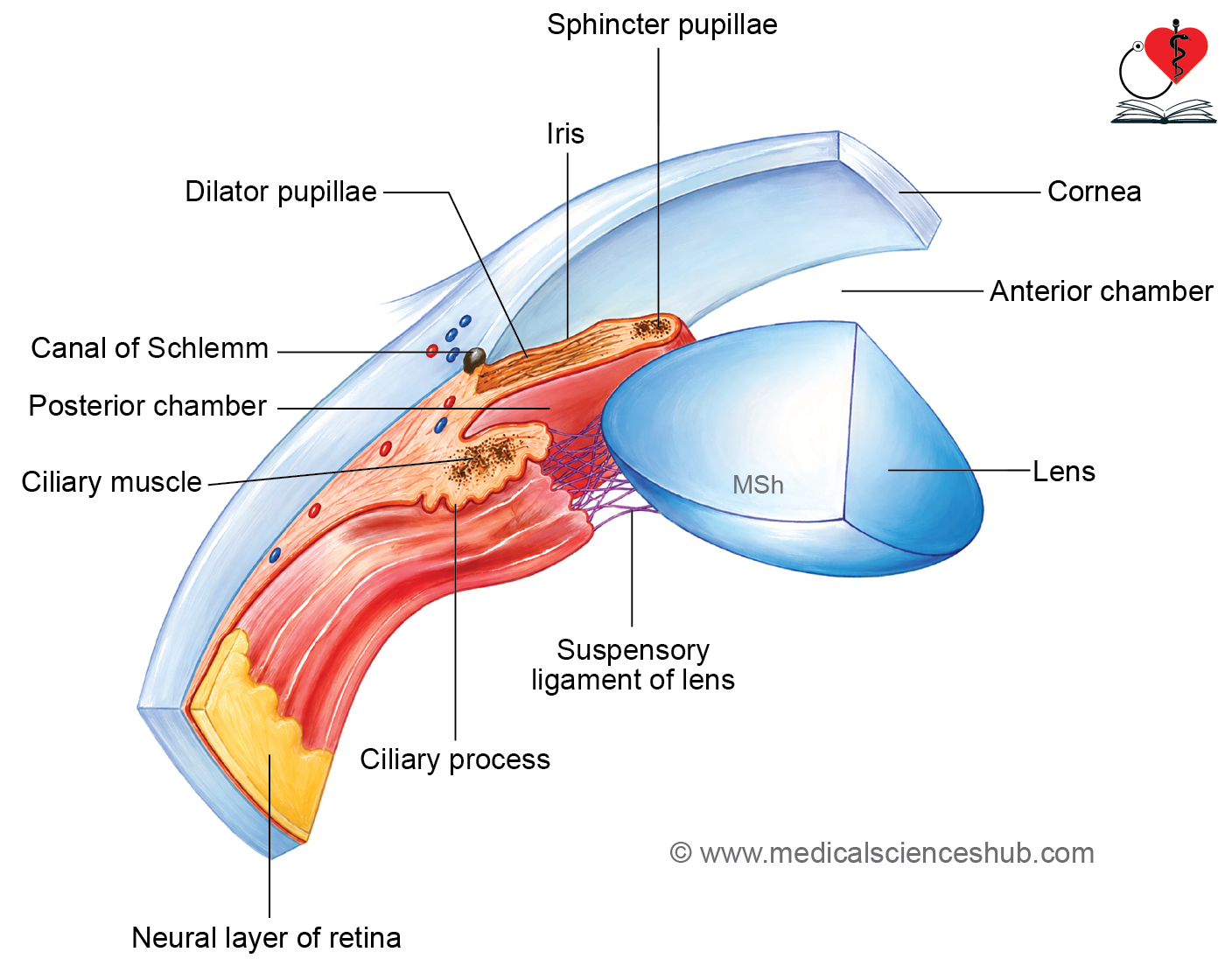
- The ciliary body is the thickened anterior continuation of the vascular layer (uvea) of the eyeball.
- It lies between the choroid posteriorly and the iris anteriorly.
- The ciliary body is attached to the lens through the zonular fibers (suspensory ligaments).
- Structurally, it consists of ciliary muscle, connective tissue, blood vessels, and ciliary processes.
Ciliary Muscle
- The ciliary muscle is composed of smooth muscle fibers arranged in three functional groups:
- Longitudinal fibers help maintain the iridocorneal angle and facilitate aqueous humor drainage.
- Radial (oblique) fibers assist in adjusting lens curvature during accommodation.
- Circular fibers reduce tension on zonular fibers, allowing the lens to become more convex for near vision.
Ciliary Processes
- The ciliary body bears approximately 70–80 ciliary processes, which project toward the lens.
- These processes are covered by a double layer of ciliary epithelium.
- The outer epithelial layer is pigmented and continuous with the retinal pigment epithelium.
- The inner nonpigmented layer actively secretes aqueous humor.
- Ciliary epithelium also contributes to the blood–aqueous barrier, helping maintain the internal environment of the eye.
- Zonular fibers arise from the ciliary processes and attach to the lens capsule, transmitting forces required for accommodation.
Iris
- The iris is the anterior-most component of the vascular coat (uvea) of the eye.
- It is attached to the sclera near the corneoscleral junction and forms a movable diaphragm in front of the lens.
- The central opening of the iris is the pupil, which regulates the amount of light entering the eye.
Structure of the Iris
- The iris consists of two principal components:
- Anterior stromal layer
- Posterior pigmented epithelium
- The posterior surface is lined by a double layer of pigmented cells:
- A posterior heavily pigmented epithelial layer.
- An anterior layer of pigmented myoepithelial cells.
- The stromal layer contains:
- Smooth muscle fibers
- Fibroblasts
- Melanocytes
- Connective tissue fibers
- Blood vessels
Muscles of the Iris
- Smooth muscle fibers are arranged into two functional groups:
- Sphincter pupillae, arranged circularly around the pupil, causes pupillary constriction.
- Dilator pupillae, arranged radially, causes pupillary dilation.
Pigmentation
- The number and distribution of melanocytes within the iris stroma determine the color of the iris.
- Greater melanin content produces darker iris coloration, whereas lower melanin content results in lighter eye colors.
Retina
- The retina is the innermost sensory layer of the eyeball and lines the posterior three-fourths of its inner surface.
- Embryologically, it develops from the two layers of the optic cup.
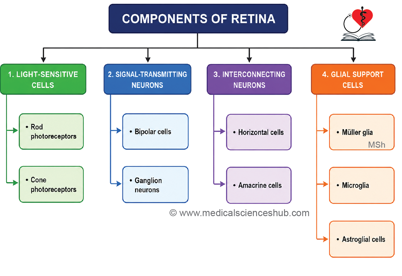
- The retina consists of:
- Neural retina, which contains the photoreceptor cells (rods and cones) responsible for vision.
- Retinal pigment epithelium (RPE), the outer pigmented layer that supports photoreceptor function.
- Retinal detachment occurs when the neural retina separates from the retinal pigment epithelium, leading to visual impairment if untreated.
Regions of the Retina
- Optic disc (optic papilla) is the site where the optic nerve exits the eyeball.
- This region lacks rods and cones and is therefore known as the blind spot.
- Fovea centralis is a small depression located approximately 2.5 mm lateral to the optic disc.
- It contains the highest density of cone cells and provides the greatest visual acuity.
- Macula lutea (macula) is a yellow-pigmented area surrounding the fovea centralis.
- It is specialized for detailed central vision and color perception.
Layers of Retina
Histologically, retina consists of ten layers as follows:
- Retinal pigmented epithelium (REP)
- Layer of rods and cones
- Outer limiting membrane
- Outer nuclear layer
- Outer plexiform layer
- Inner nuclear layer
- Inner plexiform layer
- Ganglion cell layer
- Layer of optic nerve fibers
- Inner limiting membrane
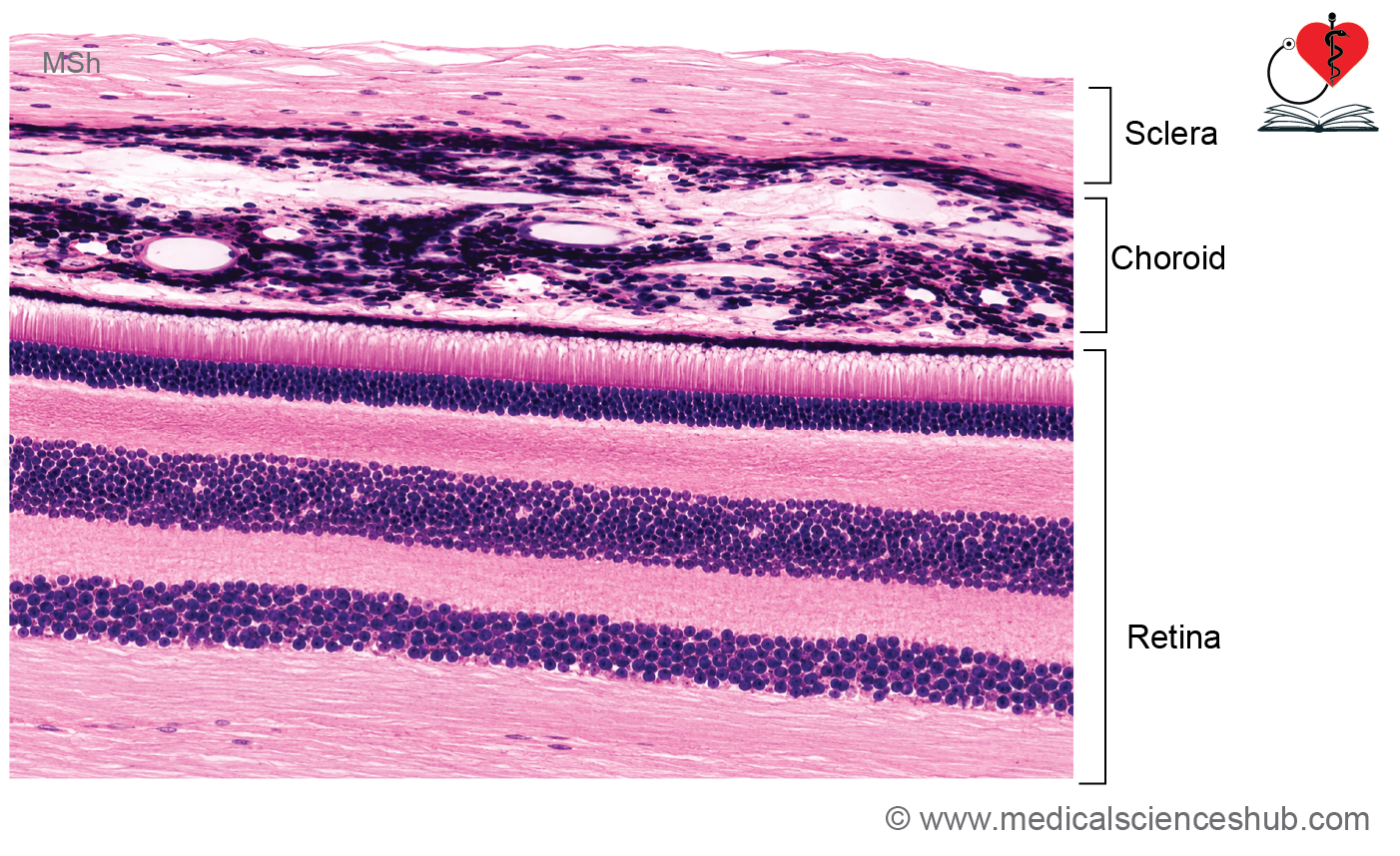
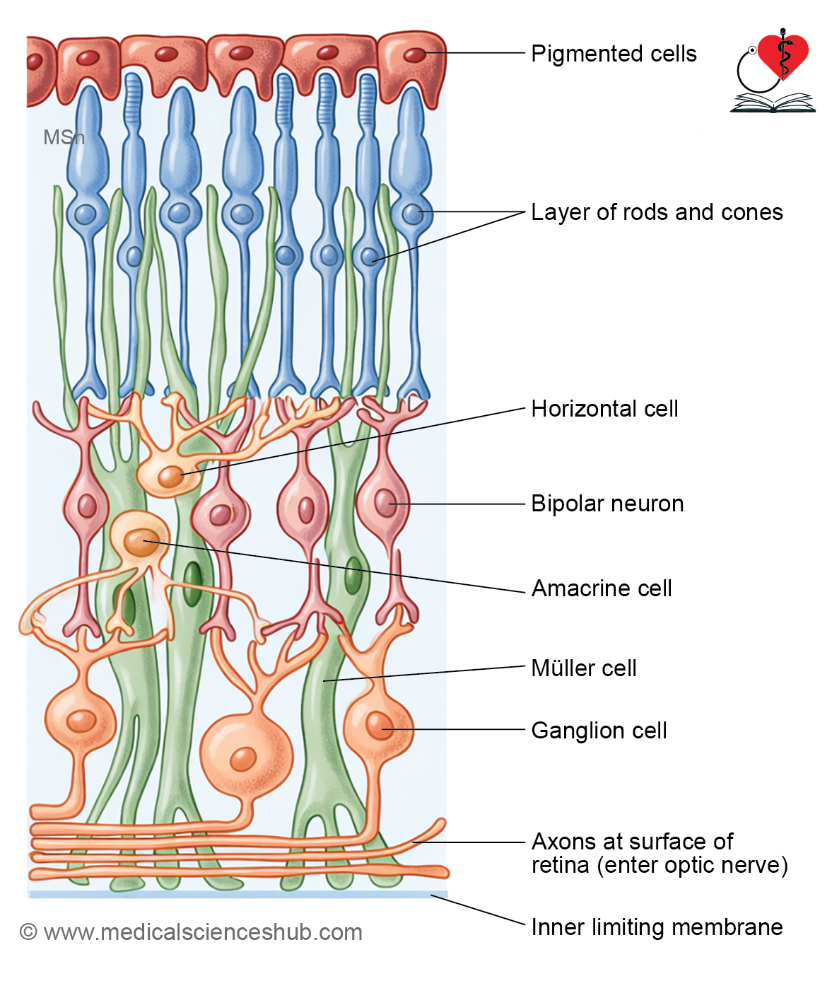
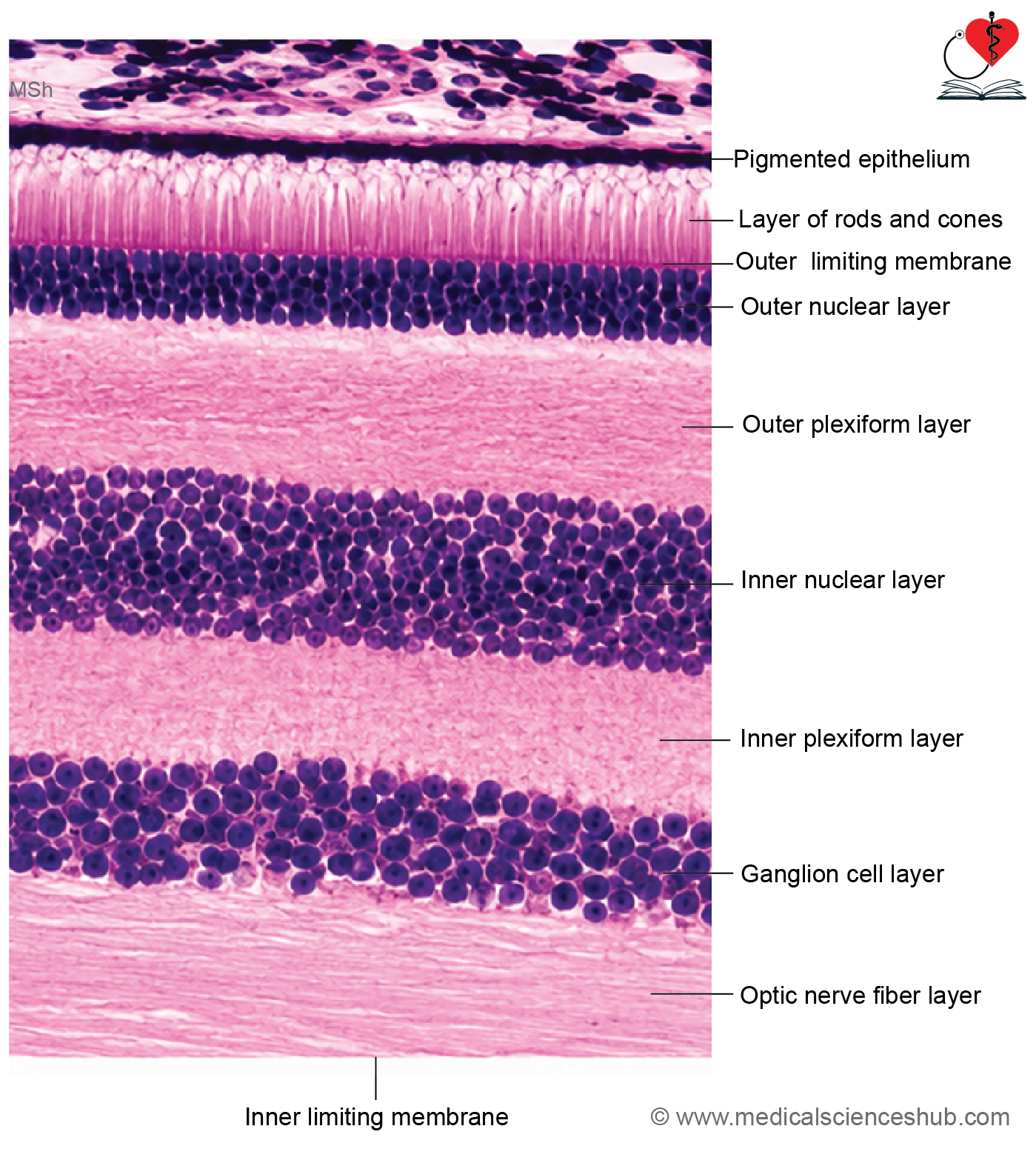
Layer 1: Retinal Pigmented Epithelium (RPE)
- The retinal pigment epithelium (RPE) consists of a single layer of low cuboidal, pigment-containing cells.
- It is separated from the choroid by Bruch’s membrane.
- RPE cells contain abundant melanin granules, which help absorb excess light.
- The apical surface of RPE cells extends short cytoplasmic processes between the outer segments of rods and cones.
Functions of RPE
- It synthesizes and stores melanin, reducing light scattering within the eye.
- It absorbs excess light, thereby improving visual clarity and contrast.
- It forms an essential component of the blood–retinal barrier, helping maintain retinal homeostasis.
- It phagocytoses and removes shed membranous discs from the outer segments of photoreceptor cells, supporting their continuous renewal and function.
Layer 2: Rod and Cones
- Photoreceptors of the retina are of two types: rods and cones. These specialized cells convert light energy into electrical signals for visual processing.
- The human retina contains approximately 120 million rods and 6–7 million cones.
- Cones are concentrated in the fovea centralis, where they provide the highest visual acuity and are responsible for color vision.
- The optic disc lacks photoreceptors and is therefore known as the blind spot.
Structure of Photoreceptors
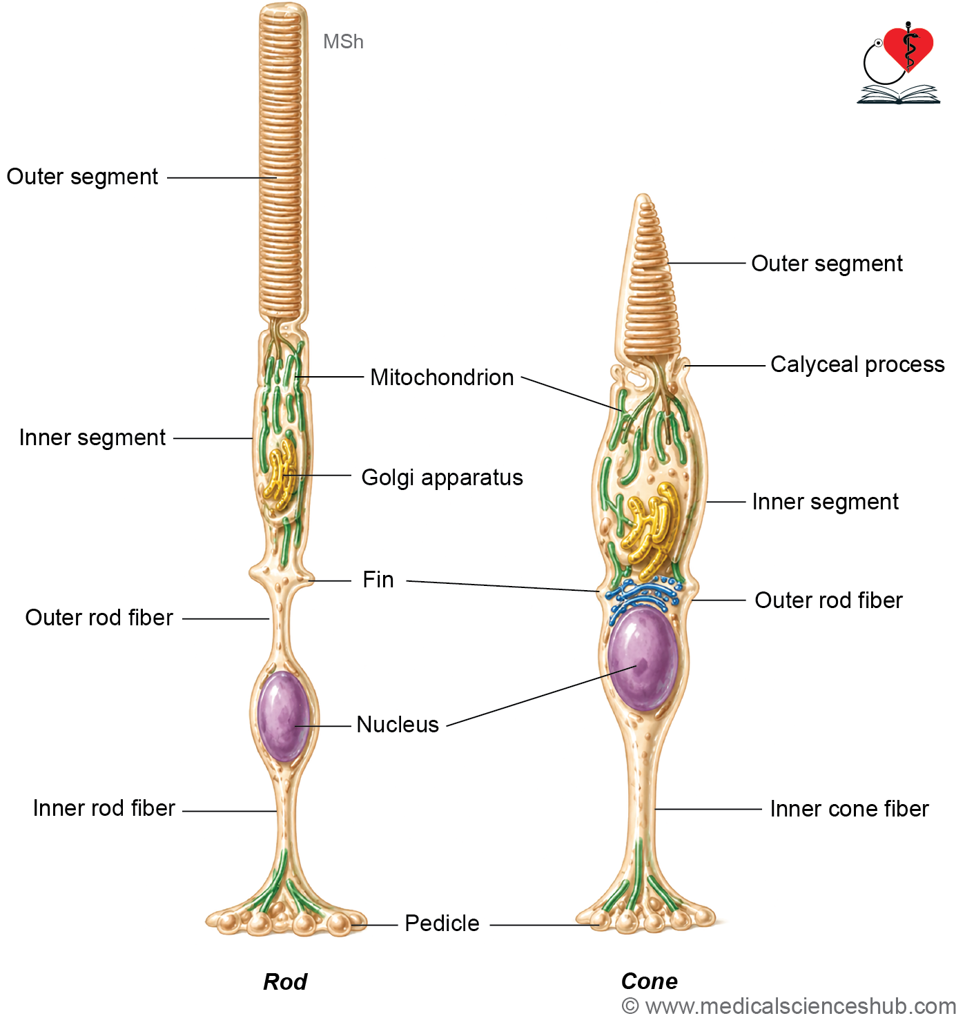
- Both rods and cones consist of three parts:
- Outer segment
- Connecting stalk (connecting cilium)
- Inner segment
- The outer segment of rods is cylindrical, whereas that of cones is tapered or conical.
- The inner segment contains abundant mitochondria and Golgi apparatus, supporting the high metabolic activity of these cells.
- The outer segment contains numerous membranous discs that house visual pigments.
- Photoreceptor discs undergo continuous renewal, and shed discs are removed by the retinal pigment epithelium (RPE) through phagocytosis.
Visual Pigments
- Rods contain rhodopsin, which is highly sensitive to dim light and supports night vision.
- Cones contain photopsins (iodopsins), which mediate color vision.
- Visual pigments consist of an opsin protein linked to a light-sensitive chromophore derived from vitamin A.
Types of Cones
- Cones are classified according to their peak wavelength sensitivity:
- Long-wavelength (red) cones: ~560–565 nm
- Medium-wavelength (green) cones: ~530–535 nm
- Short-wavelength (blue) cones: ~420–440 nm
- The combined activity of these three cone types enables normal trichromatic color vision.
Layer 3: Outer Limiting Membrane
- The outer limiting membrane is formed by adherens junctions between adjacent Müller cells and photoreceptor cells.
- It is not a true membrane but a specialized junctional complex that provides structural support to the retina.
Layer 4: Outer Nuclear Layer
- This layer contains the nuclei of rod and cone photoreceptor cells.
- Cone nuclei are generally larger and stain more lightly than rod nuclei.
- The nuclei are arranged in multiple rows, especially in rod-rich regions.
Layer 5: Outer/External Plexiform Layer
- This synaptic layer contains connections between photoreceptors and second-order neurons.
- Rod terminals form spherules, whereas cone terminals form pedicles.
- Rods often converge onto a single bipolar cell, while individual cones frequently connect with a single bipolar cell, enhancing visual acuity.
Layer 6: Inner Nuclear Layer
- This layer contains the nuclei of bipolar, horizontal, amacrine, and Müller cells.
- Bipolar cells transmit impulses from photoreceptors to ganglion cells.
- Horizontal cells modulate communication between photoreceptors and bipolar cells.
- Amacrine cells participate in synaptic interactions within the inner plexiform layer.
- Müller cells are the principal retinal glial cells and extend across almost the entire thickness of the retina, providing structural and metabolic support.
Layer 7: Inner Plexiform Layer
- This layer contains synapses between bipolar cell axons, ganglion cell dendrites, and amacrine cells.
- It serves as an important site for retinal signal processing.
Layer 8: Ganglion Cell Layer
- The layer contains cell bodies of ganglion cells, which are large neurons with pale nuclei and prominent nucleoli.
- Ganglion cell axons carry visual information toward the brain.
- In the fovea, a near one-to-one relationship exists between photoreceptors, bipolar cells, and ganglion cells, allowing high-resolution vision.
Layer 9: Layer of Optic Nerve Fibers
- This layer is formed by unmyelinated axons of ganglion cells converging toward the optic disc.
- After leaving the eyeball, these fibers become myelinated and form the optic nerve.
- Retinal blood vessels also course through this layer.
Layer 10: Inner Limiting Membrane
- The inner limiting membrane is formed by the basal lamina of Müller cells.
- It constitutes the innermost retinal boundary and separates the retina from the vitreous body.
Table 24.1: Differences between rods and cone cells
| Feature | Rod Cells | Cone Cells |
|---|---|---|
| Shape of Outer Segment | Long and cylindrical (rod-shaped) | Tapering and conical (cone-shaped) |
| Relative Number | More numerous in the retina | Less numerous than rods |
| Distribution in Retina | Widely distributed throughout the retina; absent in the fovea centralis | Highly concentrated in the fovea centralis and macular region |
| Visual Acuity | Low visual acuity due to convergence of signals | High visual acuity because of greater neuronal resolution |
| Light Sensitivity | Highly sensitive to dim light; responsible for scotopic (night) vision | Function optimally in bright light; responsible for photopic (daylight) vision |
| Color Perception | Do not mediate color vision; provide monochromatic vision | Responsible for color discrimination and perception |
| Visual Pigment | Rhodopsin | Photopsins (iodopsins) |
| Functional Types | Single functional type | Three functional types sensitive to red, green, and blue wavelengths |
| Response to Light | Slower response but greater sensitivity | Faster response with lower sensitivity |
| Primary Function | Vision under low-light conditions | Detailed and color vision under well-illuminated conditions |
Crystalline Lens
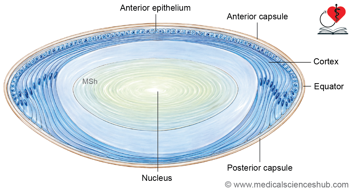
- The crystalline lens is a transparent, biconvex, and avascular structure that focuses light onto the retina.
- It is suspended from the ciliary body by zonular fibers (suspensory ligaments).
- The lens is positioned between the aqueous humor anteriorly and the vitreous body posteriorly.
Lens Capsule
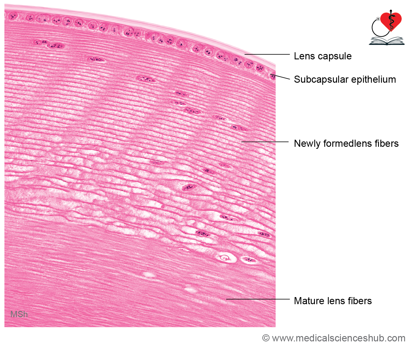
- The lens capsule is a thick, transparent basement membrane surrounding the lens.
- It is composed mainly of type IV collagen and glycoproteins.
- The capsule is continuously produced by the lens epithelial cells.
Lens Epithelium
- The anterior surface of the lens is lined by a single layer of cuboidal cells known as the lens epithelium.
- Near the equator of the lens, these cells elongate and differentiate into lens fibers.
Lens Fibers (Lens Fiber Cells)
- Lens fibers are elongated, highly specialized cells derived from lens epithelial cells.
- They are packed with soluble proteins called crystallins, which maintain lens transparency and refractive properties.
- Mature lens fibers lose their nuclei and most organelles to minimize light scattering.
CLINICAL CORRELATION
Cataract
- A cataract is an opacity of the lens resulting in reduced visual clarity.
- Common causes include aging, ocular trauma, congenital disorders, and prolonged exposure to ultraviolet radiation.
- Treatment typically involves surgical removal of the opaque lens followed by implantation of an artificial intraocular lens.
Presbyopia
- Presbyopia is an age-related reduction in the accommodative ability of the lens due to decreased elasticity.
- It usually becomes noticeable after 40 years of age and causes difficulty with near vision.
- Correction is commonly achieved with convex (plus) lenses for reading and other close work.
Optic Nerve
- The optic nerve (cranial nerve II) carries visual impulses from the retina to the brain and extends from the optic disc to the optic chiasma.
- It is formed by the axons of retinal ganglion cells along with supporting neuroglial cells.
- Embryologically, the optic nerve develops from the optic stalk, making it a central nervous system tract rather than a peripheral nerve.
Histology
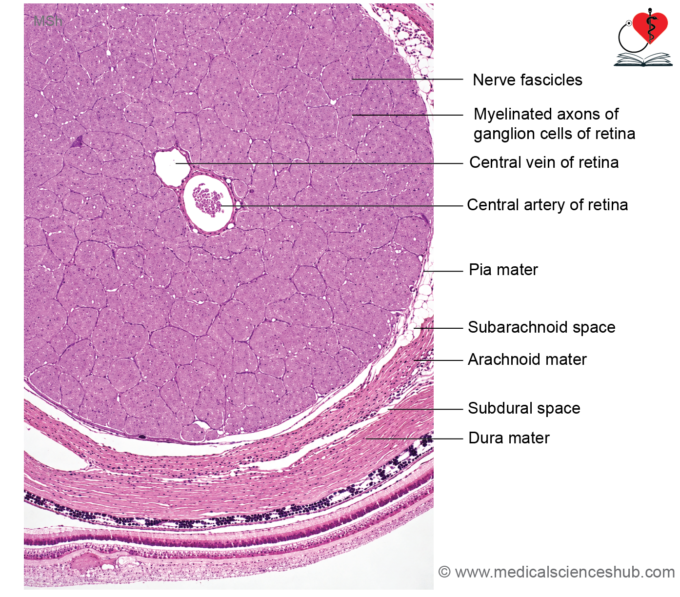
- The optic nerve is surrounded by the three meningeal layers:
- Dura mater (outer layer)
- Arachnoid mater (middle layer)
- Pia mater (inner layer)
- Unlike peripheral nerves, the optic nerve lacks:
- Endoneurium
- Perineurium
- Epineurium
- The nerve contains no Schwann cells.
- Myelination is provided by oligodendrocytes, which are central nervous system glial cells.
- Septa arising from the pia mater divide the nerve into fascicles containing bundles of myelinated axons.
- Each fascicle consists primarily of axons originating from retinal ganglion cells.
- The central retinal artery and vein pass through the center of the optic nerve and supply the retina.
CLINICAL CORRELATION
- Because myelin in the optic nerve is produced by oligodendrocytes rather than Schwann cells, disorders that affect peripheral myelin, such as Guillain–Barré syndrome, typically do not involve the optic nerve.
Eyelid
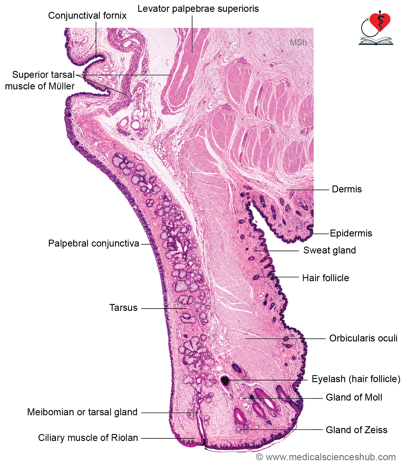
The eyelid (palpebra) is a movable fold that protects the eyeball, distributes the tear film, and helps maintain corneal moisture.
Layers of the Eyelid (from superficial to deep)
- Skin: Thin, elastic, and loosely attached.
- Subcutaneous connective tissue: Loose connective tissue with very little or no fat.
- Orbicularis oculi muscle: Skeletal muscle responsible for eyelid closure.
- Tarsal plate: Dense connective tissue that provides structural support to the eyelid.
- Palpebral conjunctiva: A mucous membrane lining the inner surface of the eyelid; it consists of stratified columnar epithelium with goblet cells and an underlying lamina propria.
- The upper eyelid also contains the levator palpebrae superioris muscle, which elevates the eyelid.
Glands of the Eyelid
- Meibomian (tarsal) glands
- Large modified sebaceous glands embedded within the tarsal plate.
- Their lipid-rich secretion reduces evaporation of the tear film and helps stabilize it.
- Glands of Zeis
- Small modified sebaceous glands associated with the eyelashes.
- They secrete oily material into eyelash follicles.
- Glands of Moll
- Modified apocrine sweat glands located near the eyelid margin.
- They open into eyelash follicles or directly onto the eyelid surface.
- Accessory lacrimal glands
- Glands of Wolfring and glands of Krause are small serous glands that contribute to tear production.
- Wolfring glands are located near the upper tarsal border, whereas Krause glands are situated within the conjunctival fornices.
- Together, these glands help maintain tear film integrity, lubrication, and ocular surface health.
CLINICAL CORRELATION
- Chalazion is a chronic blockage and inflammation of a Meibomian (tarsal) gland.
- It typically presents as a painless, slowly enlarging nodule within the eyelid, most commonly the upper eyelid.
- Many cases resolve spontaneously, although persistent lesions may require medical treatment.
- It typically presents as a painless, slowly enlarging nodule within the eyelid, most commonly the upper eyelid.
- Stye (Hordeolum) is an acute bacterial infection, usually involving a gland of Zeis or another eyelid gland.
- It causes a painful, red, and tender swelling near the eyelid margin.
- Local warmth and inflammation are characteristic features.
- Conjunctivitis (Pink Eye) is inflammation of the conjunctiva, the membrane covering the sclera and inner eyelid.
- Viral infection is the most common cause.
- Symptoms include redness, irritation, tearing, and watery discharge.
- Most viral cases are self-limiting and improve with supportive care such as artificial tears and warm compresses.
Lacrimal Gland
- The lacrimal gland is a compound tubuloalveolar serous gland responsible for the production of tears.
- Histologically, it resembles other serous exocrine glands, with secretory acini and a branching duct system.
- Myoepithelial cells are present between the secretory cells and the basal lamina of the acini and assist in the expulsion of tears.
Duct System
- Small intralobular ducts are lined by simple cuboidal epithelium.
- Larger ducts are lined by pseudostratified columnar or stratified columnar epithelium.
- The ducts open into the superior conjunctival fornix, allowing tears to spread across the ocular surface.
Accessory Lacrimal Glands
- The glands of Krause are accessory lacrimal glands located near the superior conjunctival fornix.
- These glands contribute to the continuous production of tears, helping maintain lubrication and protection of the eye.
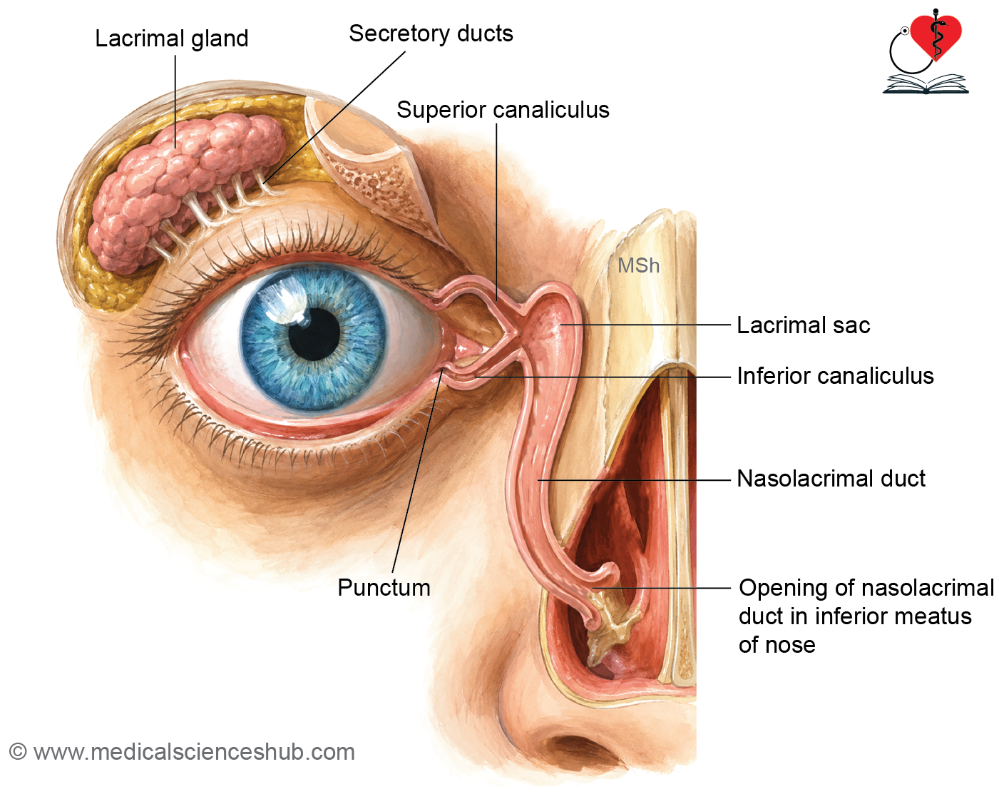
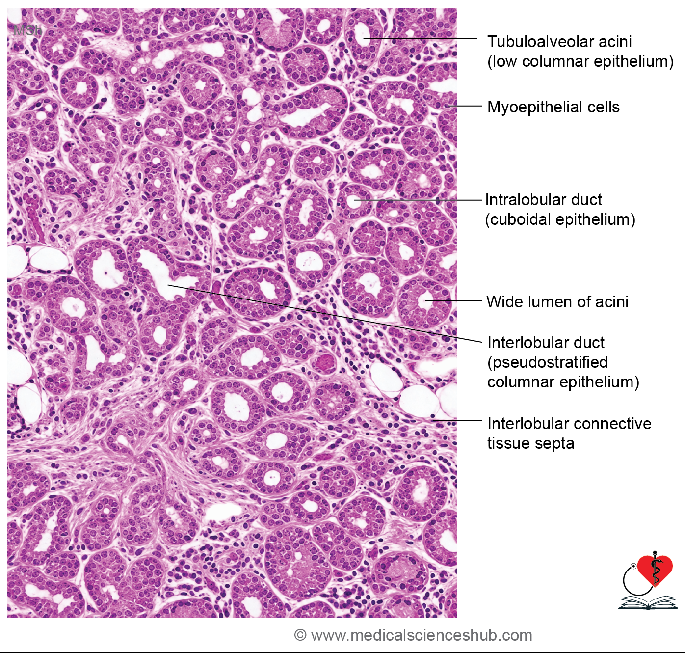
Table 24.2: Histological differences between serous salivary gland and lacrimal gland
| Feature | Lacrimal Gland | Serous Salivary Gland (e.g., Parotid Gland) |
|---|---|---|
| Acinar size | Acini are relatively larger | Acini are comparatively smaller |
| Acinar lumen | Wide and conspicuous lumen | Narrow lumen, often difficult to appreciate |
| Shape of secretory cells | Low columnar to cuboidal secretory cells | Tall pyramidal (high columnar) secretory cells |
| Staining characteristics (H&E) | Cytoplasm appears predominantly eosinophilic | Cytoplasm shows basal basophilia due to abundant rough endoplasmic reticulum and apical eosinophilia from secretory granules |
| Acinar configuration | Acini are often elongated or irregular in outline | Acini are generally rounded or oval and more uniform in shape |
| Nature of Secretion | Produces the aqueous component of tears | Produces a protein-rich serous secretion containing digestive enzymes |
| Myoepithelial cells | Present around secretory units and ducts | Present around secretory acini and ducts |
Important Questions
- List the layers of retina.
- Draw a well-labeled diagram showing histology of retina.
- List the layers of retina.
- Write a short note on histology of optic nerve.
📝 Test Your Knowledge – Practice MCQs
Attempt the chapter MCQ quiz and assess your understanding of key concepts.
Click to see: Medical Disclaimer and Copyright Notice