
Introduction
- Epithelium, also known as epithelial tissue, is one of the four basic tissues of the human body.
- It forms a continuous layer of closely packed cells called epithelial cells.
- This tissue covers the external surface of the body and lines the internal surfaces of many organs and cavities.
- Epithelium also lines the lumen (inner surface) of tubular structures such as the digestive tract, respiratory passages, blood vessels, and urinary system.
- The epithelial cells rest on a specialized extracellular layer called the basement membrane, which provides structural support and helps anchor the epithelium to the underlying connective tissue.
- Epithelial cells are tightly joined to one another by specialized intercellular junctions, resulting in very little space between adjacent cells.
- Because of this close arrangement, epithelium forms an effective barrier between different body compartments and the external environment.
- Depending on its location, epithelium performs various functions, including protection, absorption, secretion, filtration, transport, and sensory reception.
- Epithelium does not contain blood vessels within it. Nutrients and oxygen reach the epithelial cells by diffusion from the underlying connective tissue.
- The four basic tissues of the human body are:
- Epithelial tissue
- Connective tissue
- Muscular tissue
- Nervous tissue
Features of Epithelium
- Epithelium is composed of one or more layers of closely packed cells that cover body surfaces and line internal cavities, ducts, and hollow organs.
- The cells are arranged with minimal intercellular material, forming a continuous protective and functional lining.
- Epithelium is avascular, meaning it does not contain blood vessels. Nutrients and oxygen reach the epithelial cells by diffusion from the underlying connective tissue.
- Epithelial cells rest on a basement membrane, which provides structural support and attaches the epithelium to the underlying tissues.
- Adjacent epithelial cells are connected by specialized cell junctions, such as tight junctions, adherens junctions, desmosomes, gap junctions, and hemidesmosomes. These junctions maintain tissue integrity and facilitate communication between cells.
- Epithelium forms a selective barrier that separates underlying tissues from the external environment or from the contents of body cavities and organs.
- The free surface of epithelial cells may show specialized modifications according to functional requirements. For example:
- Microvilli increase the surface area for absorption in the small intestine.
- Cilia help move mucus and other materials across the epithelial surface in the respiratory tract.
- Stereocilia aid absorption and sensory functions in specific organs.
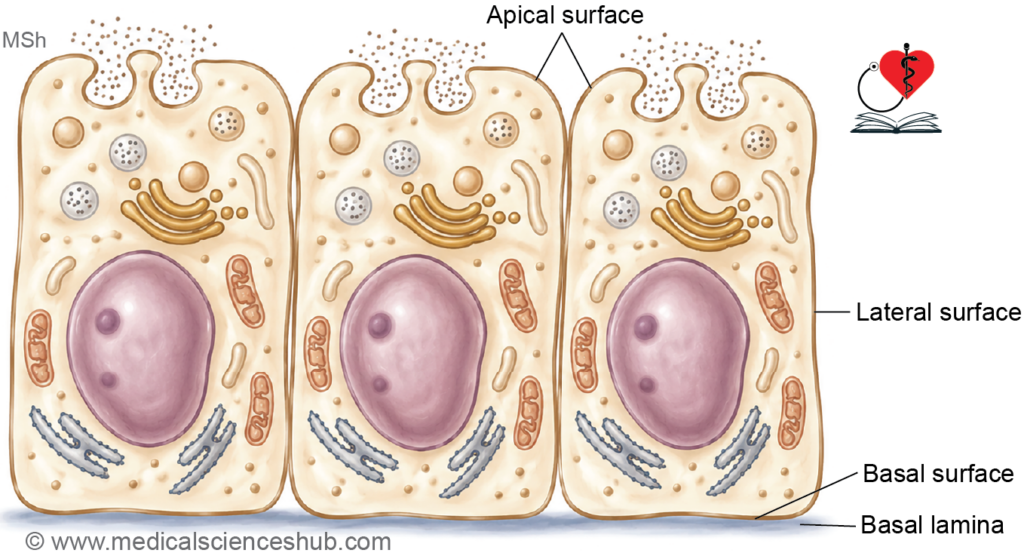
- Epithelial tissue exhibits a distinct polarity, with an apical (free) surface, lateral surfaces, and a basal surface attached to the basement membrane.
- Epithelial cells have a high capacity for cell division and regeneration, allowing rapid replacement of damaged or worn-out cells.
- The regenerative ability of epithelium helps maintain the integrity and function of surfaces that are frequently exposed to mechanical stress, chemical agents, or microbial invasion.
Functions of Epithelium
The functions of epithelium vary according to its location and structural specialization. The major functions are as follows:
1. Protection
- Epithelium forms a protective covering over body surfaces and underlying tissues.
- It helps prevent mechanical injury, dehydration, chemical damage, and microbial invasion.
- The epidermis of the skin serves as an effective protective barrier against the external environment.
2. Selective Barrier Function
- Epithelium regulates the movement of substances between different body compartments.
- It acts as a selectively permeable barrier, allowing certain substances to pass while restricting others.
- For example, the epidermis limits the entry of many microorganisms and harmful agents, while permitting the absorption of some lipid-soluble medications applied to the skin.
3. Absorption
- Specialized epithelial cells absorb nutrients, water, ions, and other substances.
- Absorptive epithelial cells often possess microvilli that increase the surface area available for absorption.
- The epithelium lining the small intestine absorbs nutrients from digested food.
- The epithelium of the renal tubules reabsorbs water and essential substances from the filtrate.
4. Secretion
- Certain epithelial cells synthesize and release substances required for various physiological functions.
- Secretory epithelial cells may occur individually or form specialized glands.
- Examples include the secretion of saliva by salivary glands, mucus by goblet cells, enzymes by digestive glands, and hormones by endocrine glands.
5. Excretion
- Some epithelial tissues participate in the removal of waste products from the body.
- The epithelium of renal tubules contributes to the excretion of metabolic waste products and the regulation of body fluid composition.
6. Transport
- Specialized epithelial cells facilitate the movement of materials across their surface.
- Ciliated epithelium transports mucus and trapped particles in the respiratory tract.
- In the female reproductive tract, ciliary activity assists in the movement of the ovum toward the uterus.
7. Sensory Reception
- Certain epithelial cells are specialized to detect stimuli from the external or internal environment.
- These cells are associated with sensory receptors and nerve endings.
- Examples include the taste buds of the tongue, the olfactory epithelium of the nasal cavity, and sensory epithelial cells of the inner ear.
8. Lubrication
- Some epithelial cells secrete mucus, which keeps surfaces moist and reduces friction.
- Mucus also helps protect epithelial surfaces from mechanical and chemical injury.
- This function is particularly important in the respiratory, gastrointestinal, and reproductive systems.
9. Filtration
- Specialized epithelial membranes allow the passage of water and small molecules while restricting larger substances.
- Filtration is an important function of the epithelium in the renal corpuscles of the kidneys, where blood plasma is filtered to form urine.
Epithelial Cells
- Epithelial cells vary in size, shape, and specialization depending on their location and function.
- These cells are closely packed with very little intercellular material between them.
- Based on their shape, epithelial cells are commonly classified into three main types:
- Squamous cells: These cells are thin and flat, resembling scales.
- Cuboidal cells: These cells have approximately equal height, width, and depth.
- Columnar cells: These cells are taller than they are wide and appear column-shaped.
- The shape of epithelial cells is closely related to their function. For example, squamous cells facilitate diffusion and filtration, whereas columnar cells are often specialized for absorption and secretion.
Polarity of Epithelial Cells
- Epithelial cells exhibit polarity, meaning that different regions of the cell have distinct structural and functional characteristics.
- Each epithelial cell has three surface domains:
- Basal surface
- Lateral surfaces
- Apical surface
1. Basal Surface
- The basal surface is the part of the cell that rests on the basement membrane.
- It anchors the epithelial cell to the underlying connective tissue.
- This surface is involved in the exchange of nutrients, metabolites, and signaling molecules between the epithelium and underlying tissues.
2. Apical Surface
- The apical surface is the free surface of the epithelial cell.
- In surface epithelium, it faces the external environment.
- In the lining of hollow organs and ducts, it faces the lumen and comes into contact with luminal contents.
- Depending on functional requirements, the apical surface may possess specialized structures such as:
- Microvilli, which increase the surface area for absorption.
- Cilia, which move substances across the epithelial surface.
- Stereocilia, which aid absorption or sensory functions in specific organs.
3. Lateral Surfaces
- The lateral surfaces lie between adjacent epithelial cells.
- These surfaces contain specialized intercellular junctions that maintain tissue integrity and facilitate communication between neighboring cells.
- Common junctions include tight junctions, adherens junctions, desmosomes, and gap junctions.
Epithelioid Cells
- Epithelioid cells, also called epithelioid histiocytes, are activated macrophages that resemble epithelial cells in appearance.
- They possess abundant pale cytoplasm and a centrally placed oval or elongated nucleus.
- Although they resemble epithelial cells morphologically, they originate from cells of the monocyte-macrophage system and are not true epithelial cells.
- Epithelioid cells often aggregate at sites of chronic inflammation.
- Multiple epithelioid cells may fuse to form multinucleated giant cells.
- These cells are a characteristic component of a granuloma, which is an organized collection of activated macrophages formed in response to persistent inflammation, infection, or foreign material.
Classification of Epithelia
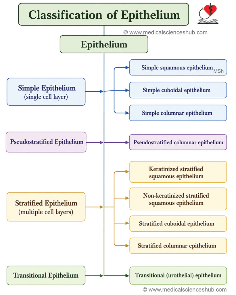
- Epithelia are classified as follows:
- Simple epithelium has only one layer of cells.
- Stratified epithelium has two or more layers of cells.
- Pseudostratified epithelium has one layer of cells resting on basal lamina, but some of these cells do not extend up to free surface of epithelium. Hence, the epithelium appears stratified.
Simple Epithelium
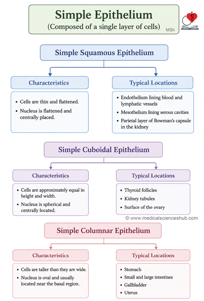
- Simple epithelium consists of a single layer of cells resting on the basement membrane.
- All cells are in direct contact with the basement membrane.
- Based on cell shape, simple epithelium is classified into:
- Simple squamous epithelium: Composed of thin, flat cells.
- Simple cuboidal epithelium: Composed of cells with approximately equal height and width.
- Simple columnar epithelium: Composed of cells that are taller than they are wide.
Simple Squamous Epithelium
Features
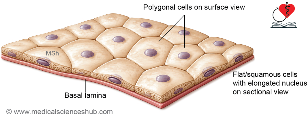
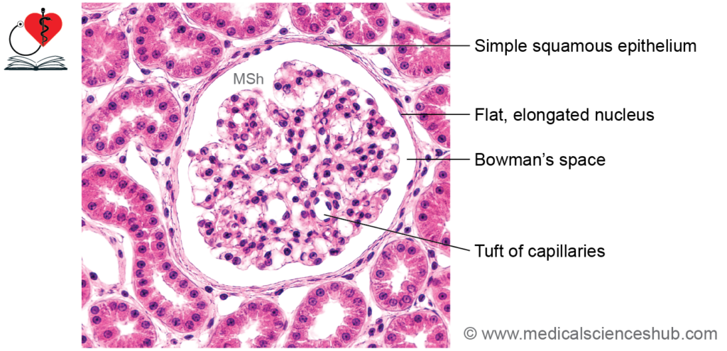
- Simple squamous epithelium consists of a single layer of thin, flat cells resting on the basement membrane.
- The cells are usually polygonal when viewed from the surface.
- Each cell contains a flattened, centrally placed nucleus.
- The cytoplasm forms a very thin layer around the nucleus.
- Because of their minimal thickness, these cells create a delicate lining that offers little resistance to the movement of substances.
Locations
- Lines the alveoli of the lungs.
- Forms the endothelium, which lines blood vessels and lymphatic vessels.
- Forms the endocardium, the inner lining of the heart.
- Forms the mesothelium, which lines the pleura, pericardium, and peritoneum.
- Lines the parietal layer of Bowman’s capsule in the kidney.
- Present in the thin segments of the loop of Henle.
Functions
- The thinness of this epithelium allows rapid diffusion of gases, especially in the lungs.
- It facilitates filtration and exchange of water and small molecules in the kidneys.
- In blood and lymphatic vessels, it provides a smooth, low-friction surface that supports efficient fluid flow.
- Mesothelial cells produce a small amount of serous fluid, which reduces friction between adjacent organs and body cavity walls.
- It is well adapted for diffusion, filtration, and selective transport rather than protection.
Simple Cuboidal Epithelium
Features
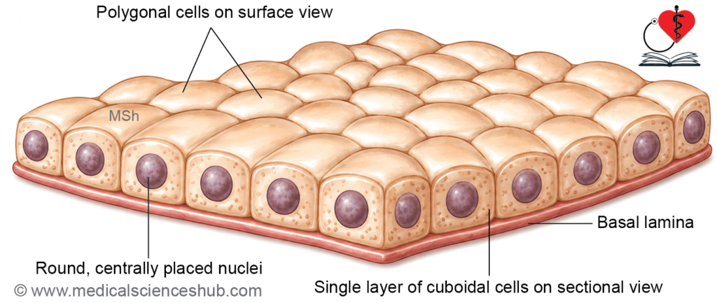
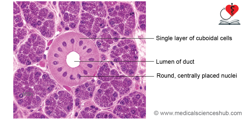
- Simple cuboidal epithelium consists of a single layer of cube-shaped cells resting on the basement membrane.
- The cells have approximately equal height and width.
- Each cell contains a centrally placed, round nucleus.
- In sections, the nuclei usually appear aligned in a single row.
- When viewed from the surface, the cells appear polygonal, often hexagonal in outline.
- The cytoplasm is moderately abundant and supports active cellular functions.
Locations
- Lines the thyroid follicles.
- Forms the lining of small ducts of exocrine glands.
- Covers the surface of the ovary (germinal epithelium).
- Forms part of the choroid plexus, which produces cerebrospinal fluid.
- Lines the posterior surface of the lens.
- Constitutes the pigmented layer of the retina.
- Lines the ducts of Bartholin glands.
- Present in parts of the kidney, especially the distal convoluted tubules.
Functions
- Simple cuboidal epithelium is specialized for absorption, secretion, transport, and excretion.
- The cells contain organelles required for active metabolic processes and synthesis of cellular products.
- This epithelium plays an important role in glandular activity and in the transport of substances across epithelial surfaces.
Simple Columnar Epithelium
Features
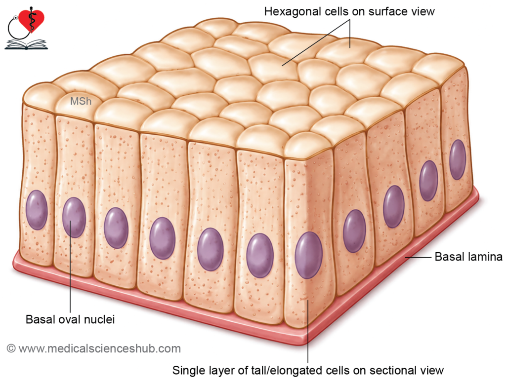
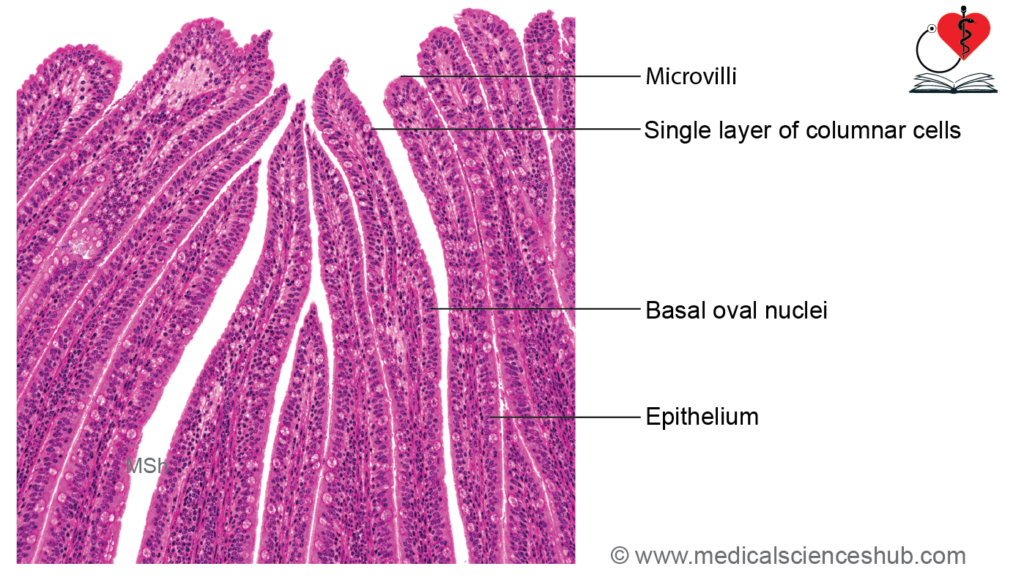
- Simple columnar epithelium consists of a single layer of tall, column-shaped cells resting on the basement membrane.
- The height of the cells is greater than their width.
- These cells contain abundant cytoplasm because they are actively involved in absorption, secretion, and synthesis.
- The nuclei are oval or elongated and are usually located near the basal part of the cells.
- The nuclei are generally arranged at a similar level, giving the epithelium a regular appearance.
- Prominent nucleoli may be present, indicating active protein synthesis.
Cytoplasmic Characteristics
- The staining pattern varies according to the organelles present within the cell.
- Cells with abundant rough endoplasmic reticulum often show basal basophilia.
- Cells rich in secretory granules and Golgi apparatus may exhibit apical eosinophilia.
- Mucus-secreting cells may appear pale or vacuolated because mucus is removed during tissue processing.
Surface Modifications
- The apical surface may show specialized structures depending on function:
- Microvilli increase the surface area for absorption.
- Closely packed microvilli may produce a striated border, whereas irregularly arranged microvilli may produce a brush border under light microscopy.
- Cilia are present in certain regions and help move materials across the epithelial surface.
Locations
- Lines the stomach.
- Lines the small and large intestines.
- Lines the gallbladder and parts of the biliary tract.
- Lines the uterus.
- Simple ciliated columnar epithelium is present in:
- Fallopian tubes
- Parts of the uterusCentral canal of the spinal cord
- Ventricular system of the brain (formed by specialized ependymal cells)
Functions
- It is specialized for absorption, secretion, transport, and protection.
- Microvilli enhance absorptive capacity, particularly in the intestine.
- Cilia facilitate the movement of mucus, fluid, or reproductive cells.
- Secretory columnar cells produce mucus, enzymes, and other biologically important substances.
Pseudostratified Epithelium
Features
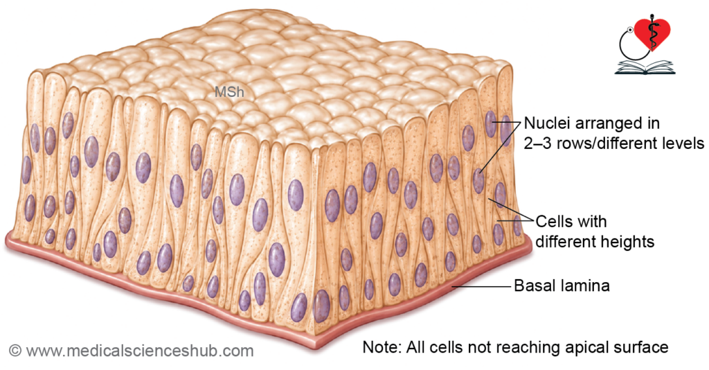
- Pseudostratified epithelium consists of a single layer of cells resting on the basement membrane.
- Although all cells are attached to the basement membrane, not all cells reach the free surface.
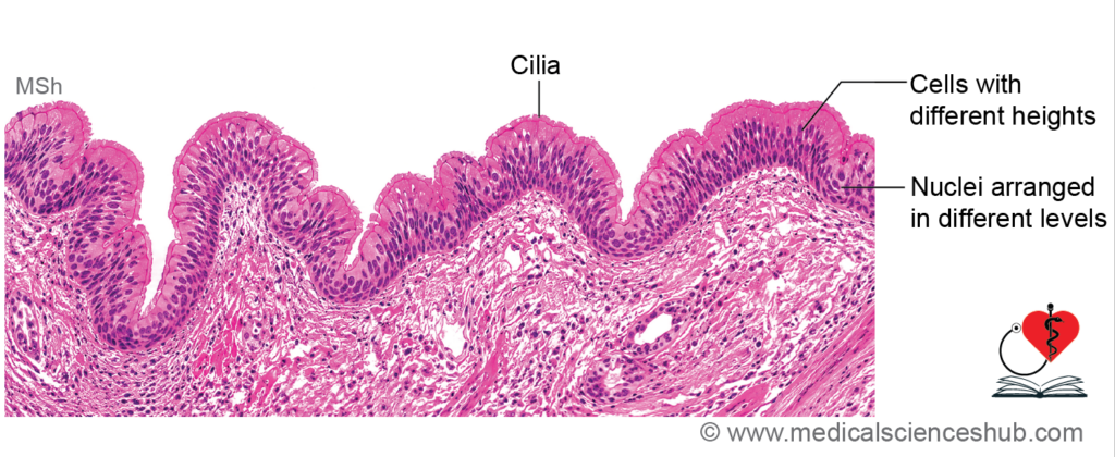
- Because the cells have different heights, their nuclei are located at different levels, giving the false appearance of multiple layers.
- Therefore, it appears stratified but is actually a type of simple epithelium.
- Small basal cells are present near the basement membrane. These cells divide and replace worn-out epithelial cells.
- The cells that extend to the free surface are usually columnar in shape; hence, the tissue is called pseudostratified columnar epithelium.
- Goblet cells may be present and secrete mucus.
Locations
- Respiratory tract: The lining of the nasal cavity, trachea, and larger bronchi is typically formed by ciliated pseudostratified columnar epithelium.
- Male reproductive tract: The epididymis and parts of the ductus deferens are lined by pseudostratified columnar epithelium with stereocilia, which are long, non-motile microvilli.
- Non-ciliated pseudostratified columnar epithelium is present in:
- Parts of the auditory (Eustachian) tube
- Ductus deferensMembranous urethra
- Penile urethra
Functions
- It provides protection to the underlying tissues.
- Cilia move mucus and trapped dust particles, microorganisms, and other foreign materials toward the pharynx for removal.
- Goblet cells secrete mucus, which traps inhaled particles and helps keep the epithelial surface moist.
- Stereocilia increase the surface area for absorption, particularly in the male reproductive tract.
- Basal cells function as stem cells and contribute to the renewal and repair of the epithelium.
Stratified Epithelium
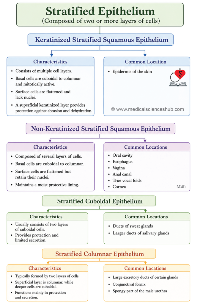
- Stratified epithelium is a multilayered epithelium. In stratified epithelium, only the basal cell layer rests on basal lamina.
- Stratified epithelium is classified as follows:
- Stratified squamous epithelium
- Stratified cuboidal epithelium
- Stratified columnar epithelium.
Stratified Squamous Epithelium
Features
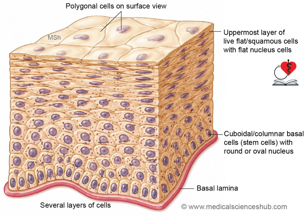
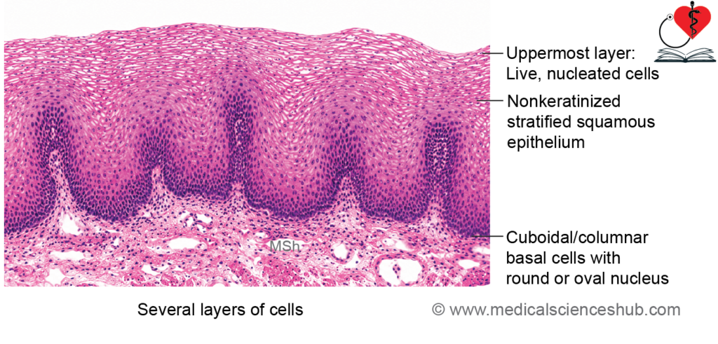
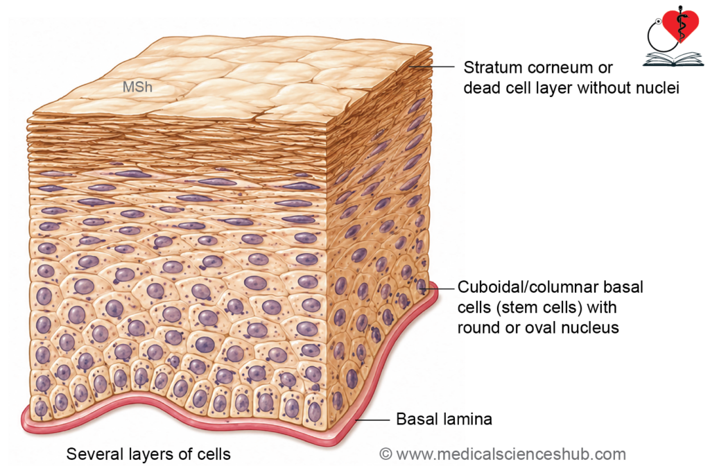
- Stratified squamous epithelium consists of multiple layers of cells resting on a basement membrane.
- The deepest (basal) layer is formed by cuboidal or columnar cells with large nuclei.
- Basal cells act as stem cells and continuously divide to replace cells lost from the surface.
- As cells move toward the surface, they become progressively flatter and acquire a squamous appearance.
- This arrangement provides excellent protection against mechanical stress and abrasion.
- Based on the presence or absence of a surface keratin layer, it is classified into:
- Keratinized stratified squamous epithelium
- Nonkeratinized stratified squamous epithelium
Nonkeratinized stratified squamous epithelium
Features
- The surface cells remain alive and retain their nuclei.
- A stratum corneum is absent.
- The superficial cells are flattened but remain moist.
- This epithelium is typically associated with underlying connective tissue called the lamina propria, together forming a mucosa.
Locations
- Oral cavity
- Oropharynx
- Laryngopharynx
- Esophagus
- Vagina
- Palatine tonsils
- True vocal folds
- Cornea
Functions
- Protects underlying tissues from abrasion while maintaining a moist surface.
- Allows flexibility and resistance to friction in regions exposed to mechanical stress.
Keratinized stratified squamous epithelium
Features
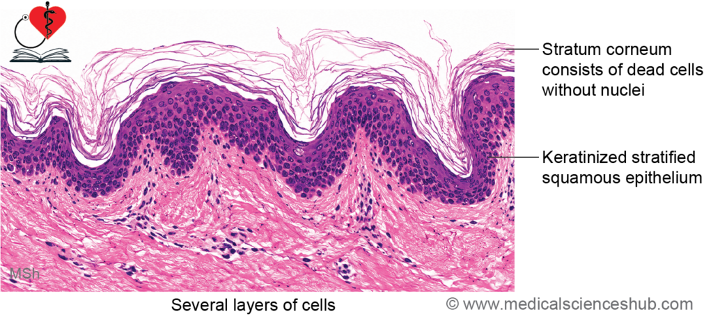
- The superficial cells undergo keratinization and accumulate keratin proteins.
- The outermost cells are dead, flattened, and lack nuclei.
- These dead cells form a protective layer called the stratum corneum.
- Keratinization is commonly associated with surfaces exposed to dryness and friction.
Location
- Forms the epidermis of the skin.
Functions
- Provides strong protection against mechanical injury.
- Acts as an effective barrier against microorganisms.
- Reduces water loss and helps prevent dehydration.
- Protects the body from environmental factors such as friction and minor trauma.
Stratified Cuboidal Epithelium
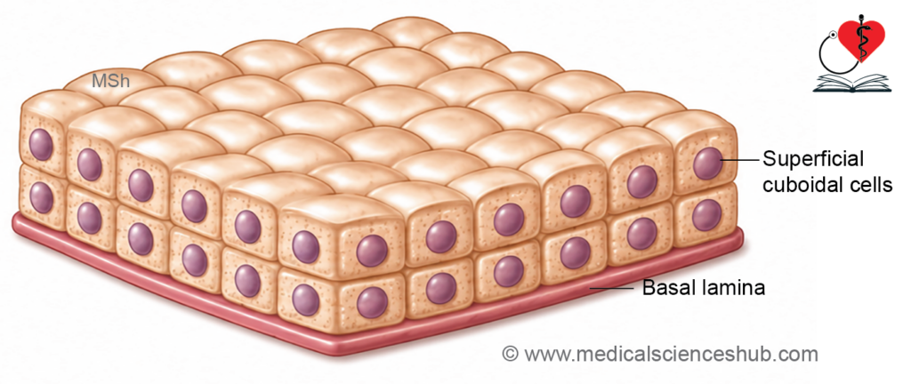
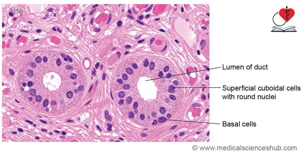
- Stratified cuboidal epithelium consists of two or more layers of cells.
- The cells of the superficial layer are cuboidal in shape.
- It is commonly found in the ducts of sweat glands, salivary glands, and sebaceous glands.
- Its main functions are to provide protection, form a barrier, and facilitate the passage of glandular secretions.
Stratified Columnar Epithelium
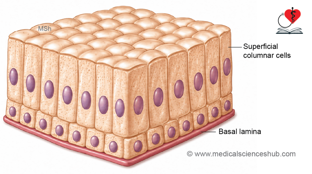
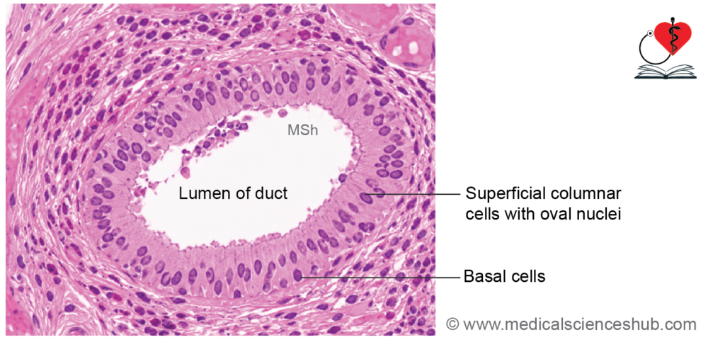
- Stratified columnar epithelium is usually composed of two cell layers.
- The superficial cells are columnar, while the basal cells are cuboidal and rest on the basement membrane.
- It lines some ducts of salivary glands, the fornix of the conjunctiva, and the spongy urethra.
- It functions in protection and the transport of secretions.
Transitional Epithelium/Urothelium
Features
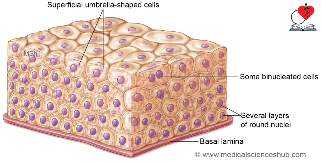
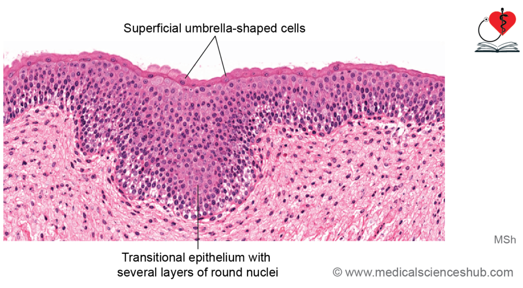
- Transitional epithelium is a specialized type of stratified epithelium that lines most parts of the urinary tract.
- Because it is characteristic of the urinary system, it is also called urothelium.
- Its appearance changes according to the degree of stretching of the organ.
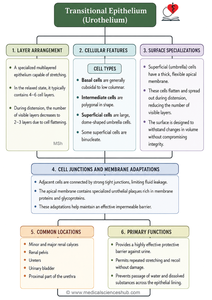
- In a distended urinary bladder, the epithelium appears thinner, usually with 2–3 cell layers.
- In an empty or contracted bladder, it appears thicker, often with 4–5 or more cell layers.
- This change in thickness occurs because the cells can alter their shape during stretching and relaxation.
- Basal cells are cuboidal and rest on the basement membrane.
- Intermediate cells are polygonal or pear-shaped.
- The superficial cells are large, dome-shaped or umbrella-shaped cells.
- Umbrella cells contain specialized membrane plaques rich in glycoproteins that strengthen the surface and reduce permeability.
- Some superficial cells may be binucleated.
Location
- Renal calyces
- Renal pelvis
- Ureters
- Urinary bladder
- Proximal part of the urethra
Functions
- It allows marked stretching and recoil of the urinary tract without damaging the epithelial lining.
- It accommodates changes in urine volume, especially in the urinary bladder.
- Tight junctions and membrane plaques form an effective barrier that prevents leakage of urine and protects underlying tissues from its potentially harmful components.
- It provides both distensibility and protection, making it uniquely suited for the urinary system.
Basement Membrane
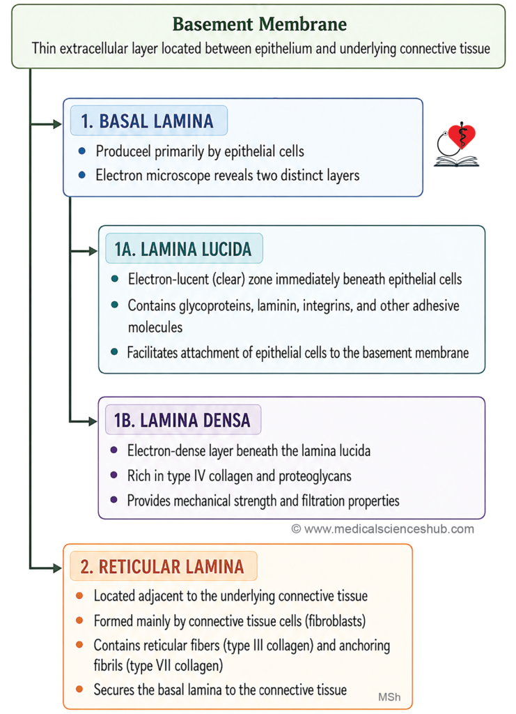
- The basement membrane is a thin layer of extracellular matrix located between the epithelium and the underlying connective tissue.
- It provides structural support and attachment for epithelial cells.
Staining Characteristics
- The basement membrane is usually not clearly visible with routine hematoxylin and eosin (H&E) staining.
- It can be demonstrated using Periodic Acid–Schiff (PAS) stain, which stains its carbohydrate-rich components a magenta color.
Structure
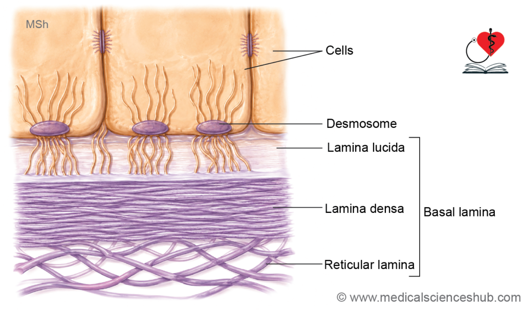
- Under the electron microscope, the basement membrane consists of two main components:
- Basal lamina
- Reticular lamina
Basal Lamina
- The basal lamina lies directly beneath the epithelial cells.
- It is produced primarily by epithelial cells.
- Traditionally, it was described as having:
- Lamina lucida (superficial layer)
- Lamina densa (deeper layer)
- Both layers contain a network of type IV collagen and specialized extracellular matrix proteins.
- Current evidence suggests that the lamina lucida is largely an artifact produced during tissue preparation, and the term basal lamina is now preferred in ultrastructural descriptions.
Reticular Lamina
- The reticular lamina lies between the basal lamina and the underlying connective tissue.
- It is produced mainly by fibroblasts.
- It contains reticular fibers and other connective tissue components.
- Type VII collagen anchoring fibrils attach the basal lamina to the reticular lamina, ensuring firm adhesion between epithelium and connective tissue.
Functions
- Provides mechanical support to the overlying epithelium.
- Anchors the epithelium to the underlying connective tissue.
- Acts as a selective filtration barrier and regulates the movement of molecules between tissues.
- Serves as a scaffold for epithelial cell migration and regeneration after injury.
- Helps maintain tissue organization and cellular polarity.
Important Questions
- List the functions of epithelium.
- Write a short note on transitional epithelium.
- Write a short note on basement membrane.
📝 Test Your Knowledge – Practice MCQs
Attempt the chapter MCQ quiz and assess your understanding of key concepts.
Click to see: Medical Disclaimer and Copyright Notice