Competencies
- AN79.4: Describe the development of somites and intraembryonic coelom.
INTRODUCTION
- The embryonic period extends from the beginning of the second week to the end of the eighth week of development. During this phase, the three germ layers—ectoderm, mesoderm, and endoderm—differentiate and give rise to the primordial tissues and organs. Because most major organs begin to form in this interval, it is also termed the organogenic period.
- Throughout these weeks, the initially flat embryonic disc undergoes cephalocaudal and lateral folding. This folding transforms it into a cylindrical, three-dimensional body form. By the end of the eighth week, the basic structural framework of the principal organ systems has been established, and the general external features of the body become identifiable.
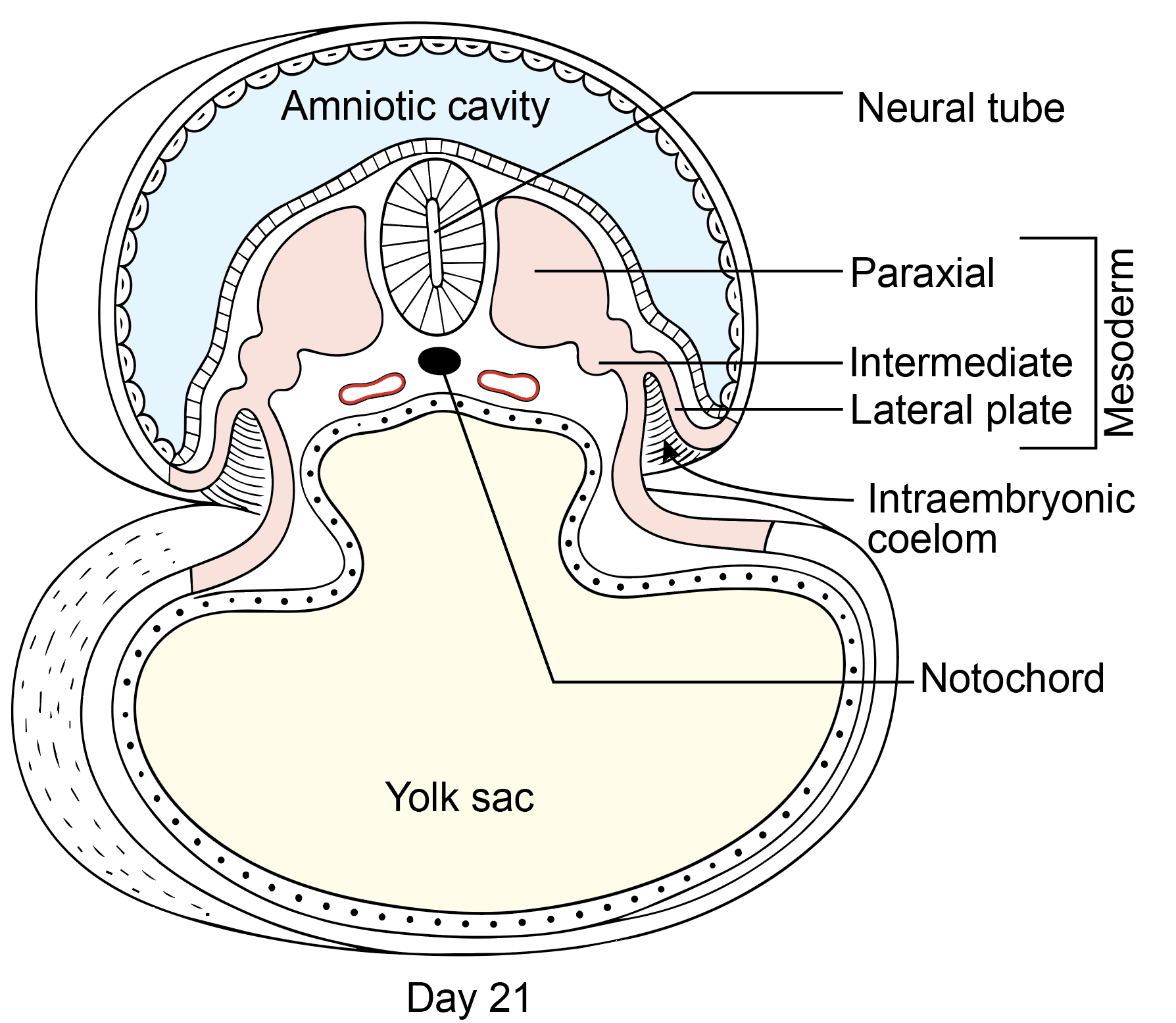
- Body folding produces a characteristic tube-within-a-tube body plan. This arrangement consists of two primary tubular structures. The outer ectodermal tube forms the epidermis of the skin, while the inner endodermal tube develops into the epithelial lining of the gastrointestinal tract. The region between these tubes is largely occupied by mesoderm, which differentiates into multiple tissues. The lateral plate mesoderm splits into somatic and splanchnic layers, creating the intraembryonic coelom, the precursor of the body cavities.
DIFFERENTIATION OF ECTODERM
- At the start of the third week, the midline ectoderm located above the developing notochord becomes thicker and forms the neural plate. This specialized region is referred to as the neuroectoderm and represents the primordium of the central nervous system.
- The rest of the ectoderm that does not participate in neural plate formation is termed the surface ectoderm.
- Thus, the ectoderm is subdivided into two components: neuroectoderm and surface ectoderm.
Neuroectoderm
- The neuroectoderm forms the entire central nervous system, including the brain and spinal cord, and major components of the peripheral nervous system, such as sensory and autonomic ganglia.
- Neurulation is the process by which the neural plate becomes the neural tube. The lateral edges of the neural plate elevate as neural folds, and the central region forms the neural groove. Fusion of the folds begins in the future cervical region and proceeds cranially and caudally, converting the plate into a closed tube.
- The cranial opening, the anterior neuropore, closes around the 25th day of intrauterine life at the 18–20 somite stage. The caudal opening, the posterior neuropore, closes around the 27th–28th day at approximately the 25-somite stage.
- Cells at the border of the neural plate form the neural crest. After closure, the neural tube separates from the surface ectoderm, and neural crest cells migrate to contribute to various structures.
- The cranial part of the neural tube enlarges to form the forebrain, midbrain, and hindbrain vesicles, while the caudal part develops into the spinal cord.
Surface Ectoderm
- The surface ectoderm shows localized depressions at the sites of the buccopharyngeal membrane and the cloacal membrane. At these regions, ectoderm is directly opposed to endoderm without intervening mesoderm. In the rest of the embryo, the intraembryonic mesoderm separates ectoderm from endoderm, except at the prechordal plate and the cloacal membrane.
- The buccopharyngeal (or oropharyngeal) membrane is located cranial to the prechordal plate during the third week. With formation of the head fold, the prechordal plate shifts to a position between the developing forebrain and the pericardial prominence. At the beginning of the fourth week, the buccopharyngeal membrane breaks down, establishing a connection between the amniotic cavity and the primitive gut.
- The cloacal membrane later subdivides into the urogenital membrane anteriorly and the anal membrane posteriorly.
- The surface ectoderm gives rise to several important structures. These include the epidermis, hair, nails, sebaceous and sweat glands; the olfactory epithelium; the lens of the eye; the otic vesicle; and the pharyngeal clefts. It also forms Rathke’s pouch, which contributes to the anterior pituitary, the epithelial lining of parts of the oral and nasal cavities including enamel of the teeth, the salivary glands, the mammary glands, and components of the pituitary gland.
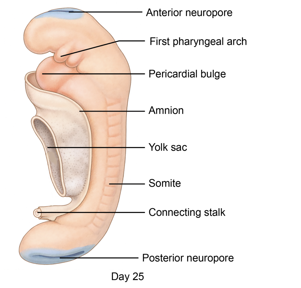
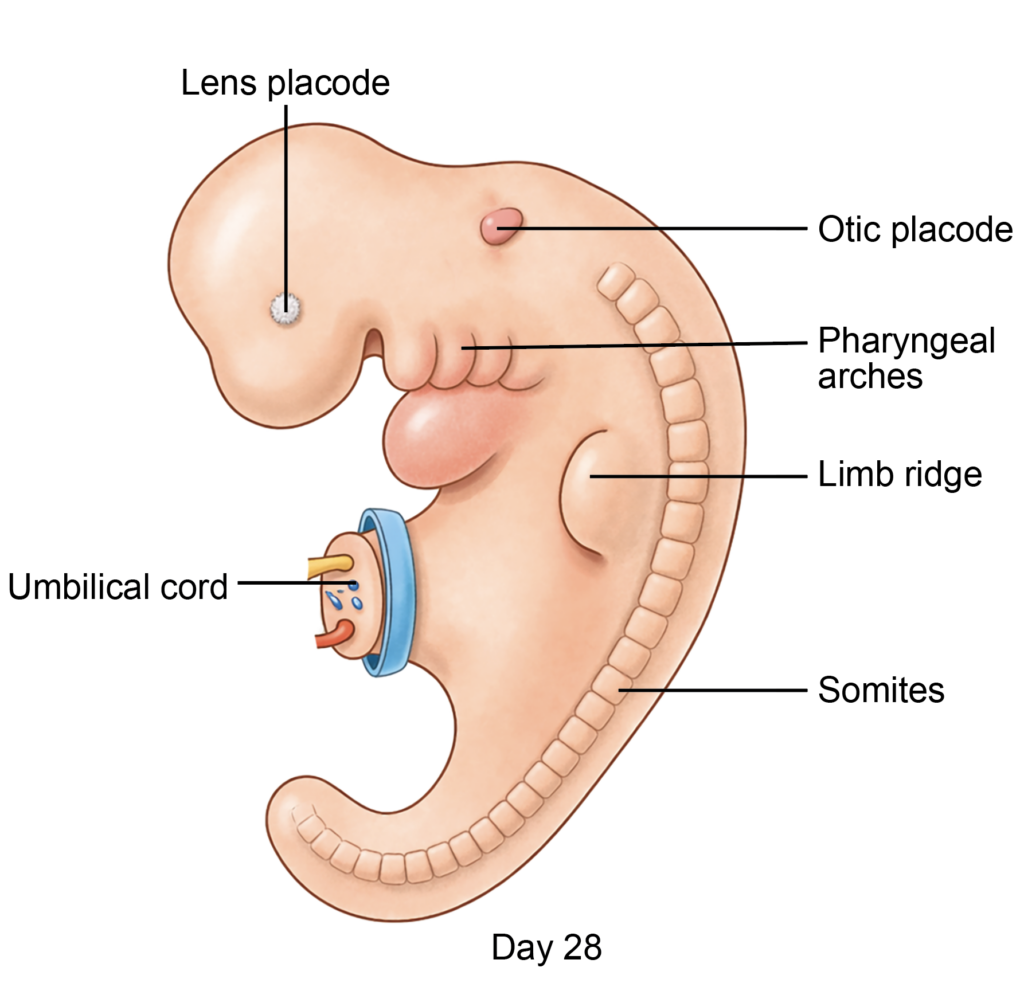
Figure 7.3a: Transverse sections showing development of the mesodermal germ layer (Click to see figure)
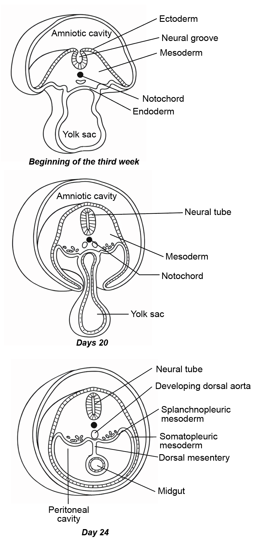
MESODERMAL DIFFERENTIATION
- By the end of the third week, the primitive streak begins to regress. Mesodermal cells that migrated through the primitive streak organize on either side of the notochord around the 17th day of intrauterine life. These cells condense to form paired, longitudinal columns of mesoderm.
- Shallow grooves then divide this mesoderm into three distinct regions.
- The paraxial mesoderm lies adjacent to the notochord and beneath the neural plate.
- Lateral to it is the intermediate mesoderm.
- The most lateral portion is the lateral plate mesoderm.
- Each of these mesodermal subdivisions differentiates into specific tissues and organ systems in the developing embryo.
- Thus, the mesoderm is organized into three parts: paraxial mesoderm, intermediate mesoderm, and lateral plate mesoderm.
Paraxial Mesoderm
- The paraxial mesoderm is located on either side of the notochord.
- It extends longitudinally from the prechordal plate to the region of the regressing primitive streak.
- This mesoderm condenses into a series of segmental, rounded structures known as somitomeres. These further organize into distinct, paired blocks called somites, which represent the metameric segmentation of the embryo and contribute to the axial skeleton, skeletal muscle, and dermis.
Intermediate Mesoderm
- The intermediate mesoderm lies between the paraxial mesoderm and the lateral plate mesoderm.
- During further development, it differentiates primarily into components of the urogenital system, including the kidneys and the gonads.
Lateral Plate Mesoderm
- The lateral plate mesoderm extends from the intermediate mesoderm laterally toward the extraembryonic mesoderm.
- Cranially, it continues into a mesodermal condensation called the pericardial bar, located cranial to the buccopharyngeal membrane.
- Within the lateral plate mesoderm, small intercellular spaces appear and coalesce to form the intraembryonic coelom. A similar cavity develops in the pericardial bar and contributes to the formation of the pericardial cavity.
Formation of Coelomic Cavity
- The developing pericardial cavity initially communicates with the intraembryonic coelom, creating an inverted U-shaped tubular space within the embryo. As development progresses, this coelomic space undergoes partitioning and differentiation.
- The single coelomic cavity ultimately gives rise to three major body cavities: the pericardial cavity, the pleural cavities, and the peritoneal cavity.
Formation of Layers of Lateral Plate Mesoderm
- The appearance of the intraembryonic coelom splits the lateral plate mesoderm into two distinct layers.
- The outer somatopleuric layer is the parietal layer and lies adjacent to the ectoderm.
- The inner splanchnopleuric layer is the visceral layer and lies next to the endoderm.
- As the intraembryonic coelom enlarges, it separates these two layers laterally and temporarily communicates with the extraembryonic coelom. This connection supports early exchange of nutrients before the uteroplacental circulation becomes fully established.
- In the cranial region, a horseshoe-shaped mass of mesoderm connects the somatopleuric and splanchnopleuric layers. This structure is known as the septum transversum, which later contributes to the formation of the liver and part of the diaphragm.
Fate of Lateral Plate Mesoderm
The lateral plate mesoderm differentiates into two layers, each forming specific structures.
A. Somatopleuric layer
- The somatopleuric (parietal) layer contributes to the parietal linings of the pericardial, pleural, and peritoneal cavities. It also forms the dermis of the body wall and limbs, the pectoral and pelvic girdles, and the skeletal elements of the limbs. The muscles of the limbs are derived from migrating cells of the myotomes, which originate from the paraxial mesoderm.
B. Splanchnopleuric layer
- The splanchnopleuric (visceral) layer forms the visceral linings of the pericardial, pleural, and peritoneal cavities. It also gives rise to the smooth muscle and connective tissue of the gastrointestinal tract, respiratory tract, and the heart.
SOMITES
- Along the length of the embryo, from the prechordal plate to the region of the regressing primitive streak, the paraxial mesoderm condenses to form somitomeres. These segment further into paired, block-like structures called somites.
- The cranial somitomeres located rostral to the otic vesicle do not segment into typical somites. Instead, they contribute to the striated muscles of the face, jaw, and pharynx. The mesoderm of the prechordal plate region forms preoccipital somites, which later give rise to the extraocular muscles.
- The first pair of somites appears around the 20th day of development near the cranial end of the notochord. New somites form in a craniocaudal sequence until about the 30th day.
- In humans, 42 to 44 pairs initially develop = 3 preoccipital + 4 occipital + 8 cervical + 12 thoracic + 5 lumbar + 5 sacral + 8 to 10 coccygeal pairs
- The first cervical and several coccygeal somites regress, resulting in approximately 37 persistent pairs. The remaining somites contribute primarily to the axial skeleton, associated skeletal muscles, and dermis.
- Counting of somites: The first pair of somites appears around the 20th day of development. After this stage, approximately three additional pairs form each day until the end of the fifth week. Because somites develop in a regular and predictable sequence, their number is used to estimate the embryonic age during early development.
Structure of Somite
- A somite is a segmented, triangular mass of mesenchymal tissue derived from the paraxial mesoderm.
- Each somite differentiates into three distinct components:
- The sclerotome, located ventromedially, surrounds the developing neural tube and contributes to the formation of the vertebrae and ribs.
- The intermediate portion forms the myotome, which gives rise to skeletal muscles. Thus, skeletal muscles of the body wall and limbs originate from the myotomes of somites.
- The dorsolateral part becomes the dermatome, which forms the dermis of the skin of the back.
- A small central cavity, known as the myocele, is initially present within each somite but disappears as the cells proliferate and reorganize.
- Each somite is associated with a single spinal nerve, establishing segmental innervation. Some descriptions group the dermatome and myotome together as a dermomyotome before they separate into distinct layers.
Embryonic Age and Somite Count
- In the early embryonic period, the number of somite pairs increases in a regular sequence. For this reason, counting somites provides a reliable method to estimate embryonic age, especially during the third and fourth weeks.
Table 8.1: The correlation between embryo age with number of somites
| Approximate Age (in days) | Somite Pairs (approximate range) |
|---|---|
| 20 | 1–4 |
| 21 | 4–7 |
| 22 | 7–10 |
| 23 | 10–13 |
| 24 | 13–17 |
| 25 | 17–20 |
| 26 | 20–23 |
| 27 | 23–26 |
| 28 | 26–29 |
| 30 | 34–35 |
| End of 5th week | 42–44 |
FOLDING OF EMBRYO
- In the third week of development, the embryo is a trilaminar germ disc formed by ectoderm, mesoderm, and endoderm. Rapid growth in the central region compared to the margins produces mechanical forces that initiate embryonic folding. This process converts the flat disc into a cylindrical body form.
- Folding occurs in two directions. Cephalocaudal folding in the median plane creates the cranial fold and caudal fold. Lateral folding in the transverse plane brings the right and left edges toward the midline.
- Before folding begins, the septum transversum lies at the cranial end and later shifts ventrally, contributing to diaphragm development.
Effects of folding
- Embryonic folding produces important structural changes in the developing embryo.
- Folding converts the flat trilaminar germ disc into a cylindrical body form, establishing the basic body outline.
- The cranial, caudal, and lateral folds move toward the ventral surface, creating a narrow, hourglass-shaped constriction at the yolk sac.
- The amniotic cavity enlarges and gradually covers the ventral aspect of the embryo. The amniotic membrane also surrounds the connecting stalk, forming a tubular covering.
- A portion of the yolk sac becomes enclosed within the embryo and forms the primitive gut, while the remaining external part becomes the umbilical vesicle.
- The vitellointestinal duct (omphaloenteric duct) temporarily connects the midgut to the umbilical vesicle. The segment of the primitive gut cranial to this duct is the foregut, and the segment caudal to it is the hindgut.
Head Fold
- Rapid growth of the cranial part of the germ disc, especially around the developing brain region, causes the embryo to bend forward over the cranial end of the notochord. This bending forms the head fold and contributes to the early shaping of the forebrain region.
- The head fold produces several structural changes:
- A portion of the yolk sac becomes enclosed within the fold and forms the foregut, which represents the cranial part of the primitive gut.
- The opening between the foregut and the remaining midgut is called the anterior intestinal portal.
- On the ventral side of the foregut lie the buccopharyngeal membrane, the developing pericardial cavity, and the septum transversum.
- On the dorsal side are the notochord and the developing brain vesicles.
- The rapidly enlarging forebrain vesicle is situated cranial to the foregut.
Tail Fold
- Continued growth of the caudal region of the germ disc causes ventral bending around the caudal end of the notochord. This movement forms the tail fold and shapes the posterior part of the embryo.
- The tail fold produces the following changes:
- A portion of the yolk sac becomes enclosed within the fold to form the hindgut, which represents the caudal part of the primitive gut.
- The allantois extends from the hindgut and forms the allantoenteric diverticulum, which lies ventral to the hindgut.
- The cloacal membrane, a bilaminar structure, lies caudal to the connecting stalk. It later divides into the urogenital membrane and the anal membrane.
- The notochord and neural tube are located dorsal to the developing hindgut.
- The regressing primitive streak and primitive node lie caudal to the hindgut.
- The allantoenteric diverticulum divides the hindgut into preallantoic and postallantoic segments.
- The opening between the midgut and the preallantoic hindgut is called the posterior intestinal portal.
Lateral Folds
- Rapid expansion of the central region of the germ disc produces right and left lateral folds. These folds move ventrally and meet in the midline. Their fusion with the head and tail folds at the primitive umbilical ring converts the flat disc into a cylindrical embryo.
- Changes produced by lateral folding
- The amnioectodermal junction shifts ventrally and becomes located on the developing umbilical cord (connecting stalk).
- A portion of the yolk sac becomes enclosed within the embryo and forms the midgut.
- The midgut remains temporarily connected to the remaining yolk sac, now called the umbilical vesicle, through the vitellointestinal duct.
- The splanchnopleuric intraembryonic mesoderm covers the ventrolateral surface of the gut and reflects dorsally to form the dorsal mesentery.
- Fusion of the somatopleuric mesoderm in the ventral midline transforms the intraembryonic coelom into the developing peritoneal cavity.
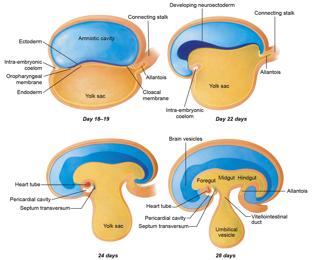
Figure 7.4a: Formation of craniocaudal (head and tail) folding of the embryo (Click to see figure)
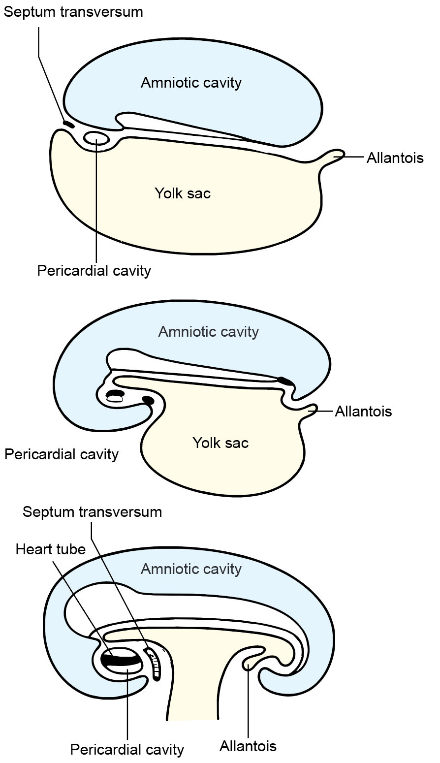
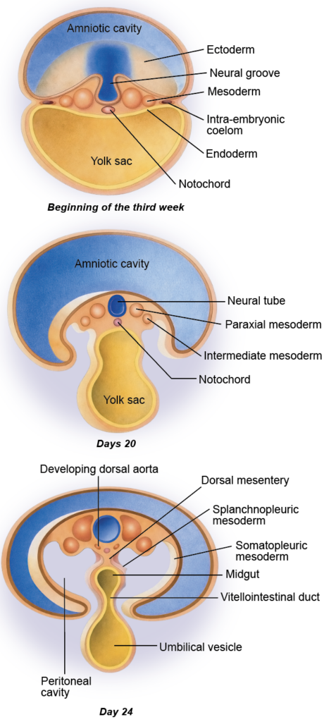
Figure 7.5a: Lateral folding of the developing embryo (Click to see figure)
DIFFERENTIATION OF ENDODERM
- During embryonic folding, the flat endoderm is transformed into a tubular structure that forms the primitive gut. This tube is divided into three regions: foregut, midgut, and hindgut. Each part gives rise to specific epithelial linings and glandular parenchyma
Derivatives of the Primitive Gut
Foregut
- Epithelial lining of the pharynx, oesophagus, stomach, and proximal duodenum up to the ampulla of Vater
- Epithelial lining of the respiratory tract, auditory tube, and tympanic cavity
- Parenchyma of the thyroid, parathyroid, thymus, liver, pancreas, and palatine tonsil
Midgut
- Epithelial lining of the distal duodenum, jejunum, ileum, caecum, appendix, ascending colon, and proximal two-thirds of the transverse colon
Hindgut
- Epithelial lining of the distal one-third of the transverse colon, descending colon, sigmoid colon, rectum, and upper anal canal up to the mucocutaneous junction
- Epithelial lining of the urinary bladder (except trigone), most of the urethra, and the vagina
- Parenchyma of the prostate (except fibromuscular stroma) and bulbourethral glands
MAJOR CHANGES IN 4TH TO 8TH WEEKS
- At the start of the fourth week, the embryo is still relatively flat and shows about 4 to 12 somites. Rapid growth and differentiation during the next few weeks establish the basic body form and major organ systems.
Fourth Week
During this week, body folding becomes evident and organ development accelerates.
- The number of somites increases progressively.
- The neural tube differentiates into three primary brain vesicles: forebrain, midbrain, and hindbrain.
- Head and tail folds become prominent, giving the embryo a curved shape.
- Around the 21st day, the primitive heart begins to beat.
- By the 24th day, the first pharyngeal arch appears.
- By the 26th day, three pairs of pharyngeal arches are visible.
- Around the 26th to 27th day, the upper limb bud appears as a small projection.
Sixth Week
Growth and external features become more distinct.
- The embryo shows spontaneous movements.
- The brain vesicles enlarge rapidly.
- Nasal processes appear on the developing face.
- The buccopharyngeal membrane ruptures, establishing communication between the foregut and amniotic cavity.
- The caecum and appendix begin to develop, and the spleen appears in the dorsal mesogastrium.
- Digital differentiation starts in the upper limb and occurs slightly later in the lower limb.
- Temporary physiological umbilical herniation of the midgut is common.
Seventh and Eighth Weeks
By these weeks, the embryo shows clear human features.
- The face, external ear (auricle), and eyes become well defined.
- Limbs and digits are clearly formed.
- The metanephric kidneys develop.
- The testes and ovaries begin to differentiate.
- External genitalia start showing early differentiation.
- In the eighth week, limb movements become coordinated.
- Primary ossification centers appear in several bones.
- The temporary tail-like eminence regresses and disappears.
Important Questions
- Write a short note explaining the formation, structure, and developmental significance of somites in the embryo.
- Write a brief note describing the formation and importance of the cephalic (head) fold during early embryonic development.
- Write a short note outlining the formation and developmental role of the caudal (tail) fold of the embryo.