INTRODUCTION
- The ear is a specialized sensory organ responsible for hearing and the maintenance of equilibrium (balance). Structurally, it is divided into three main parts: the external ear, middle ear, and internal ear.
- The external ear comprises the auricle and the external acoustic meatus. It is separated from the middle ear by the tympanic membrane.
- The middle ear is an air-filled cavity that communicates anteriorly with the nasopharynx via the auditory (pharyngotympanic) tube and posteriorly with the mastoid antrum through the aditus.
- The internal ear consists of the bony labyrinth, located within the petrous part of the temporal bone, and the membranous labyrinth contained within it.
- The bony labyrinth encloses perilymph, while the membranous labyrinth contains endolymph and includes the cochlear duct, saccule, utricle, and three semicircular ducts.
- The organ of Corti within the cochlear duct is responsible for hearing.
- The maculae (in the saccule and utricle) and the cristae ampullaris (in semicircular ducts) are sensory receptors essential for maintaining balance.
- The development of the ear involves contributions from all three germ layers.
- The surface ectoderm gives rise to the internal and external ear structures, the endoderm forms the epithelial lining of the middle ear cavity, and the mesoderm contributes to the formation of connective tissues, ossicles, and associated muscles.
DEVELOPMENT OF INTERNAL EAR
- The internal ear originates from an ectodermal structure called the otic vesicle (otocyst). It ultimately differentiates into the membranous labyrinth and is later surrounded by the bony labyrinth.
Membranous Labyrinth
Stages of Development
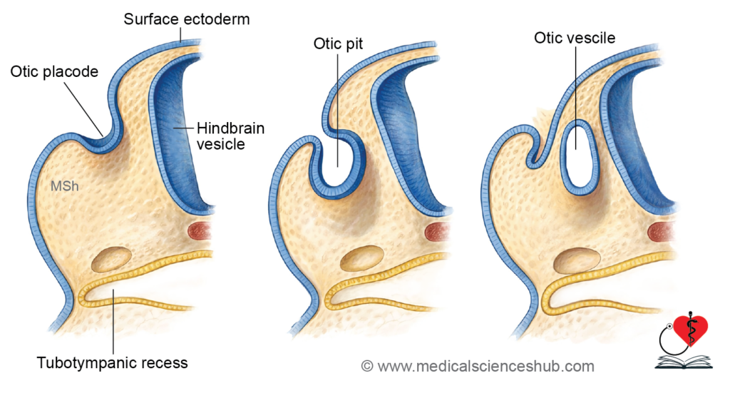
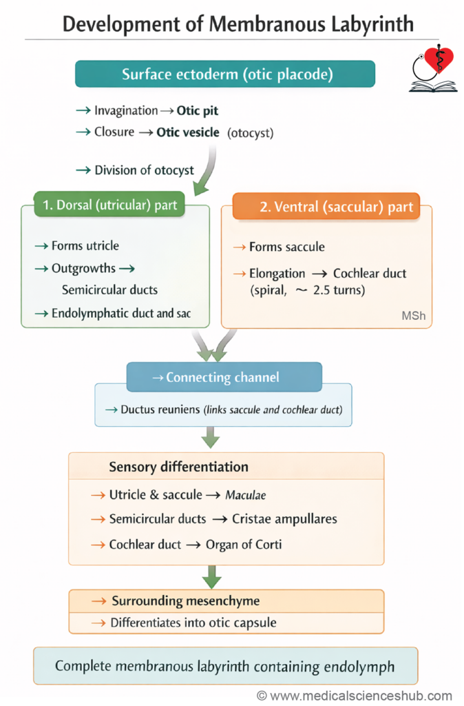
- During the 4th week, the surface ectoderm adjacent to the rhombencephalon forms bilateral thickenings known as otic placodes. These placodes invaginate to form otic pits, which then detach from the surface to become otic vesicles (otocysts). These vesicles migrate deeper and give rise to the membranous labyrinth.
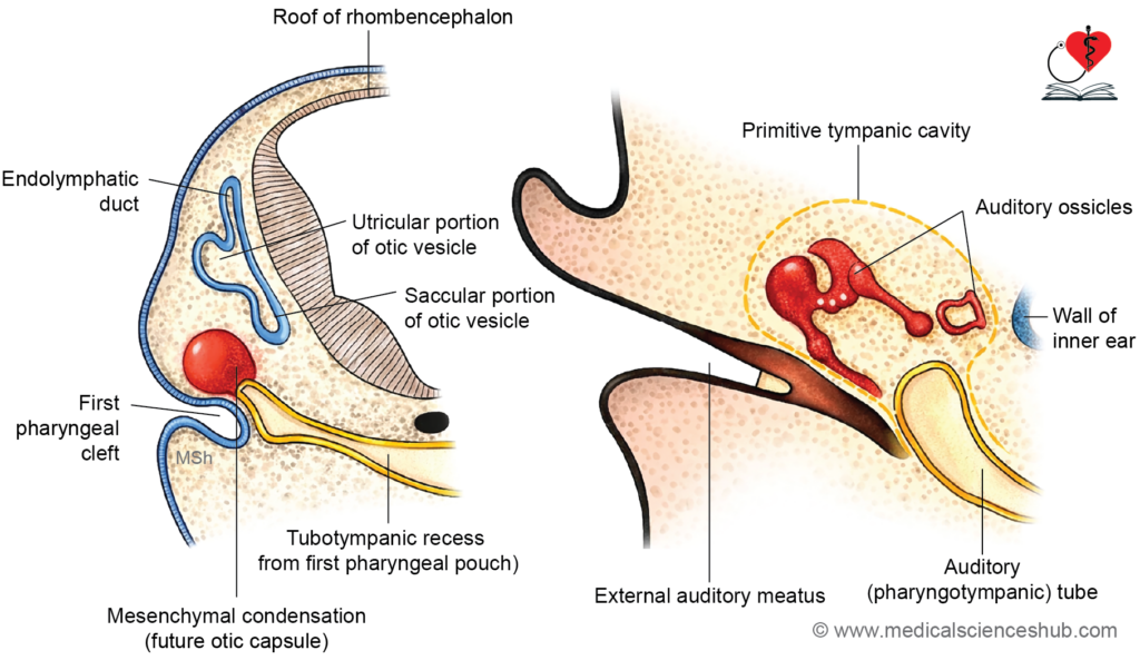
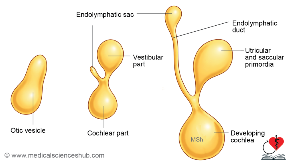
- The otic vesicle differentiates into two main regions:
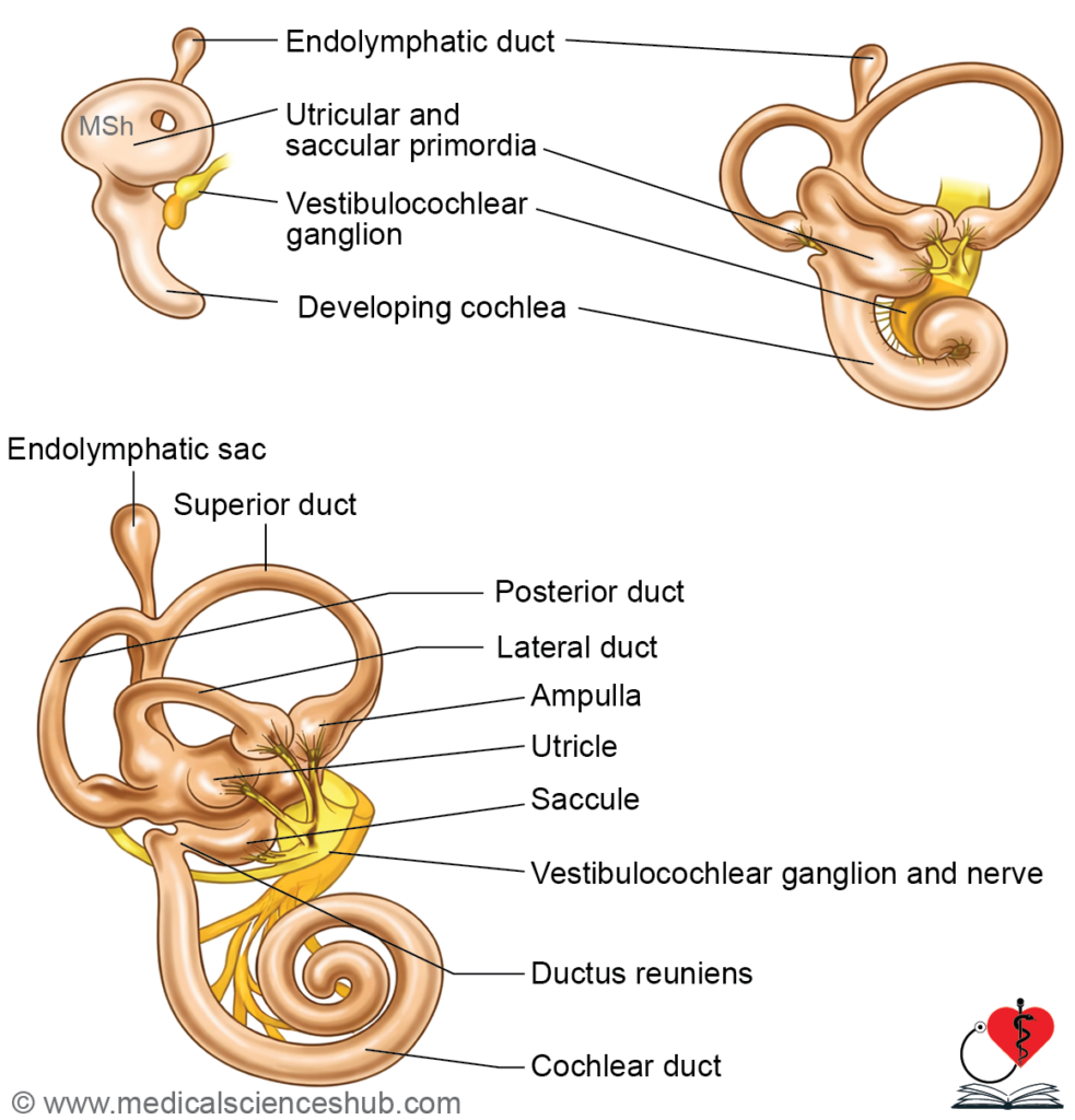
- Dorsal (vestibular) part → forms the utricle, semicircular ducts, and the endolymphatic duct and sac.
- Ventral (cochlear) part → gives rise to the saccule and cochlear duct, which contains the organ of Corti.
- The epithelial lining of the membranous labyrinth becomes specialized to form sensory receptors:
- Cristae ampullaris in semicircular ducts (detect angular acceleration)
- Maculae in utricle and saccule (detect linear acceleration and gravity)
- Organ of Corti in the cochlear duct (hearing)
- Neural crest cells migrate toward the otic vesicle and differentiate into bipolar neurons of the vestibulocochlear ganglion. Their peripheral processes innervate the sensory regions of the membranous labyrinth, while central processes project to the vestibular and cochlear nuclei of the hindbrain, forming the vestibulocochlear nerve (CN VIII).
- This coordinated development establishes both the sensory and neural components required for hearing and balance.
Bony Labyrinth
The bony labyrinth develops from mesenchymal tissue surrounding the otic vesicle (membranous labyrinth).
- The surrounding mesenchyme condenses to form the otic capsule, which later undergoes chondrification to become cartilage and subsequently ossifies.
- A layer of periotic mesenchyme initially separates the otic capsule from the membranous labyrinth. This tissue gradually degenerates, creating a space that fills with perilymph, while the membranous labyrinth becomes filled with endolymph.
- Selective resorption of periotic tissue leads to the formation of key spaces:
- Around the utricle and saccule → vestibule
- Around the semicircular ducts → semicircular canals
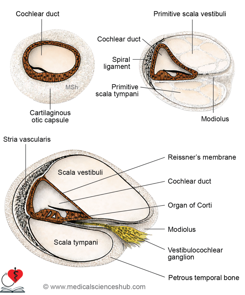
- In the cochlear region, incomplete resorption of periotic tissue results in two perilymph-filled channels:
- Scala vestibuli (above)
- Scala tympani (below)
These are separated from the cochlear duct by the vestibular (Reissner’s) membrane and basilar membrane, respectively.
- At the apex of the cochlea, the scala vestibuli and scala tympani communicate through the helicotrema. The cochlear duct is anchored to the bony wall by the spiral ligament, and the membranes converge toward the central axis, the modiolus.
This process establishes the rigid framework and fluid-filled compartments essential for sound transmission and balance.
Histogenesis of Internal Ear
Histogenesis of Semicircular Canals
- The otic (auditory) vesicle is initially lined by low columnar epithelium, which later becomes flattened except in the ampullary regions.
- In the ampullae, specialized epithelial cells differentiate into hair cells and a gelatinous structure called the cupula. Together, they form the crista ampullaris, the sensory receptor for detecting angular (rotational) movements.
- In the utricle and saccule, similar hair cells with an overlying gelatinous layer form the maculae, which are responsible for sensing linear acceleration and gravity.
Histogenesis of Cochlea
- In the cochlear duct, epithelial cells adjacent to the scala tympani thicken to form two ridges separated by the spiral sulcus:
- The inner ridge gives rise to the membrana tectoria, a gelatinous structure that interacts with hair cells.
- The outer ridge differentiates into inner hair cells, multiple rows of outer hair cells, and supporting cells (rod cells), forming the organ of Corti.
- The vestibular (Reissner’s) membrane develops from the dorsal wall of the cochlear duct. The lateral wall forms a highly vascular region known as the stria vascularis, which plays a key role in endolymph production.
- Histological differentiation of the cochlea is largely complete by the sixth month of intrauterine life (IUL).
- These changes establish the specialized sensory structures required for hearing and balance.
MIDDLE EAR
- The middle ear cavity is primarily derived from endoderm. During the 5th week, a diverticulum called the tubotympanic recess arises from the dorsal part of the first pharyngeal pouch, with a minor contribution from the second pouch. This recess grows laterally toward the developing external ear.
- By the 6th month of intrauterine life (IUL), the tubotympanic recess approaches the first pharyngeal cleft (future external acoustic meatus). A thin layer of mesenchyme remains between them. The tympanic membrane is formed by the fusion of all three germ layers: ectoderm (external), mesoderm (middle), and endoderm (internal).
- Growth and rearrangement of pharyngeal arch mesenchyme alter the region. The third pharyngeal arch shifts to lie adjacent to the first arch, and a narrow segment of the tubotympanic recess between these arches forms the auditory (pharyngotympanic) tube. The lateral expanded part becomes the primary tympanic cavity.
Formation of definitive tympanic cavity
- The primary tympanic cavity enlarges to surround the ear ossicles, along with associated muscles, nerves, and blood vessels, forming the definitive middle ear cavity.
- A dorsal outgrowth of this cavity develops into the tympanic antrum, which later communicates with mastoid air cells.
- This sequence establishes the air-filled middle ear space essential for sound conduction.
Ear Ossicles
- By the end of the 7th week, mesenchymal condensations appear in the roof of the developing tympanic cavity. Two condensations arise from the first pharyngeal arch (Meckel’s cartilage) and differentiate into the malleus and incus. A third condensation from the second pharyngeal arch (Reichert’s cartilage) forms the stapes.
- Initially, the ossicles remain embedded within surrounding mesenchyme. By approximately the 8th month of intrauterine life, this mesenchyme undergoes degeneration, allowing the mucosal lining of the middle ear cavity to extend around and envelop the ossicles.
- This process enables the ossicles to become freely mobile, which is essential for efficient transmission of sound vibrations.
Muscles of Middle Ear
- The tensor tympani muscle develops from mesenchyme of the first pharyngeal arch. Accordingly, it is innervated by the trigeminal nerve (mandibular division), which is the nerve of this arch.
- The stapedius muscle arises from mesenchyme of the second pharyngeal arch and is supplied by the facial nerve, the nerve associated with this arch.
- This pattern reflects the typical relationship between pharyngeal arch origin and nerve supply of head and neck muscles.
Formation of Round and Oval Windows
- During development, the bony labyrinth remains thin at two specific sites:
- The area opposite the stapes forms the oval window (fenestra vestibuli).
- A second thin area, located inferior to the stapes, forms the round window (fenestra cochleae).
- The oval window opens into the vestibule and is sealed by the footplate of the stapes, enabling transmission of sound vibrations.
- The round window communicates with the scala tympani and is closed by the secondary tympanic membrane, which allows pressure dissipation within the cochlea.
- These structures are essential for effective mechanical transmission and modulation of sound within the inner ear.
Formation of Tubal Tonsil
The pharyngeal opening of the tubotympanic recess persists as the opening of the auditory (Eustachian) tube in the nasopharynx. This region becomes surrounded by an aggregation of lymphoid tissue, which develops into the tubal tonsil.
The tubal tonsil contributes to local immune defense by protecting the upper respiratory tract near the auditory tube opening.
EXTERNAL EAR
External Acoustic Meatus
- The external acoustic meatus develops from the dorsal part of the first pharyngeal cleft, which invaginates to form a funnel-shaped primary meatus extending toward the primary tympanic cavity. The medial cells proliferate to form a solid epithelial mass known as the meatal plug (medial plate).
- By the 7th month of intrauterine life, this solid plug undergoes canalization to create a lumen. The newly formed passage, called the secondary meatus, establishes continuity with the tympanic cavity.
- The cells of the meatal plug contribute to the outer ectodermal (cutaneous) layer of the tympanic membrane. The handle of malleus and chorda tympani nerve become embedded within the fibrous (mesodermal) layer between the ectodermal and endodermal components.
- At birth, the tympanic membrane is relatively horizontal. With growth, it assumes an oblique orientation, facing downward, forward, and medially.
Auricle(Pinna)
- Around the 6th week of intrauterine life, six mesenchymal swellings known as auricular hillocks appear around the dorsal part of the first pharyngeal cleft. Three develop from the first (mandibular) arch, and three from the second (hyoid) arch.
- These hillocks enlarge, fuse, and remodel to form the auricle (pinna) as follows:
- First hillock → tragus
- Second hillock → crus of helix
- Third hillock → helix
- Fourth hillock → antihelix
- Fifth hillock → antitragus
- Sixth hillock → lower helix and lobule
- Development of the auricle is largely completed by the 4th month of intrauterine life.
- The internal ear develops earlier (early 4th week) than the middle and external ear.
- The middle ear cavity, ear ossicles, and internal ear reach near adult size at birth.
- Mastoid air cells develop postnatally, becoming prominent by about 2 years of age.
- Failure of canalization of the meatal plug is a common cause of congenital deafness
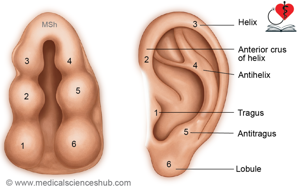
Table 24.1: Development of ear
| Adult Structure | Embryonic Origin |
|---|---|
| External Ear | |
| Auricle (Pinna) | Six ectodermal hillocks: 1st arch → tragus, helix; 2nd arch → antihelix, antitragus, lobule |
| External Acoustic Meatus | 1st pharyngeal cleft (ectoderm) |
| Middle Ear | |
| Tympanic Cavity | Tubotympanic recess (endoderm of 1st pouch) |
| Auditory Tube & Mastoid Antrum | Tubotympanic recess |
| Ossicles | Malleus, incus → 1st arch; stapes → 2nd arch |
| Muscles | Tensor tympani → 1st arch; stapedius → 2nd arch |
| Tympanic Membrane | Three layers: ectoderm (outer), mesoderm (middle), endoderm (inner) |
| Internal Ear | |
| Membranous Labyrinth | From otic vesicle → dorsal (utricle, semicircular ducts) and ventral (saccule, cochlear duct) parts |
| Bony Labyrinth | From otic capsule (mesenchyme) → vestibule, semicircular canals, cochlea |
CLINICAL EMBRYOLOGY
DEVELOPMENTAL ANOMALIES
External Ear
- Developmental defects may arise from failure of fusion or absence of auricular hillocks, leading to partial or complete absence of the auricle or formation of accessory nodules.
- The position of the auricle may be abnormal. Since its ascent depends on mandibular growth, conditions such as micrognathia or agnathia can result in a low-set ear, as seen in mandibulofacial dysostosis.
- Microtia refers to underdevelopment of the auricle, whereas anotia denotes complete absence.
External of Auditory Meatus
- Atresia of the external acoustic meatus occurs due to failure of canalization of the meatal plug, resulting in conductive hearing loss.
- Congenital exaggeration of the meatal curvature may obscure visualization of the tympanic membrane.
Middle Ear
- Ossicular malformations, particularly of the malleus and incus, are associated with first arch syndromes.
- Congenital fixation of the stapes at the oval window (fenestra vestibuli) leads to significant conductive deafness.
- An abnormal course or bulging of the facial nerve canal may be present within the middle ear.
Internal Ear
- Maternal infection with the rubella virus, especially during the second month of pregnancy, can impair development of the cochlea, vestibular apparatus, and organ of Corti, resulting in hearing deficits (often limited to low-frequency perception).
- Exposure to thalidomide may cause malformations of the semicircular canals.
- These anomalies highlight the importance of precise embryological development for normal auditory and vestibular function.