INTRODUCTION
The pectoral region is situated on the front of the trunk, anterior to the thoracic cage.
It serves as a connecting area between the upper limb and the anterolateral thoracic wall.
Contents
- The principal contents include the following structures:
- Mammary gland
- Muscles
- Pectoralis major
- Pectoralis minor
- Subclavius
- The pectoral and clavipectoral fasciae form the supporting fascial layers of this region.
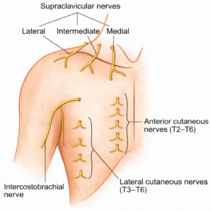
Cutaneous Nerves of the Pectoral Region
- The skin over the front of the chest wall receives sensory supply from two main groups of nerves. These nerves arise from cervical and thoracic spinal segments.
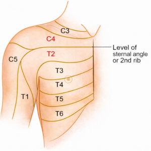
1. Supraclavicular Nerves
- These are branches of the cervical plexus. They arise from spinal nerve roots C3 and C4.
- They are divided into:
- Medial supraclavicular nerve
- Intermediate supraclavicular nerve
- Lateral supraclavicular nerve
- Area supplied:
- Skin above a horizontal line drawn through the sternal angle
- Upper part of the pectoral region
2. Intercostal Nerves
- These are the anterior rami of thoracic spinal nerves. The 2nd to 6th intercostal nerves contribute to the skin supply of this region.
- Their cutaneous supply is through:
- Lateral cutaneous branches
- Anterior cutaneous branches
- Root value: T2 to T6
- Area supplied:
- Skin below the horizontal line passing through the sternal angle
- Lower part of the pectoral region
Cutaneous Blood Vessels of the Pectoral Region
- The skin over the front of the chest receives blood from branches of arteries present in the thoracic and cervical regions. These vessels form a rich network to maintain adequate circulation to the skin and subcutaneous tissue.
1. Branches of the Internal Thoracic Artery
- The perforating branches arise from the internal thoracic artery. They pass forward through the intercostal spaces.
- These branches supply:
- Skin near the sternum
- Anterior part of the pectoral region
2. Lateral Cutaneous Branches of Posterior Intercostal Arteries
- These branches arise from the posterior intercostal arteries. They emerge along the lateral chest wall. They travel with the lateral cutaneous nerves.
- They supply:
- Skin on the lateral aspect of the chest
- Side of the pectoral region
3. Supraclavicular Artery
- This artery is a branch of the transverse cervical artery.
- It provides blood to:
- Skin over the lateral end of the clavicle
- Uppermost part of the pectoral region
MUSCLES OF PECTORAL REGION
- The pectoral region has three muscles:
- 1. Pectoralis major
- 2. Pectoralis minor
- 3. Subclavius
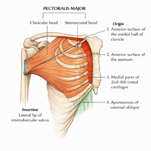
Pectoralis Major Muscle
- The pectoralis major is the largest and most prominent muscle of the pectoral region. It is a thick, broad, fan-shaped muscle located on the front of the chest wall. It forms most of the bulk of the anterior chest and contributes to the anterior axillary fold.
Origin
The muscle has two main heads:
- Clavicular head: It forms smaller portion. It arises from the anterior surface of the medial half of the clavicle.
- Sternocostal head: It forms the larger portion. It composed of manubrial, sternal, costal, and aponeurotic fibers.
- Manubrial fibers: arise from the lateral part of the anterior surface of the manubrium.
- Sternal fibers: arise from the anterior surface of the sternum up to the level of the 6th costal cartilage.
- Costal fibers: arise from the 2nd to 6th costal cartilages.
- Aponeurotic fibers: arise from the aponeurosis of the external oblique muscle of the abdomen.
Insertion
- The muscle fibers converge to form a strong, U-shaped tendon. This tendon attaches to the lateral lip of the intertubercular sulcus (bicipital groove) of the humerus.
- The tendon is made of two layers (bilaminar):
- Anterior lamina is the shorter layer. It is formed mainly by clavicular fibres.
- Posterior lamina is formed mainly by sternocostal fibres.
Direction of Fibers
- Clavicular fibers run downward and laterally.
- Sternocostal fibers run upward and laterally. The lower fibres twist before insertion so that the lowest fibers attach at a higher level on the humerus. This twisting arrangement contributes to the formation of the anterior axillary fold.
Nerve Supply
- Supplied by both:
- Medial pectoral nerve
- Lateral pectoral nerve
Actions of Pectoralis Major
- Overall action when the entire muscle acting together: It brings the arm toward the body (adduction). It rotates the arm inward (medial rotation). It helps pull the shoulder girdle forward, working with the serratus anterior during pushing movements.
- Clavicular head: It flexes the arm at the shoulder joint. Especially active when lifting the arm forward from the anatomical position.
- Sternocostal head: It pulls the arm downward when it is already raised. It extends the flexed arm, particularly against resistance, such as during climbing or a pull-down.
- Functional importance: It is strongly involved in activities like pushing, hugging, lifting heavy objects, and performing push-ups.
Clinical Integration
- Clinical Testing
- Testing the clavicular head:
- Ask the person to lift a heavy rod or push the arm forward against resistance.
- The clavicular part becomes clearly visible and palpable during this action.
- Testing the sternocostal head:
- Ask the person to press downward on a fixed rod or perform a downward pushing movement against resistance.
- Testing the clavicular head:
- Congenital Variations
- Pectoralis major is one of the muscles most often absent at birth.
- The absence may be:
- Complete, where the entire muscle is missing.
- Partial, affecting only the clavicular head or the sternocostal head.
- In some individuals, a distinct gap or cleft may be present between the clavicular and sternocostal portions.
- Absence or underdevelopment of this muscle can lead to chest wall asymmetry. It may be associated with conditions such as Poland syndrome.