
Competencies
- PY1.4 Describe apoptosis – programmed cell death
Introduction
Genetics has transformed modern medicine by revealing how genes control health and disease. Advances in genomics, gene therapy, and cancer-related genes have reshaped treatment strategies. Alongside this, understanding apoptosis—the programmed death of cells—opens new possibilities in cancer care and regenerative medicine.
Physiology Of Genetics
- Genetics is the scientific study of heredity and biological variation. It explains how traits are transmitted from parents to offspring through genes and how these traits interact with environmental influences.
- Genomics is a rapidly advancing field that examines the structure, function, and regulation of the entire set of genes within an organism.
- Understanding genetic mechanisms has led to the development of medical genetics, which focuses on identifying the genetic basis of diseases and improving diagnosis, prevention, and treatment strategies.
- Modern research has introduced gene therapy, a therapeutic approach in which functional genes are introduced or modified to correct defective genes responsible for certain genetic disorders.
- Scientific discoveries established that hereditary characteristics are carried by discrete units called genes, which are organized along chromosomes inside the cell nucleus.
Chromosomes
- Chromosomes are specialized nuclear structures that carry genetic information in the form of deoxyribonucleic acid.
- In human somatic cells, the nucleus contains 46 chromosomes, arranged in 23 pairs.
- These include 22 pairs of autosomes and one pair of sex chromosomes, which determine biological sex.
- In contrast, gametes such as sperm and ova contain only 23 chromosomes, ensuring that the normal diploid number is restored after fertilization.
Structure of Chromosomes
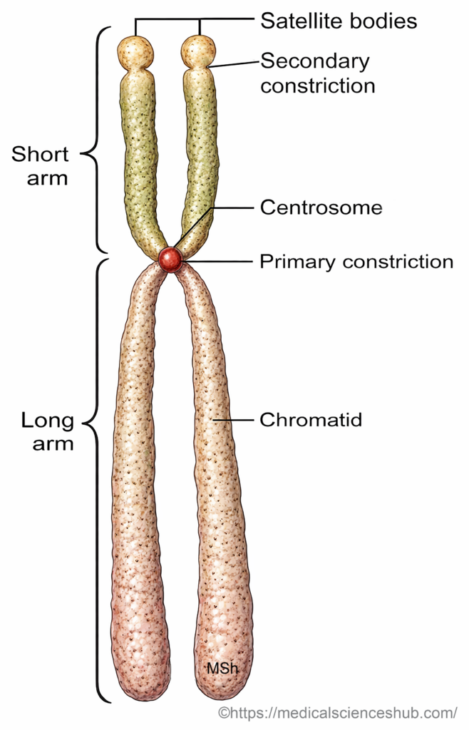
- Each chromosome is composed of two identical sister chromatids that are joined together at a constricted region called the centromere.
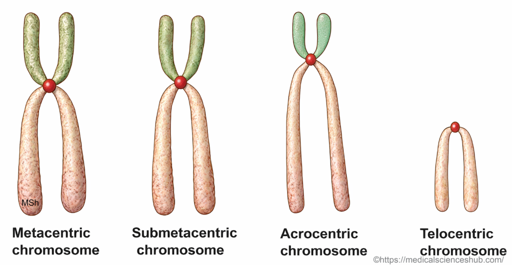
- Chromosomes are classified according to the position of the centromere. They may be metacentric, submetacentric, acrocentric, or telocentric.
- During mitosis, chromosomes become condensed and clearly visible under a microscope.
- During the interphase stage of the cell cycle, chromosomes appear as extended thread-like structures known as chromatin.
- Chromatin consists mainly of deoxyribonucleic acid, associated proteins such as histones, small amounts of ribonucleic acid, and other regulatory molecules that help maintain chromosome structure and function.
Clinical Physiology
- Karyotyping is a laboratory method used to analyze the number and structural features of chromosomes in human cells. It helps detect chromosomal abnormalities associated with genetic disorders. Peripheral blood lymphocytes are commonly used for this test. Karyotyping aids in diagnosing conditions such as Down syndrome and certain infertility disorders.
Deoxyribonucleic Acid (DNA)
- Deoxyribonucleic acid is the principal genetic material present in bacteria and in the nuclei and mitochondria of eukaryotic cells.
- DNA forms the structural and functional basis of chromosomes, which carry hereditary information.
- In human somatic cells, chromosomes occur in pairs, whereas gametes contain only one set of chromosomes.
- Each chromosome contains a very long DNA molecule that is tightly packed within the nucleus.
- DNA is organized by winding around histone proteins, forming repeating structural units called nucleosomes.
- The complex formed by DNA and histone proteins is known as chromatin.
- During cell division, chemical modifications of histone proteins loosen the chromatin structure, allowing chromosomes to condense and become visible under a microscope.
DNA Content of a Cell
- The total DNA in a human somatic cell contains approximately six billion nucleotide base pairs.
- Different chromosomes contain different amounts of DNA.
- The largest chromosome carries a large proportion of genetic material, whereas the smallest chromosome contains significantly fewer nucleotide pairs.
DNA Nucleotides
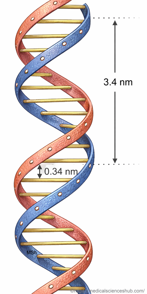
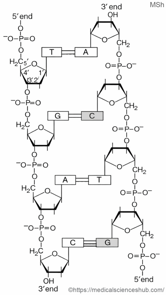
- DNA consists of two long polynucleotide strands arranged in a double helix configuration.
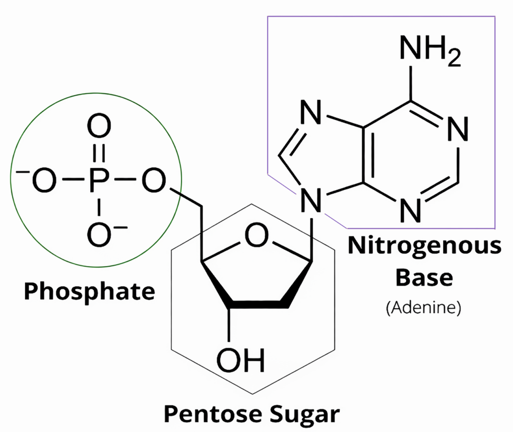
- Each nucleotide is composed of three components: a nitrogenous base, a deoxyribose sugar, and a phosphate group.
- Nitrogenous bases are classified into purines (adenine and guanine) and pyrimidines (cytosine and thymine).
- These bases form specific pairs that encode genetic information required for cellular structure, function, and inheritance.
Double Helix Structure
- Deoxyribonucleic acid consists of two long polynucleotide strands arranged in a double helix configuration.
- Each strand has a backbone formed by alternating deoxyribose sugar and phosphate groups.
- Nitrogenous bases project inward from the backbone and pair with bases on the opposite strand.
- Base pairing occurs through hydrogen bonds between complementary bases.
- Adenine pairs with thymine, while guanine pairs with cytosine, forming complementary base pairs. This specific pairing maintains the stability and accurate replication of the DNA molecule.
Functions of DNA
- DNA carries genetic information that is transmitted from parents to offspring.
- It contains instructions for the synthesis of ribonucleic acid and cellular proteins, including enzymes.
- DNA regulates important cellular processes such as cell growth and cell division.
Genome
- Deoxyribonucleic acid is the primary component of chromosomes and stores the genetic instructions required for cellular structure and function.
- The complete set of genetic material present in a cell is called the genome.
- In humans, the genome contains approximately three billion nucleotide base pairs distributed among 23 pairs of chromosomes in somatic cells.
- Each chromosome pair consists of one chromosome inherited from the mother and the other from the father.
- Among these pairs, 22 pairs are autosomes and one pair forms the sex chromosomes.
Clinical Physiology
- Genetic Message and Protein Synthesis
- Genetic information in deoxyribonucleic acid is encoded by the sequence of nitrogenous bases.
- These bases include purines (adenine and guanine) and pyrimidines (cytosine and thymine).
- The order of these bases forms the genetic code that determines the sequence of amino acids in proteins.
- During gene expression, genetic information is copied into ribonucleic acid through transcription.
- Messenger ribonucleic acid transports this information from the nucleus to the ribosome, where protein synthesis occurs.
- The proteins produced include structural proteins and enzymes that regulate cellular metabolism and physiological processes.
- Genes and Protein Formation
- A gene is a specific segment of deoxyribonucleic acid that contains information required for the synthesis of a protein or functional ribonucleic acid.
- A single gene may produce different protein products through processes such as alternative ribonucleic acid processing.
- These proteins may perform different physiological functions within the body.
- Nuclear Sexing
- Nuclear sexing is a laboratory method used to determine genetic sex by identifying sex chromatin in somatic cell nuclei.
- In females, one X chromosome becomes inactive and forms a condensed structure known as the Barr body.
- Barr bodies can be detected in stained epithelial cells obtained from the oral cavity or in neutrophils of peripheral blood.
- The presence of sex chromatin in a significant proportion of cells indicates a genetically female individual.
- Chromosomal Disorders
- Chromosomal abnormalities may involve either autosomes or sex chromosomes and often lead to congenital disorders.
- Autosomal abnormality
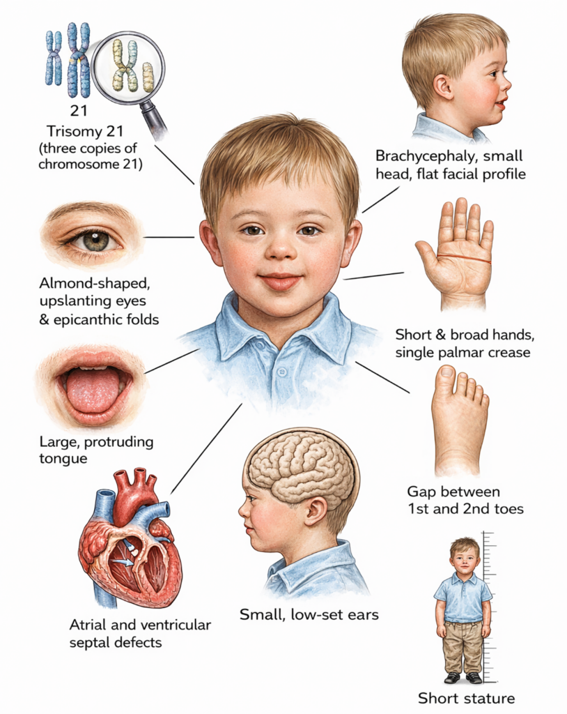
- Trisomy 21 occurs when three copies of chromosome 21 are present instead of two.
- This condition results in Down syndrome, which is characterized by intellectual disability, distinctive facial features, and congenital anomalies.
- Sex chromosome abnormalities
- Turner syndrome occurs when one X chromosome is absent, resulting in a single X chromosome.
- Individuals typically present with short stature, ovarian failure, and primary amenorrhea.
- Klinefelter syndrome occurs in males who possess an additional X chromosome.
- Common features include infertility, testicular atrophy, and development of breast tissue.
- X-Linked Disorders
- X-linked disorders result from mutations in genes located on the X chromosome.
- These disorders usually manifest in males because they possess only one X chromosome.
- Common examples include hemophilia, glucose-6-phosphate dehydrogenase deficiency, and nephrogenic diabetes insipidus.
Genes
- A gene is the functional unit of deoxyribonucleic acid that carries information required for the synthesis of a ribonucleic acid molecule or a protein.
- Genes are arranged along chromosomes, and each chromosome contains a large number of genes.
- In multicellular organisms, most cells contain the same genetic material, but they perform different functions.
- Functional differences between tissues arise because only specific genes are selectively expressed in each cell type.
- Regulation of gene activity ensures that proteins required for a particular cell function are produced at the appropriate time.
Gene Expression
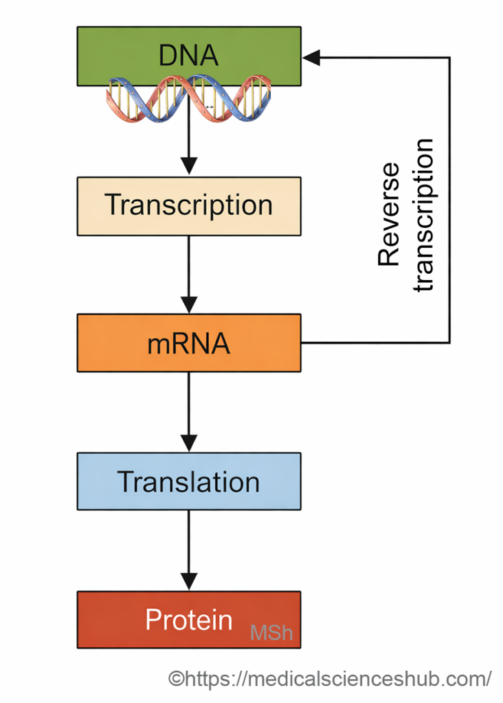
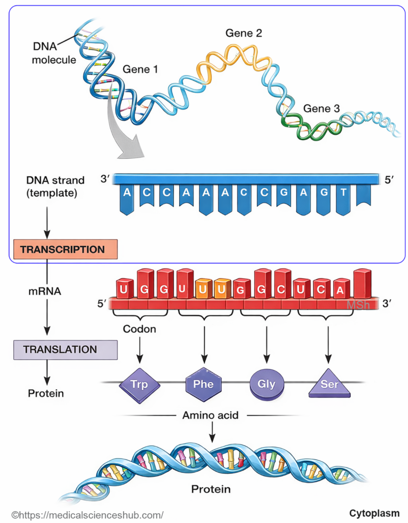
- Gene expression refers to the process through which genetic information is used to synthesize functional proteins. This process occurs in two major stages: transcription and translation.
Transcription
- Transcription is the process in which ribonucleic acid is synthesized using deoxyribonucleic acid as a template.
- During transcription, a specific segment of the deoxyribonucleic acid double helix unwinds to expose the gene sequence.
- One strand of deoxyribonucleic acid serves as the template strand, guiding the formation of messenger ribonucleic acid through complementary base pairing.
- The enzyme ribonucleic acid polymerase binds to a regulatory region known as the promoter to initiate transcription.
Translation
- Translation is the process by which the genetic message carried by messenger ribonucleic acid is converted into a polypeptide chain. This process occurs at ribosomes in the cytoplasm.
- Before translation, messenger ribonucleic acid undergoes processing in which introns are removed and exons are joined to form a mature transcript.
- The final messenger ribonucleic acid sequence directs the assembly of amino acids into a specific protein.
Gene Unit
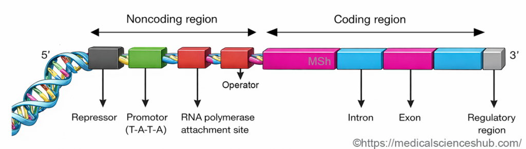
- A gene unit is a functional segment of DNA that directs the synthesis of a specific ribonucleic acid molecule.
- Each gene contains two main components: regulatory sequences and structural sequences.
Regulatory DNA Sequences
- Regulatory DNA sequences are non-coding regions that control the initiation and rate of transcription.
- The promoter is located upstream of the gene and serves as the binding site for RNA polymerase.
- Many promoters contain a TATA sequence, which helps position the enzyme near the transcription start site.
- The operator is a short regulatory segment located close to the promoter region.
- This region must remain free of regulatory proteins for RNA polymerase to proceed along the gene.
- Under certain conditions, a repressor protein binds to the operator and blocks transcription.
- Additional regulatory elements may also occur in upstream or downstream regions and influence gene activity.
Structural Components of Genes
- Exons are nucleotide sequences that remain in the final ribonucleic acid molecule and determine the amino acid sequence of a polypeptide.
- Introns are intervening nucleotide sequences located between exons.
- During transcription, both introns and exons are copied into precursor ribonucleic acid.
- Introns are removed by ribonucleic acid splicing, and adjacent exons are joined to form mature messenger ribonucleic acid ready for translation.
Steps of Protein Synthesis
Protein synthesis occurs in three major steps.
Transcription:
- Transcription is the process by which genetic information in deoxyribonucleic acid is copied into messenger ribonucleic acid. During this process, the two strands of deoxyribonucleic acid separate in the region of the gene.
- Ribonucleic acid polymerase synthesizes a complementary messenger ribonucleic acid strand using one deoxyribonucleic acid strand as a template.
- Each group of three nucleotides on messenger ribonucleic acid forms a codon, which specifies a particular amino acid.
- The messenger ribonucleic acid molecule then leaves the nucleus and attaches to a ribosome in the cytoplasm.
Amino acid activation and translation:
- Transfer ribonucleic acid molecules carry specific amino acids to the ribosome.
- Each transfer ribonucleic acid contains an anticodon that is complementary to the codon on messenger ribonucleic acid.
- Amino acids are activated by the enzyme aminoacyl transfer ribonucleic acid synthetase using energy from adenosine triphosphate.
- Translation proceeds through three stages.
- Initiation begins when the ribosome assembles on messenger ribonucleic acid and the first transfer ribonucleic acid binds.
- Elongation occurs when amino acids are added sequentially to form a growing polypeptide chain.
- Termination occurs when a stop codon signals the release of the completed polypeptide.
Post-translational modification:
- Newly formed polypeptides often undergo chemical modifications to become functional proteins. Common modifications include glycosylation, hydroxylation, and proteolytic cleavage.
Regulation of Gene Expression
- Gene expression can be controlled at several molecular levels.
- Gene amplification increases the number of copies of a gene and enhances protein production.
- Gene rearrangement allows the formation of diverse antigen-specific immunoglobulins.
- Transcription factors regulate the interaction between regulatory proteins and specific deoxyribonucleic acid sequences.
- Regulation may also occur through changes in messenger ribonucleic acid processing, stability, or translation efficiency.
Ribonucleic Acid (RNA)
- Ribonucleic acid (RNA) is a nucleic acid composed of a single chain of ribonucleotides arranged in a polynucleotide strand.
- Unlike deoxyribonucleic acid, RNA is usually single-stranded and therefore shorter and more flexible in structure.
- RNA is present in several cellular locations, including the nucleus, cytoplasm, ribosomes, and to a lesser extent the mitochondria.
- The backbone of RNA contains the sugar ribose, whereas deoxyribonucleic acid contains deoxyribose.
- In RNA, the nitrogenous base uracil replaces thymine, which is present in deoxyribonucleic acid.
Types of RNA
- Messenger RNA (mRNA) is synthesized in the nucleus and carries genetic information from deoxyribonucleic acid to the cytoplasm. It serves as a template for protein synthesis at the ribosomes.
- Transfer RNA (tRNA) transports specific amino acids to the ribosome and aligns them according to the sequence encoded in messenger RNA.
- Ribosomal RNA (rRNA) is produced in the nucleolus and combines with proteins to form ribosomes, which function as the cellular sites of protein synthesis.
Application of Genetics in Medicine
- Advances in molecular genetics have enabled precise analysis, diagnosis, and treatment of many diseases.
- Modern genetic techniques allow scientists to identify specific genes, analyze their structure, and study their function in health and disease.
- These methods are widely applied in medical research, disease diagnosis, therapeutic development, and forensic investigations.
Recombinant Deoxyribonucleic Acid Technology
- Recombinant deoxyribonucleic acid technology is a genetic engineering method used to combine deoxyribonucleic acid from different sources.
- In this process, a selected gene is inserted into a vector, which allows the gene to replicate within a host cell.
- Specialized enzymes such as restriction endonucleases and deoxyribonucleic acid ligase are used to cut and join deoxyribonucleic acid fragments.
Main stages of the procedure
- Gene isolation: A copy of the required gene is obtained from cellular deoxyribonucleic acid or synthesized using complementary deoxyribonucleic acid derived from messenger ribonucleic acid.
- Insertion into vector: The isolated gene is attached to a vector molecule that can replicate inside a host cell. Common vectors include plasmids and bacteriophage deoxyribonucleic acid.
- Introduction into host cell: The recombinant vector is transferred into a suitable host cell such as bacteria, forming recombinant deoxyribonucleic acid.
- Cloning and multiplication: The host cell replicates the recombinant molecule, producing many identical copies of the inserted gene. The amplified gene can then be isolated and analyzed.
Medical and Scientific Uses of Recombinant Technology
- Recombinant methods allow large-scale production of therapeutic proteins and hormones, including insulin, growth hormone, and erythropoietin.
- These techniques support accurate laboratory diagnosis by detecting genetic material associated with infectious or inherited diseases.
- Gene therapy aims to treat genetic disorders by introducing functional genes into affected cells.
- Genetic analysis is used in forensic medicine, particularly in deoxyribonucleic acid profiling for identification.
- Recombinant technology is also applied in agriculture to develop genetically improved crops with better yield and resistance.
Polymerase Chain Reaction
- Polymerase chain reaction is a laboratory method used to rapidly amplify a specific sequence of deoxyribonucleic acid.
- The technique produces millions of copies of a selected deoxyribonucleic acid segment within a short time.
- The process involves repeated cycles of three steps:
- Denaturation: Heating separates the double-stranded deoxyribonucleic acid into single strands.
- Annealing: Short primers bind to complementary sequences on the template strands.
- Extension: Deoxyribonucleic acid polymerase synthesizes new strands using deoxyribonucleotide triphosphates as substrates.
Applications
- Polymerase chain reaction is highly sensitive and can detect extremely small quantities of deoxyribonucleic acid.
- It is widely used in molecular diagnosis of infections, genetic disease testing, and deoxyribonucleic acid fingerprinting.
Blotting Techniques
- Blotting techniques are laboratory methods used to identify specific nucleic acids or proteins within complex biological samples.
- The technique involves separation of molecules by electrophoresis followed by transfer onto a membrane and detection using labeled probes.
Southern blotting
- This method identifies specific deoxyribonucleic acid fragments.
- Deoxyribonucleic acid is extracted from cells, cut with restriction enzymes, and separated by gel electrophoresis.
- The fragments are transferred to a membrane and hybridized with labeled complementary probes.
- It is commonly used for deoxyribonucleic acid fingerprinting and detection of disease-causing gene mutations.
Northern blotting
- This technique analyzes ribonucleic acid molecules instead of deoxyribonucleic acid.
- It is mainly used to study gene expression in different tissues or developmental stages.
Western blotting
- This method detects specific proteins in a biological sample.
- Proteins are separated by electrophoresis, transferred to a membrane, and identified using labeled antibodies.
- Western blotting is often used as a confirmatory test in the diagnosis of certain viral infections.
Cloning
- Cloning refers to the production of multiple genetically identical copies of a cell, gene, or organism.
- It is an important technique in modern genetics, biotechnology, and biomedical research.
- Major types include gene cloning, reproductive cloning, embryo cloning, and tissue cloning.
Gene Cloning
- Gene cloning is the process of producing many identical copies of a specific deoxyribonucleic acid fragment. The selected gene is inserted into a cloning vector, such as a bacterial plasmid, which can replicate within a host cell. After insertion, the recombinant molecule is introduced into a host organism where the gene is replicated repeatedly.
- Gene cloning is widely used in genetic engineering, genome sequencing, and certain forms of gene therapy.
H5: Reproductive Cloning
- Reproductive cloning produces an organism that possesses the same nuclear genetic material as the donor organism.
- The technique is based on somatic cell nuclear transfer.
- In this method, the nucleus from a somatic donor cell is transferred into an egg cell whose nucleus has been removed.
- The reconstructed egg is stimulated chemically or electrically to initiate cell division.
- The developing embryo is then implanted into the uterus of a surrogate female, where it develops into a fetus.
- This method may be used to reproduce animals that are difficult to breed naturally.
Embryo Cloning
- Embryo cloning, often termed therapeutic cloning, involves the creation of embryos for scientific and medical research.
- The objective is not the production of a cloned individual but the generation of stem cells for research.
- Stem cells are commonly obtained from the blastocyst stage of embryonic development.
- These cells have the capacity to differentiate into many specialized cell types.
- Stem cell research contributes to the study of human development and potential treatments for degenerative diseases.
Tissue Cloning
- Tissue cloning refers to the growth of cells in controlled laboratory conditions through tissue culture techniques.
- Cells are maintained in a suitable nutrient medium that supports their growth and multiplication.
- Cloned cells are used for studying the effects of hormones, antibiotics, and pharmaceutical agents.
- Tissue culture methods also support biomedical research and regenerative medicine.
Mutation
- Mutation is a permanent alteration in the deoxyribonucleic acid sequence of a gene.
- Mutations may occur spontaneously or may be induced by mutagens, such as ultraviolet radiation, X-rays, and certain chemicals.
- Genetic mutations can alter the structure or function of proteins and may lead to inherited disorders.
- Two major categories include point mutation and frame shift mutation.
Point Mutation
- Point mutation involves the replacement of one nucleotide base pair by another in the deoxyribonucleic acid sequence.
- In a transition, a purine base is replaced by another purine, or a pyrimidine is replaced by another pyrimidine.
- In a transversion, a purine base is replaced by a pyrimidine, or a pyrimidine is replaced by a purine.
Frame-shift Mutations
- Frame shift mutation occurs when nucleotide base pairs are inserted into or deleted from the gene sequence. These changes alter the reading frame of messenger ribonucleic acid during translation. As a result, the synthesized protein contains an abnormal amino acid sequence and may lose its normal function.
Genetic Screening
- Genetic screening refers to the detection of variations or abnormalities in the genetic material of an individual. It is used to identify inherited disorders, detect disease susceptibility, and guide preventive medical care.
- Genetic screening may be applied before birth, in healthy carriers, or in individuals at risk of developing certain disorders.
Prenatal Diagnosis
- Prenatal diagnosis is performed to identify genetic or developmental abnormalities in the fetus before birth.
- Early detection allows appropriate medical management and informed clinical decisions.
- Common diagnostic methods include chorionic villus sampling, amniocentesis, and preimplantation genetic testing.
Diagnosis of Carrier States
- Some individuals carry a disease-causing gene but do not show clinical symptoms.
- Identification of such carriers is important for genetic counseling and prevention of disease transmission.
- Examples include carrier detection for sickle cell disease, cystic fibrosis, and phenylketonuria.
Prognostic Diagnosis
- Certain disorders develop later in life in genetically susceptible individuals.
- Prognostic genetic testing helps identify individuals at risk of developing such diseases and supports preventive strategies.
Genetic Basis of Cancer
- Cancer develops when genetic alterations disrupt the normal regulation of cell growth and division.
- Mutations in deoxyribonucleic acid or structural chromosomal abnormalities can promote uncontrolled cellular proliferation.
- Certain cancers show a hereditary predisposition, including cancers of the colon and breast, retinoblastoma, and some forms of leukemia.
- Environmental agents such as ionizing radiation and mutagenic chemicals can damage genetic material and contribute to cancer development.
Cancer Genes
- Genes involved in cancer development can be broadly classified into oncogenes and tumor suppressor genes.
- Oncogenes promote abnormal cell proliferation, whereas tumor suppressor genes normally restrict cell division and maintain genomic stability.
Oncogenes
- Oncogenes are altered forms of normal cellular genes called proto-oncogenes.
- Proto-oncogenes normally regulate cell growth, differentiation, and survival.
- Genetic alterations can convert proto-oncogenes into oncogenes, leading to excessive cellular proliferation.
- Mechanisms of oncogene activation
- Chromosomal translocation can relocate a gene to a different chromosome, altering its regulation and increasing its activity.
- Missense mutation may change the amino acid sequence of a protein, resulting in abnormal signaling pathways that stimulate cell growth.
- Gene amplification produces multiple copies of a gene, causing overproduction of growth-promoting proteins.
Defective P53 Gene
Normally, stimulation of P53 gene results in formation of P53 protein. P53 protein serves as a transcription factor for many physiological functions that prevent malignancy. These are:
- P53 protein activates genes that promote DNA repair.
- It activates genes that arrest cell division.
- It also stimulates genes that help apoptosis.
About half of malignancies are associated with defects in P53 gene.
Tumor Suppressor Gene
- Tumor suppressor genes normally inhibit cell proliferation and maintain genomic integrity. Loss or inactivation of these genes removes important regulatory controls on the cell cycle.
- Key examples
- The p53 gene produces a protein that regulates cell cycle control, promotes deoxyribonucleic acid repair, and initiates apoptosis when genetic damage is severe.
- Mutations in the p53 gene are present in a large proportion of human cancers.
- The retinoblastoma gene regulates progression of the cell cycle. Mutation or deletion of this gene can result in uncontrolled cell division and development of retinoblastoma.
Additional Genetic Factors
- Mutator genes are defective genes involved in deoxyribonucleic acid repair. Loss of their function increases the accumulation of mutations in the genome.
- Telomerase, an enzyme that maintains the length of telomeres, is often highly active in cancer cells.
- Increased telomerase activity allows malignant cells to continue dividing without undergoing normal cellular aging.
Gene Therapy
- Gene therapy is a medical technique used to treat inherited disorders caused by defective genes.
- The objective is to introduce a functional gene or modify the existing genetic material to restore normal cellular function.
- Three main strategies are used in gene therapy:
- Gene replacement: A defective gene is replaced with a normal functional gene.
- Gene correction: Specific nucleotide errors in deoxyribonucleic acid are repaired.
- Gene augmentation: An additional functional gene is inserted to compensate for a defective gene.
Somatic Cell Therapy
- In humans, gene therapy is generally performed on somatic cells, which are non-reproductive cells.
- Genetic modifications in somatic cells affect only the treated individual and are not inherited.
- The usual procedure involves several steps:
- Cells carrying the defective gene are isolated from the patient and grown in laboratory culture.
- A therapeutic gene construct is introduced into these cells using suitable vectors.
- The genetically modified cells are then returned to the patient.
- This approach has been explored in disorders such as cystic fibrosis and severe combined immunodeficiency.
Apoptosis
- Apoptosis is a genetically regulated process of programmed cell death that eliminates unwanted or damaged cells.
- It is a controlled and energy-dependent process that differs from necrosis, which results from acute cellular injury.
- Apoptosis maintains tissue homeostasis by balancing cell proliferation and cell removal.
- It plays important roles in both physiological development and various disease processes.
Physiological examples
- During brain development, excess neurons are removed to refine neural connections.
- In embryonic development, apoptosis eliminates tissues such as the webbing between developing digits.
- Many circulating cells, including eosinophils, undergo apoptosis after completing their functional lifespan.
Mechanisms of Apoptosis
Stimuli that initiate apoptosis
- Apoptosis is triggered by activation of intracellular enzymes known as caspases, which are cysteine proteases.
- Caspases normally exist in inactive forms and become activated by internal or external signals.
Internal stimuli
- Mitochondria release proteins such as cytochrome c, which activate caspase pathways.
- Mitochondria may also release apoptosis-inducing factors that enter the nucleus and promote deoxyribonucleic acid fragmentation.
External stimuli
- Certain ligands bind to cell surface receptors and initiate apoptotic signaling.
- For example, binding of tumor necrosis factor receptors activates caspase pathways.
- These signals result in chromatin condensation and fragmentation of genetic material.
Molecular Regulation of Apoptosis
Initiation signals
- Apoptosis may begin when cells lose essential survival signals such as growth factors, hormones, or cytokines.
- Activation of membrane receptors involved in cell death signaling can initiate the process.
- Intracellular stress factors, including radiation, hypoxia, heat, or genetic damage, may also trigger apoptosis.
- Certain programmed genetic pathways regulate the initiation of cell death during development.
Regulatory proteins
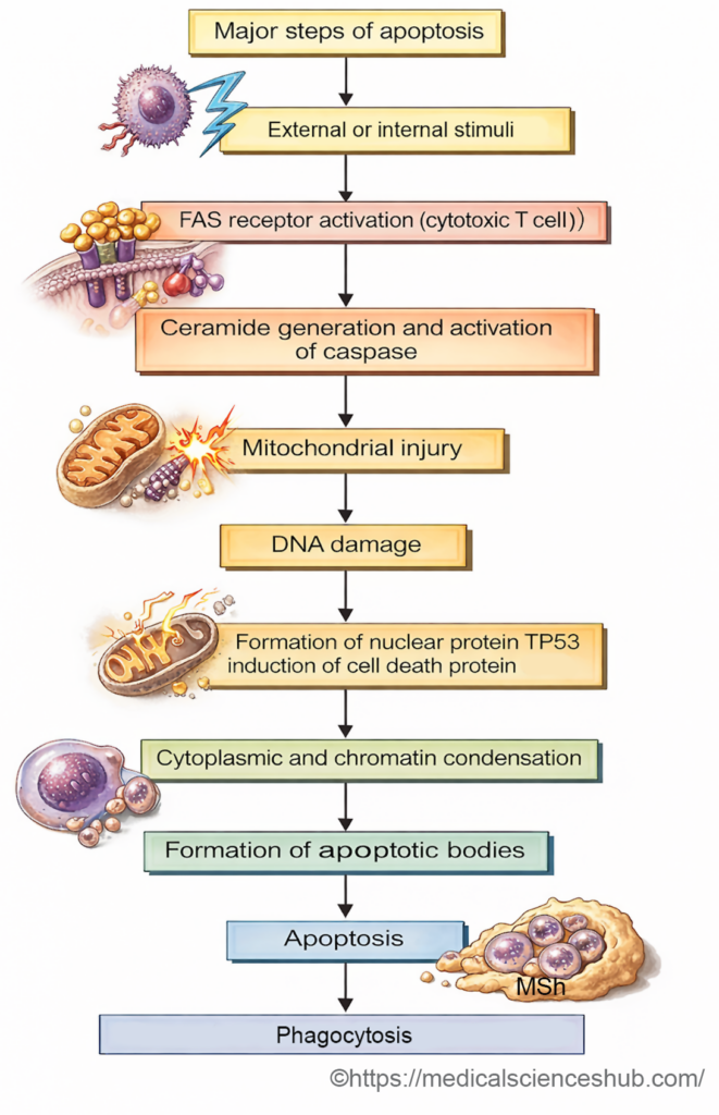
- FAS receptor, the surface receptor present on cytotoxic T cells when comes in contact with the target cell, is activated. This leads to activation of caspases and subsequent proteolysis.
- The BCL-2 family of proteins plays a major role in controlling mitochondrial pathways of apoptosis.
- Some members of this family inhibit apoptosis, whereas others promote it.
- The balance between pro-apoptotic proteins such as BAX and anti-apoptotic proteins such as BCL-2 determines whether the cell survives or undergoes programmed death.
- The p53 protein also contributes by activating genes that promote apoptosis when severe genetic damage occurs.
Major Cellular Events in Apoptosis
- Activation of death receptors on the cell membrane triggers intracellular caspase activation.
- Cellular stress or damage promotes mitochondrial injury and release of pro-apoptotic factors.
- Nuclear damage and activation of regulatory proteins lead to initiation of the apoptotic pathway.
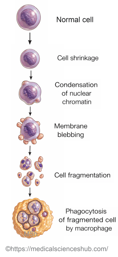
- The affected cell undergoes cell shrinkage and cytoplasmic condensation.
- The nucleus shows chromatin condensation and fragmentation of deoxyribonucleic acid.
- The cell membrane forms membrane-bound fragments known as apoptotic bodies. These fragments are rapidly recognized and removed by phagocytic cells, usually without inflammation.
Physiological and Pathological Roles
Physiological processes
- Apoptosis contributes to hormone-dependent tissue regression, such as endometrial shedding during the menstrual cycle. It occurs during regression of the lactating mammary gland after cessation of breastfeeding. Continuous turnover of intestinal epithelial cells also involves programmed cell death.
- The thymus undergoes progressive involution with age through apoptotic loss of lymphocytes.
Pathological processes
- Apoptosis contributes to the death of tumor cells exposed to chemotherapeutic agents.
- Cytotoxic lymphocytes can induce apoptosis in transplanted tissues during transplant rejection.
- Viral infections may trigger apoptosis of infected cells, as observed in certain forms of viral hepatitis.
- Withdrawal of hormonal or growth stimuli can cause pathological atrophy in affected tissues.
- Cell death may also occur after exposure to radiation, severe hypoxia, or thermal injury.
- Apoptosis is involved in several neurodegenerative disorders, including degenerative diseases of the central nervous system.
Structural and Biochemical Features
Morphological changes
- Apoptosis usually affects individual cells or small clusters of cells. Cells become smaller and rounder, and the cytoplasm appears dense and eosinophilic.
- Nuclear chromatin condenses along the nuclear membrane.
- The plasma membrane forms blebs that give rise to apoptotic bodies containing cellular organelles. These fragments are rapidly engulfed by macrophages, and inflammatory reaction is minimal or absent.
Biochemical changes
- Activation of caspases causes proteolysis of cytoskeletal proteins.
- Nuclear enzymes fragment deoxyribonucleic acid into characteristic segments.
- Specific molecules, including phosphatidylserine, appear on the cell surface and signal phagocytes to remove apoptotic cells.
Clinical Physiology
Significance in Medicine
- Abnormal regulation of apoptosis contributes to several diseases, including cancer, autoimmune disorders, and degenerative diseases.
- Excessive apoptosis may result in tissue loss, whereas insufficient apoptosis may permit survival of abnormal cells.
- Understanding the molecular control of apoptosis has become important for developing targeted therapies, particularly in oncology.
- Research on apoptosis also supports advances in regenerative medicine and strategies for controlling tissue damage.
Important Questions
- Describe the structure and functions of chromosomes.
- Explain the structure and functions of DNA and RNA.
- Define genome and explain its significance in human genetics.
- Describe the process of gene expression and protein synthesis.
- Classify different types of genetic mutations with examples.
- Explain the principle and significance of nuclear sex determination (nuclear sexing).
- Explain the basic principles and applications of recombinant DNA technology.
- Describe the principle and applications of polymerase chain reaction (PCR).
- Explain different blotting techniques and their uses.
- Describe the types of cloning with suitable examples.
- Explain the concept and significance of gene amplification.
- Describe the role of oncogenes in the development of cancer.
- Explain the function of tumor suppressor genes in preventing cancer.
- Define gene therapy and describe its different types and medical applications.
- Define apoptosis and describe the cellular changes associated with it.
📝 Test Your Knowledge – Practice MCQs
Attempt the chapter MCQ quiz and assess your understanding of key concepts.
Click to see: Medical Disclaimer and Copyright Notice