
Competencies
- PY1.5 Describe and discuss transport mechanisms across cell membranes
Introduction
- The cell membrane regulates the movement of fluids, electrolytes, and other molecules between the intracellular and extracellular environments.
- Membrane transport occurs through specialized channels and carrier proteins embedded in the lipid bilayer.
- The selective permeability of the membrane maintains differences in composition between intracellular fluid and extracellular fluid.
- Transport of substances such as drugs, chemical molecules, and hormones can influence cellular activities and physiological functions.
Direct Diffusion Through the Lipid Bilayer
- The membrane consists primarily of a lipid bilayer, which allows lipid-soluble molecules to diffuse directly across it.
- Substances such as gases, fatty acids, alcohols, ketone bodies, aldehydes, and small uncharged molecules cross the membrane easily.
- Water molecules can also move across the membrane by diffusion.
Transport Through Proteins
- Water-soluble substances cannot readily cross the lipid bilayer.
- Molecules such as electrolytes, glucose, and amino acids pass through protein channels or bind to carrier proteins for membrane transport.
Protein Channels In The Membrane
- Protein channels are membrane proteins that form tubular pathways connecting the extracellular and intracellular environments.
- These channels are selectively permeable, allowing only specific substances to cross the membrane.
- The permeability of each channel depends on the size, shape, and electrical charge of the molecule or ion.
- Most channels are formed by several polypeptide subunits that surround a water-filled pore.
Types of Protein Channels
Membrane channels are broadly classified into ion channels and water channels.
Ion Channels
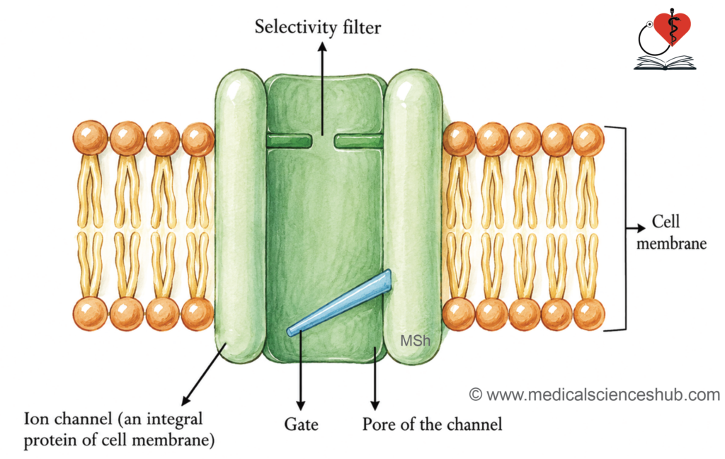
- Ion channels are integral membrane proteins that extend across the entire lipid bilayer. They contain an aqueous pore through which ions move from one side of the membrane to the other. Ions pass through this pore without entering the lipid portion of the membrane.
- Many ion channels contain a gating mechanism that opens or closes in response to specific stimuli such as voltage changes, ligand binding, or mechanical stretch. When the gate opens, ions can move rapidly through the channel.
- A selectivity filter within the pore ensures that only particular ions are allowed to pass.
Examples of Ion Channels
- Sodium channels contain several subunits surrounding a pore approximately 0.5 nanometers in diameter. The inner surface of the pore carries a negative charge, which favors the passage of sodium ions.
- Potassium channels are typically about 0.3 nanometers in diameter and exist in several structural forms.
- Calcium channels may be voltage-gated, ligand-gated, or sensitive to mechanical stretch.
- Chloride channels occur in different structural forms, including channels formed by multiple protein subunits. The cystic fibrosis transmembrane conductance regulator is an important chloride channel.
Water Channels
- Aquaporins are specialized membrane proteins that facilitate rapid movement of water across the cell membrane.
- Multiple aquaporin types are present in different tissues and contribute to water transport in many organs.
Gating of Ion Channels
- Ion channels regulate the movement of ions across the cell membrane through specialized gating mechanisms.
- Some channels remain continuously open and are called non-gated channels.
- Many channels possess gates that open or close in response to specific stimuli.
- The opening or closure of these gates controls the passage of ions through the channel.
- Three principal gating mechanisms are recognized: voltage gating, ligand gating, and mechanical gating.
Voltage-gating
- Voltage-gated channels open or close when the membrane potential changes beyond a critical threshold.
- These channels are common in excitable tissues, such as nerve and muscle cells.
- Sodium, potassium, and calcium channels are typical examples of voltage-gated channels.
- They also occur in cardiac pacemaker tissues and participate in electrical activity of the heart.
- These channels play an essential role in the generation and propagation of action potentials.
Mechanism of Voltage Gating
- Changes in membrane potential cause movement of charged amino acids within the channel protein.
- This movement produces a conformational change that opens or closes the channel gate.
- When the gate opens, ions move rapidly through
Sodium Channel Gating
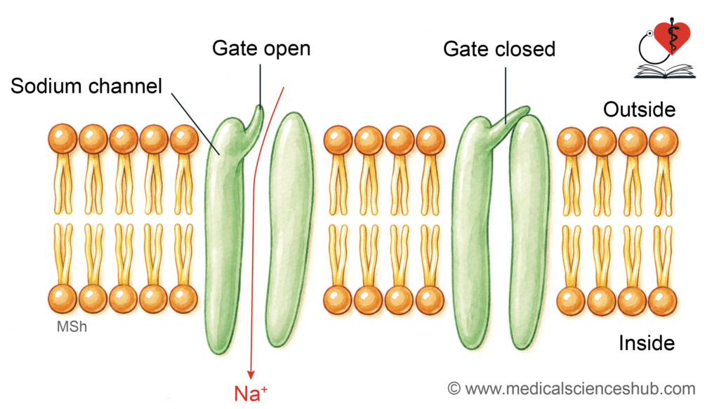
- In many sodium channels, the gate is located near the outer opening of the channel.
- At the resting membrane potential, the gate remains closed.
- When the membrane becomes less negative, the gate opens and sodium ions enter the cell.
- This sodium influx contributes to membrane depolarization and initiation of an action potential.
Potassium Channel Gating
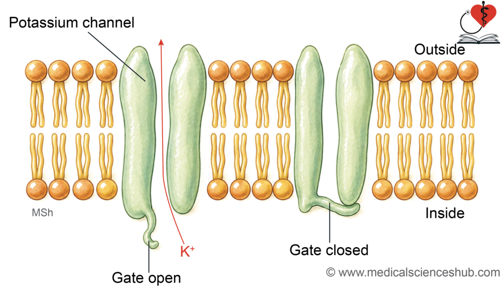
- Potassium channel gates are usually located near the inner cytoplasmic opening.
- At resting membrane potential, these gates remain closed.
- When the membrane becomes more positive, the channels open and potassium ions leave the cell.
- Potassium efflux contributes to repolarization of the membrane following depolarization.
Clinical Physiology
- Sodium channels open rapidly during an action potential, producing a steep depolarization phase.
- Potassium channels open more slowly, so repolarization occurs more gradually.
- Alterations in sodium or potassium channel function can change electrical activity and may contribute to cardiac arrhythmias and neuromuscular disorders.
Ligand Gating
Interaction of the channel with a ligand (hormone or neurotransmitter) causes opening or closure of the channel. Hence, the channels are called ligand-gated channels. As the ligand is a chemical agent, the gating is also called chemical gating.
Mechanism of ligand gating:
- The chemical agent binds with the specific receptor protein on the membrane and brings about conformational change in the protein that, in turn, directly or indirectly opens the channel.
- Nicotinic cholinergic receptor channel in postsynaptic neuromuscular junction is a ligand-gated ion channel. Acetylcholine released at the nerve ending binds with these receptors and opens the ion channels that generate postsynaptic potential.
- Acetylcholine at the parasympathetic endings binds with muscarinic cholinergic receptors on the membrane of GI smooth muscle cells and activates G protein, which, in turn, activates the enzyme that causes production of second messengers such as cyclic AMP or Ca++. The second messengers activate enzymes like kinase that act on the ion channel by phosphorylation.
- Other examples are the action of noradrenaline on β-adrenergic receptors on the smooth muscle cell.
Direct opening of channels: The receptor for the hormone or neurotransmitter may also be the channel itself, so that the action by the ligand is direct on the channel. For example, acetylcholine released at parasympathetic nerve endings acts on the receptor in the postsynaptic membrane in the cardiac pacemaker cells. This causes direct opening of K+ channels in the pacemaker tissue and causes hyperpolarization that decreases the discharge rate of the pacemaker.
Mechanical Gating
- Some ion channels are mechanosensitive and open in response to mechanical forces such as stretch or pressure on the cell membrane.
- Mechanical deformation of the membrane alters the structure of the channel and allows ions to pass through it. These channels play an important role in sensory transduction.
- Examples include channels in hair cells of the cochlea and vestibular system, which are involved in hearing and balance. Stretch-sensitive channels are also present in cardiac ventricular muscle, where they respond to mechanical stretching of the myocardium.
Patch Clamping
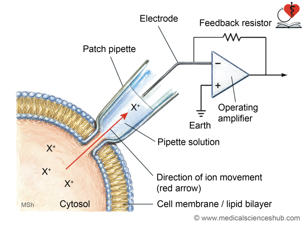
- The patch clamp technique is a highly sensitive method used to study individual membrane ion channels.
- A fine micropipette with a very small tip is placed on the cell membrane and gentle suction is applied. A tiny patch of membrane seals the pipette tip, allowing detailed recording of single-channel ion movement.
Types of Patch Clamping
- Patch clamp recording is used to analyze the electrical activity of individual ion channels in the cell membrane. The membrane patch studied is extremely small and usually contains only one or a few channels.
- Cell-attached configuration: The membrane patch remains attached to the intact cell while recordings are obtained from the channels present in that region.
- Whole-cell configuration: The pipette establishes electrical continuity with the cell interior, allowing measurement of ionic currents across the entire cell membrane.
- Inside-out configuration: A small patch of membrane is detached and oriented so that the inner surface of the membrane faces the external solution, enabling study of intracellular regulatory factors
Uses of Patch-clamp Technique
- The technique is widely used to investigate the function and regulation of membrane ion channels.
- It helps explain how channel abnormalities contribute to diseases such as cystic fibrosis.
- It is also valuable for studying the effects of drugs that block or modify ion channels
Neher and Sakmann Model
- A very fine glass micropipette with a narrow opening is placed against a small region of the cell membrane.
- Gentle suction creates a tight seal between the pipette tip and the membrane patch.
- The isolated patch usually contains only one ion channel, allowing precise measurement of channel activity.
- Ions passing through the channel enter the pipette and generate tiny ionic currents.
- A highly sensitive electrode detects and records these small electrical changes. This method shows that ion channels open and close in a controlled manner to permit selective ion movement.
- It also helps identify structural regions responsible for channel gating and ion selectivity by studying genetic or functional changes in channel proteins.
Types of Carrier Proteins
- Carrier proteins are membrane proteins that bind specific molecules and move them across the cell membrane.
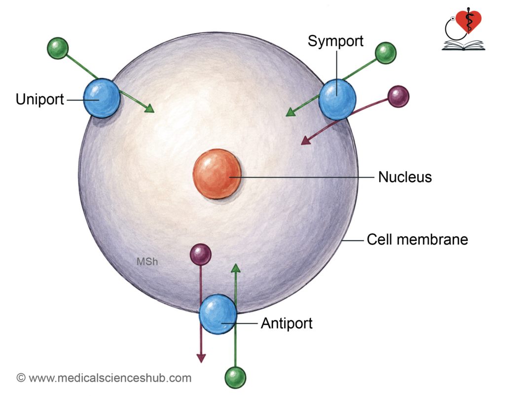
- Based on the direction and number of substances transported, they are classified into uniport, symport, and antiport systems.
Uniport
- Uniport carriers transport only one type of substance across the membrane.
- The molecule moves independently without coupling to the movement of other substances.
- An example is a carrier that transports a single ion or molecule across the membrane.
- For example, Na+ channels transport Na+ and K+ channels transport K+.
Symport
- Symport carriers transport two or more substances together in the same direction across the membrane.
- One substance often moves along its gradient and drives the transport of another molecule.
- A common example is the sodium–glucose co-transporter in intestinal and renal epithelial cells.
Antiport
- Antiport carriers move two substances in opposite directions across the membrane.
- One substance enters the cell while another leaves through the same carrier.
- The sodium–potassium pump exchanges sodium and potassium ions using energy from adenosine triphosphate.
- The sodium–hydrogen exchanger in renal tubular cells removes hydrogen ions while sodium ions enter the cell.
Major Categories of Membrane Transport
- Passive transport: simple diffusion, facilitated diffusion, osmosis, filtration, bulk flow, and solvent drag.
- Active transport: primary active transport and secondary active transport.
- Vesicular transport: exocytosis and endocytosis.
- Epithelial transport: transcellular and paracellular pathways.
Table 6.1: Differences between active and passive transport processes.
| Feature | Active Transport | Passive Transport |
|---|---|---|
| Direction of movement | Substances move against the electrochemical gradient, from a region of lower concentration to a region of higher concentration. | Substances move along the electrochemical gradient, from a region of higher concentration to a region of lower concentration. |
| Energy requirement | Requires metabolic energy, usually obtained from adenosine triphosphate (ATP) hydrolysis. | Does not require cellular energy because movement occurs spontaneously along the gradient. |
| Transport proteins and saturation | Usually involves specific carrier proteins or pumps. Transport shows saturation kinetics because the number of carriers is limited. | May occur through simple diffusion across the lipid bilayer or through channels and carriers. Saturation may occur only when carrier proteins are involved; simple diffusion does not show saturation. |
Characteristics of Carrier-mediated Transport
Carrier-mediated transport depends on specific membrane carrier proteins that bind and move substances across the cell membrane. These systems exhibit two important properties: competitive inhibition and saturation kinetics.
- Competitive inhibition: Molecules with similar chemical structures compete for the same carrier protein. This competition reduces the transport rate of one or more molecules. For example, certain sugars can compete for intestinal transport pathways.
- Saturation kinetics: The number of carrier proteins in the membrane is limited. Therefore, the transport rate reaches a maximum level called the transport maximum. Increasing substrate concentration beyond this level does not further increase the rate of transport.
Transport Processes
Types of Transport Processes
Movement of substances across the cell membrane occurs through specialized transport mechanisms. These mechanisms are broadly classified into passive transport, active transport, and vesicular transport. Transport across epithelial layers also occurs through specialized epithelial pathways.
Special Features
Passive Transport
- Passive transport occurs when substances move down the electrochemical gradient, from higher to lower concentration. This process does not require metabolic energy or adenosine triphosphate.
- Movement occurs spontaneously through the membrane or through channels and carriers.
- Common examples include simple diffusion, facilitated diffusion, osmosis, filtration, bulk flow, and solvent drag.
Active Transport
- Active transport moves substances against the electrochemical gradient. This process requires energy, usually derived from adenosine triphosphate (ATP) hydrolysis.
- Transport systems show saturation kinetics because carrier proteins are limited.
- Active transport mechanisms include primary active transport and secondary active transport
Vesicular Transport
- Vesicular transport moves materials using membrane-bound vesicles. This process is generally independent of concentration gradients and often requires metabolic energy.
- The two major mechanisms are endocytosis and exocytosis.
Simple Diffusion
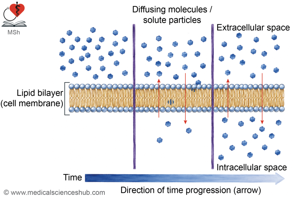
- Simple diffusion is a form of passive transport in which molecules move from a region of higher concentration to a region of lower concentration.
- The movement occurs due to the random kinetic motion of molecules. This process does not require metabolic energy or carrier proteins.
- Diffusion across a membrane occurs when a concentration gradient exists between the two sides of the membrane. Net movement continues until diffusion equilibrium is reached.
- Common substances that diffuse through membranes include oxygen, carbon dioxide, urea, and ammonia.
Factors that Determine Rate of Diffusion
· The rate of diffusion across the cell membrane depends on two major groups of factors.
- These include the properties of the substance, such as molecular size, lipid solubility, and electrical charge.
- The properties of the membrane, including thickness, surface area, and number of channels, also influence diffusion
Properties of the Substance
The movement of molecules across the cell membrane depends mainly on the concentration gradient, electrical gradient, and membrane permeability of the substance.
Effect of Concentration and Electrical Gradients
- The concentration gradient determines the primary direction of diffusion, with molecules moving from higher to lower concentration.
- Charged particles are also influenced by the electrical gradient, which depends on the membrane potential. The interior of most cells is negatively charged compared with the exterior.
- Positively charged ions move more easily toward the negatively charged intracellular environment. The combined influence of chemical and electrical forces is called the electrochemical gradient, which is particularly important for ion movement.
- For gases, diffusion depends on differences in partial pressure, while water movement may depend on hydrostatic pressure.
Thermal energy contributes to random motion:
- Molecules in liquids and gases show continuous random motion due to thermal energy. This random molecular movement forms the physical basis for diffusion in biological fluids.
Unidirectional flux:
- When a semipermeable membrane separates two solutions, molecules move in both directions across the membrane.
- Movement from each side is called unidirectional flux and is proportional to the concentration of the substance.
- Net diffusion represents the difference between the opposing fluxes.
- Diffusion stops when equal movement occurs in both directions and equilibrium is achieved.
Relationship Between Concentration and Diffusion
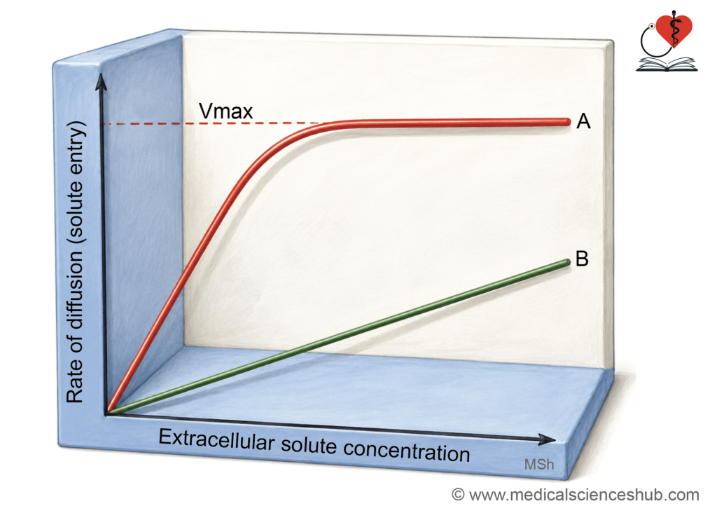
- In simple diffusion, the rate of diffusion increases linearly with the concentration difference across the membrane.
Permeability of the Substance
- Membrane permeability refers to the ability of a substance to pass through the cell membrane.
- The permeability of a molecule depends on several structural and physical factors.
Lipid Solubility
- The membrane consists mainly of a lipid bilayer.
- Substances that are highly lipid-soluble diffuse more easily across the membrane than water-soluble molecules.
Molecular Size
- Smaller molecules generally cross the membrane faster than larger molecules.
- For ions that move through aqueous channels, permeability decreases as molecular size increases.
- Hydrated sodium ions are effectively larger than hydrated potassium ions because they attract more water molecules.
- As a result, potassium permeability at rest is greater than sodium permeability.
Electrical Charge
- The cell membrane normally maintains a negative interior potential.
- Positively charged ions tend to move toward the intracellular space, whereas negatively charged ions are relatively restricted.
Temperature
- Increased temperature enhances molecular motion and therefore increases the rate of diffusion.
Channel Distribution
- The number and type of membrane ion channels strongly influence the permeability of specific substances.
Pressure Gradient
- Differences in hydrostatic pressure can also promote movement of molecules across the membrane from higher to lower pressure regions.
Properties of the Membrane
- The rate of diffusion across a membrane depends on both concentration difference and membrane structure. Diffusion increases when the surface area available for transfer is larger.
- Diffusion decreases as membrane thickness increases, because the distance for movement becomes greater.
- According to Fick’s law, diffusion rate is directly proportional to surface area and concentration gradient, and inversely proportional to thickness.
- The diffusion coefficient (D) reflects how easily a substance moves through the medium.
- In biological membranes, thickness is relatively constant; therefore, permeability is expressed as the permeability coefficient (P).
- Net movement always occurs down the concentration gradient.
- Net rate of diffusion
Facilitated Diffusion
· Facilitated diffusion uses carrier proteins for passive transport. It moves substances down the concentration gradient without energy.
Features of Facilitated Diffusion
It differs from simple diffusion by four special features:
- Faster rate of transport
- Saturation kinetics
- Competitive inhibition
- Specificity.
Faster Rate of Transport
· Facilitated diffusion transports hydrophilic molecules faster than simple diffusion. Carrier proteins enhance movement beyond that predicted by partition properties. Although transport shows saturation, the overall rate remains higher than simple diffusion.
Clinical Physiology
- Partition coefficient: Partition coefficient compares solubility in lipid and water. Lipophilic substances have high values; hydrophilic substances have low values. Hydrophilic molecules diffuse faster via carrier-mediated transport across membranes
Saturation Kinetics
- In simple diffusion, transport rate increases continuously with concentration and shows no saturation.
- In facilitated diffusion, transport depends on the number of carrier proteins.
- When all carrier binding sites are occupied, the system reaches maximum transport capacity. This limit is called the saturation point. Despite saturation, facilitated diffusion usually maintains a higher transport rate than simple diffusion
Competitive Inhibition
- Multiple substances may share the same carrier protein. These substances compete for binding, reducing each other’s transport rate. For example, glucose and galactose compete for the same transporter.
- Sodium ions and calcium ions may also compete for shared transport mechanisms. This process differs from co-transport, where substances are transported together. It also differs from exchange mechanisms, where movement of one substance promotes movement of another in the opposite direction.
Clinical Physiology
- Salt is mixed with glucose in ORS: Sodium enhances glucose absorption through intestinal epithelial cells. This occurs via co-transport mechanisms. Therefore, oral rehydration solution contains sodium and glucose in appropriate proportions.
Specificity
- Carrier proteins selectively transport specific molecules across membranes. Some carriers can transport structurally related substances. For example, glucose shares transport pathways with sodium in certain systems.
Factors Affecting Facilitated Diffusion
- Factors influencing simple diffusion also affect facilitated diffusion. The number of carrier proteins is the primary limiting factor. Hormones can regulate transport by modifying carrier availability. For example, insulin increases glucose uptake via GLUT-4 transporters in muscle and adipose tissue
Table 6.2: Differences between simple diffusion and facilitated diffusion
| Feature | Simple Diffusion | Facilitated Diffusion |
|---|---|---|
| Transport mechanism | Occurs without carrier involvement. | Requires specific carrier proteins or channels. |
| Saturation behavior | Shows no saturation; rate increases linearly with concentration. | Exhibits saturation kinetics due to limited carriers. |
| Competition | No competitive inhibition occurs. | Shows competitive inhibition among similar substrates. |
| Transport rate | Generally slower, especially for polar molecules. | Faster due to carrier-mediated transport. |
| Specificity | Lacks specificity for molecules. | Demonstrates molecular specificity for substrates. |
Osmosis
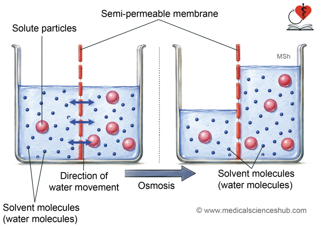
- Osmosis is the movement of solvent across a semipermeable membrane toward higher solute concentration. Solvent molecules move continuously due to thermal motion.
- A semipermeable membrane allows solvent passage but restricts solute movement. Water moves from regions of higher water activity to lower water activity. Solutions with higher solute concentration have lower water activity. Net solvent movement occurs from dilute to concentrated solutions until equilibrium is reached.
Osmotic Effectiveness of a Substance
- Osmotic effectiveness depends on a substance remaining confined to one side of the membrane. Freely diffusible substances, such as urea, are osmotically ineffective. Substances that do not easily cross membranes are osmotically active.
- Glucose shows temporary osmotic effect because it is metabolized. Plasma proteins are highly effective because they remain within the compartment.
- Large molecules, such as dextran, also produce strong osmotic effects.
- Sodium chloride contributes to osmotic balance despite partial membrane permeability.
Clinical Physiology
- Normal saline is effective in hypovolemia: Effective treatment requires fluids with sustained osmotic effectiveness. Solutions should remain within the vascular compartment for longer duration. Glucose solutions are rapidly metabolized and provide limited volume expansion. Normal saline (0.9% sodium chloride) remains in circulation and effectively restores blood volume.
Osmotic Pressure
- Osmotic pressure is the minimum pressure required to stop solvent movement across a semipermeable membrane. Water moves toward higher solute concentration when the solute cannot cross the membrane. This movement continues until balanced by opposing hydrostatic pressure.
- Osmotic pressure depends on the number of dissolved particles, not their chemical nature. Non-dissociating substances produce pressure based on their molecular concentration. Dissociating substances generate greater pressure due to increased particle number. In body fluids, proteins contribute to colloid osmotic pressure, which helps maintain fluid balance.
Clinical Physiology
- Oncotic pressure determines rate of capillary filtration: Oncotic pressure is osmotic pressure generated by plasma proteins. It normally measures about 25 millimeters of mercury. It regulates capillary filtration and fluid balance. Reduced protein levels decrease oncotic pressure and may cause edema.
Terms Used in Osmosis
Mole
- A mole is the molecular weight of a substance expressed in grams. It represents a fixed number of molecules of that substance.
Osmole and Milliosmole
- An osmole represents the number of osmotically active particles in solution. A milliosmole is one-thousandth of an osmole. Non-ionizing substances, such as glucose, produce one osmole per mole. Ionizing substances, such as sodium chloride, produce multiple osmoles due to dissociation.
- For example, sodium chloride forms two particles, while calcium chloride forms three particles
Osmolality and Osmolarity
- Osmolality is the number of osmoles per kilogram of solvent. Osmolarity is the number of osmoles per liter of solution. Osmolality is independent of temperature and solute volume.
- Osmolarity varies with temperature and solution volume. Both values are usually similar in physiological conditions.
Plasma Osmolality
- Plasma osmolality depends on the total concentration of dissolved particles. Sodium chloride contributes the majority of plasma osmolality. Plasma proteins contribute minimally despite their large size. Normal plasma osmolality is approximately 290 milliosmoles per kilogram.
Clinical Physiology
- Osmoles determine osmotic pressure: Osmotic pressure depends on the number of dissolved particles. Particle size, shape, and charge do not significantly influence osmotic pressure.
Measurement of Osmotic Pressure
By Freezing Point Depression
- Osmometers estimate osmotic pressure using colligative properties.
- Dissolved solutes lower the freezing point of a solution in proportion to particle concentration.
- One mole of solute lowers water’s freezing point by about 1.86 degrees Celsius. Normal plasma shows a freezing point near minus 0.54 degrees Celsius, reflecting about 290 milliosmoles per kilogram.
Tonicity
- Tonicity compares solution osmolality with plasma. Isotonic solutions have similar osmolality and do not alter cell volume. Hypotonic solutions cause cell swelling and possible lysis. Hypertonic solutions cause cell shrinkage due to water loss.
Dynamic Changes in Tonicity
- Some solutions change tonicity after administration. Glucose solutions may become hypotonic after metabolism of glucose. Normal saline remains effective for maintaining extracellular volume.
Clinical Physiology
- Hyperosmolal coma: Increased plasma osmolality causes cellular dehydration. Severe hyperosmolality may lead to neurological impairment, including coma. Elevated urea and creatinine can contribute to brain dysfunction in renal failure.
Using Van’t Hoff Equation
- Osmotic pressure can be estimated using the Van’t Hoff relation. It depends on particle concentration, temperature, and a constant.
Measuring Equivalent Hydrostatic Pressure
- Osmotic pressure can also be determined by the hydrostatic pressure needed to oppose water movement across a semipermeable membrane.
Other Transport Phenomena
Filtration, Bulk Flow and Solvent Drag
- Movement of fluid across capillary walls depends on hydrostatic pressure and oncotic pressure. Fluid leaves capillaries when hydrostatic pressure exceeds osmotic forces. Fluid enters capillaries when osmotic forces are greater than hydrostatic pressure.
Filtration
- Filtration is the movement of fluid across a membrane driven by pressure differences.
Bulk Flow
- Bulk flow refers to the movement of large volumes of fluid during filtration.
Solvent Drag
- During bulk flow, water carries dissolved solutes; this is called solvent drag.
Donnan Effect
- Presence of non-diffusible ions causes unequal distribution of permeable ions across membranes. This results in electrochemical imbalance between compartments.
Gibbs-Donnan Effect
- Non-diffusible intracellular proteins create an electrical difference across the membrane. This increases intracellular osmotic particles and promotes water entry. The sodium–potassium pump maintains cell volume and prevents swelling.
Nonionic Diffusion
- Nonionized molecules cross membranes more easily than charged ions. Nonionic diffusion occurs when substances diffuse in their uncharged form. After crossing, molecules may dissociate into ions. This process is important in the kidney and gastrointestinal epithelium.
Active Transport
- Active transport moves substances against their electrochemical gradient. This process requires cellular energy to maintain differences between intracellular and extracellular environments. It preserves cell volume and essential solute concentrations.
Key Characteristics
- Transport occurs in an uphill direction against concentration or electrical gradients. Energy is supplied by adenosine triphosphate, and inhibition of energy production impairs transport. The process shows saturation kinetics due to limited carrier proteins and energy availability.
Types of Active Transport
- Two main types exist: primary active transport and secondary active transport.
Primary Active Transport
- Primary active transport uses energy directly from adenosine triphosphate. Transport proteins, called ion pumps, move substances across membranes. These pumps hydrolyze adenosine triphosphate to release energy for transport. The sodium–potassium pump is a major example that maintains ionic gradients. Other examples include calcium pumps and hydrogen ion pumps.
Classes of Transport Proteins
- P-type transporters function through phosphorylation during transport.
- V-type transporters regulate ion movement in intracellular compartments.
- F-type transporters are involved in energy coupling within mitochondria.
- ATP-binding cassette transporters move various molecules across membranes.
CFTR protein:
- The cystic fibrosis transmembrane conductance regulator is important for chloride transport. The multidrug resistance protein exports drugs from cells and contributes to treatment resistance.
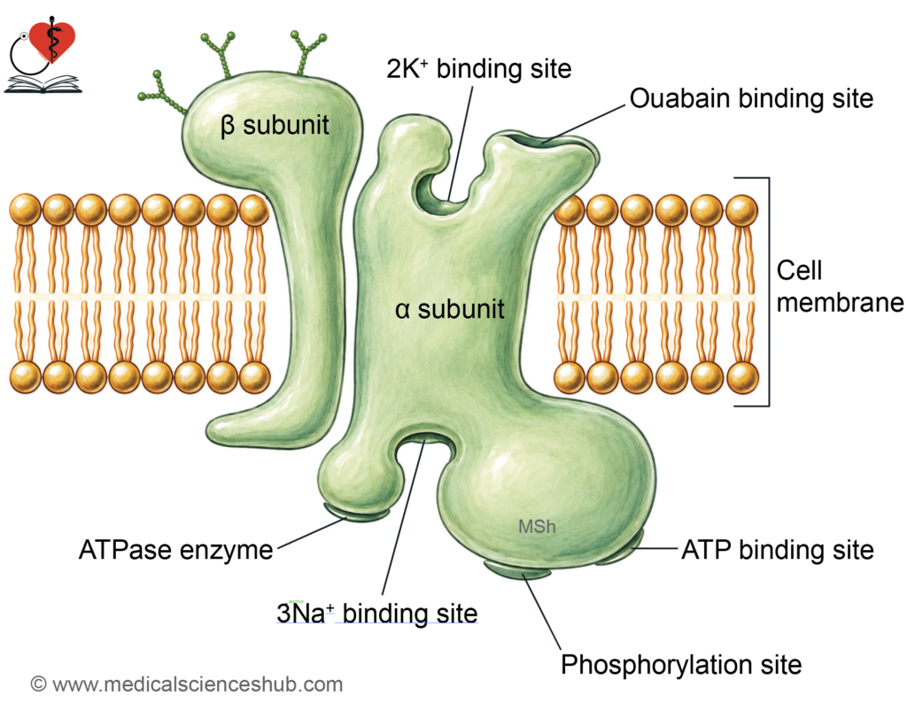
Na+–K+ ATPase
- The sodium–potassium pump is present in all eukaryotic cells. It functions as an antiport system, moving sodium out and potassium into the cell. This process maintains high intracellular potassium and low intracellular sodium levels.
Structure and its Functional Aspects
- The pump is a heterodimer with alpha and beta subunits. The alpha subunit is catalytic and contains binding sites for sodium, potassium, and adenosine triphosphate. The beta subunit supports proper membrane localization and stability.
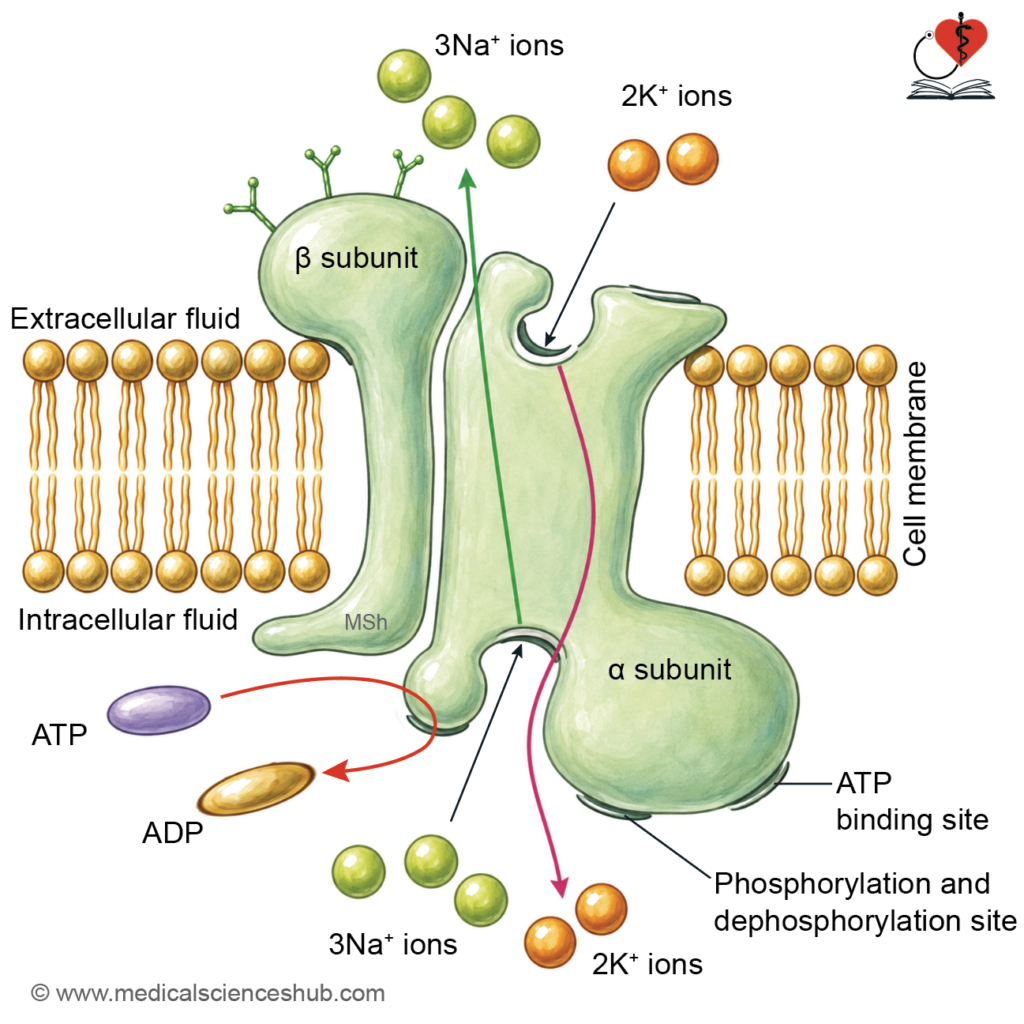
- On the cytoplasmic side, the pump binds three sodium ions and adenosine triphosphate. Hydrolysis of adenosine triphosphate provides energy for transport. Phosphorylation of the protein triggers conformational changes. Sodium ions are released outside, and two potassium ions bind externally. The pump then returns to its original state and releases potassium inside the cell.
Functional Significance
- The pump transports three sodium ions out and two potassium ions into the cell per cycle. It is electrogenic, as it creates a net outward positive charge. This activity is essential for maintaining membrane potential and cell volume.
Table 6.3: Types of ATPases, their location and functions
| ATPase Type | Representative Examples | Cellular Location | Primary Function |
|---|---|---|---|
| P-type ATPases | Sodium–potassium pump, calcium pump, hydrogen–potassium pump | Plasma membrane; sarcoplasmic reticulum | Maintain ion gradients, regulate intracellular calcium, and control acid–base balance |
| V-type ATPases | Hydrogen ion pump | Lysosomal and vesicular membranes | Acidify intracellular compartments for enzymatic activity |
| F-type ATPases | Adenosine triphosphate synthase | Inner mitochondrial membrane | Generate adenosine triphosphate during oxidative phosphorylation |
| ATP-binding cassette transporters | Cystic fibrosis transmembrane conductance regulator, multidrug resistance protein | Plasma membrane | Transport ions and organic molecules; contribute to ion balance and drug efflux |
Clinical Physiology
- Importance of electrogenic pump: The electrogenic pump generates membrane potential essential for nerve and muscle signaling.
Functions
- The sodium–potassium pump is a P-type ATPase that exchanges three sodium ions for two potassium ions.
- It maintains low intracellular sodium and high intracellular potassium concentrations.
- It regulates cell volume by controlling osmotic water movement.
- It prevents cellular swelling and structural damage.
- It supports protein synthesis by maintaining adequate intracellular potassium levels.
- It contributes to the resting membrane potential essential for electrical activity.
- It participates in the action of hormones such as insulin, aldosterone, and thyroid hormones.
Mechanism of Action
- The sodium–potassium pump operates through cyclic phosphorylation and dephosphorylation of its carrier protein.
- Three sodium ions and adenosine triphosphate bind to the intracellular side of the pump.
- Hydrolysis of adenosine triphosphate causes phosphorylation, leading to a conformational change. This change releases sodium ions outside the cell. Two potassium ions then bind to the extracellular surface of the protein.
- Dephosphorylation restores the original conformation of the carrier. Potassium ions are released into the cytoplasm. Each cycle transports three sodium ions out and two potassium ions into the cell using one adenosine triphosphate molecule.
Regulation of Na+-K+ Pump Activity
- Activation: Hormones such as insulin, thyroxine, and aldosterone increase pump activity.
- Inhibition: Substances like digitalis, dopamine, and metabolic inhibitors reduce activity.
- Conditions such as hypoxia and hypothermia also suppress pump function.
Clinical Physiology
- Digitalis inhibits Na+-K+ pump: Digitalis inhibits the sodium–potassium pump by binding to its external surface. This increases intracellular sodium and reduces potassium entry. Reduced sodium gradient decreases calcium efflux via exchange mechanisms. Increased intracellular calcium enhances myocardial contractility.
Other ATPases
Ca++ATPase
- The calcium pump is a P-type ATPase present in cell membrane and intracellular organelles. It actively transports calcium out of the cytoplasm. This maintains very low intracellular calcium concentration.
- In the sarcoplasmic reticulum and endoplasmic reticulum, it stores calcium for later use. Stored calcium is essential for processes such as muscle contraction and cellular signaling.
H+-K+ ATPase
- The hydrogen–potassium pump is located in gastric parietal cells and renal tubular cells.
- In the stomach, it secretes hydrogen ions into the lumen in exchange for potassium ions. This process is essential for gastric acid secretion and digestion.
- In the kidney, it contributes to hydrogen ion secretion and potassium reabsorption. It plays a key role in maintaining acid–base balance.
H+ATPase or Proton ATPase
- The proton pump is present in lysosomes, endoplasmic reticulum, and mitochondria.
- In lysosomes and endoplasmic reticulum, it is a V-type ATPase that acidifies these organelles. Acidic conditions are necessary for enzymatic activity and cellular processing.
- In mitochondria, it functions as an F-type ATPase. It uses a proton gradient to generate adenosine triphosphate during cellular respiration.
ABC Transporters
- ATP-binding cassette transporters are membrane proteins that utilize adenosine triphosphate for transport. They contain multiple transmembrane domains for substrate movement. They export diverse substances, including ions, drugs, peptides, and bile acids.
- Multidrug resistance proteins remove drugs from cells, contributing to resistance.
- The cystic fibrosis transmembrane conductance regulator functions as a chloride channel and belongs to this family.
Clinical Physiology
- ATP-binding cassette transporters expel anticancer drugs from cells. This reduces intracellular drug levels and leads to chemotherapy resistance. Examples include multidrug resistance protein and breast cancer resistance protein.
Secondary Active Transport
- Secondary active transport moves substances against their gradient using energy from another solute gradient.
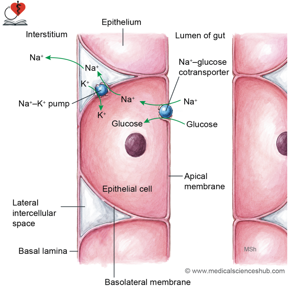
- The sodium gradient is the main driving force in most cells. This gradient is created and maintained by the sodium–potassium pump.
- The transport system does not directly use adenosine triphosphate but depends on primary active transport. Inhibition of the sodium–potassium pump stops this process.
Mechanism
- Low intracellular sodium promotes sodium entry from the lumen into the cell. Carrier proteins couple sodium movement with transport of other solutes. In symport, sodium and substances such as glucose or amino acids move in the same direction. This enables glucose absorption in the intestine and renal tubules.
Properties
- Transport proteins show specificity, saturation kinetics, and competitive inhibition. The transported substance moves against its own electrochemical gradient. The process is indirectly dependent on cellular energy through sodium gradient maintenance.
Clinical Physiology
- The carrier protein may also be an antiport: Antiport transporters move substances in opposite directions across membranes. The sodium–hydrogen exchanger and sodium–calcium exchanger are common examples.
Vesicular Transport
· Vesicular transport moves substances via formation or fusion of membrane-bound vesicles.
- Endocytosis involves vesicle formation to bring materials into the cell.
- Exocytosis involves vesicle fusion to release substances outside the cell. This mechanism transports large molecules that cannot cross membranes by diffusion or carriers. It carries proteins, hormones, neurotransmitters, nutrients, and cellular waste.
Role of Vesicular Transport Proteins
Specific proteins regulate vesicle formation, movement, and fusion.
Clathrin
- Clathrin forms a coated structure that helps vesicle formation. It participates in transport from Golgi apparatus and in endocytic pathways.
Coating Proteins (COP)
- Coating proteins assist vesicle transport between endoplasmic reticulum and Golgi apparatus. They ensure proper sorting and direction of vesicles.
Dynamin
- Dynamin mediates pinching off of vesicles from the plasma membrane. It is essential for clathrin-mediated endocytosis.
Docking Proteins
- Docking proteins enable vesicles to attach to target membranes before fusion. Specific proteins ensure accurate vesicle targeting.
Types of Vesicular Transports
- Vesicular transport includes endocytosis, exocytosis, and transcytosis.
Endocytosis
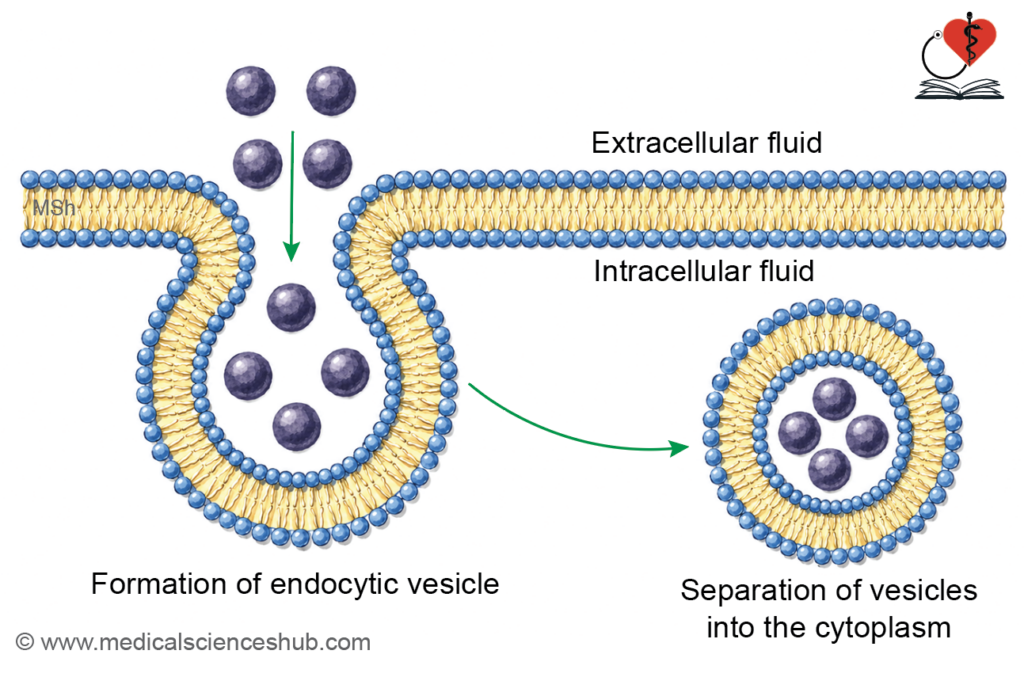
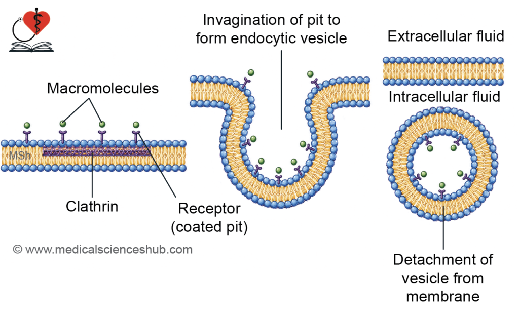
- Endocytosis internalizes substances through membrane invagination. Vesicles enclose fluids, solutes, or particles of varying sizes.
Mechanisms of Endocytosis
- Constitutive endocytosis occurs continuously without external signals. The membrane invaginates, encloses material, and forms a vesicle. The vesicle separates and enters the cytoplasm for further processing.
- Clathrin-mediated endocytosis occurs at specialized membrane regions called coated pits. Macromolecules bind to specific receptors concentrated in these pits.
- Clathrin assembles beneath the membrane and promotes vesicle formation. The membrane invaginates and encloses extracellular material into a vesicle. The vesicle detaches from the membrane with the help of contractile proteins. Clathrin then dissociates and is recycled for further use.
- The vesicle fuses with lysosomes, where enzymes degrade the contents. This process internalizes substances such as lipoproteins, growth factors, and microorganisms. Caveolae, formed by caveolin, represent another pathway for endocytosis.
Types of Endocytosis
- Endocytosis is of three types: phagocytosis, receptor-mediated endocytosis and pinocytosis.
Phagocytosis
- Phagocytosis is the ingestion of large particles or microorganisms by specialized cells. Cells such as macrophages and neutrophils engulf and digest foreign material. The process begins with attachment of particles to the cell membrane. The membrane surrounds the particle to form a vesicle. The vesicle fuses with lysosomes for degradation and destruction of contents.
Receptor-mediated Endocytosis
- Receptor-mediated endocytosis is a selective uptake mechanism using specific receptors. Receptors are concentrated in membrane regions called coated pits. Binding of ligands triggers rapid vesicle formation and internalization. It transports hormones, proteins, toxins, and viruses into cells.
Pinocytosis
- Pinocytosis is the uptake of extracellular fluid and dissolved substances. It occurs continuously in most cells. Vesicles formed contain fluid rather than solid particles.
Exocytosis
- Exocytosis is the process of releasing substances from the cell.
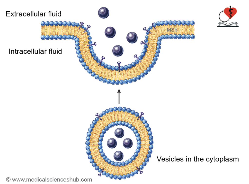
- Materials are synthesized in the endoplasmic reticulum and packaged in the Golgi apparatus. Vesicles move to the plasma membrane and fuse with it. Their contents are released into the extracellular fluid. This process requires adenosine triphosphate and calcium ions.
Mechanism of Exocytosis
- Constitutive exocytosis occurs continuously in most cells for routine secretion.
- Regulated exocytosis occurs in response to specific stimuli. It enables rapid release of hormones, neurotransmitters, and enzymes.
Transcytosis
- Transcytosis involves vesicular transport across a cell. Vesicles carry substances from one side of the cell to the other. It is important for transport across epithelial and endothelial cells.
Transport Across The Epithelia
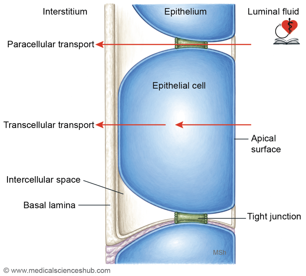
- Epithelium is a cell layer resting on a basement membrane.
- The basolateral membrane contains active transport systems that create ion gradients. These gradients drive movement of solutes across the apical surface.
- Tight junctions permit selective ion passage and restrict large molecules.
Mechanisms of Transport
- Transcellular transport occurs through the cell from apical to basolateral side.
- Paracellular transport occurs between cells through tight junctions.
Examples and Special Mechanism
- Sodium and glucose absorption in intestine and kidney are typical examples.
- Ultrafiltration in renal glomerulus depends mainly on pressure differences across epithelium.
📝 Test Your Knowledge – Practice MCQs
Attempt the chapter MCQ quiz and assess your understanding of key concepts.
Click to see: Medical Disclaimer and Copyright Notice