
Competency
- AN10.12: Describe and demonstrate shoulder joint for type, articular surfaces, capsule, synovial membrane, ligaments, relations, movements, muscles involved, blood supply, nerve supply, and applied anatomy.
Introduction
- The shoulder joint is also called the glenohumeral joint.
- It is formed between the glenoid cavity of the scapula and the head of the humerus.
- It is the most freely movable joint in the human body.
- Because of its wide range of movement, it is the joint most commonly affected by dislocation.
Type
- The shoulder joint is a ball-and-socket synovial joint.
- It is a multiaxial joint, allowing movements in multiple planes.
Articular surfaces
- The shoulder joint has two articular surfaces:
Proximal articular surface (Glenoid cavity of the scapula)
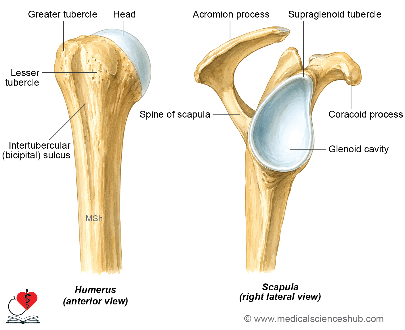
- It is a shallow, pear-shaped depression on the scapula.
- It is covered by articular hyaline cartilage.
Distal articular surface (Head of the humerus)
- It is smooth, rounded, and globular.
- It forms about one-third of a sphere.
- It is covered by articular hyaline cartilage.
- The head of the humerus is much larger than the glenoid cavity.
- This size difference allows a wide range of shoulder movements, but it also makes the joint less stable and more prone to dislocation.
Ligaments
The shoulder joint is strengthened and supported by the following ligaments:
- Capsular ligament
- Glenohumeral ligaments
- Coracohumeral ligament
- Transverse humeral ligament
- Glenoid labrum (fibrocartilaginous rim that deepens the glenoid cavity and improves joint stability)
Capsular ligament
- The capsular ligament is a thin fibrous capsule that surrounds the shoulder joint.
- It extends from the glenoid cavity of the scapula to the humerus.
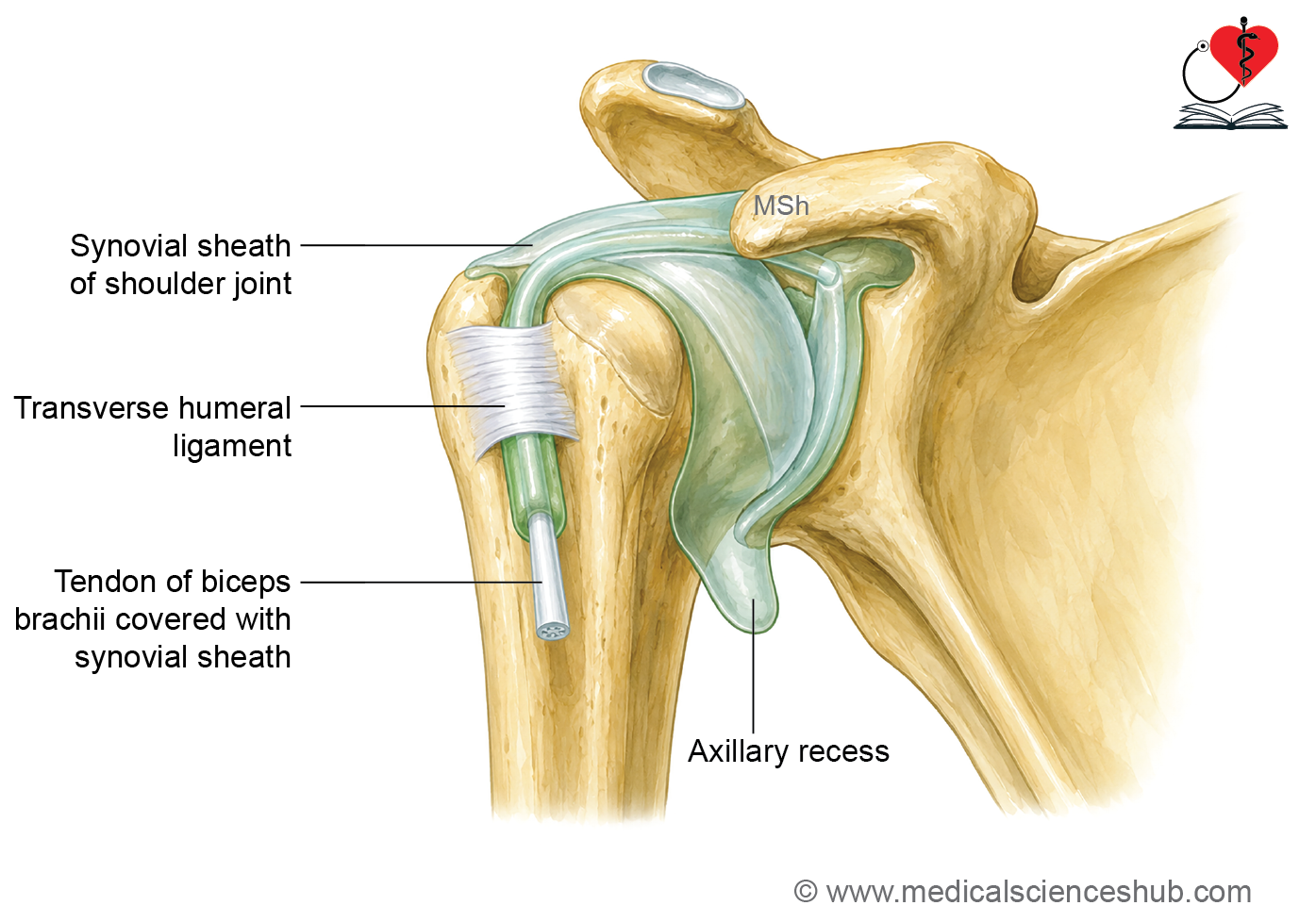
- The capsule is loose (lax), allowing the shoulder a wide range of movements.
Attachments
- Proximal attachment: Attached to the margin of the glenoid cavity, enclosing the glenoid labrum and supraglenoid tubercle.
- Distal attachment: Attached to the anatomical neck of the humerus. Medially, the attachment extends down the shaft to the surgical neck.
Synovial membrane
- The synovial membrane lines the inner surface of the fibrous capsule.
Glenohumeral ligaments
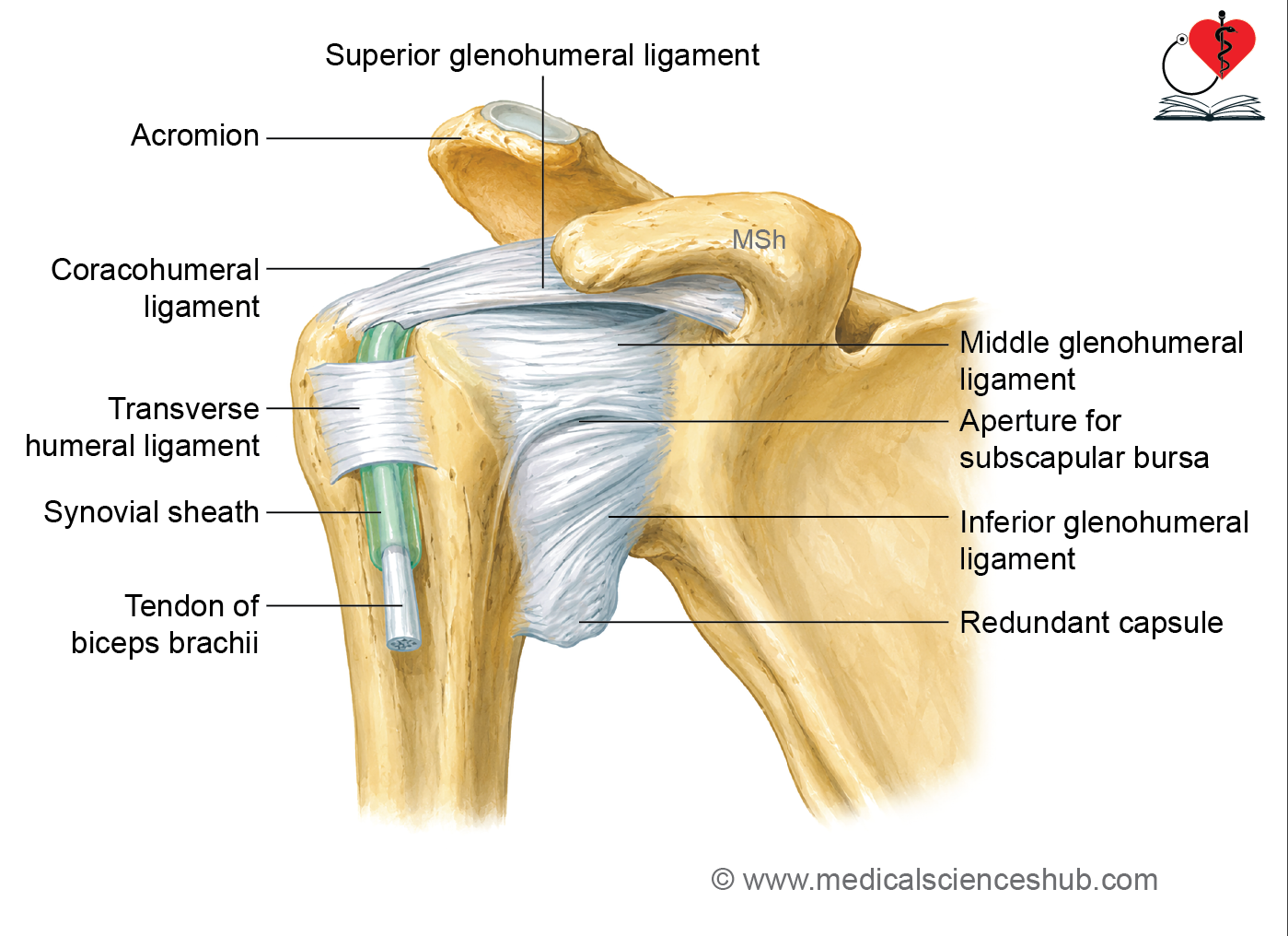
- Three thickenings of the anterior capsule form the:
- Superior glenohumeral ligament
- Middle glenohumeral ligament
- Inferior glenohumeral ligament
- These ligaments help strengthen and stabilize the shoulder joint.
Coracohumeral ligament
- Extends from the root of the coracoid process to the anterior part of the greater tubercle of the humerus.
- It helps support and strengthen the upper part of the joint capsule.
Transverse humeral ligament
- A fibrous band that bridges the bicipital (intertubercular) groove between the greater and lesser tubercles.
- It holds the tendon of the long head of biceps brachii within the groove.
- It contributes to stability of the shoulder joint.
Glenoidal labrum
- The glenoid labrum is a fibrocartilaginous rim attached to the margin of the glenoid cavity.
- It deepens the glenoid cavity, increasing the stability of the shoulder joint while allowing a wide range of movement.
Bursae Related to the Shoulder Joint
The important bursae around the shoulder joint are:
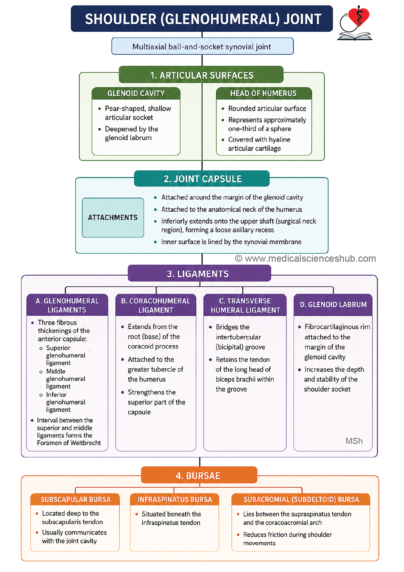
1. Subscapular Bursa
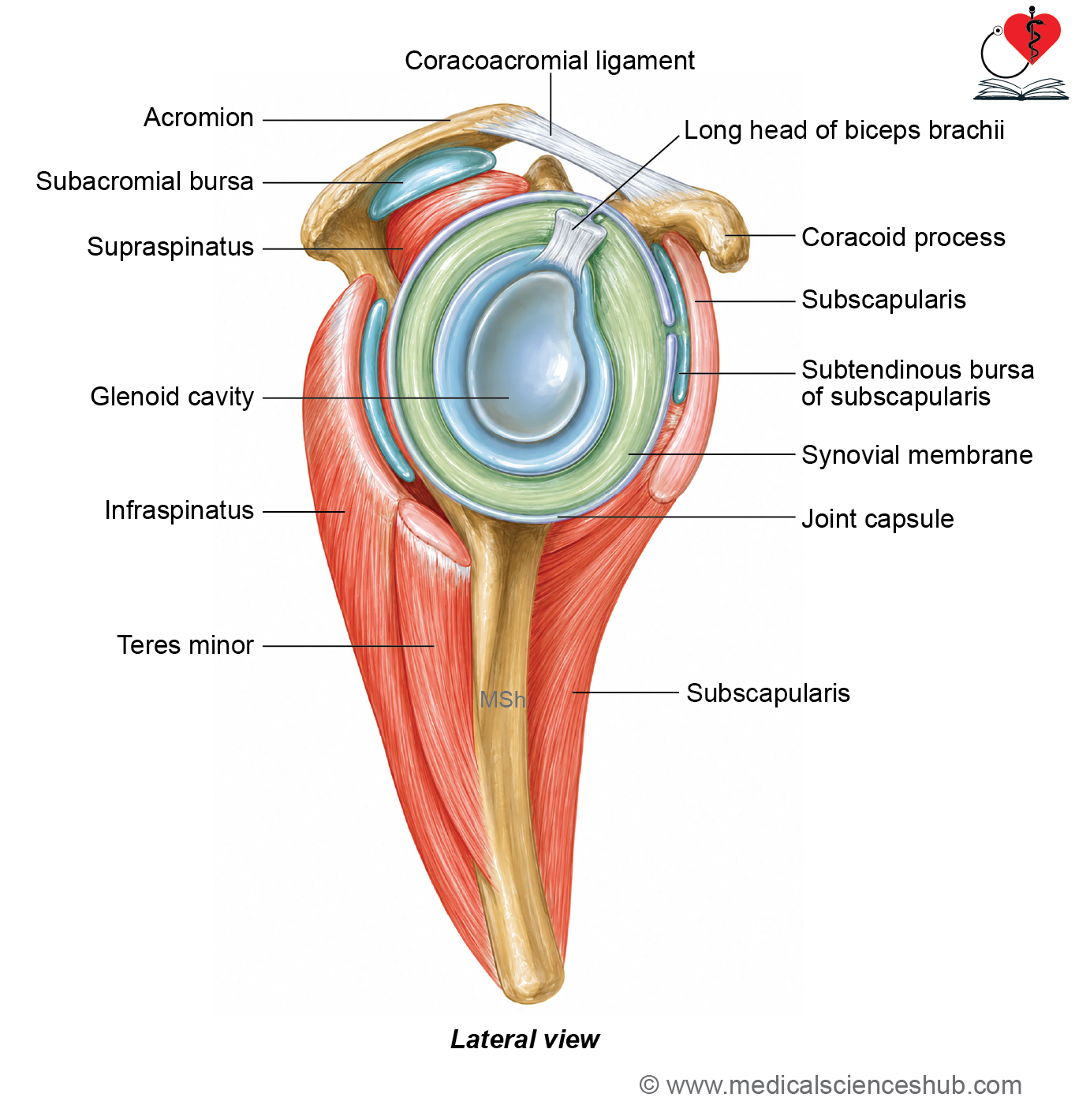
- Lies deep to the subscapularis muscle and anterior to the neck of the scapula.
- Communicates with the shoulder joint cavity.
- Reduces friction during movement of the subscapularis tendon.
2. Infraspinatus Bursa
- Lies between the tendon of the infraspinatus muscle and the joint capsule.
- Communicates with the shoulder joint cavity.
- Reduces friction between the tendon and the joint.
3. Subacromial (Subdeltoid) Bursa
- Lies between the coracoacromial arch and the tendon of the supraspinatus muscle.
- It is the largest bursa in the body.
- Allows the supraspinatus tendon to glide smoothly beneath the coracoacromial arch.
- Usually does not communicate with the shoulder joint cavity.
Functions of the bursae
- Reduce friction between tendons and bones.
- Facilitate smooth movement of the shoulder joint.
Relations
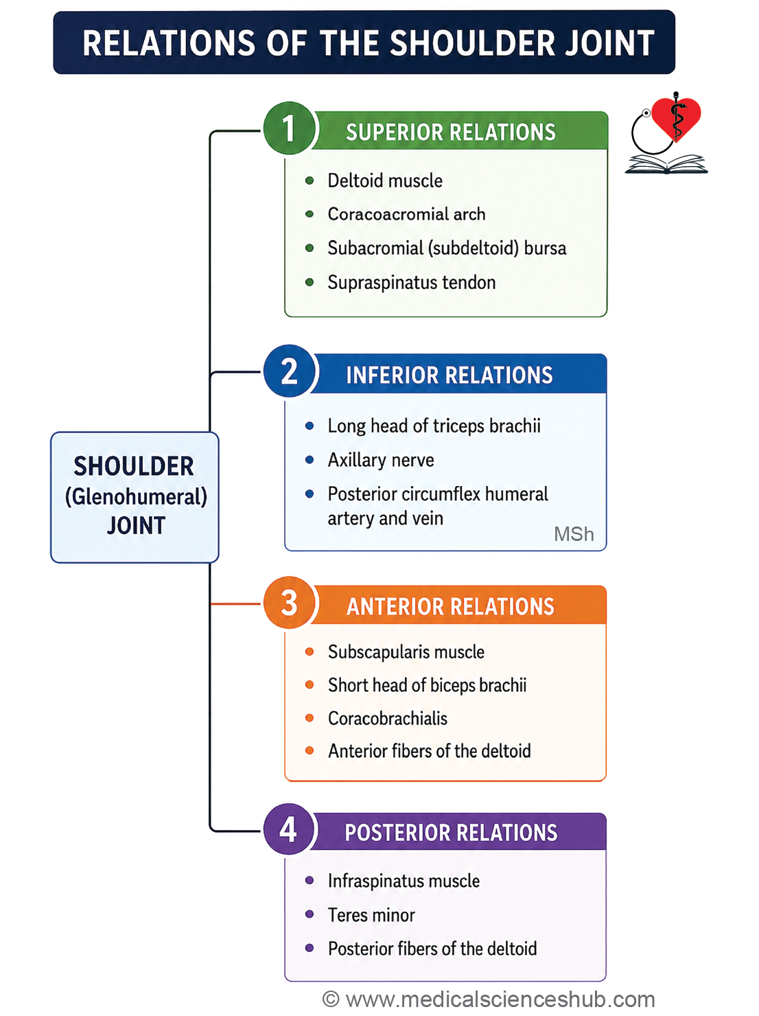
Superior
- Subacromial bursa
- Supraspinatus muscle
- Coracoacromial arch
- Lateral fibers of the deltoid muscle
Inferior
- Long head of triceps brachii (arises from the infraglenoid tubercle)
- Axillary nerve
- Posterior circumflex humeral vessels
Anterior
- Coracobrachialis muscle
- Short head of biceps brachii
- Subscapular bursa
- Subscapularis muscle
- Anterior fibers of the deltoid muscle
Posterior
- Infraspinatus muscle
- Teres minor muscle
- Posterior fibers of the deltoid muscle
Blood and Nerve Supply
Blood supply
The shoulder joint receives blood from the following arteries:
- Anterior circumflex humeral artery
- Posterior circumflex humeral artery
- Subscapular artery
- Suprascapular artery
Nerve supply
The shoulder joint is supplied by articular branches from:
- Axillary nerve
- Musculocutaneous nerve
- Suprascapular nerve
Movements of the Shoulder Joint
The shoulder joint permits a wide range of movements because it is a multiaxial ball-and-socket joint.
Flexion and extension
Flexion
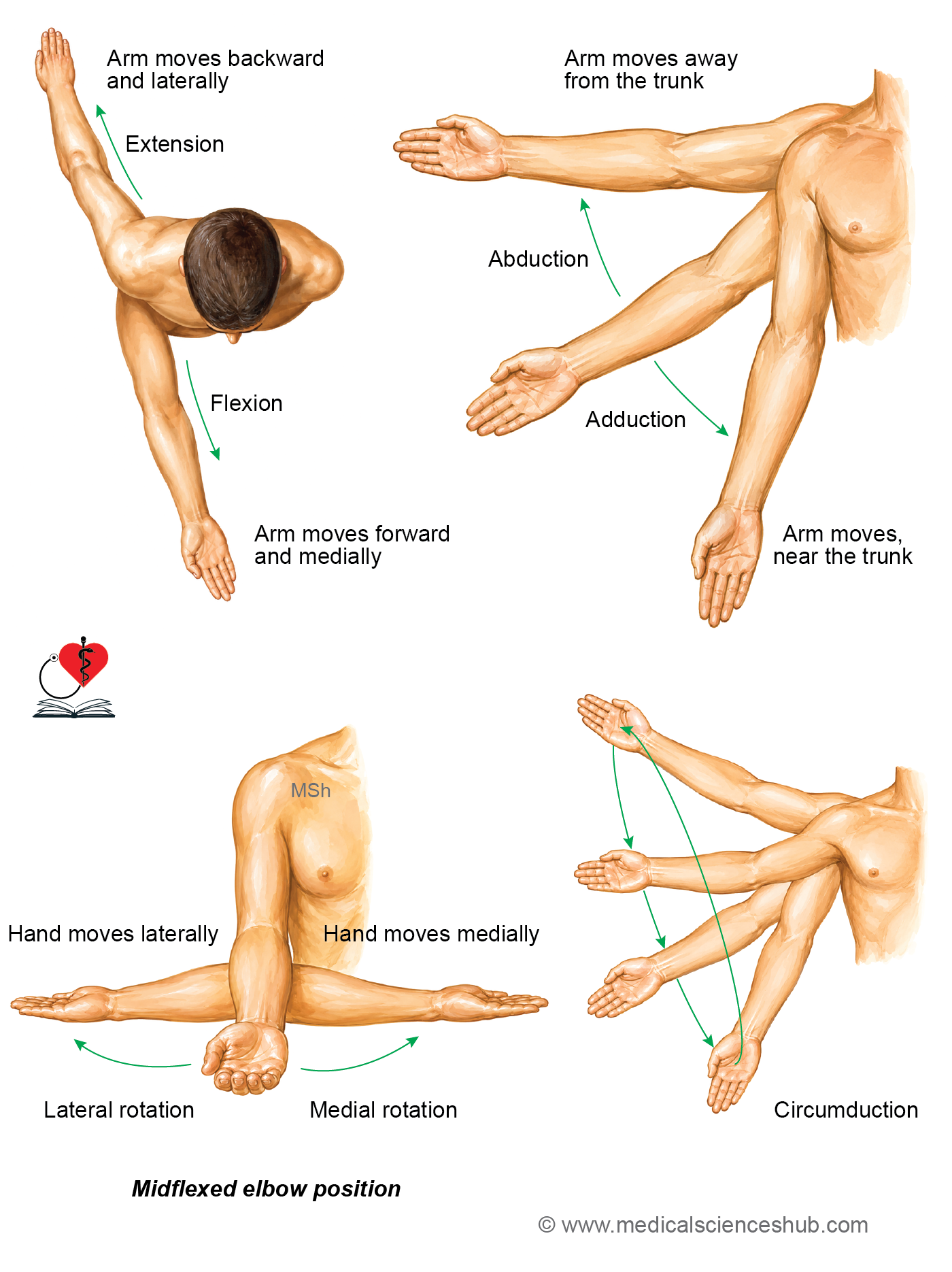
- During flexion, the arm moves forward and towards the midline.
- The movement occurs parallel to the surface of the glenoid cavity.
- It takes place around a transverse axis passing through the head of the humerus.
Main muscles
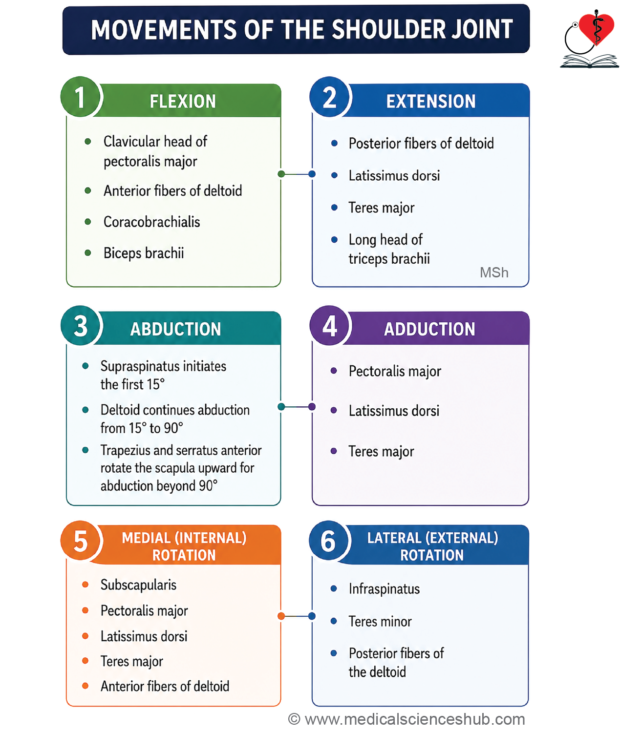
- Clavicular head of pectoralis major
- Anterior fibers of deltoid
Accessory muscles
- Short head of biceps brachii
- Coracobrachialis
- Sternocostal head of pectoralis major
Extension
- During extension, the arm moves backward and away from the midline.
- The movement occurs in the same plane and axis as flexion.
Main muscles
- Posterior fibers of deltoid
- Latissimus dorsi
Accessory muscles
- Teres major
- Long head of triceps brachii
Adduction and abduction
Abduction
- During abduction, the arm moves away from the trunk.
- The movement occurs in a plane midway between the sagittal and coronal planes.
- It takes place around an anteroposterior axis through the head of the humerus.
Muscles
- 0° to 90°: Supraspinatus initiates the movement, and the middle (acromial) fibers of deltoid continue it.
- Above 90°: Serratus anterior and trapezius rotate the scapula upward to allow overhead abduction.
Clinical note: Full overhead abduction requires upward rotation of the scapula.
Adduction
- During adduction, the arm moves towards the trunk.
Main muscles
- Sternocostal part of pectoralis major
- Latissimus dorsi
Accessory muscles
- Teres major
- Coracobrachialis
- Biceps brachii
Medial and lateral rotation
Medial Rotation
- During medial rotation, the anterior surface of the humerus turns towards the midline.
- When the elbow is flexed to 90°, the hand moves medially.
- The movement occurs in the transverse plane around a vertical axis through the humerus.
Main muscle
- Subscapularis
Accessory muscles
- Pectoralis major
- Latissimus dorsi
- Anterior fibers of deltoid
- Teres major
Lateral Rotation
- During lateral rotation, the anterior surface of the humerus turns away from the midline.
- When the elbow is flexed to 90°, the hand moves laterally.
- The movement occurs in the same plane and axis as medial rotation.
Main muscle
- Infraspinatus
Accessory muscles
- Teres minor
- Posterior fibers of deltoid
Circumduction
- Circumduction is a circular (conical) movement of the arm.
- It is produced by the combined sequence of flexion, extension, abduction, and adduction.
Scapulohumeral rhythm
- During abduction of the arm, two movements occur simultaneously:
- Abduction at the glenohumeral (shoulder) joint
- Upward rotation of the scapula
- These two movements work together to produce full overhead abduction.
- For every 2° of movement at the shoulder joint, the scapula rotates upward by 1°.
- This 2:1 ratio between glenohumeral movement and scapular rotation is known as scapulohumeral rhythm.
Factors providing stability to shoulder joint
The stability of the shoulder joint is maintained by the following structures:
- Rotator cuff (musculotendinous cuff): The tendons of the rotator cuff muscles surround the joint. They hold the head of the humerus firmly within the glenoid cavity.
- Coracoacromial arch: It forms a protective arch above the shoulder joint. It acts as a secondary socket and helps prevent upward dislocation of the humeral head.
- Long head of biceps brachii: The tendon of the long head of the biceps helps stabilize the humeral head. It also resists upward displacement of the humerus.
- Glenoid labrum: It is a fibrocartilaginous rim attached to the margin of the glenoid cavity. It deepens the glenoid cavity, improving the stability of the joint.
Joint capsule: The capsule is loose, especially inferiorly, to allow a wide range of movements. As a result, the inferior aspect of the joint is the weakest part, making inferior dislocation of the humeral head the most common type of shoulder dislocation.
CLINICAL INTEGRATION
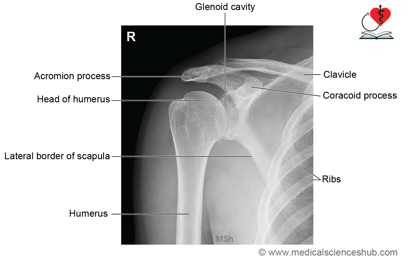
Radiograph of the Shoulder
- A shoulder X-ray (radiograph) is commonly used to assess the bones and joint.
- It helps diagnose fractures, dislocations, arthritis, and other shoulder disorders.
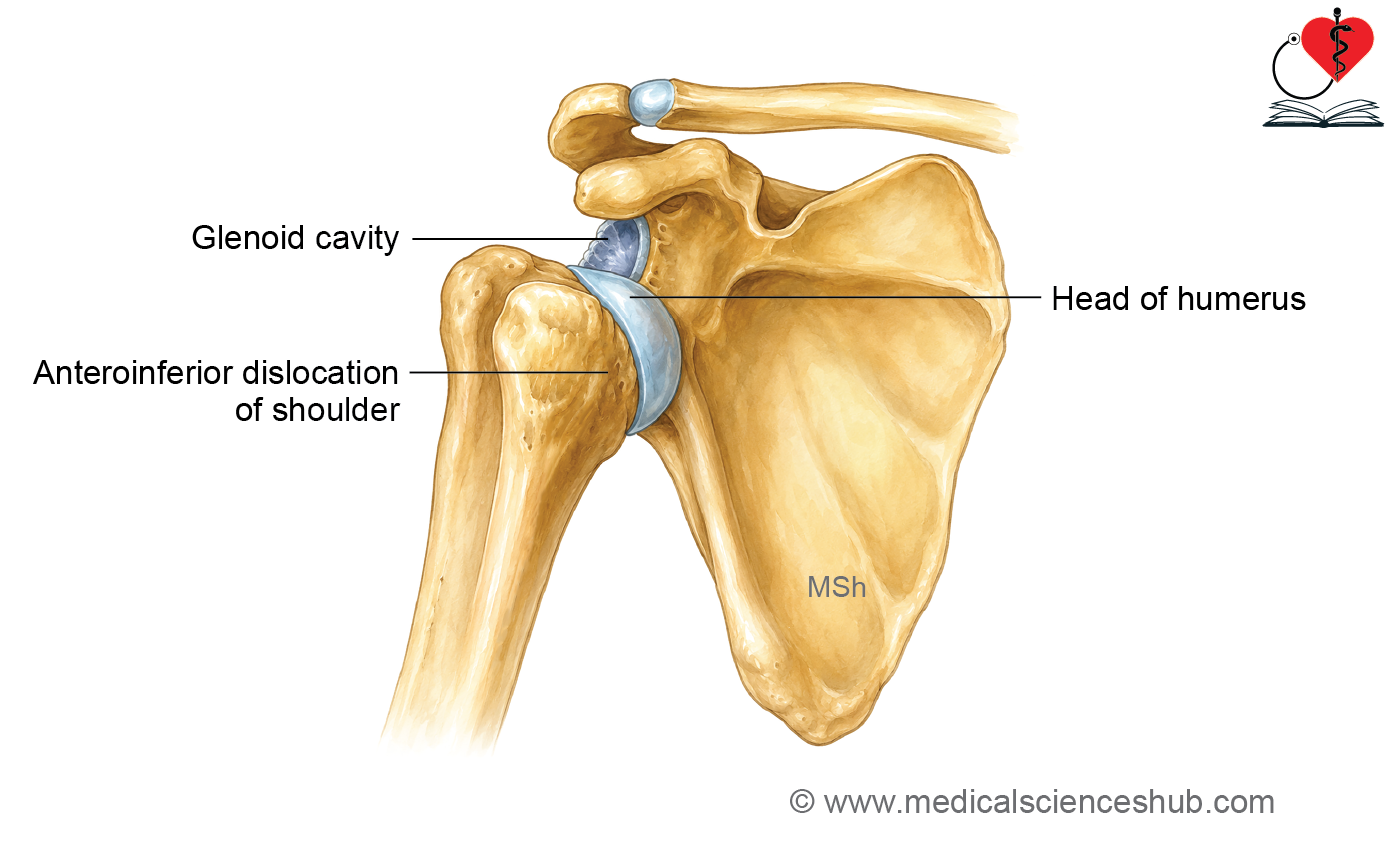
Dislocation of the Shoulder
- The shoulder joint most commonly dislocates inferiorly.
- This occurs because the joint capsule is loose and weakest on the inferior side.
- An inferior dislocation may injure the axillary nerve, leading to weakness of the deltoid muscle and loss of sensation over the upper lateral arm.
Frozen Shoulder (Adhesive Capsulitis)
- Frozen shoulder is caused by inflammation, thickening, and adhesion of the joint capsule.
- It results in pain, stiffness, and marked restriction of shoulder movements.
Calcific Tendinitis of the Shoulder
- Calcific tendinitis is caused by deposition of calcium salts around the shoulder.
- It may occur due to:
- Calcification and inflammation of the subacromial bursa (calcific subacromial bursitis).
- Calcification of the supraspinatus tendon, which may also cause subacromial bursitis.
Rotator Cuff Injury
- Rotator cuff injuries usually result from repetitive overhead activities, overuse, or trauma.
- They cause pain, weakness, and difficulty in lifting or rotating the arm.
Glenoid Labrum Tear
- Tears of the glenoid labrum are common in athletes and individuals with shoulder instability.
- They cause shoulder pain, clicking, and painful or restricted movements.
Sprengel Shoulder
- Sprengel shoulder is a congenital condition in which the scapula is abnormally elevated.
- As a result, the shoulder appears higher than normal and shoulder movements may be limited.
Shoulder Tip Pain
- Pain felt at the tip of the shoulder may not always arise from the shoulder joint.
- Irritation of the diaphragm, peritoneum, or gallbladder can produce referred pain at the shoulder tip.
- This occurs because these structures and the shoulder region share the C3 and C4 spinal nerve segments (via the phrenic nerve).
Important Questions
- Describe a shoulder joint under the following heads: (1) Type,(2) articular surfaces, (3) ligaments, (4) bursa, (5) relations,(6) movements, and (7) applied aspects.
- Draw a well-labeled diagram of relations of shoulder joint.
- List the various movements and responsible muscles at the shoulder joint.
- List the factors providing stability to the shoulder joint.
📝 Test Your Knowledge – Practice MCQs
Attempt the chapter MCQ quiz and assess your understanding of key concepts.
Click to see: Medical Disclaimer and Copyright Notice