
Competencies
- AN11.1: Describe and demonstrate muscle groups of upper arm with emphasis on biceps and triceps brachii.
- AN11.2: Identify and describe origin, course, relations, branches (or tributaries), and termination of important nerves and vessels in arm.
- AN11.4: Describe the anatomical basis of Saturday night paralysis.
- AN13.7: ….. Palpation of brachial artery, ……, Testing of muscles: Biceps brachii, ….. .
- The arm is the part of the upper limb that extends from the shoulder joint to the elbow joint.
- In everyday language, the word “arm” is often used to describe the entire upper limb. However, in anatomy, the term “arm” refers only to the region between the shoulder and the elbow.
- The humerus is the only bone present in the arm.
- The arm is divided into two compartments:
- Anterior (flexor) compartment
- Posterior (extensor) compartment
- The Latin word brachium means arm.
- Many anatomical structures in the arm are named using the term brachial or brachi-, such as the brachial artery and the brachialis muscle
Surface Landmarks
Bony Landmarks
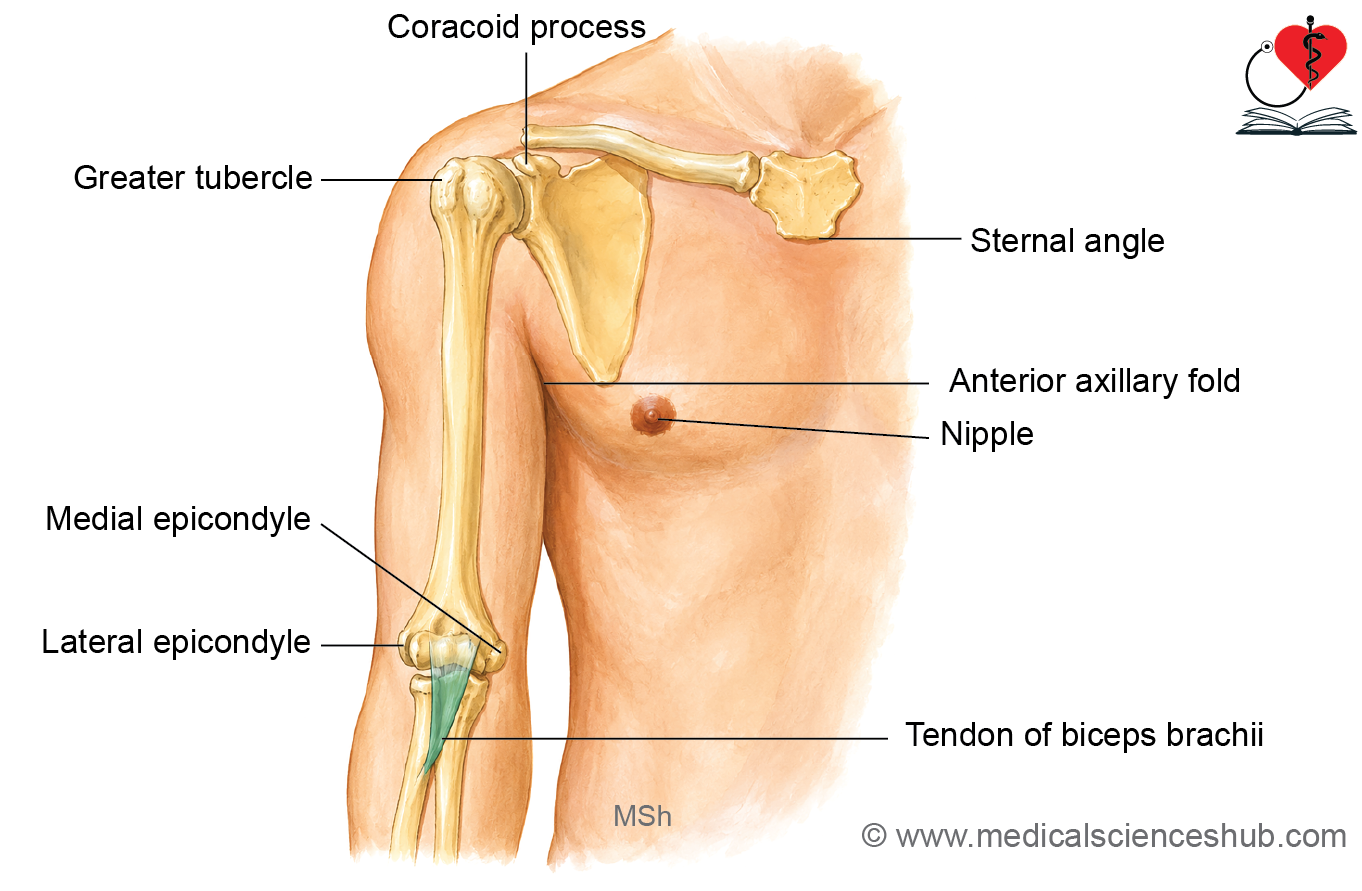
- The greater tubercle of the humerus can be felt deep to the deltoid muscle, just below the acromion.
- The shaft of the humerus can be palpated through the muscles of the arm.
- The medial epicondyle of the humerus is the most prominent bony point on the medial side of the elbow. It is easily seen and felt when the elbow is partially flexed.
- The lateral epicondyle of the humerus can be felt in a depression on the posterolateral side of the elbow when the elbow is fully extended.
- The medial and lateral supracondylar ridges can be felt in the lower part of the arm, just above their respective epicondyles.
- The head of the radius can be palpated just below the lateral epicondyle in a depression on the posterolateral side of the elbow when the elbow is extended.
Soft Tissue Landmarks
- The deltoid muscle forms the rounded contour of the shoulder.
- The belly of the biceps brachii muscle becomes prominent in the middle of the arm when the elbow is flexed.
- The tendon of the biceps brachii can be felt in front of the elbow.
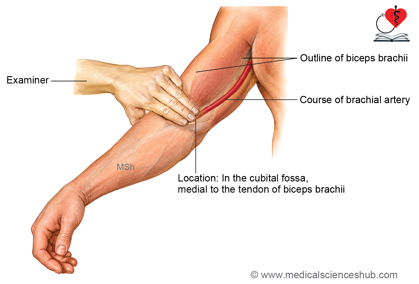
- The brachial artery pulse can be palpated just medial to the tendon of the biceps brachii in the front of the elbow.
- The ulnar nerve passes behind the medial epicondyle of the humerus. It can be palpated by gently rolling it against the medial epicondyle.
The deep fascia of the arm is called the brachial fascia
- The brachial fascia surrounds the muscles of the arm and forms medial and lateral intermuscular septa, which extend inward and attach to the humerus.
- The medial intermuscular septum extends from the brachial fascia to the medial supracondylar ridge and the medial border of the humerus.
- The lateral intermuscular septum extends from the brachial fascia to the lateral supracondylar ridge and the lateral border of the humerus.
- The intermuscular septa divide the arm into two compartments:
- Anterior (flexor) compartment
- Posterior (extensor) compartment
- A transverse intermuscular septum separates the biceps brachii from the brachialis muscle.
- An anteroposterior intermuscular septum separates the muscles arising from the lateral supracondylar ridge from the brachialis muscle.
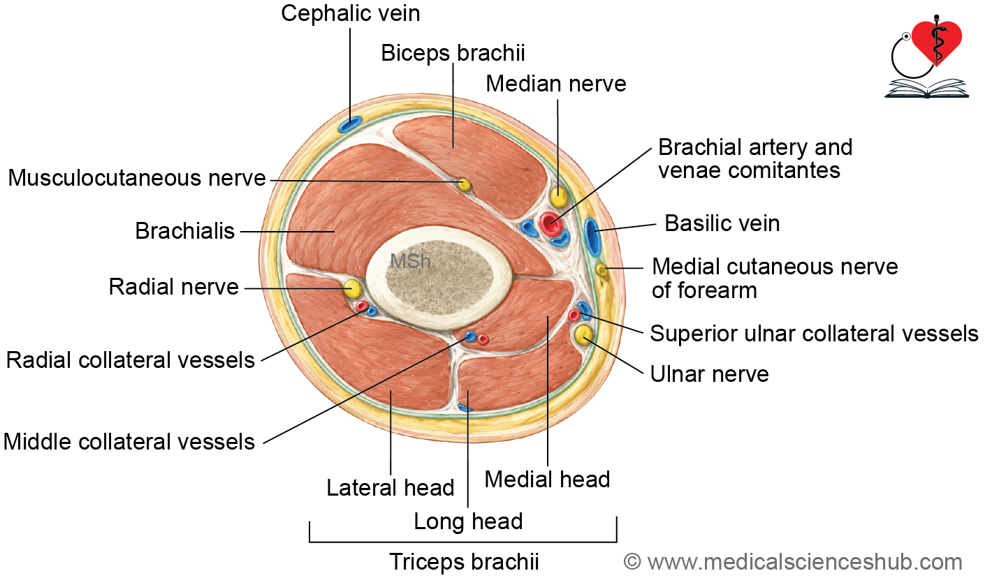
Anterior Compartment of Arm
- he anterior compartment of the arm contains muscles, nerves, and blood vessels.
- The muscles present in this compartment are:
- Coracobrachialis
- Biceps brachii
- Brachialis
- The musculocutaneous nerve is the main nerve of the anterior compartment.
- The median, ulnar, and radial nerves also pass through the anterior compartment but do not supply its muscles.
- The brachial artery is the main artery of the anterior compartment.
Muscles of Anterior Compartment
- The anterior (flexor) compartment contains three muscles:
- Coracobrachialis
- Biceps brachii
- Brachialis
- All three muscles are supplied by the musculocutaneous nerve.
Coracobrachialis
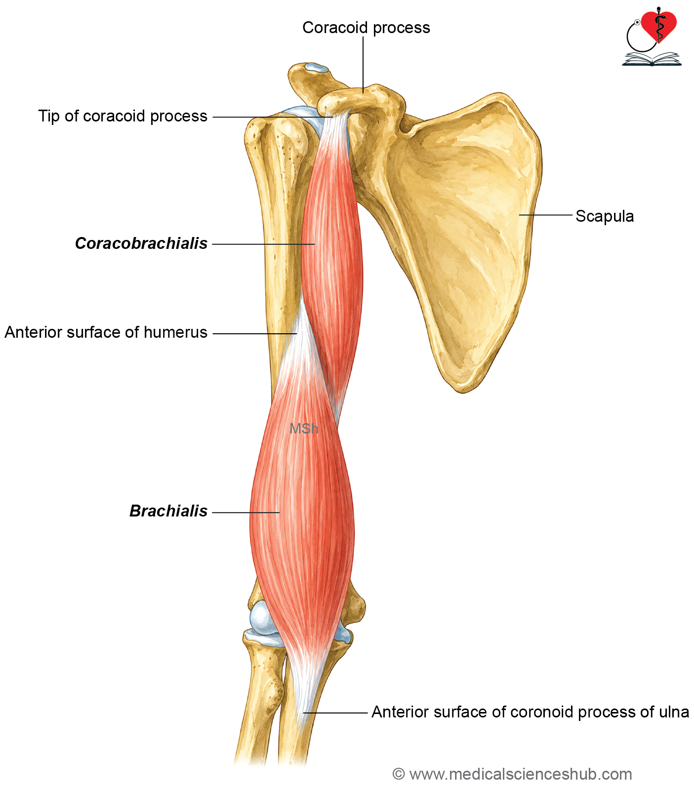
- The coracobrachialis is a small muscle located in the anterior compartment of the arm.
- Morphologically, it represents the medial compartment of the arm.
Origin
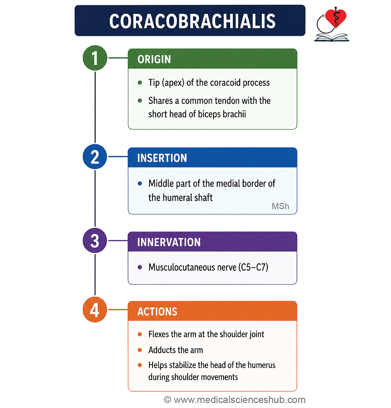
- The coracobrachialis arises from the tip of the coracoid process of the scapula, along with the short head of the biceps brachii.
Insertion
- It is inserted into the middle 5 cm of the medial border of the humerus.
Nerve Supply
- The muscle is supplied by the musculocutaneous nerve.
Actions
- It flexes the arm at the shoulder joint.
- It adducts the arm at the shoulder joint.
CLINICAL ANATOMY
- Morphologically, the coracobrachialis represents the medial compartment of the arm. In contrast, the lower limb has a well-developed medial (adductor) compartment in the thigh.
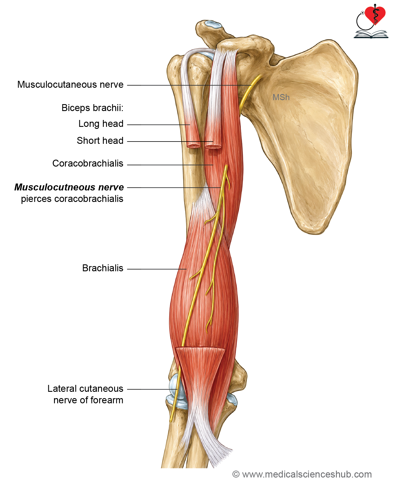
- The musculocutaneous nerve pierces the coracobrachialis muscle. This is an important anatomical feature used in identification.
- Developmentally, the coracobrachialis has two heads, and the musculocutaneous nerve passes between them.
- Because it is pierced by the musculocutaneous nerve, the coracobrachialis is also known as Casser’s perforated muscle.
- The ligament of Struthers is an occasional fibrous band that extends from the supracondylar process (trochlear spine) of the humerus to the medial epicondyle. It is an anatomical variation and may be associated with compression of nearby neurovascular structures.
Biceps Brachii
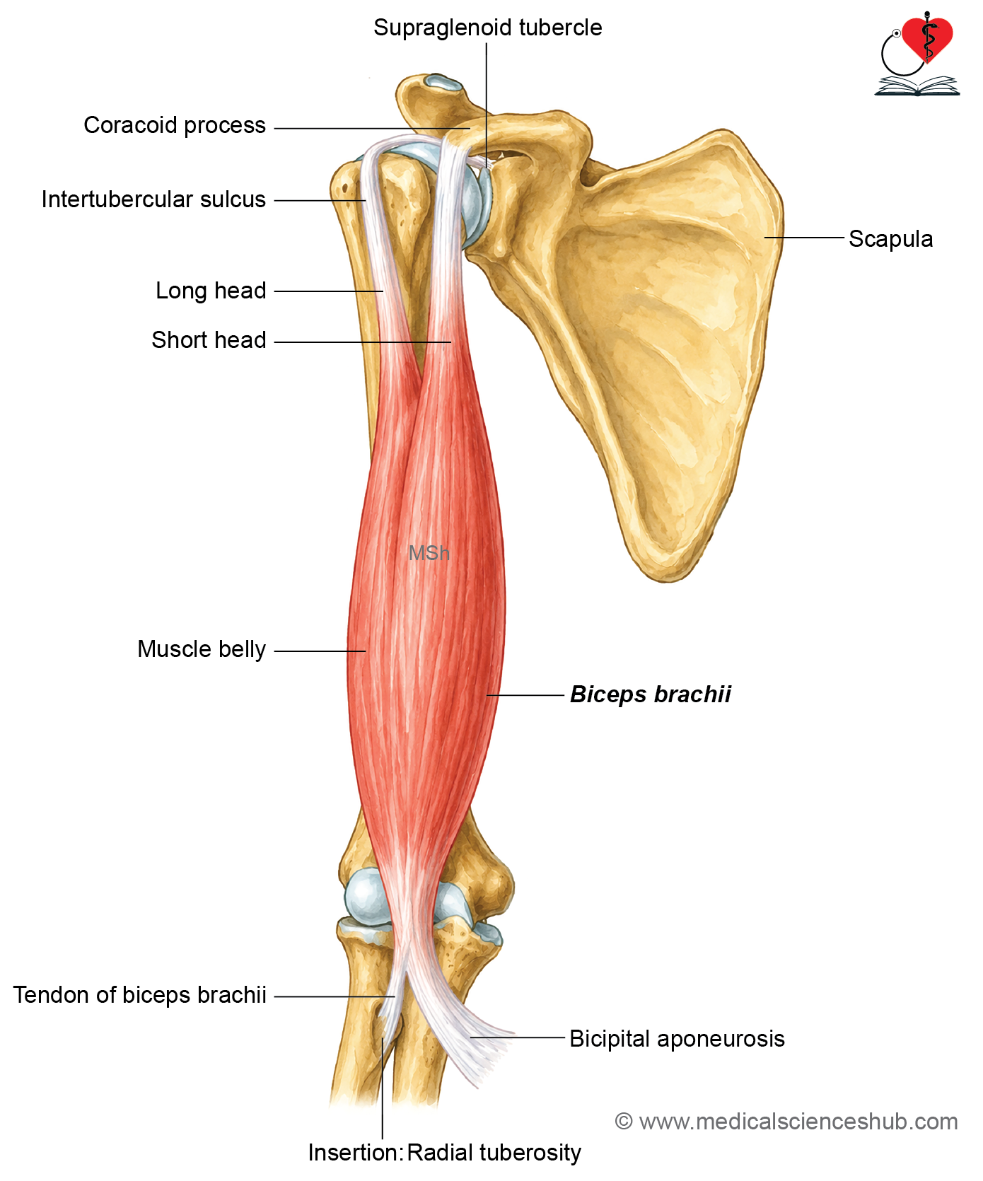
- The biceps brachii is a large muscle located in the anterior compartment of the arm.
- It has two heads: the long head and the short head.
Origin
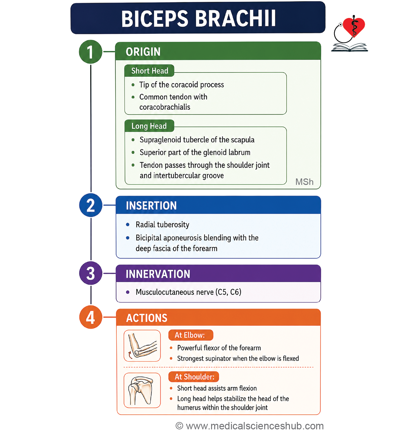
- The long head arises from the supraglenoid tubercle of the scapula.
- The short head arises from the tip of the coracoid process of the scapula.
- The two heads unite in the middle of the arm to form a fusiform muscle belly.
Insertion
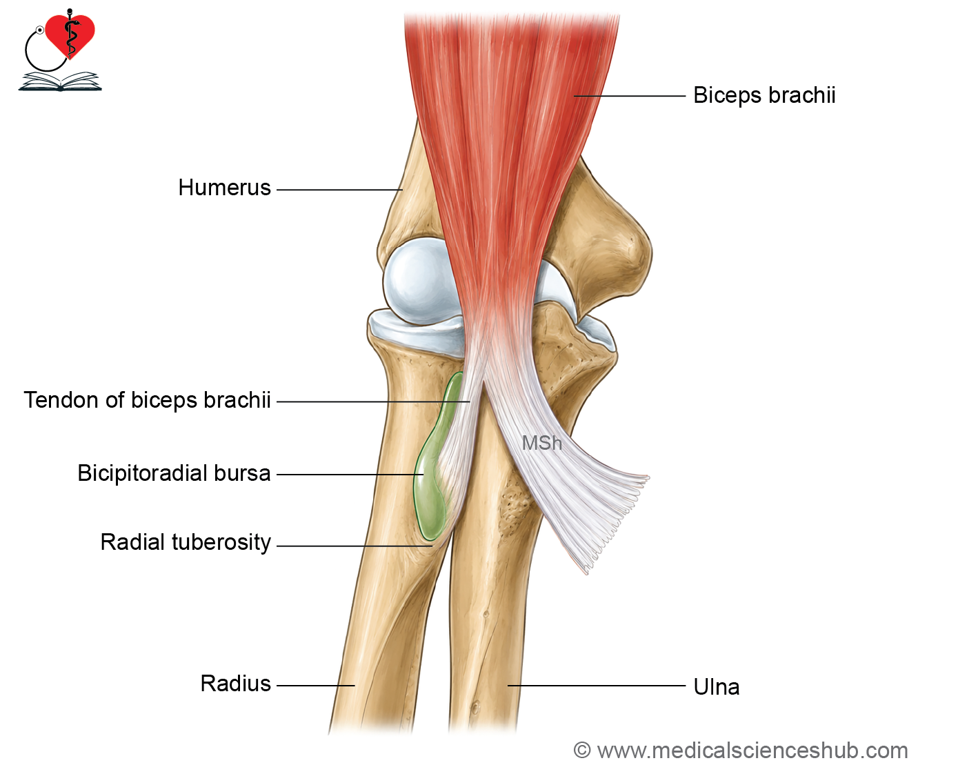
- The tendon of the biceps brachii is inserted into the posterior part of the radial tuberosity.
- A broad fibrous expansion called the bicipital aponeurosis extends medially from the tendon, blends with the deep fascia of the forearm, and is attached to the ulna.
Nerve Supply
- The muscle is supplied by the musculocutaneous nerve.
Actions
- It is a powerful supinator of the forearm, especially when the elbow is flexed.
- It is a strong flexor of the elbow joint.
- It acts as a weak flexor of the shoulder joint.
- The long head helps stabilize the shoulder joint by preventing upward displacement of the head of the humerus.
Clinical Testing
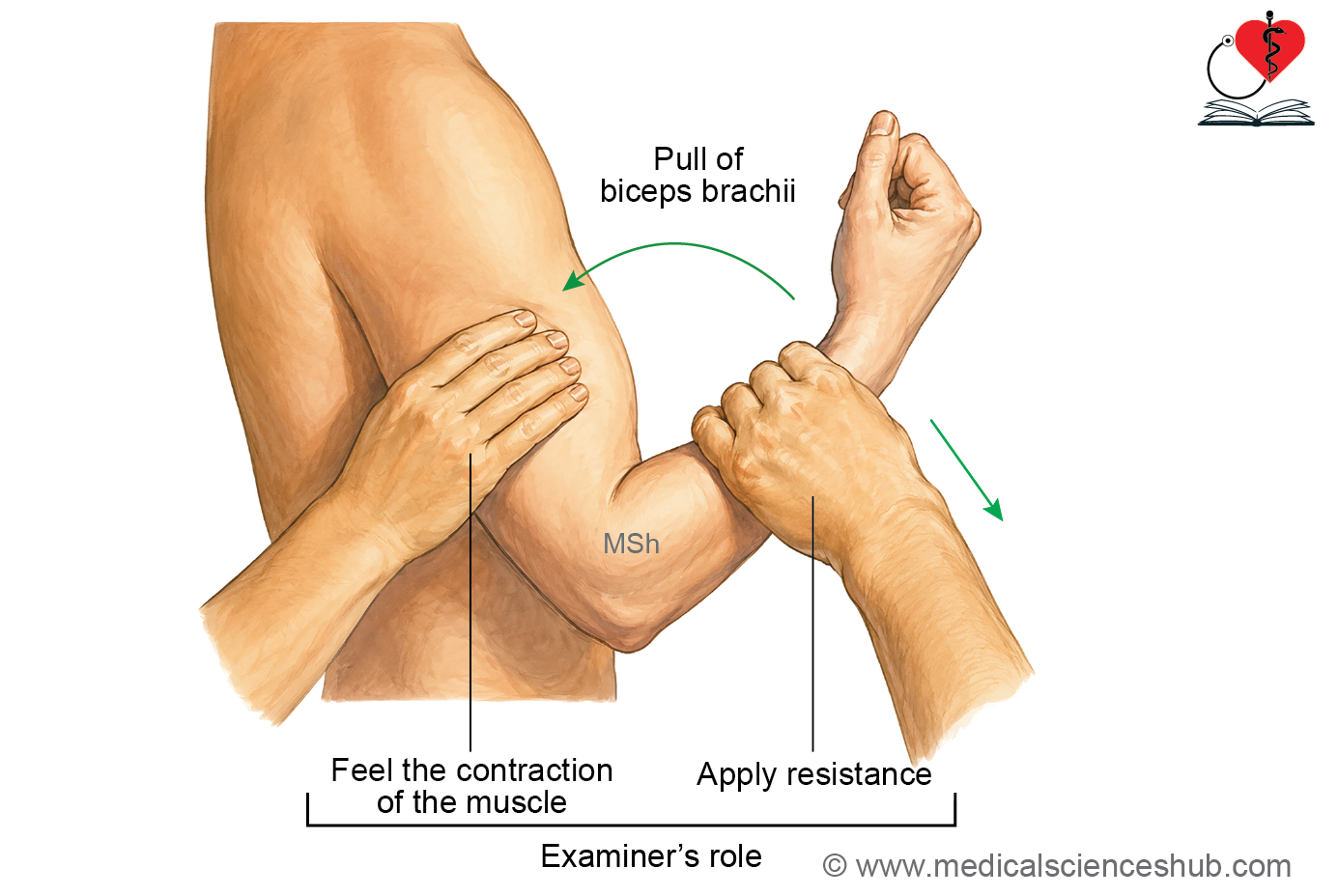
- The function of the biceps brachii is tested by asking the patient to flex the elbow against resistance with the forearm in the supinated position.
- During this movement, the contracted muscle and tendon can be easily palpated.
CLINICAL INTEGRATION
Additional Features of the Biceps Brachii
- The biceps brachii crosses both the shoulder joint and the elbow joint. Therefore, it acts on both joints.
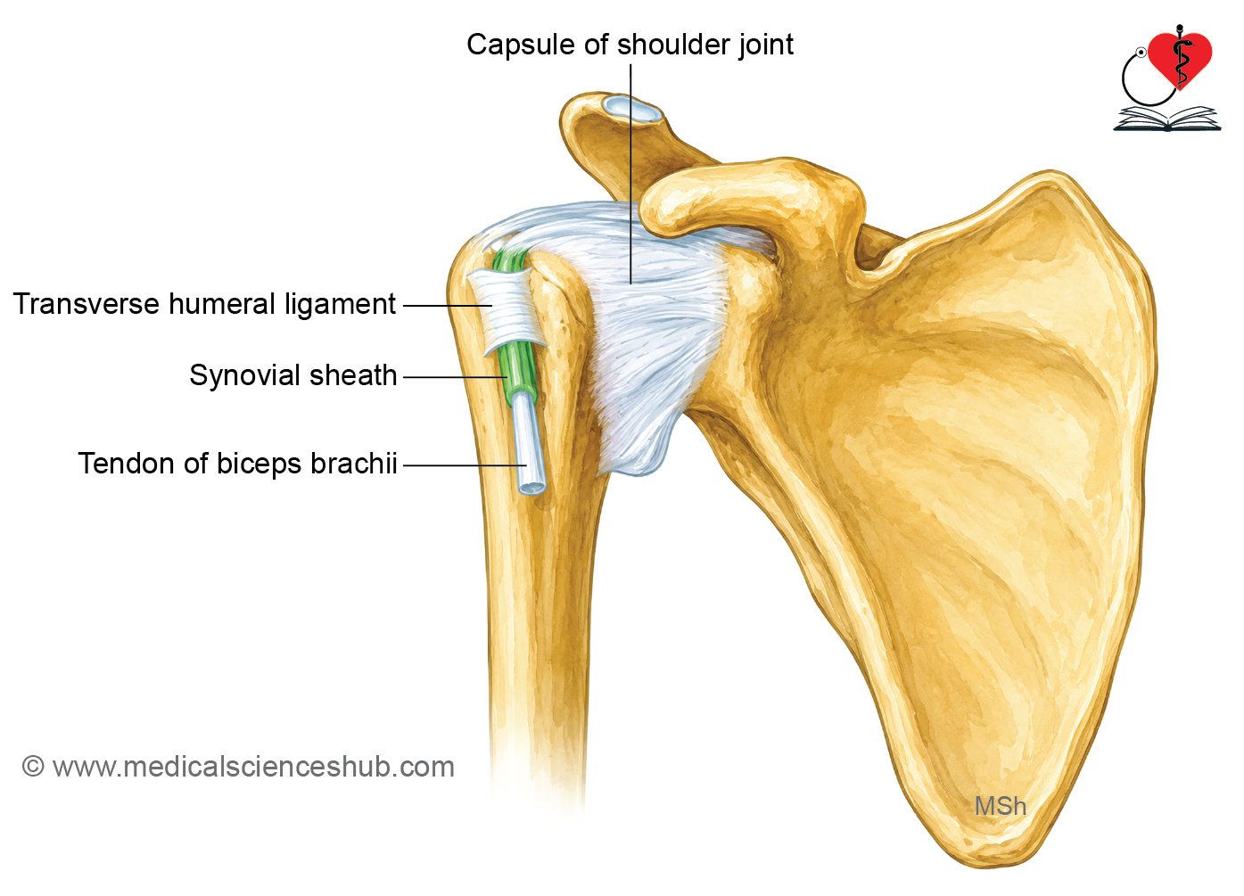
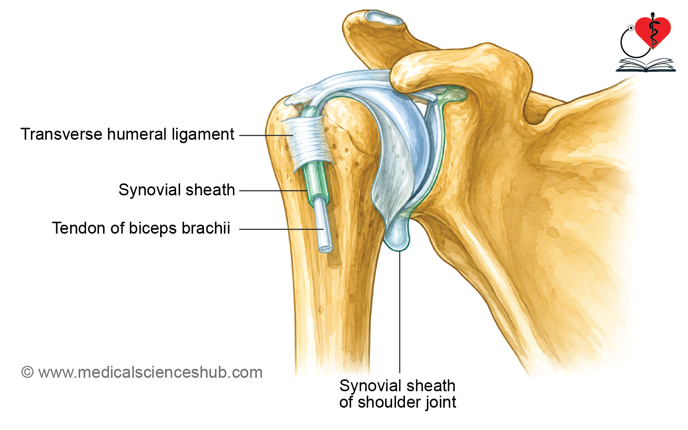
- The long head of the biceps brachii originates inside the capsule of the shoulder joint (intracapsular) but remains outside the synovial membrane (extrasynovial).
- The tendon of the long head passes through the intertubercular (bicipital) groove of the humerus.
- The name biceps is derived from Latin, where “bi” means two and “caput” means head, referring to its two heads.
- Near its insertion, the muscle forms:
- A strong tendon that attaches to the radial tuberosity.
- A bicipital aponeurosis that blends with the deep fascia of the forearm.
- A bursa lies between the tendon of the biceps brachii and the anterior part of the radial tuberosity. It reduces friction during forearm movements.
- The word muscle is derived from the Latin word musculus, meaning little mouse, because the contracting biceps was thought to resemble a small mouse moving beneath the skin.
Tendon Rupture
- Rupture of the biceps tendon may occur during strenuous athletic activities or heavy lifting.
- Mild cases are usually treated conservatively, whereas severe tendon ruptures may require surgical repair.
Biceps Reflex
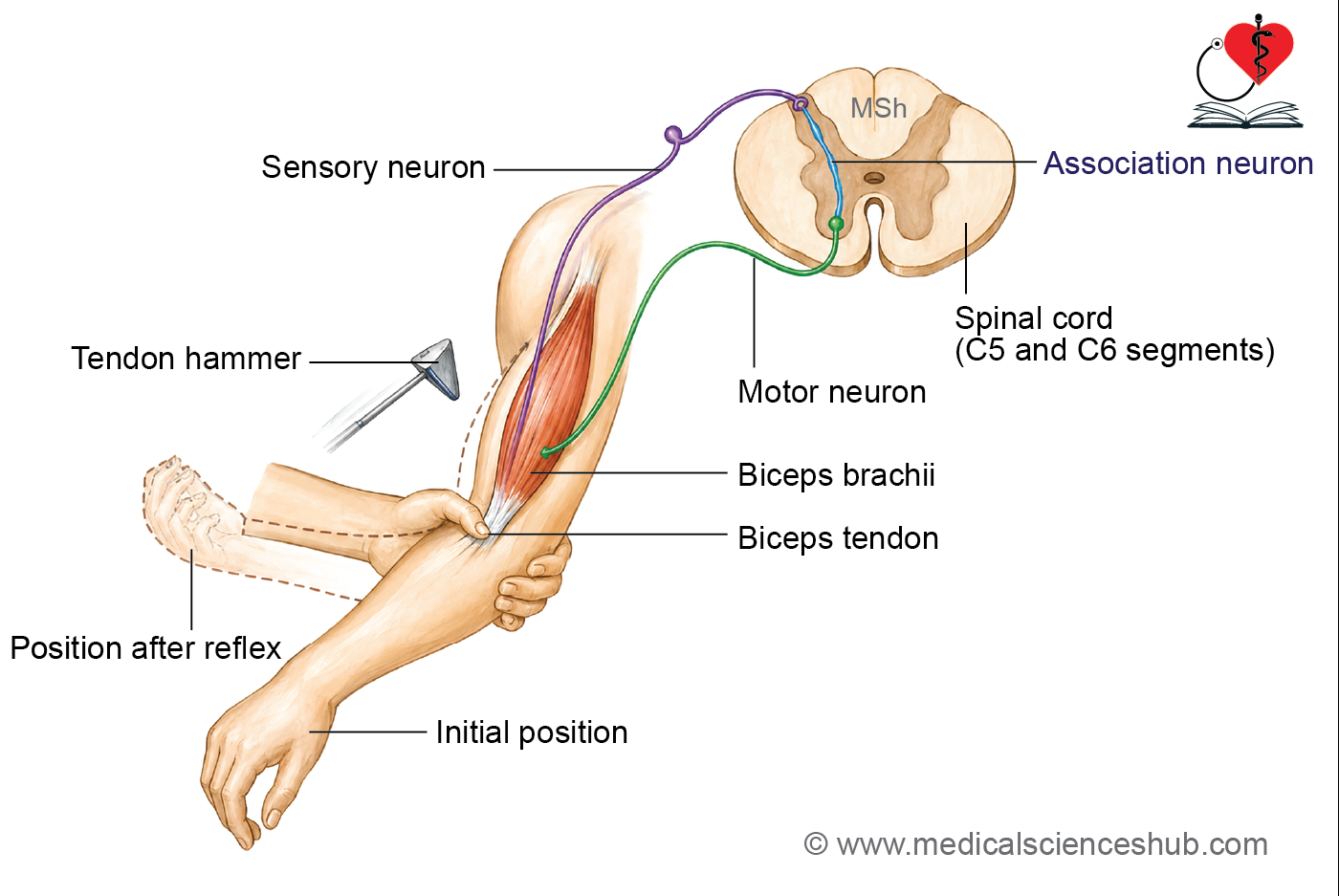
- The biceps reflex is used to assess the integrity of the musculocutaneous nerve and the C5 and C6 spinal cord segments.
- The reflex pathway is:
- Stretch of the biceps tendon → sensory fibres of the musculocutaneous nerve → C5 and C6 spinal cord segments → motor fibres of the musculocutaneous nerve → contraction of the biceps brachii.
- Procedure:
- The patient’s forearm is placed in a partially extended and pronated position.
- The examiner taps the biceps tendon with a tendon hammer and observes for contraction of the muscle.
- Interpretation:
- Absent reflex: Suggests musculocutaneous nerve injury or a lower motor neuron lesion.
- Exaggerated (brisk) reflex: Suggests an upper motor neuron lesion.
Brachialis
Origin
- The brachialis arises from:
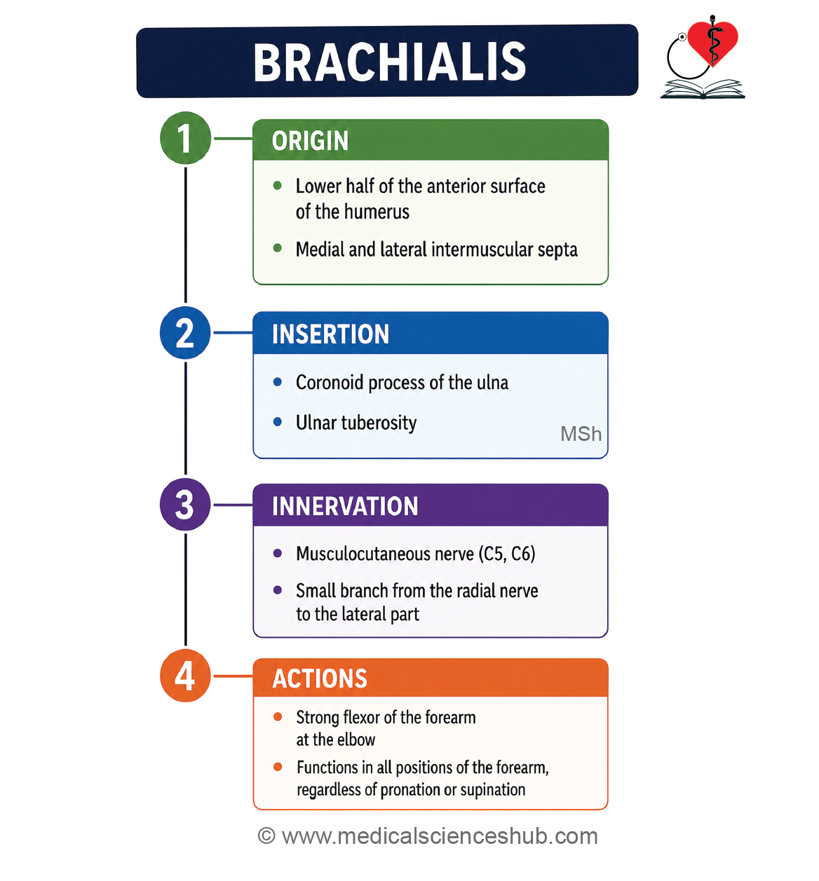
- The lower half of the anteromedial and anterolateral surfaces of the humerus.
- The anterior surfaces of the medial and lateral intermuscular septa.
- The brachialis is a broad, bulky muscle that lies deep to the lower part of the biceps brachii.
- Its fibres run vertically downward and converge towards the coronoid process of the ulna.
Insertion
- The muscle is inserted into the anterior surface of the coronoid process and the ulnar tuberosity of the ulna.
Nerve Supply
- The musculocutaneous nerve supplies the medial and major part of the muscle.
- The radial nerve supplies a small lateral part of the muscle.
Actions
- The brachialis is the strongest flexor of the elbow joint.
- Because it is the primary muscle responsible for elbow flexion, it is known as the “workhorse of the elbow joint.”
Clinical Testing
- The function of the brachialis is tested by asking the patient to flex the elbow against resistance.
- The examiner palpates the contracted muscle during elbow flexion to assess its strength and function.
Musculocutaneous Nerve
- The musculocutaneous nerve is the main nerve of the anterior compartment of the arm.
- It supplies all the muscles in the anterior compartment of the arm.
Beginning
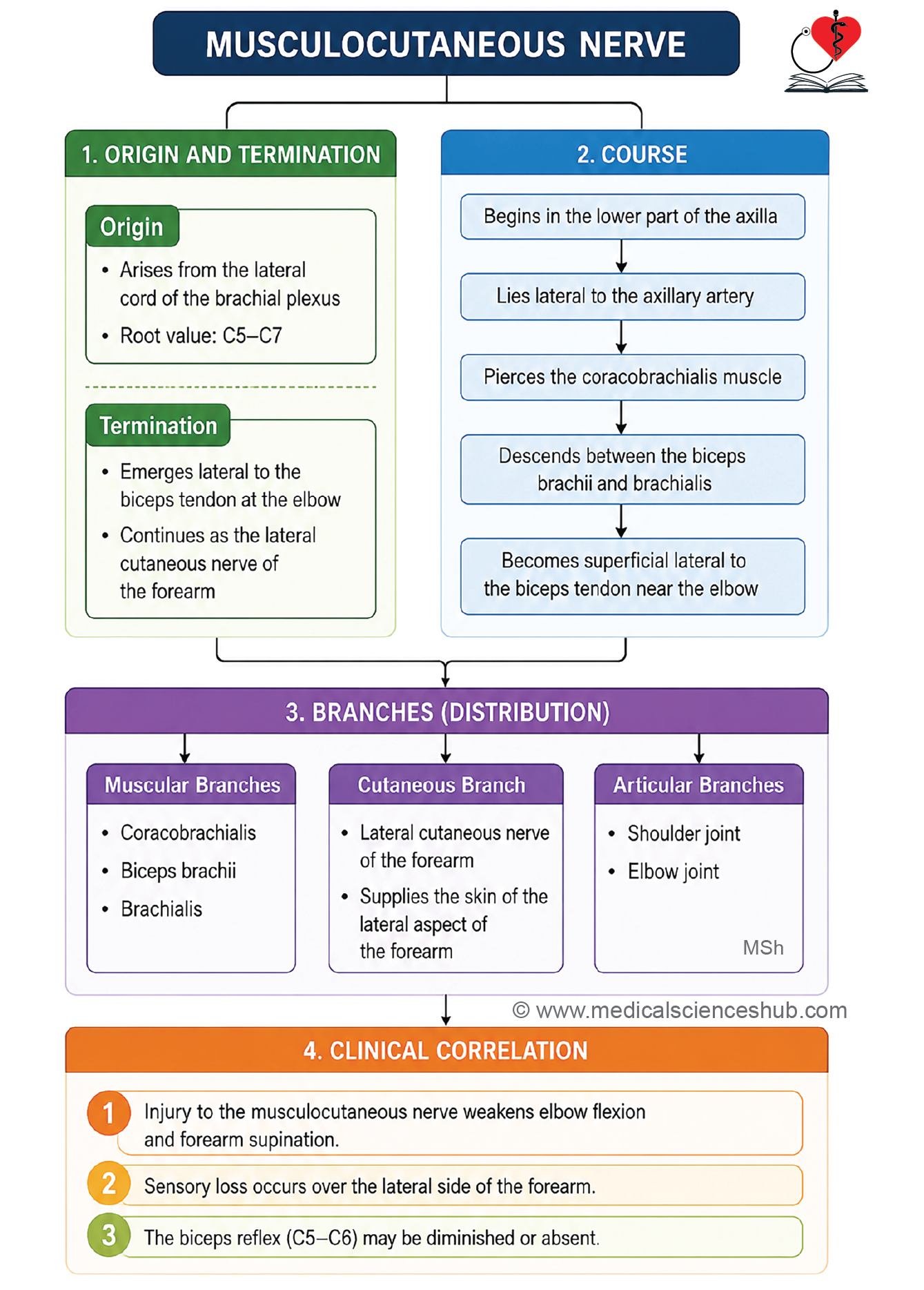
The musculocutaneous nerve arises from the lateral cord of the brachial plexus.
Root Value
- It carries fibres from the C5, C6, and C7 spinal nerves.
Termination
About 2 cm above the elbow joint, the musculocutaneous nerve continues as the lateral cutaneous nerve of the forearm.
Course
- The nerve begins in the lower part of the axilla.
- It passes through the coracobrachialis muscle, an important identifying feature.
- It then descends downward and laterally between the biceps brachii and brachialis muscles.
- Near the elbow, it emerges lateral to the tendon of the biceps brachii.
- It pierces the deep fascia about 2 cm above the elbow and continues as the lateral cutaneous nerve of the forearm.
Relations
In the Lower Part of the Axilla
- Anterior: Pectoralis major
- Posterior: Subscapularis
- Medial: Axillary artery
- Lateral: Coracobrachialis
In the Upper Part of the Arm
- The musculocutaneous nerve lies within the coracobrachialis muscle after piercing it.
In the Middle Part of the Arm
- The nerve lies between the biceps brachii and brachialis muscles.
- It pierces the deep fascia approximately 2 cm above the elbow joint before continuing into the forearm.
Branches and distribution
Muscular Branches
- The musculocutaneous nerve supplies the following muscles of the anterior compartment of the arm:
- Coracobrachialis
- Biceps brachii
- Brachialis (major medial part of the muscle)
Cutaneous Branch
- Near the elbow, the nerve continues as the lateral cutaneous nerve of the forearm.
- This nerve provides sensory innervation to the skin on the lateral side of the forearm.
Articular Branches
- The musculocutaneous nerve gives articular branches to:
- The shoulder joint
- The elbow joint
CLINICAL INTEGRATION
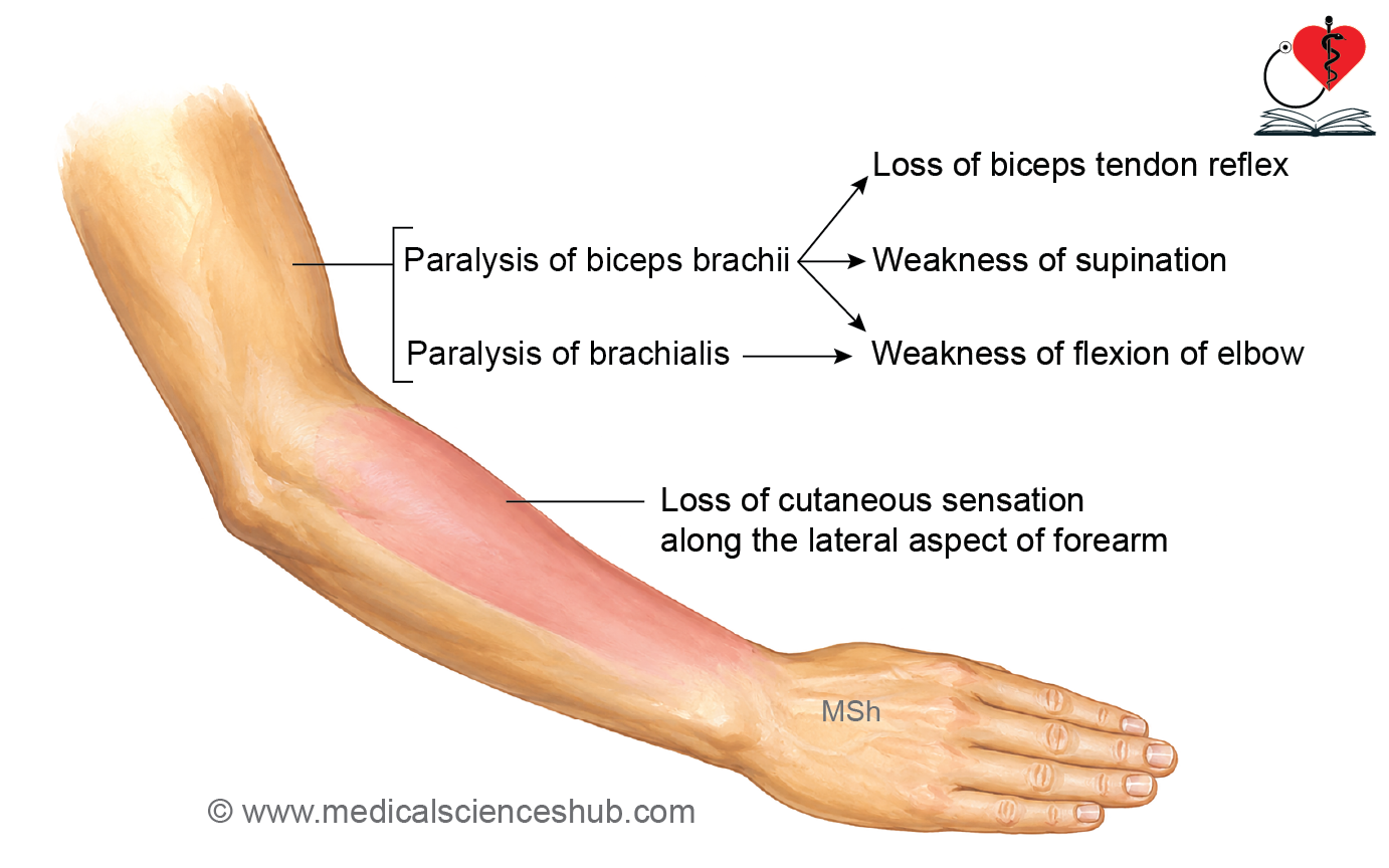
Damage to the musculocutaneous nerve produces motor and sensory deficits because it supplies the muscles of the anterior compartment of the arm and the skin of the lateral forearm.
Motor Effects
- Paralysis of the biceps brachii causes:
- Weakness of forearm supination, especially when the elbow is flexed.
- Loss or reduction of the biceps tendon reflex.
- Paralysis of the brachialis causes:
- Weakness of elbow flexion.
Sensory Effects
- There is loss of sensation over the lateral aspect of the forearm due to involvement of the lateral cutaneous nerve of the forearm, the terminal branch of the musculocutaneous nerve.
Brachial Artery
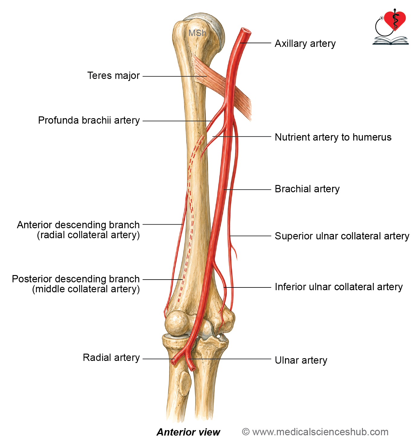
- The brachial artery is the main artery of the arm and is the primary source of blood supply to the forearm.
Beginning
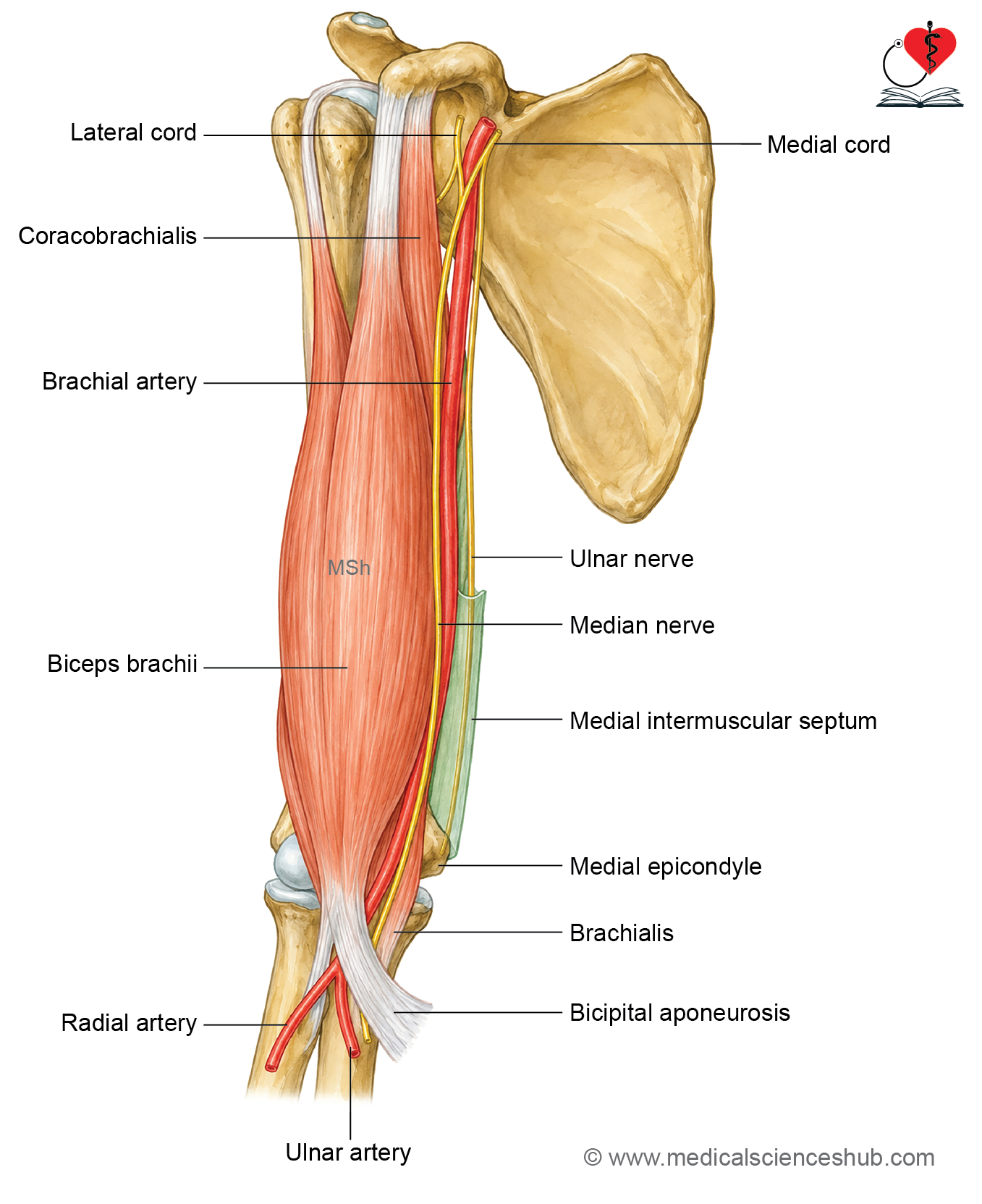
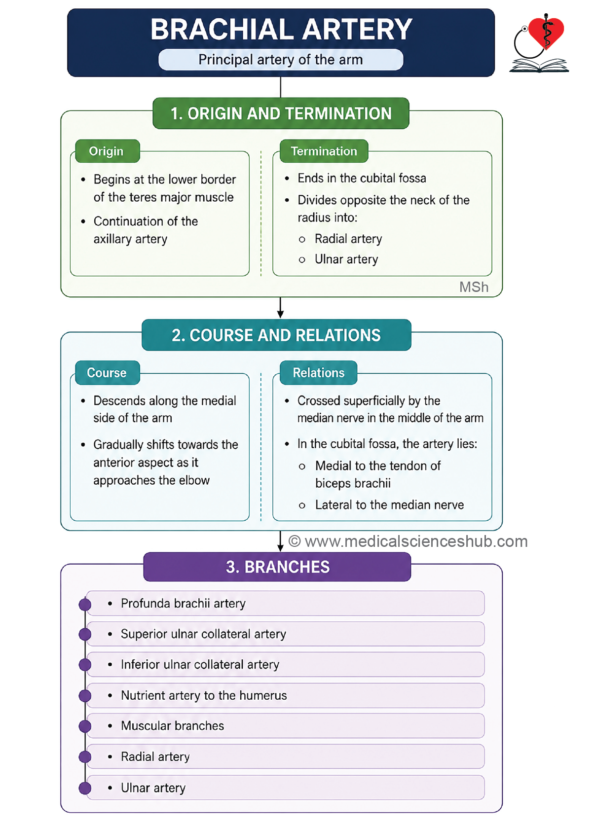
- It begins as the continuation of the axillary artery at the lower border of the teres major muscle.
Course
- The artery passes downward through the arm with a slight lateral course. It reaches the cubital fossa in front of the elbow, where it lies near the neck of the radius.
Termination
- At the level of the neck of the radius in the cubital fossa, the brachial artery ends by dividing into its two terminal branches: the radial artery and the ulnar artery.
Relations
In the arm
- Anteriorly: The median nerve crosses the brachial artery from the lateral side to the medial side.
- Posteriorly: The artery is related to the triceps brachii in the upper part of the arm and the brachialis muscle in the lower part.
- Medially: In the upper part, it is related to the ulnar nerve and the basilic vein. In the lower part, it is related to the median nerve.
- Laterally: In the upper part, it is related to the median nerve and the coracobrachialis muscle. In the lower part, it is related to the biceps brachii muscle.
In cubital fossa
- Anteriorly: It is covered by the bicipital aponeurosis and the median cubital vein.
- Posteriorly: It lies on the brachialis muscle.
- Medially: It is related to the median nerve.
- Laterally: It is related to the tendon of the biceps brachii.
Branches
- The brachial artery gives rise to the following branches:
- Profunda brachii artery.
- Superior ulnar collateral artery.
- Inferior ulnar collateral artery.
- Nutrient artery to the humerus.
- Several unnamed muscular branches that supply the muscles of the arm.
- Terminal Branches: In the cubital fossa, the brachial artery ends by dividing into two terminal branches:
- Ulnar artery.
- Radial artery.
CLINICAL INTEGRATION
Pulsation of the Brachial Artery
- The pulsation of the brachial artery can be felt in the cubital fossa.
- It is palpated just medial to the tendon of the biceps brachii.
Recording Blood Pressure
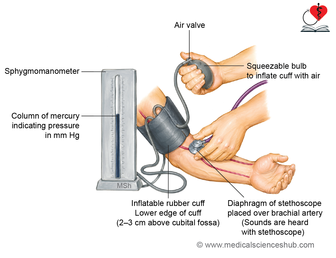
- Blood pressure is commonly measured over the brachial artery.
- When the cuff pressure is raised above the systolic blood pressure, the brachial artery is compressed and blood flow temporarily stops.
- As the cuff pressure falls below the systolic pressure, blood begins to flow through the artery again.
- This blood flow produces Korotkoff sounds, which are heard with a stethoscope placed over the brachial artery in the cubital fossa.
- The appearance and disappearance of these sounds are used to determine systolic and diastolic blood pressure.
Compression of the Brachial Artery
- The brachial artery can be compressed against the shaft of the humerus to control bleeding from injuries of the forearm or hand.
- Compression may be achieved manually or by using a tourniquet or a blood pressure cuff.
Posterior Compartment of Arm
The posterior compartment of the arm contains the following important structures:
- Triceps brachii muscle.
- Radial nerve.
- Profunda brachii artery.
- Ulnar nerve.
- Superior and inferior ulnar collateral arteries pass through this compartment.
.
Triceps Brachii Muscle
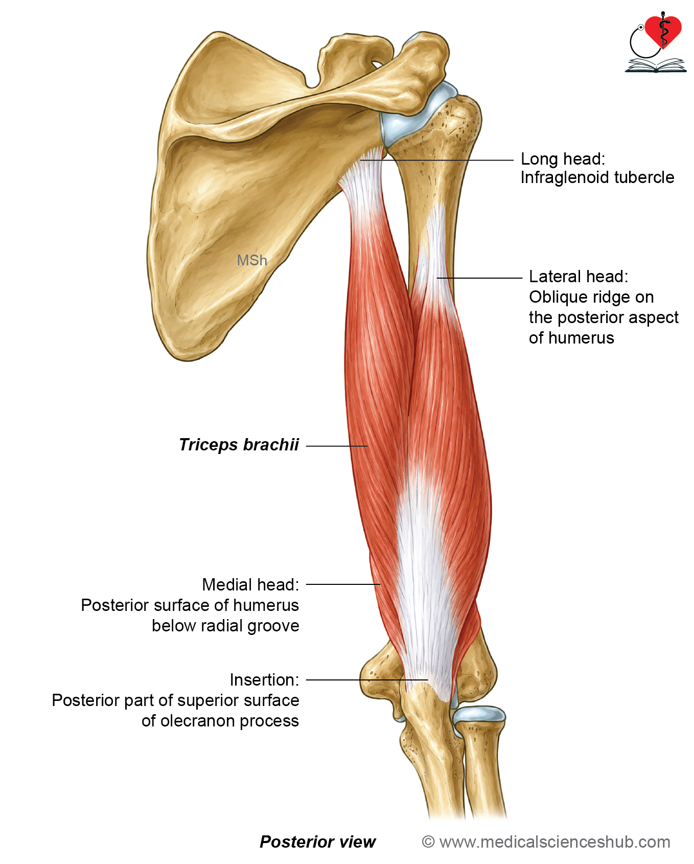
- The triceps brachii is a large muscle that occupies most of the posterior compartment of the arm.
- It has three heads: the long head, lateral head, and medial head.
Origin
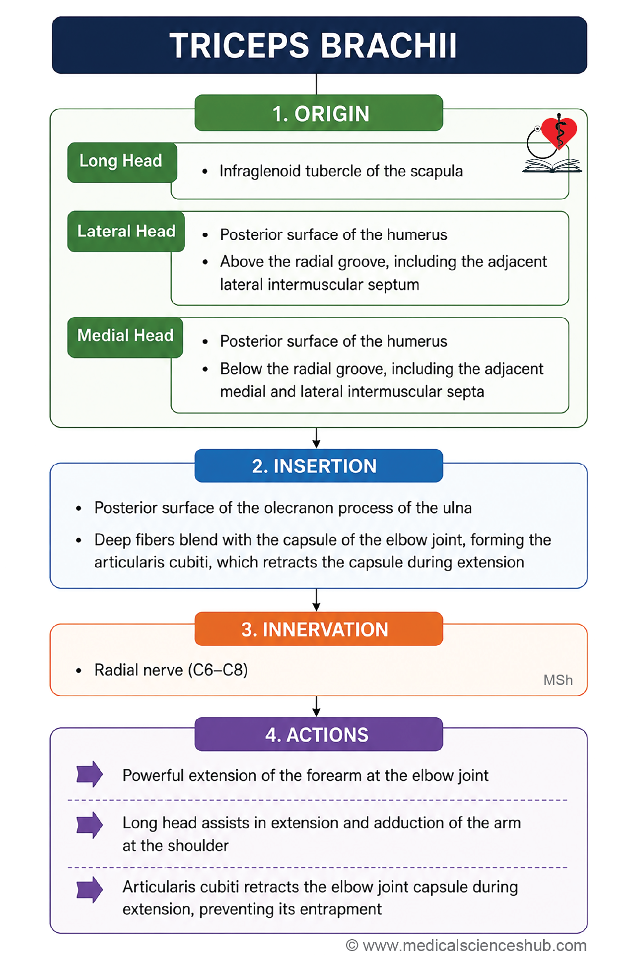
- The long head arises from the infraglenoid tubercle of the scapula.
- The lateral head arises from the posterior surface of the humerus, above and lateral to the radial groove.
- The medial head arises from the posterior surface of the humerus below and medial to the radial groove. The medial head also takes origin from the medial and lateral intermuscular septa.
Insertion
- All three heads unite to form a common tendon that inserts onto the posterior part of the superior surface of the olecranon process of the ulna.
Innervations
The triceps brachii is supplied by the radial nerve.
Actions
- The triceps brachii is the chief extensor of the forearm at the elbow joint.
- Although elbow extension is often assisted by gravity, the triceps is the main muscle responsible for powerful active extension.
- The long head also helps in extension and adduction of the arm at the shoulder joint.
CLINICAL INTEGRATION
Clinical Testing
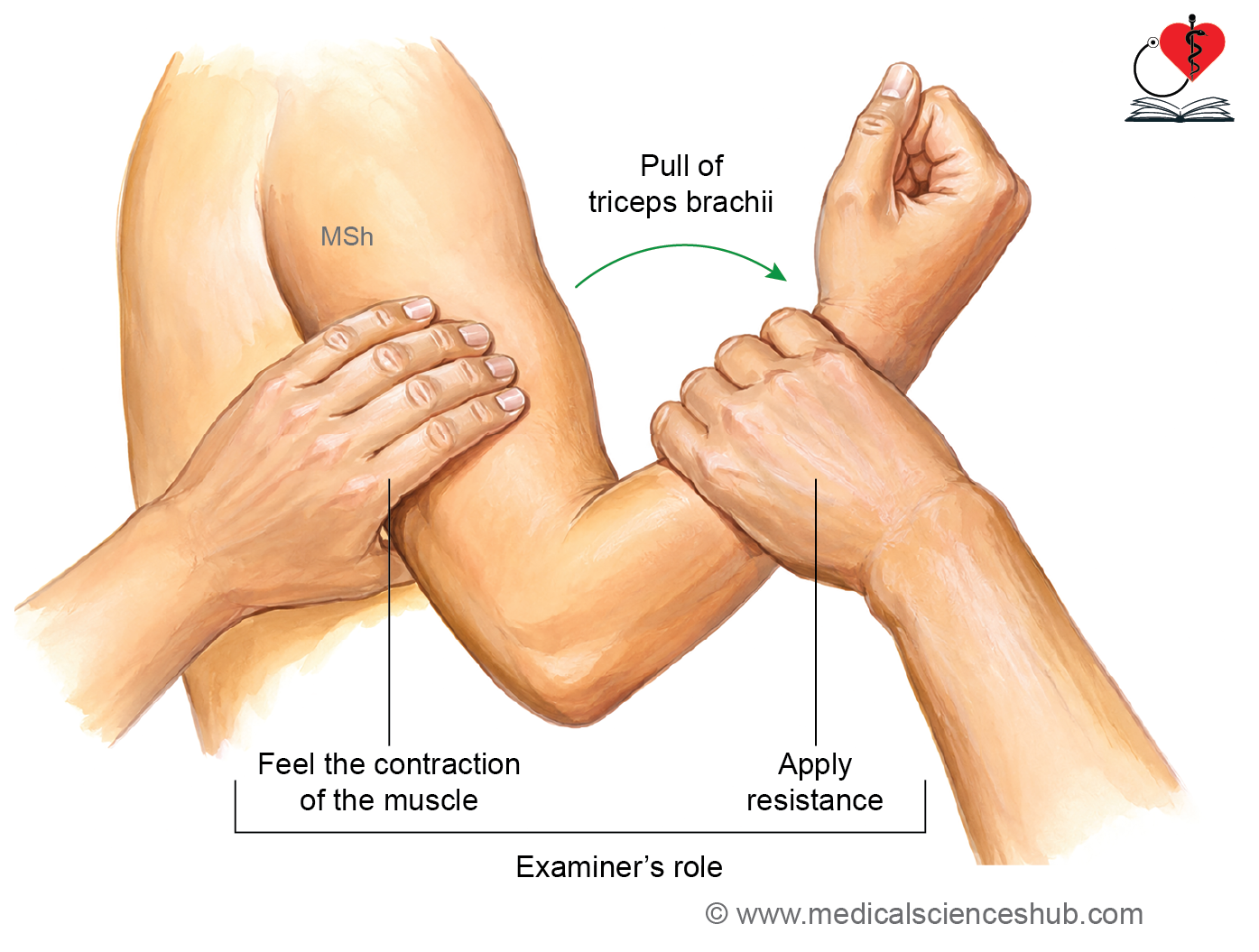
- The triceps brachii is tested by asking the patient to extend the elbow against resistance.
- Contraction of the muscle can be felt over the back of the arm during this movement.
Triceps Sparing in Radial Nerve Injury
- In injuries to the radial nerve within the radial (spiral) groove, the triceps brachii is not completely paralyzed.
- This is because the nerve supply to the long head and most of the medial head arises from branches of the radial nerve that are given off in the axilla, proximal to the site of injury.
- As a result, elbow extension is usually weakened but not completely lost.
Radial Nerve
- The radial nerve is the main nerve of the posterior compartment of the arm, forearm, and the dorsum of the hand.
- It is the continuation of the posterior cord of the brachial plexus.
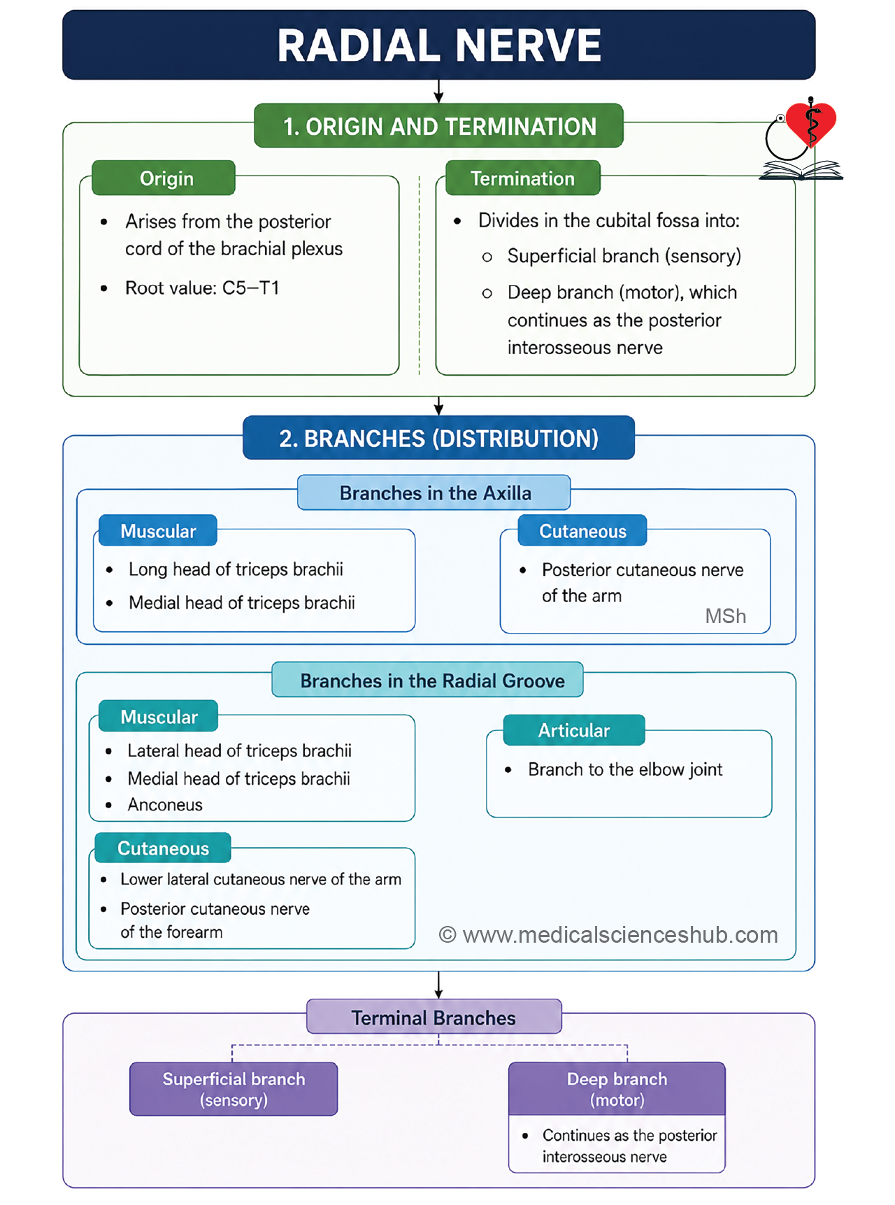
- The radial nerve carries fibers from the C5, C6, C7, C8, and T1 spinal nerve roots.
Beginning
- The radial nerve arises as the continuation of the posterior cord of the brachial plexus.
- It begins behind the third part of the axillary artery in the lower part of the axilla.
- It is the largest terminal branch of the brachial plexus.
Course and relations
- The radial nerve begins in the lower part of the axilla.
- It passes behind the axillary artery and then lies behind the upper part of the brachial artery for a short distance.
- The nerve enters the posterior compartment of the arm through the lower triangular interval along with the profunda brachii artery.
- It runs downward and laterally in the radial (spiral) groove on the posterior surface of the humerus.
- The radial nerve then pierces the lateral intermuscular septum to enter the anterior compartment of the arm.
- In the anterior compartment, it lies deep to the brachioradialis and extensor carpi radialis longus muscles.
- Just below the lateral epicondyle of the humerus, the radial nerve divides into its two terminal branches:
- The superficial branch.
- The deep branch (posterior interosseous nerve).
Branches
Branches in the Axilla
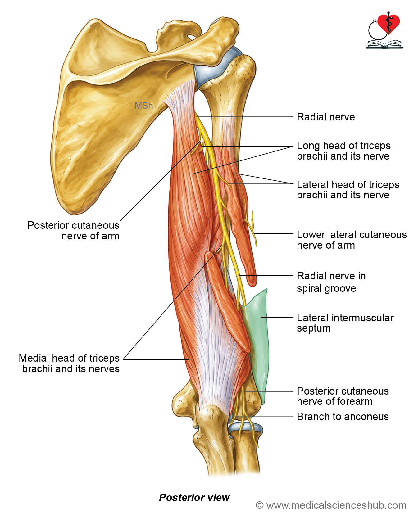
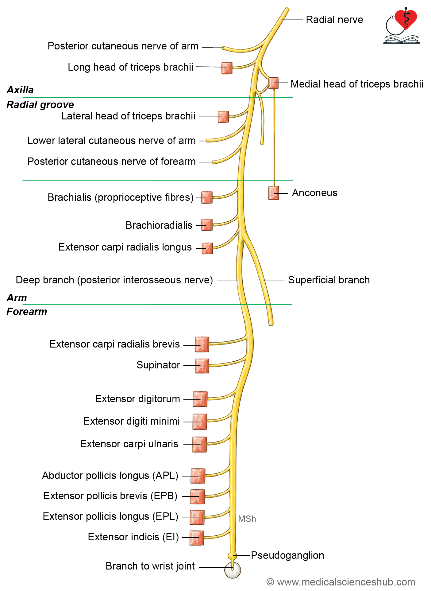
- The radial nerve gives muscular branches to the long head and medial head of the triceps brachii.
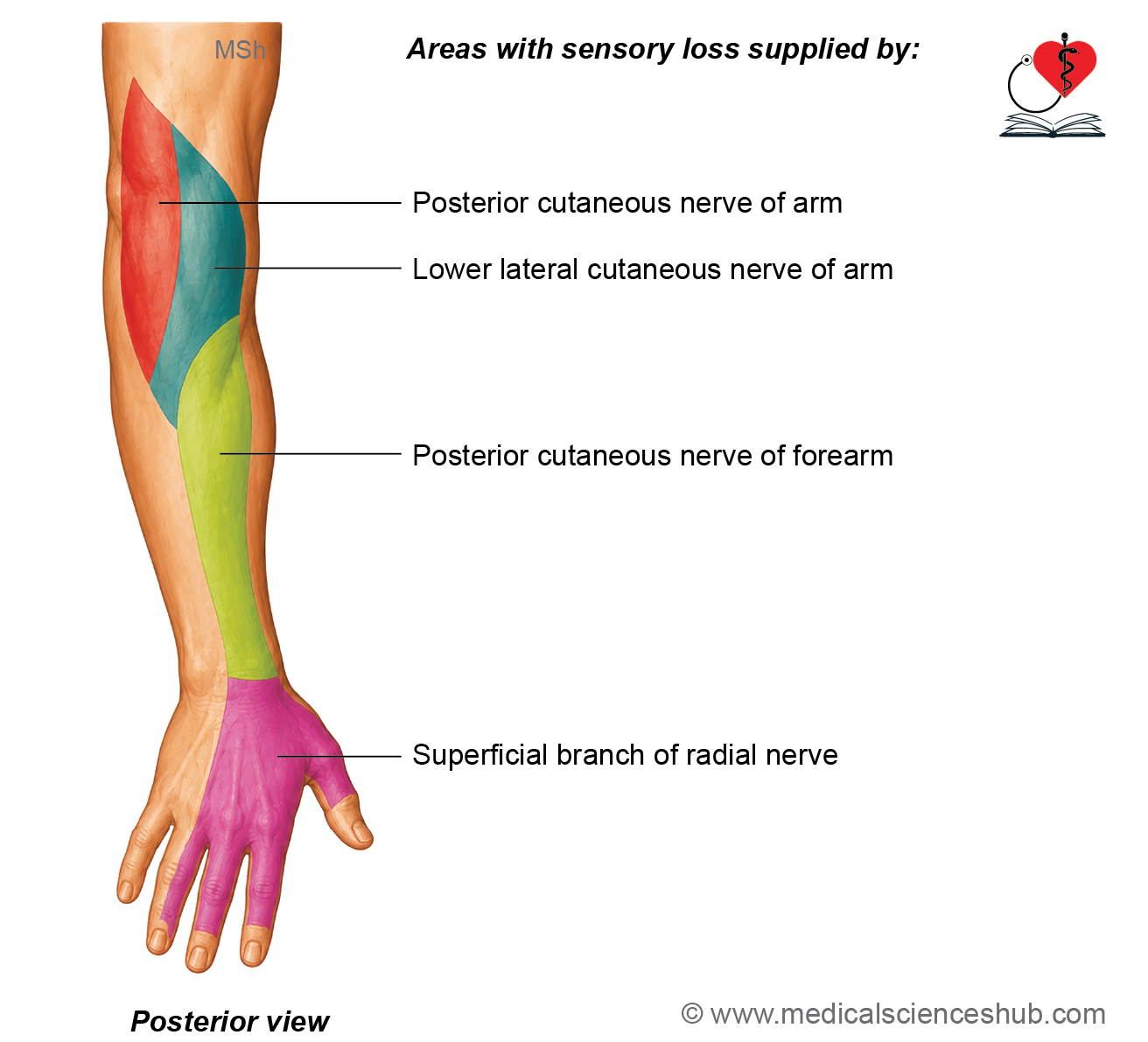
- It gives the posterior cutaneous nerve of the arm, which supplies the skin on the back of the arm.
Branches in the Radial Groove
- It gives a muscular branch to the lateral head of the triceps brachii.
- It gives another branch to the medial head of the triceps brachii. This branch is also known as the ulnar collateral nerve because it accompanies the ulnar nerve.
- A branch to the anconeus muscle arises through the nerve supplying the medial head of the triceps.
- It gives the lower lateral cutaneous nerve of the arm, which supplies the skin over the lower lateral part of the arm.
- It gives the posterior cutaneous nerve of the forearm, which supplies the skin on the back of the forearm.
Branches in the Lower Part of the Arm
- The radial nerve gives muscular branches to the following muscles:
- Brachialis (lateral part).
- Brachioradialis.
- Extensor carpi radialis longus.
- It also gives an articular branch to the elbow joint.
Terminal Branches
- The radial nerve ends by dividing into two terminal branches:
- Superficial branch: A sensory branch that supplies the skin of the dorsum of the hand and the posterior aspect of the lateral 3½ fingers (excluding the nail beds).
- Deep branch (posterior interosseous nerve): A motor branch that pierces the supinator muscle and supplies the remaining muscles of the posterior compartment of the forearm.
CLINICAL INTEGRATION
Injury to the Radial Nerve
- The clinical features of a radial nerve injury depend on the level at which the nerve is damaged.
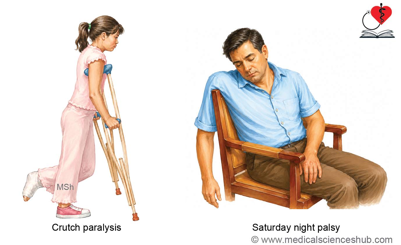
Injury in the Axilla (Saturday Night Palsy)
- This injury may occur due to prolonged pressure on the nerve, such as sleeping with the arm hanging over the back of a chair or from improper use of crutches.
- It produces the following clinical features:
- Loss of extension of the forearm at the elbow due to paralysis of the triceps brachii.
- Weakness of supination of the forearm.
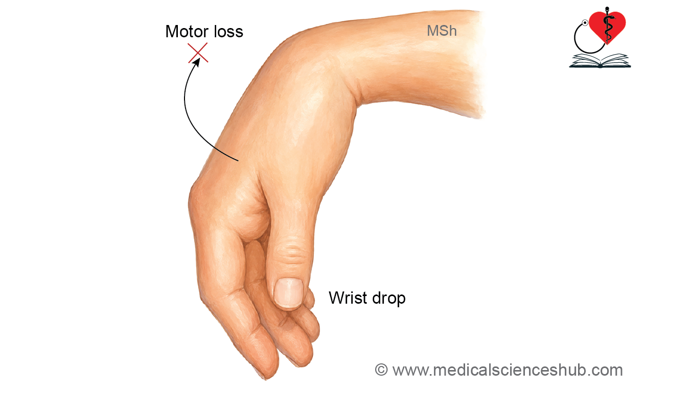
- Loss of extension of the wrist and fingers, resulting in wrist drop.
- Loss of sensation over the lateral and posterior aspects of the arm, the posterior aspect of the forearm, the dorsum of the hand, and the posterior aspect of the lateral 3½ fingers.
Injury in the Radial Groove
- A radial nerve injury in the radial (spiral) groove produces most of the features seen in axillary injury.
- The long head and most of the medial head of the triceps brachii are usually spared because they receive their nerve supply before the nerve enters the radial groove.
- As a result, elbow extension is weakened but not completely lost.
- Wrist drop, weakness of finger extension, and sensory loss over the radial nerve distribution are still present.
- Injuries to the radial nerve in the forearm produce different clinical features and are discussed separately.
Profunda Brachii Artery
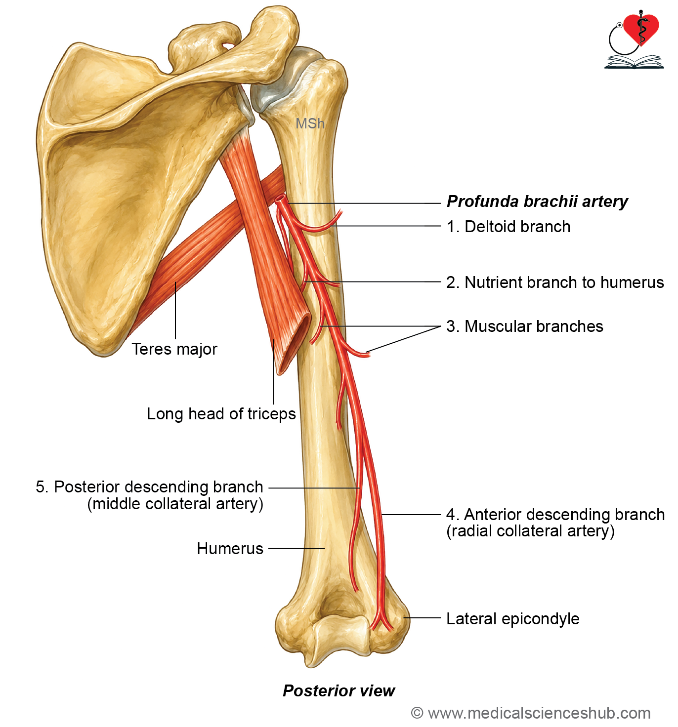
- The profunda brachii artery (deep artery of the arm) is the main artery supplying the posterior compartment of the arm.
- It is the largest branch of the brachial artery.
Beginning
The profunda brachii artery arises from the brachial artery just below the lower border of the teres major muscle.
Course and relations
- The artery passes from the posteromedial side of the brachial artery into the lower triangular interval.
- It accompanies the radial nerve through the radial (spiral) groove on the posterior surface of the humerus.
Termination
The profunda brachii artery ends by dividing into:
- Anterior descending (radial collateral) artery.
- Posterior descending (middle collateral) artery.
Branches
The profunda brachii artery gives the following branches:
- Deltoid (ascending) branch, which ascends to anastomose with the descending branch of the posterior circumflex humeral artery.
- Nutrient artery to the humerus.
- Muscular branches to the muscles of the posterior compartment of the arm.
- Anterior descending (radial collateral) artery, which accompanies the radial nerve and anastomoses with the radial recurrent artery in front of the lateral epicondyle of the humerus.
- Posterior descending (middle collateral) artery, which anastomoses with the interosseous recurrent artery behind the lateral epicondyle of the humerus.
Important Questions
- Write a short note on biceps brachii.
- Write a short note on bicipital aponeurosis.
- Write a short note on brachialis.
- Write a short note on musculocutaneous nerve.
- List the anatomical changes occurring at the level of insertion of coracobrachialis muscle.
- Write a short note on triceps brachii.
- Write a short note on radial (musculospiral) nerve in radial groove.
- Write a short note on wrist drop.
- Write a short note on arteria profunda brachii.
- Write a short note on biceps brachii.
- Write a short note on bicipital aponeurosis.
- Write a short note on brachialis.
📝 Test Your Knowledge – Practice MCQs
Attempt the chapter MCQ quiz and assess your understanding of key concepts.
Click to see: Medical Disclaimer and Copyright Notice