
Competencies
- AN12.1: Describe and demonstrate important muscle groups of ventral forearm with attachments, nerve supply, and actions.
- AN12.2: Identify and describe origin, course, relations, branches (or tributaries), termination of important nerves and vessels of forearm.
- AN13.7: ………, palpation of radial artery, …….. .
Introduction
- The forearm is the part of the upper limb located between the elbow and the wrist.
- It contains two long bones:
- Radius, which lies on the lateral (thumb) side.
- Ulna, which lies on the medial (little finger) side.
- The radius and ulna are connected at three sites:
- Superior radioulnar joint.
- Interosseous membrane, which joins the shafts of the two bones.
- Inferior radioulnar joint.
- The anatomical name for the forearm is antebrachium, meaning the region between the arm (brachium) and the wrist.
.
Fascial Compartments of Forearm
- The forearm contains both superficial fascia and deep fascia.
- The superficial fascia is thin and contains only a small amount of fat.
- The deep fascia of the forearm is called the antebrachial fascia.
- The antebrachial fascia surrounds and encloses all the structures of the forearm.
- Unlike the deep fascia of the thigh, the antebrachial fascia is not very thick.
- Its thin and flexible nature allows the forearm muscles to move freely during different movements.
- The deep fascia is attached to the posterior subcutaneous border of the ulna.
- At the front of the wrist, the deep fascia becomes thickened to form the flexor retinaculum.
- At the back of the wrist, the deep fascia forms the extensor retinaculum.
- A lateral intermuscular septum extends from the deep fascia to the radius.
- The forearm is divided into anterior (flexor) and posterior (extensor) compartments.
- These two compartments are formed by the deep fascia, radius, ulna, interosseous membrane, and lateral intermuscular septum.
Anterior Compartment of Forearm
- Anterior compartment of forearm is separated from the posterior compartment of forearm by deep fascia , ulna, interosseous membrane, radius, and lateral intermuscular septum.
Contents of Anterior Compartment of Forearm
The anterior compartment of the forearm contains the following structures:
Muscles
Superficial group
- The pronator teres muscle.
- The flexor carpi radialis muscle.
- The palmaris longus muscle.
- The flexor digitorum superficialis muscle.
- The flexor carpi ulnaris muscle.
Deep group
- The flexor pollicis longus muscle.
- The flexor digitorum profundus muscle.
- The pronator quadratus muscle.
Arteries
- The radial artery.
- The ulnar artery.
Nerves
- The median nerve.
- The ulnar nerve.
- The radial nerve (its branches supply structures in this region).
Surface Landmarks
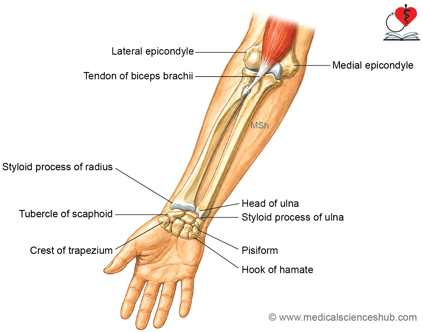
- The tendon of the biceps brachii can be felt in the cubital fossa, in front of the elbow.
- The biceps tendon becomes more prominent when the elbow is flexed against resistance.
- The brachial artery can be palpated just medial to the biceps tendon.
- The head of the radius can be felt in a small depression on the posterolateral side of the extended elbow, just below the lateral epicondyle.
- The olecranon process is easily palpable when the elbow is slightly flexed.
- The styloid process of the radius can be felt on the lateral side of the wrist, at the proximal end of the anatomical snuffbox.
- The head of the ulna can be felt as a rounded prominence on the medial side of the wrist.
- The styloid process of the ulna can be felt on the posteromedial side of the wrist.
- The styloid process of the ulna lies about 1 cm proximal to the styloid process of the radius.
- The pisiform bone can be felt on the medial side of the base of the hypothenar eminence.
- The hook of the hamate can be felt only by deep palpation, just below the pisiform bone.
- The tubercle of the scaphoid can be felt at the base of the thenar eminence.
- The tubercle (crest) of the trapezium can be felt by deep palpation just below the scaphoid tubercle.
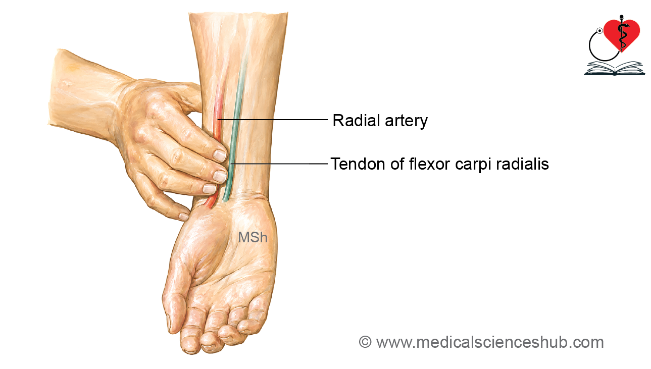
- The radial artery pulse can be felt on the front of the distal forearm between the anterior border of the radius laterally and the tendon of the flexor carpi radialis medially.
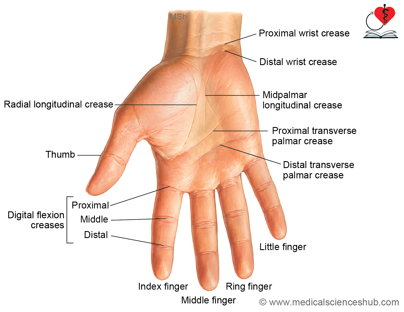
Transverse Creases in Front of Wrist
- The proximal transverse wrist crease lies at the level of the wrist joint.
- The distal transverse wrist crease is the more prominent wrist crease.
- The distal transverse wrist crease corresponds to the proximal border of the flexor retinaculum.
Superficial Muscles of Front of Forearm
- The anterior compartment of the forearm contains five superficial muscles.
- These muscles are:
- Pronator teres
- Flexor carpi radialis
- Palmaris longus
- Flexor digitorum superficialis
- Flexor carpi ulnaris
Common flexor origin
- All the superficial flexor muscles arise from the anterior surface of the medial epicondyle of the humerus.
- This common attachment is known as the common flexor origin.
- In addition to the common flexor origin, some of these muscles also take origin from the radius or ulna.
- Since these muscles share a common origin, they are best identified away from their proximal attachment, where they can be distinguished more easily.
Pronator Teres
- The pronator teres is a small muscle located on the lateral side of the superficial flexor group.
- It runs obliquely across the upper part of the anterior forearm.
- The pronator teres forms the medial boundary of the cubital fossa.
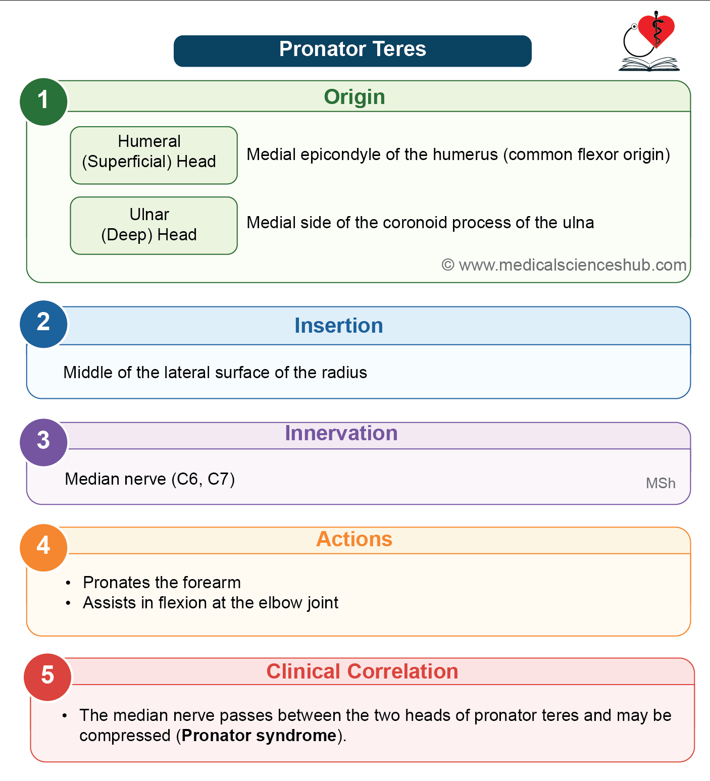
- The median nerve passes between the two heads of the pronator teres.
- The ulnar artery passes deep to the ulnar (deep) head of the muscle.
Origin
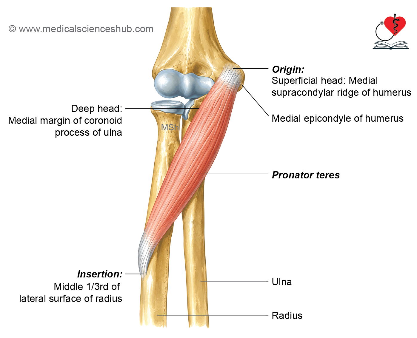
- The pronator teres has two heads.
- The humeral (superficial) head arises from the medial epicondyle of the humerus.
- The ulnar (deep) head arises from the medial border of the coronoid process of the ulna.
- The muscle fibres run downward and laterally towards the middle of the radius.
Insertion
The muscle is inserted into the middle one-third of the lateral surface of the radius.
Innervation (nerve supply)
The pronator teres is supplied by the median nerve.
Actions
- It pronates the forearm.
- The humeral head also assists in weak flexion of the elbow joint.
Clinical testing
To test the muscle, ask the patient to pronate the forearm against resistance while the elbow is flexed.
Flexor Carpi Radialis
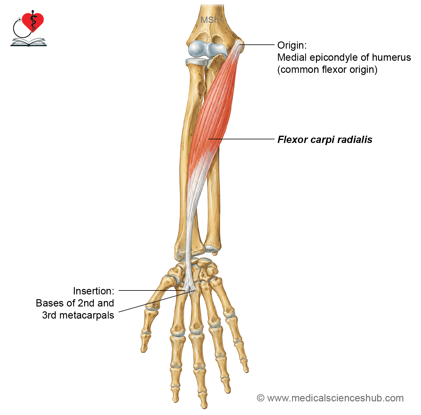
The flexor carpi radialis (FCR) is a long, spindle-shaped muscle located in the front of the forearm. It has a large muscular belly and a long, narrow tendon.
Origin
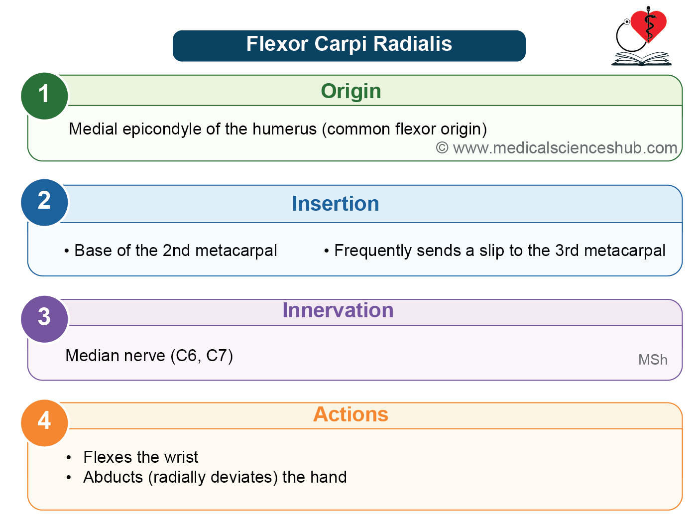
Arises from the medial epicondyle of the humerus through the common flexor tendon.
- The muscle fibers pass downward and slightly outward (laterally).
- The muscular part extends to about the middle of the forearm, after which it continues as a long tendon.
Insertion
- It Inserts into the bases of the second and third metacarpal bones.
Innervation
It is supplied by the median nerve.
Actions
At the wrist joint, the flexor carpi radialis:
- Flexes the hand along with the flexor carpi ulnaris.
- Abducts (radially deviates) the hand along with the brachioradialis and the extensor carpi radialis longus and brevis.
CLINICAL ANATOMY
- The radial artery lies immediately lateral to the tendon of the flexor carpi radialis near the lower end of the forearm. Therefore, the tendon serves as an important landmark for locating and palpating the radial pulse.
- The tendon of the flexor carpi radialis passes through its own separate compartment beneath the flexor retinaculum, unlike the flexor tendons that pass through the carpal tunnel, before inserting into the second and third metacarpals.
Palmaris Longus
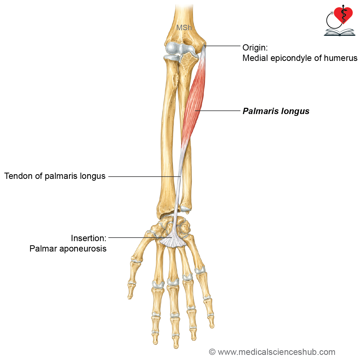
The palmaris longus is a slender muscle located in the superficial layer of the front of the forearm. It has a small muscle belly and a long, thin tendon.
Origin
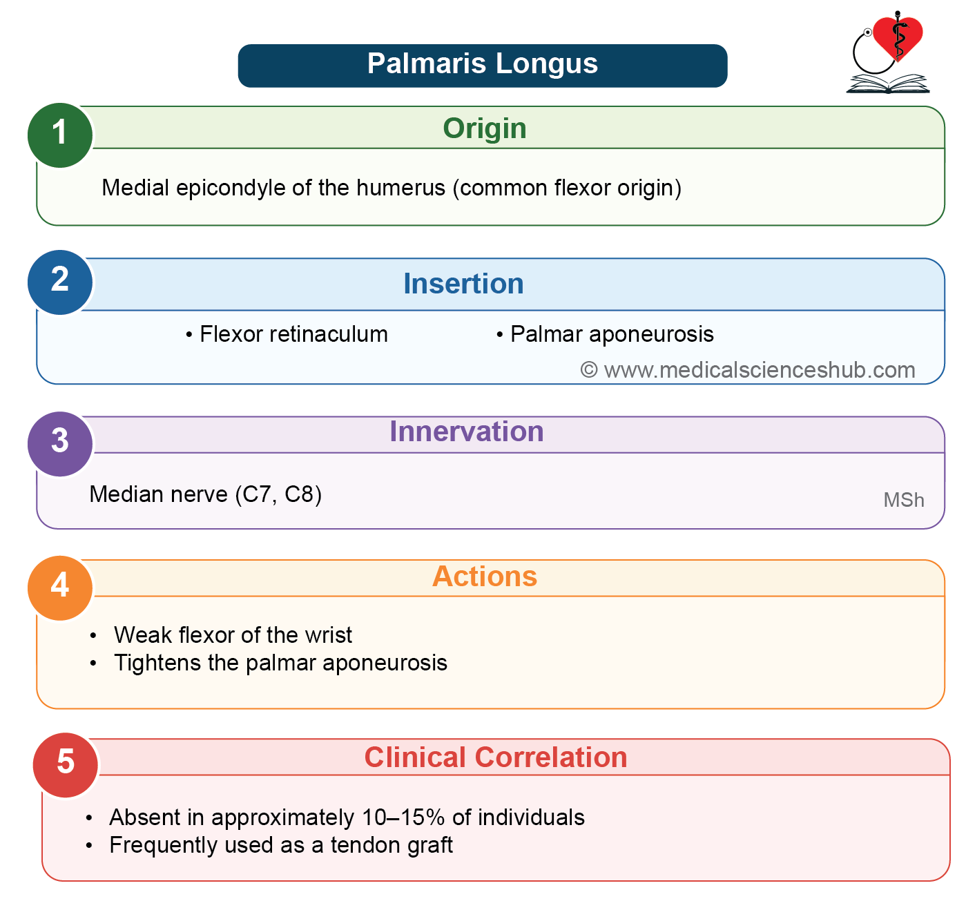
The palmaris longus originates from the medial epicondyle of the humerus.
The muscle fibers pass downward and continue as a long tendon in the middle of the front of the forearm. The tendon crosses superficial to the flexor retinaculum at the wrist.
Insertion
The tendon is attached to the flexor retinaculum and the apex of the palmar aponeurosis.
Innervation
The palmaris longus is supplied by the median nerve.
Actions
The palmaris longus helps in flexion of the wrist. It also tightens the flexor retinaculum and the palmar aponeurosis, which helps improve grip during hand movements.
CLINICAL ANATOMY
- The palmaris longus is considered a vestigial muscle because it has a small muscle belly and a long tendon, indicating that it has lost much of its original function during evolution.
- The palmar aponeurosis is regarded as the degenerated distal part of the tendon of the palmaris longus.
- The palmaris longus is absent in about 14% of individuals. Its absence does not cause any significant loss of function.
- The tendon of the palmaris longus is commonly used for tendon grafting because it can be removed without causing noticeable weakness or impairment of hand movements.
Flexor Digitorum Superficialis (Sublimis)
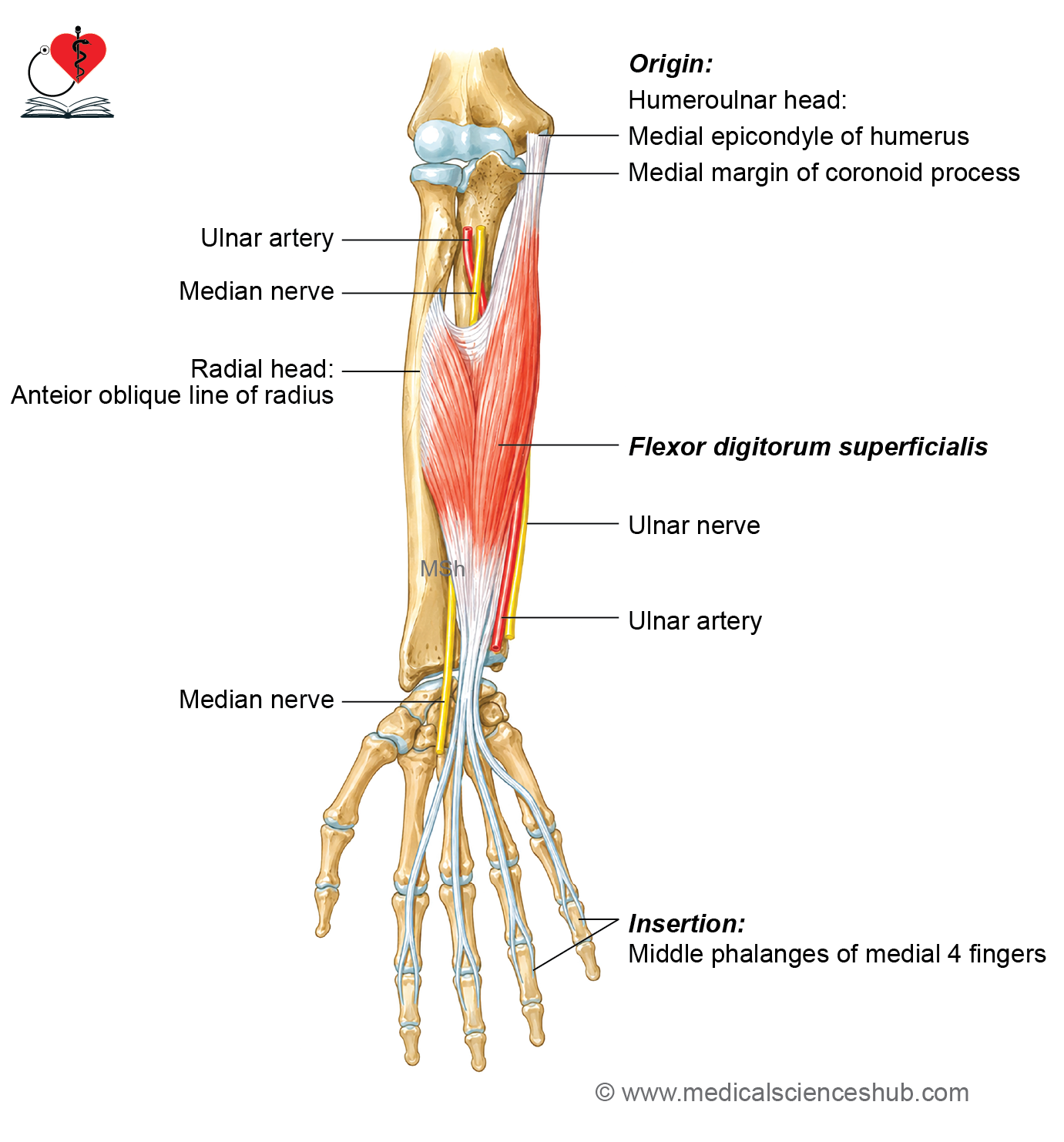
The flexor digitorum superficialis (FDS) is the largest muscle among the superficial flexor muscles of the forearm. It is located between the superficial and deep muscle groups and is therefore considered the muscle of the intermediate compartment of the forearm.
Origin
The flexor digitorum superficialis has two heads of origin.
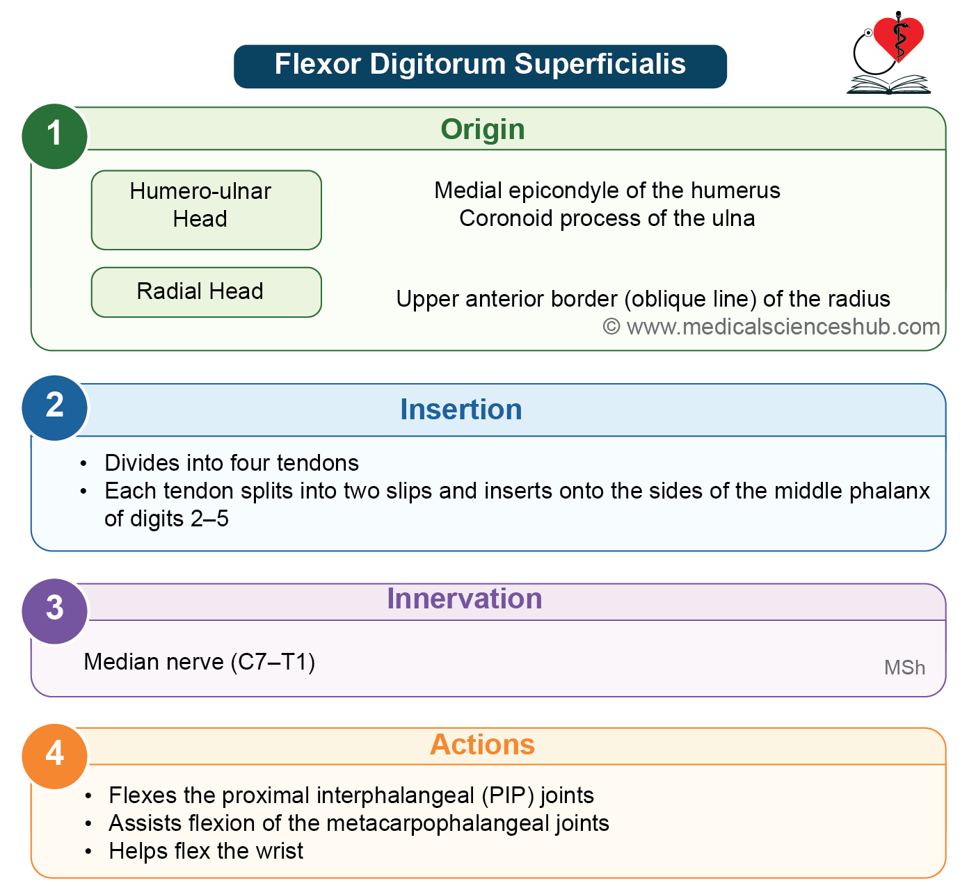
The humeroulnar head arises from the medial epicondyle of the humerus, the medial border of the coronoid process of the ulna, and the ulnar collateral ligament of the elbow.
The radial head arises from the anterior oblique line of the radius, which is present on the upper part of its anterior border.
Direction of fibers
The muscle fibers descend to form a broad muscle belly. In the lower part of the forearm, the belly divides into superficial and deep layers.
The superficial layer gives rise to two tendons for the middle and ring fingers. The deep layer forms two tendons for the index and little fingers.
As each tendon reaches the base of the proximal phalanx, it splits into medial and lateral slips. These slips allow the tendon of the flexor digitorum profundus to pass through to the distal phalanx.
Insertion
Each tendon divides into two slips that are attached to the sides of the middle phalanx of the index, middle, ring, and little fingers.
Innervation
The flexor digitorum superficialis is supplied by the median nerve.
Actions
The flexor digitorum superficialis flexes the proximal interphalangeal (PIP) joints of the medial four fingers. It also helps in flexion of the metacarpophalangeal (MCP) joints and assists in flexion of the wrist.
CLINICAL ANATOMY
- A tendinous arch is formed between the two heads of the flexor digitorum superficialis. The median nerve and the ulnar artery pass deep to this arch.
- The flexor digitorum superficialis has independent action on each finger, allowing the index, middle, ring, and little fingers to be flexed separately at the proximal interphalangeal joints.
Flexor Carpi Ulnaris
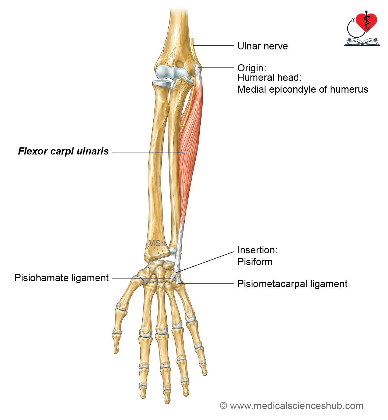
- The flexor carpi ulnaris (FCU) is the most medial muscle in the superficial layer of the front of the forearm.
- It is a long, spindle-shaped (fusiform) muscle.
Origin
- The flexor carpi ulnaris has two heads of origin.
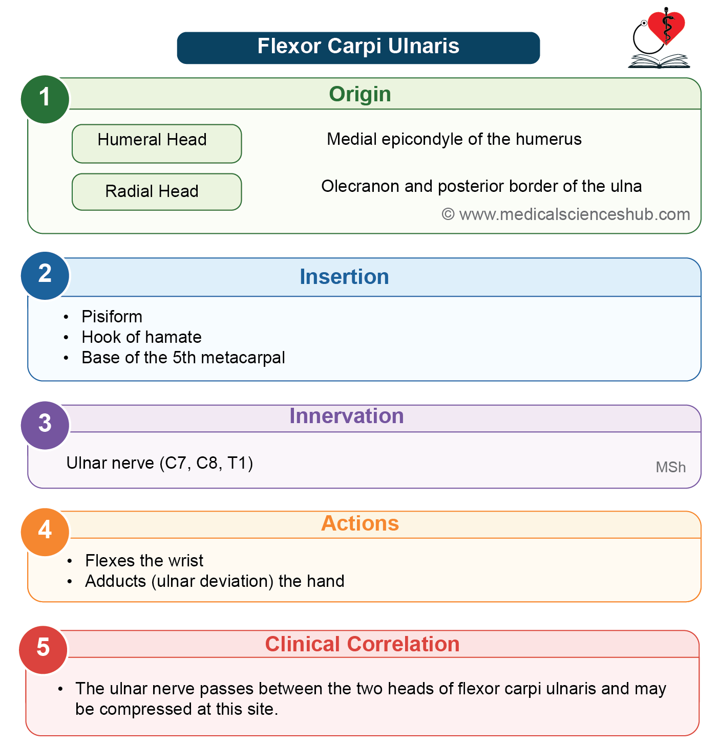
- The small humeral head arises from the medial epicondyle of the humerus.
- The larger ulnar head arises from the medial side of the olecranon process and the upper two-thirds of the posterior border of the ulna.
Direction of Fibers
- The muscle fibers run downward and form a fusiform muscle belly.
- The belly continues as a long tendon in the lower part of the forearm.
Insertion
- The tendon is attached to the pisiform bone.
- It also reaches the hook of the hamate through the pisohamate ligament.
- It is further attached to the base of the fifth metacarpal through the pisometacarpal ligament.
Innervation
The flexor carpi ulnaris is supplied by the ulnar nerve.
Actions
- At the wrist joint, the flexor carpi ulnaris produces flexion along with the flexor carpi radialis.
- It also produces adduction (ulnar deviation) of the hand along with the extensor carpi ulnaris.
CLINICAL ANATOMY
- The ulnar nerve passes between the two heads of the flexor carpi ulnaris.
- In the lower part of the forearm, the ulnar nerve and ulnar artery lie lateral to the tendon of the flexor carpi ulnaris. This relationship is an important anatomical landmark.
- The pisiform is a sesamoid bone that develops within the tendon of the flexor carpi ulnaris.
- All the superficial muscles of the anterior compartment of the forearm arise from the medial epicondyle of the humerus.
- All these muscles are supplied by the median nerve, except the flexor carpi ulnaris, which is supplied by the ulnar nerve.
Deep Muscles Of Front Of Forearm
- The deep muscles of the anterior compartment of the forearm are arranged in two layers.
Superficial Layer
- The superficial layer consists of:
- Flexor pollicis longus
- Flexor digitorum profundus
Deep Layer
- The deep layer contains only one muscle:
- Pronator quadratus
- Most of the deep muscles of the anterior compartment are supplied by the anterior interosseous nerve, which is a branch of the median nerve.
- An exception is the medial half of the flexor digitorum profundus, which is supplied by the ulnar nerve.
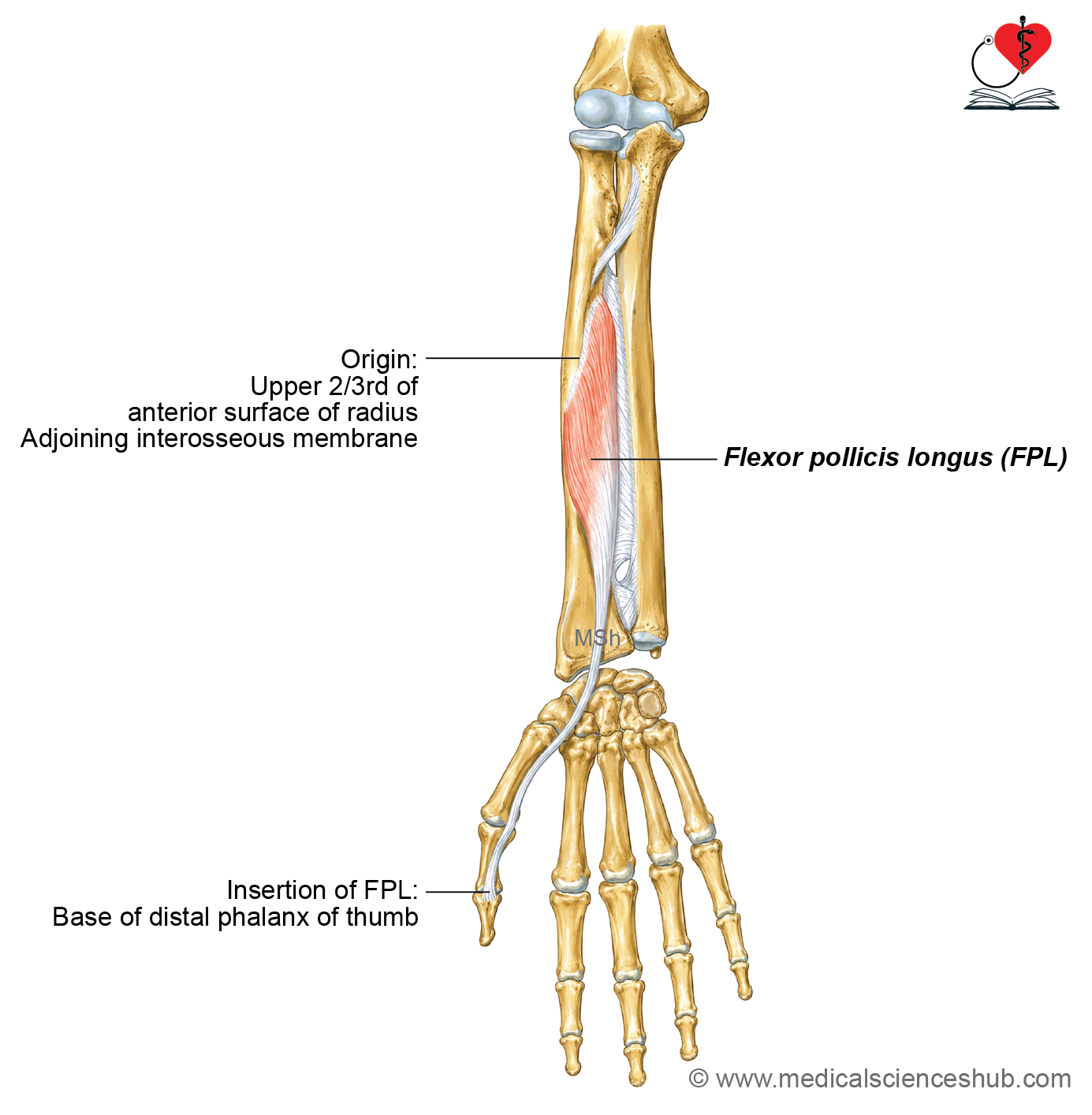
Flexor Pollicis Longus
- The flexor pollicis longus (FPL) is a long and slender muscle located in the deep layer of the front of the forearm.
- It covers the upper three-fourths of the anterior surface of the radius.
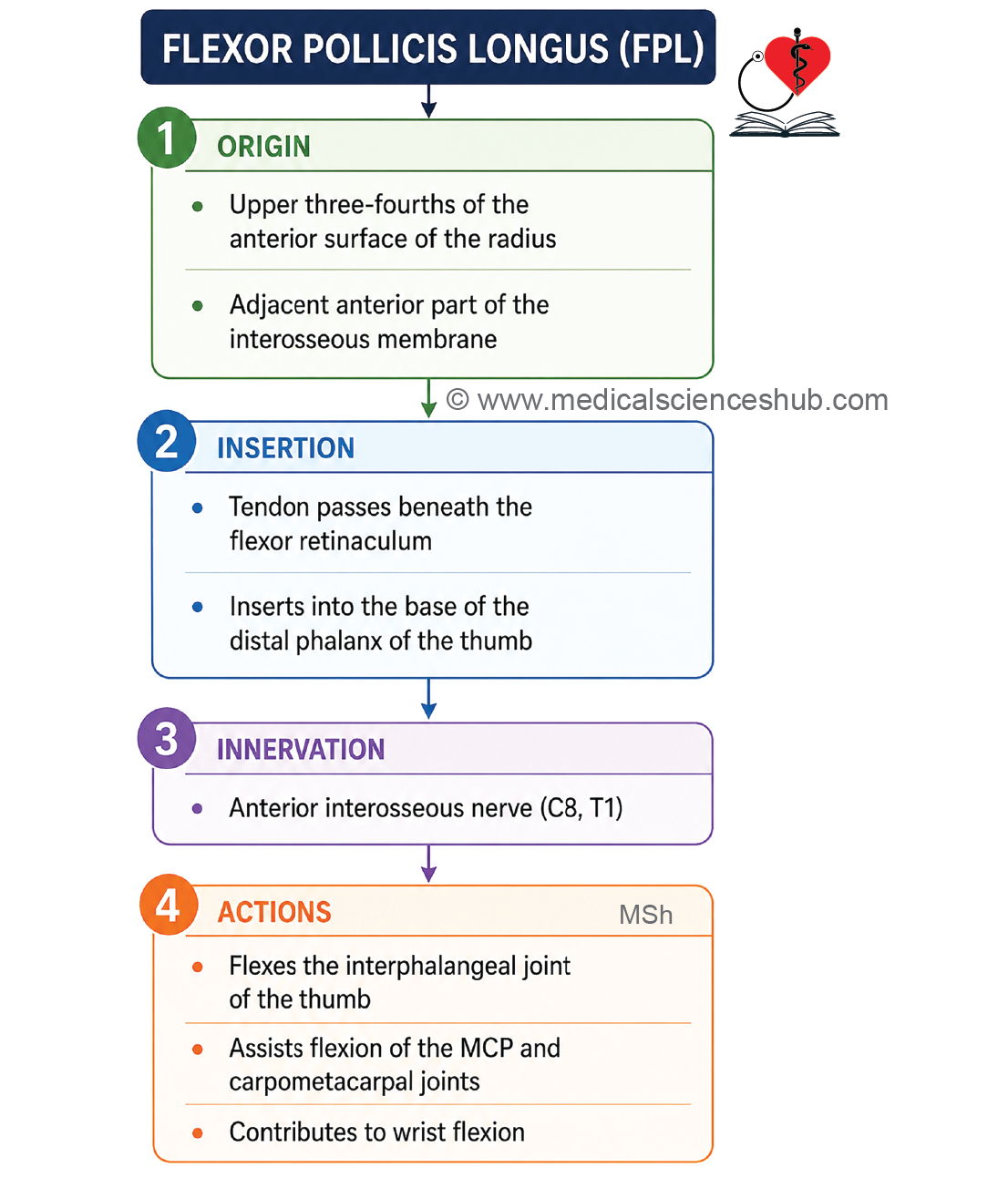
Origin
- The flexor pollicis longus arises from:
- The upper three-fourths of the anterior surface of the radius, below the anterior oblique line.
- The adjacent part of the interosseous membrane.
Direction of Fibers
- The muscle fibers pass downward and form a flattened tendon.
- The tendon passes deep to the flexor retinaculum and enters the palm.
Insertion
The tendon is attached to the palmar surface of the base of the distal phalanx of the thumb.
Innervation
The flexor pollicis longus is supplied by the anterior interosseous nerve, which is a branch of the median nerve.
Actions
- The flexor pollicis longus flexes the interphalangeal (IP) joint of the thumb.
- It also helps in flexion of the metacarpophalangeal (MCP) joint of the thumb.
Clinical testing
The function of the flexor pollicis longus is tested by asking the patient to flex the interphalangeal joint of the thumb against resistance.
CLINICAL ANATOMY
The tendon of the flexor pollicis longus passes deep to the flexor retinaculum within a separate synovial sheath known as the radial bursa.
Flexor Digitorum Profundus
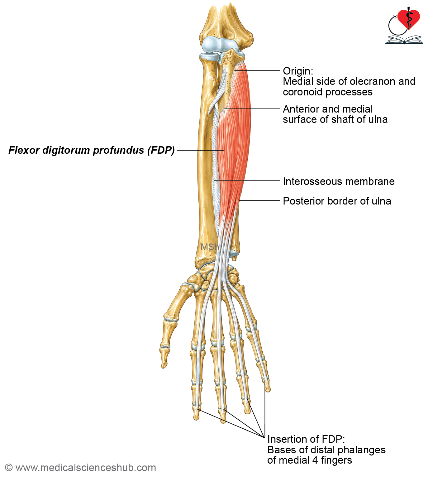
- The flexor digitorum profundus (FDP) is a large and powerful muscle located in the deep layer of the front of the forearm.
- It covers most of the anterior surface of the ulna.
Origin
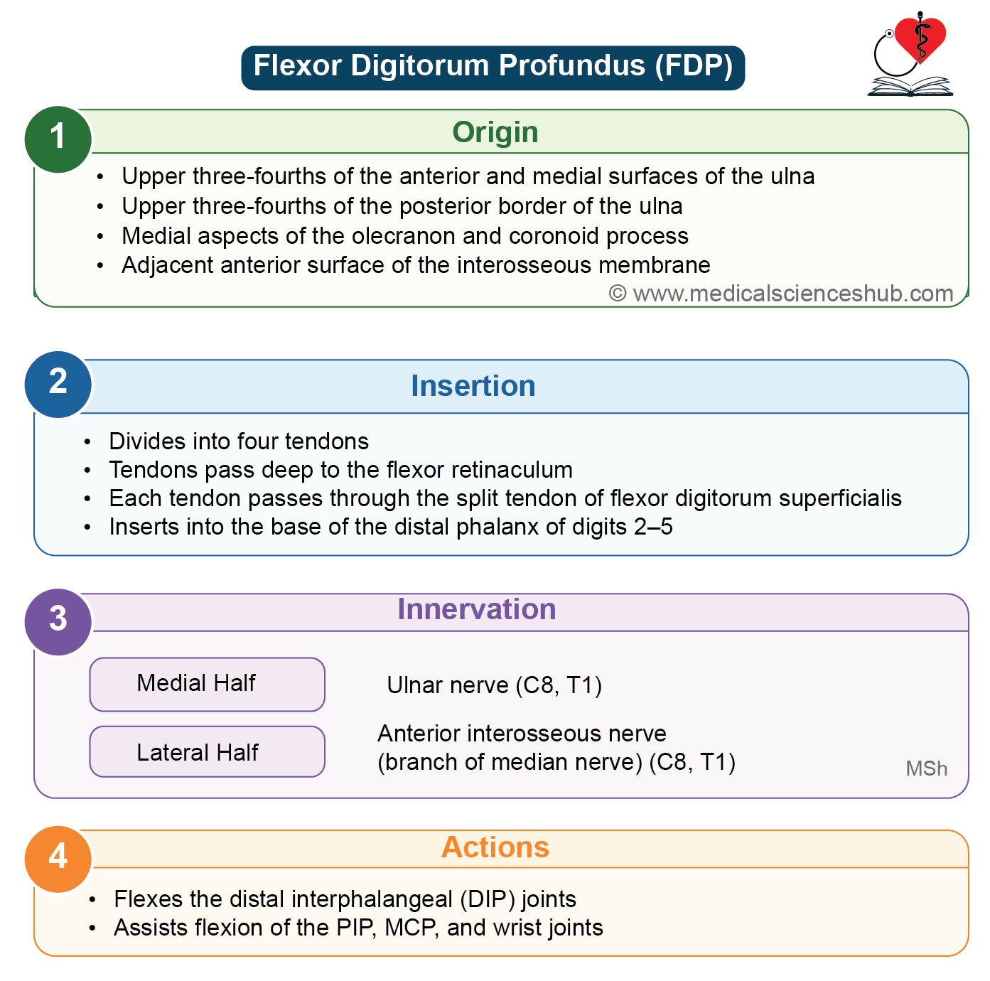
- The flexor digitorum profundus arises from:
- The upper three-fourths of the anterior and medial surfaces of the ulna.
- The upper three-fourths of the posterior border of the ulna.
- The medial surface of the olecranon process.
- The adjacent part of the interosseous membrane.
Direction of Fibers
- The muscle fibers descend to form four tendons for the index, middle, ring, and little fingers.
- The tendon to the index finger is more separate from the others, allowing it to move more independently.
- All four tendons pass deep to the flexor retinaculum before entering the palm.
Insertion
Each tendon is attached to the palmar surface of the base of the distal phalanx of the medial four fingers.
Innervation
- The medial half of the flexor digitorum profundus is supplied by the ulnar nerve.
- The lateral half is supplied by the anterior interosseous nerve, which is a branch of the median nerve.
Actions
- The flexor digitorum profundus produces flexion of the distal interphalangeal (DIP) joints, which is its main and exclusive action.
- It also assists in flexion of the proximal interphalangeal (PIP) joints, metacarpophalangeal (MCP) joints, and the wrist.
CLINICAL ANATOMY
- Clinical testing: The muscle is tested by asking the patient to flex the distal interphalangeal joint of a finger while the proximal interphalangeal joint is held in extension.
- The flexor digitorum profundus is called a hybrid (composite) muscle because it receives nerve supply from both the ulnar nerve and the median nerve.
- The tendons of the flexor digitorum profundus pass deep to the flexor retinaculum within a synovial sheath known as the ulnar bursa.
- The flexor digitorum profundus is the main muscle responsible for the powerful gripping action of the hand.
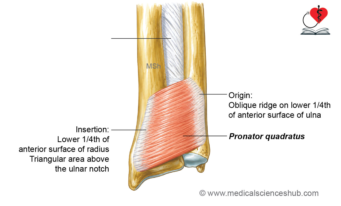
Pronator Quadratus
- The pronator quadratus is the deepest muscle of the anterior compartment of the forearm.
- It is located in front of the lower parts of the radius and ulna.
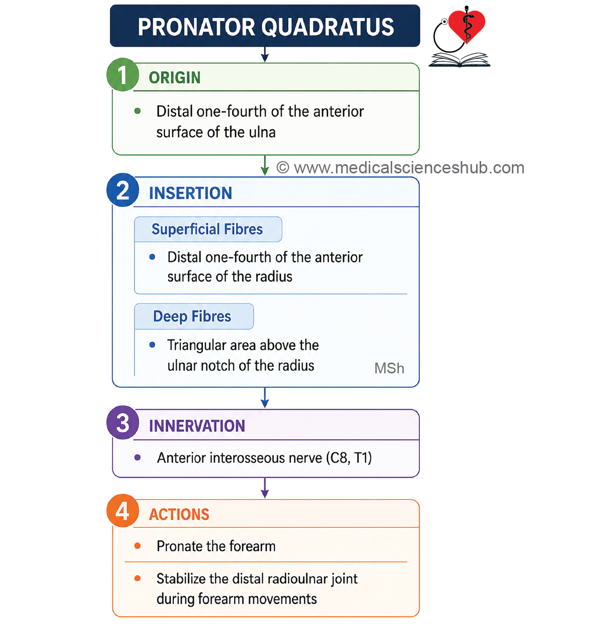
Origin
- The pronator quadratus arises from the oblique ridge on the lower one-fourth of the anterior surface of the ulna and the adjacent area on its medial side.
Direction of Fibers
- The muscle fibers run horizontally from the ulna to the radius. This horizontal arrangement is a distinctive feature of the muscle.
Insertion
- The superficial fibers are attached to the anterior surface and anterior border of the lower one-fourth of the radius.
- The deep fibers are attached to the triangular area above the ulnar notch of the radius.
Innervation
The pronator quadratus is supplied by the anterior interosseous nerve, which is a branch of the median nerve.
Actions
- The superficial fibers produce pronation of the forearm.
- The deep fibers help stabilize the lower ends of the radius and ulna, especially during movements of the forearm.
CLINICAL ANATOMY
- Clinical Testing: The function of the pronator quadratus is tested by asking the patient to pronate the forearm against resistance.
- To expose the pronator quadratus during dissection, the lower part of the flexor digitorum profundus is retracted medially and the flexor pollicis longus is retracted laterally.
- The pronator quadratus is the main muscle responsible for pronation of the forearm. Its action is assisted by the pronator teres.
Arteries of Front of Forearm
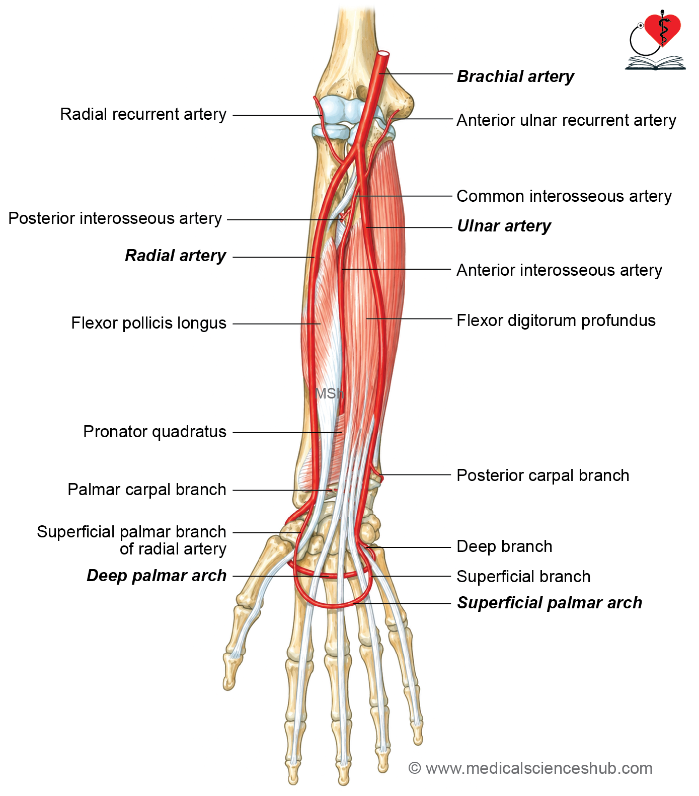
- Anterior compartment of the forearm contains the following main arteries:
- Ulnar artery
- Anterior interosseous artery
- Radial artery
Ulnar Artery
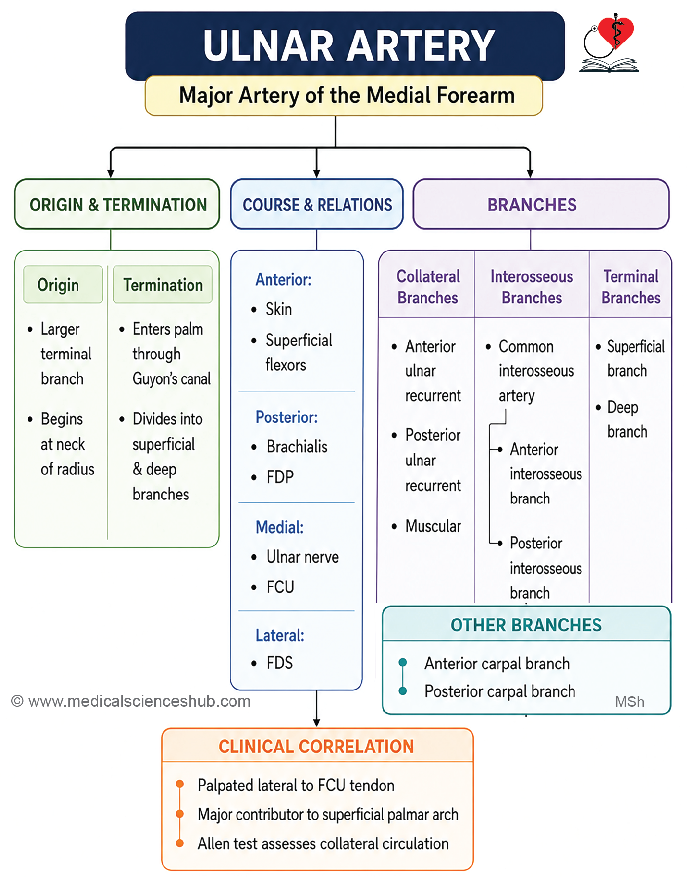
- The ulnar artery is the larger terminal branch of the brachial artery.
- It is the main artery supplying the medial side of the forearm and a large part of the hand.
Beginning
- The ulnar artery begins in the cubital fossa as a terminal branch of the brachial artery.
- It arises at the level of the neck of the radius.
Course in the forearm
- In the upper one-third of the forearm, the ulnar artery passes downward and medially deep to the pronator teres muscle.
- In the lower two-thirds of the forearm, it descends vertically toward the wrist.
- At the wrist, the artery passes superficial to the flexor retinaculum and enters the palm.
Termination
In the palm, the ulnar artery divides into two terminal branches:
- Superficial branch, which forms the superficial palmar arch.
- Deep branch, which contributes to the deep palmar arch and supplies the muscles of the palm.
Relations
Anterior
- In the upper part of the forearm, the artery is covered by the five superficial flexor muscles.
- In the lower part of the forearm, it lies just beneath the skin and fascia.
Posterior
- In the cubital fossa, it lies on the brachialis muscle.
- In the rest of the forearm, it lies on the flexor digitorum profundus.
- At the wrist, it lies in front of the flexor retinaculum.
Medial
- Flexor carpi ulnaris
- Ulnar nerve
Lateral
- Flexor digitorum superficialis
Branches
The ulnar artery gives off the following branches:
- Anterior ulnar recurrent artery
- Posterior ulnar recurrent artery
- Common interosseous artery
- Anterior ulnar carpal branch
- Posterior ulnar carpal branch
- Terminal branches
Superficial branch, which continues as the superficial palmar arch
Deep branch, which contributes to the deep palmar arch
Key Description
- The ulnar artery passes deep to the pronator teres, lies lateral to the ulnar nerve and the flexor carpi ulnaris, and crosses the flexor retinaculum superficially.
- The artery should not be pulled during dissection near its origin because it is firmly fixed by the posterior interosseous artery.
- The anterior and posterior ulnar recurrent arteries take part in the arterial anastomosis around the elbow.
- The common interosseous artery arises from the posterolateral side of the ulnar artery and quickly divides into the anterior and posterior interosseous arteries.
- The anterior interosseous artery runs on the anterior surface of the interosseous membrane between the flexor pollicis longus and flexor digitorum profundus, along with the anterior interosseous nerve.
- The posterior interosseous artery enters the posterior compartment of the forearm through an opening above the interosseous membrane. It gives off the interosseous recurrent artery, which also participates in the arterial anastomosis around the elbow.
- The anterior and posterior ulnar carpal branches contribute to the arterial anastomosis around the wrist joint.
Anterior Interosseous Artery
The anterior interosseous artery is a terminal branch of the common interosseous artery.
It is also known as the volar interosseous artery.
Course
- The artery descends along the anterior surface of the interosseous membrane.
- It travels together with the anterior interosseous nerve.
- Near the upper border of the pronator quadratus, the artery pierces the interosseous membrane to enter the posterior compartment of the forearm.
- It then passes deep to the extensor retinaculum and joins the dorsal carpal arch.
Branches
The anterior interosseous artery gives off the following branches:
- Muscular branches to the deep muscles of the forearm.
- Nutrient arteries to the radius and ulna.
- A median artery, present in about 8% of individuals, which accompanies the median nerve.
Radial Artery
- The radial artery is the smaller terminal branch of the brachial artery.
- It supplies the lateral side of the forearm and contributes to the blood supply of the hand.
- The radial artery is the most commonly used artery for palpating the pulse.
Beginning
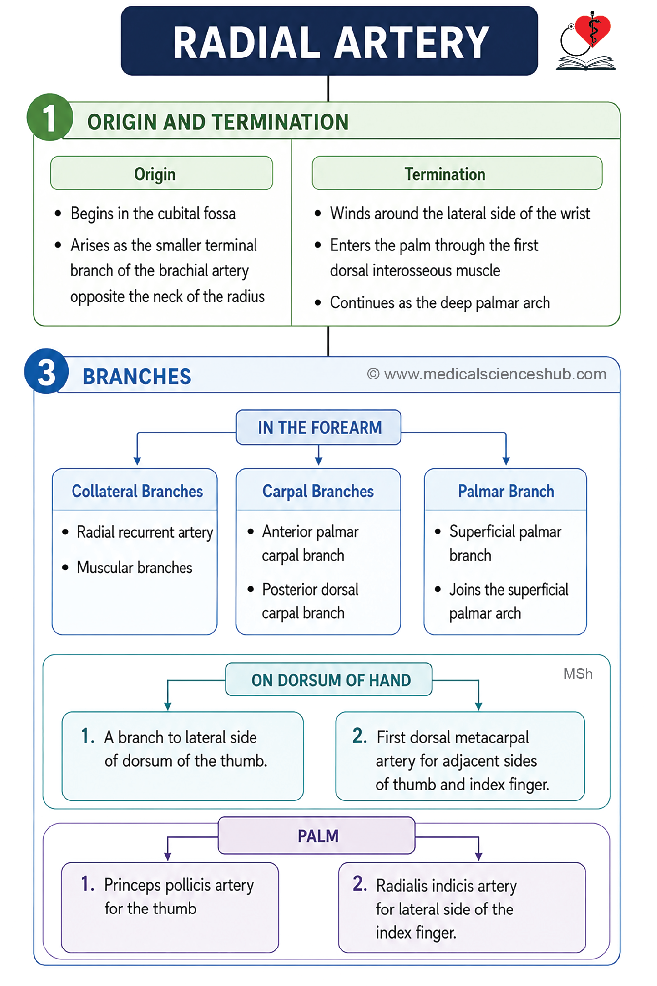
- The radial artery begins in the cubital fossa as a terminal branch of the brachial artery.
- It arises at the level of the neck of the radius.
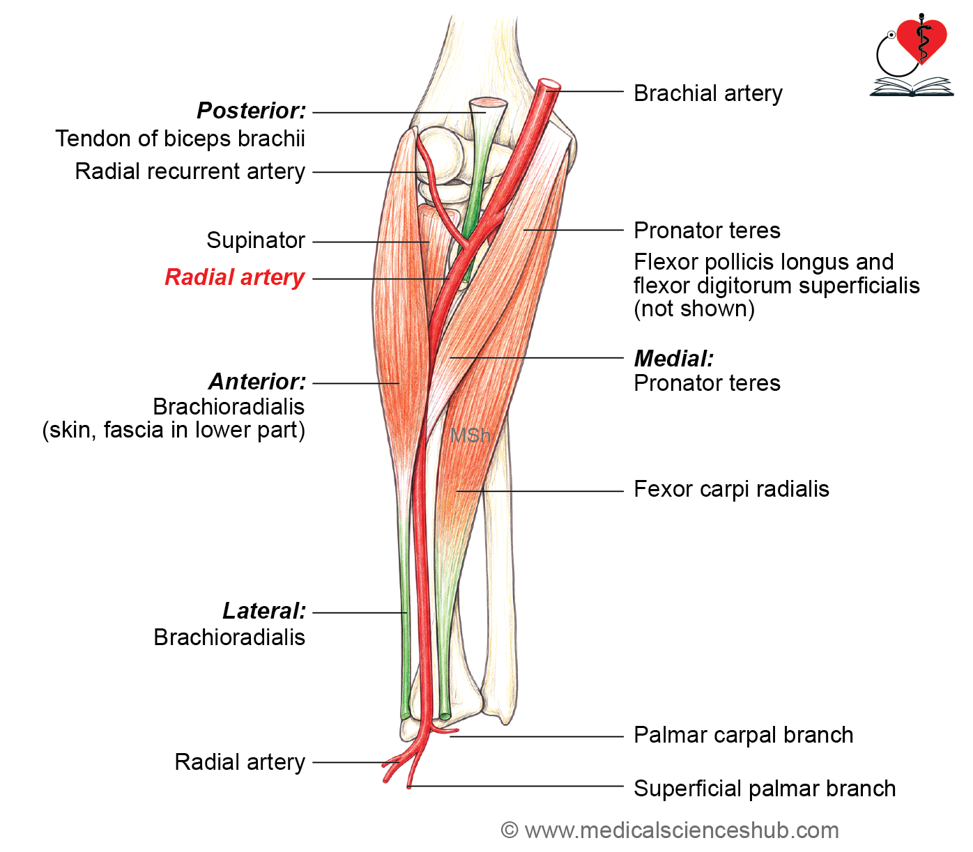
Course
- The artery descends toward the wrist with a slight lateral curve.
- Near the wrist, it leaves the front of the forearm by winding around the lateral side of the wrist.
- It then passes through the anatomical snuffbox.
- From the anatomical snuffbox, the artery enters the first interosseous space.
- It passes between the two heads of the first dorsal interosseous muscle and enters the palm.
- In the palm, it continues as the deep palmar arch.
Termination
The radial artery ends by continuing as the deep palmar arch.
Relations in forearm
Anterior
- In the upper part of the forearm, it is covered by the brachioradialis.
- In the lower part of the forearm, it lies beneath the skin and fascia.
Posterior
From above downward, it lies on:
- Biceps brachii tendon
- Supinator
- Pronator teres
- Flexor pollicis longus
- Flexor digitorum superficialis
Medial
- Pronator teres in the upper part of the forearm.
- Tendon of flexor carpi radialis in the lower part of the forearm.
Lateral
- Brachioradialis
Branches
The radial artery gives off the following branches:
- Muscular branches
- Radial recurrent branch
- Anterior (palmar) carpal branch
- Posterior (dorsal) carpal branch
- Superficial palmar branch – completes superficial palmar arch
- The radial recurrent artery arises just below the elbow and participates in the arterial anastomosis around the elbow.
- The anterior (palmar) carpal branch arises near the lower border of the pronator quadratus. It joins the corresponding branch of the ulnar artery deep to the flexor tendons.
- The posterior (dorsal) carpal branch joins the dorsal carpal branch of the ulnar artery to form the dorsal carpal arch.
- The superficial palmar branch arises just above the wrist. It passes superficial to the flexor retinaculum and contributes to the superficial palmar arch.
CLINICAL INTEGRATION
- The radial pulse is the pulse most commonly examined during clinical assessment.
- It is felt in the lower part of the forearm, where the radial artery lies against the anterior surface of the lower end of the radius.
- The artery is palpated just lateral to the tendon of the flexor carpi radialis.
Nerves of Front of Forearm
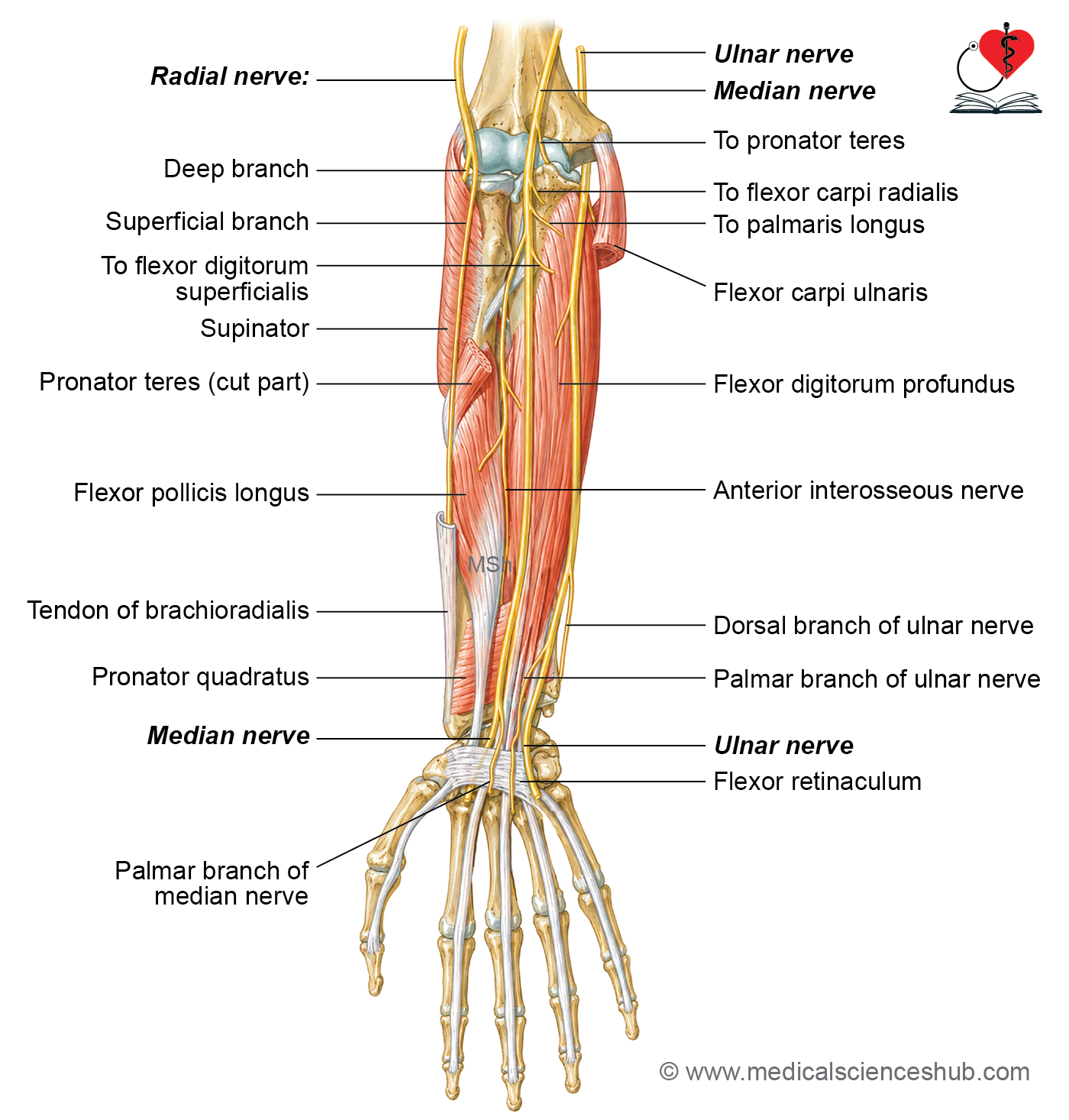
- The front of the forearm has ulnar nerve, median nerve, and radial nerve.
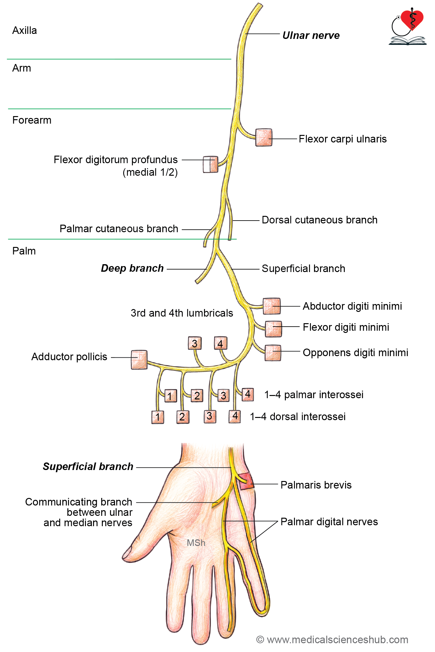
Ulnar Nerve
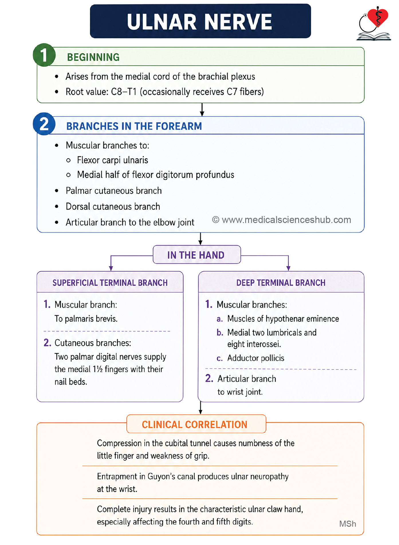
- The ulnar nerve is a terminal branch of the medial cord of the brachial plexus.
- Its root value is C8 and T1, and it may receive a contribution from C7.
Course and main relations
In the Axilla
- The ulnar nerve arises in the lower part of the axilla.
- It runs between the axillary artery and the axillary vein.
In the Arm
- The nerve descends along the medial side of the brachial artery up to the insertion of the coracobrachialis.
- It then pierces the medial intermuscular septum along with the superior ulnar collateral artery to enter the posterior compartment of the arm.
- It continues downward behind the medial epicondyle of the humerus.
In the Forearm
- The ulnar nerve enters the forearm by passing between the two heads of the flexor carpi ulnaris. This passage is called the cubital tunnel.
- It then descends vertically toward the wrist.
- At the wrist, it passes superficial to the flexor retinaculum, just lateral to the pisiform bone, and medial to the ulnar artery.
In the Palm
- At the distal border of the flexor retinaculum, the ulnar nerve divides into superficial and deep terminal branches.
Branches and distribution
In the Axilla and Arm
- The ulnar nerve does not give any branches in the axilla or the arm.
In the Forearm
- It gives muscular branches to:
- Flexor carpi ulnaris
- Medial half of the flexor digitorum profundus
- It gives articular branches to the elbow joint.
- It gives a palmar cutaneous branch, which supplies the skin over the hypothenar eminence.
- It gives a dorsal cutaneous branch, which supplies the proximal part of the dorsum of the medial two and a half fingers and the adjacent area of the dorsum of the hand.
In the Palm
- The nerve ends by dividing into superficial and deep terminal branches, which supply the muscles and skin of the hand.
CLINICAL ANATOMY
- The palmar cutaneous branch arises just above the wrist and enters the palm by passing superficial to the flexor retinaculum along with the ulnar nerve.
- The dorsal cutaneous branch arises about 7.5 cm above the wrist beneath the flexor carpi ulnaris. It winds around the ulna to reach the back of the forearm and hand.
- In the lower part of the forearm, the ulnar nerve lies superficially. It is medial to the tendon of the flexor carpi ulnaris and medial to the ulnar artery, while the ulnar artery lies lateral to the nerve.
- Ulnar paradox refers to the phenomenon in which a high lesion of the ulnar nerve produces a less obvious claw hand deformity than a low lesion, because paralysis of the flexor digitorum profundus reduces flexion of the distal interphalangeal joints of the ring and little fingers.
Median Nerve
- The median nerve is the main nerve of the anterior compartment of the forearm.
- It is sometimes called the “laborer’s nerve” because it supplies most of the muscles responsible for powerful movements of the forearm and thumb.
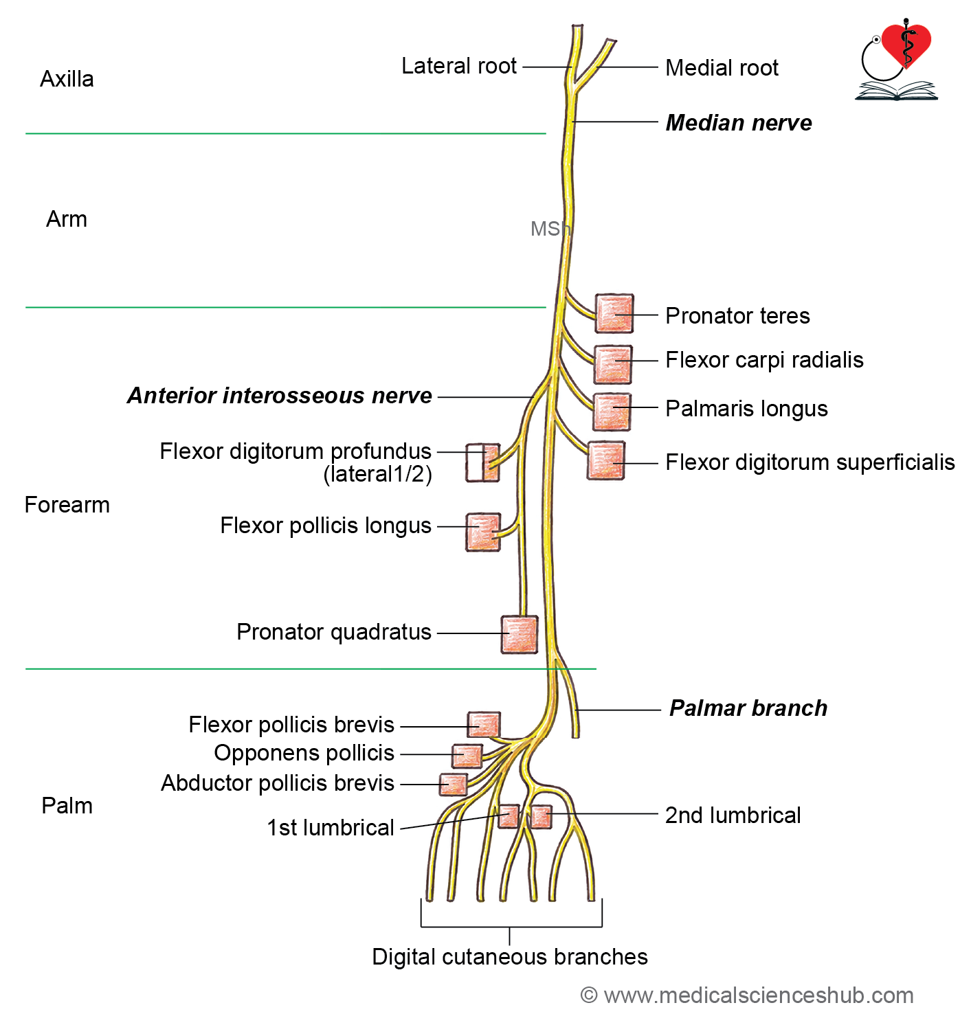
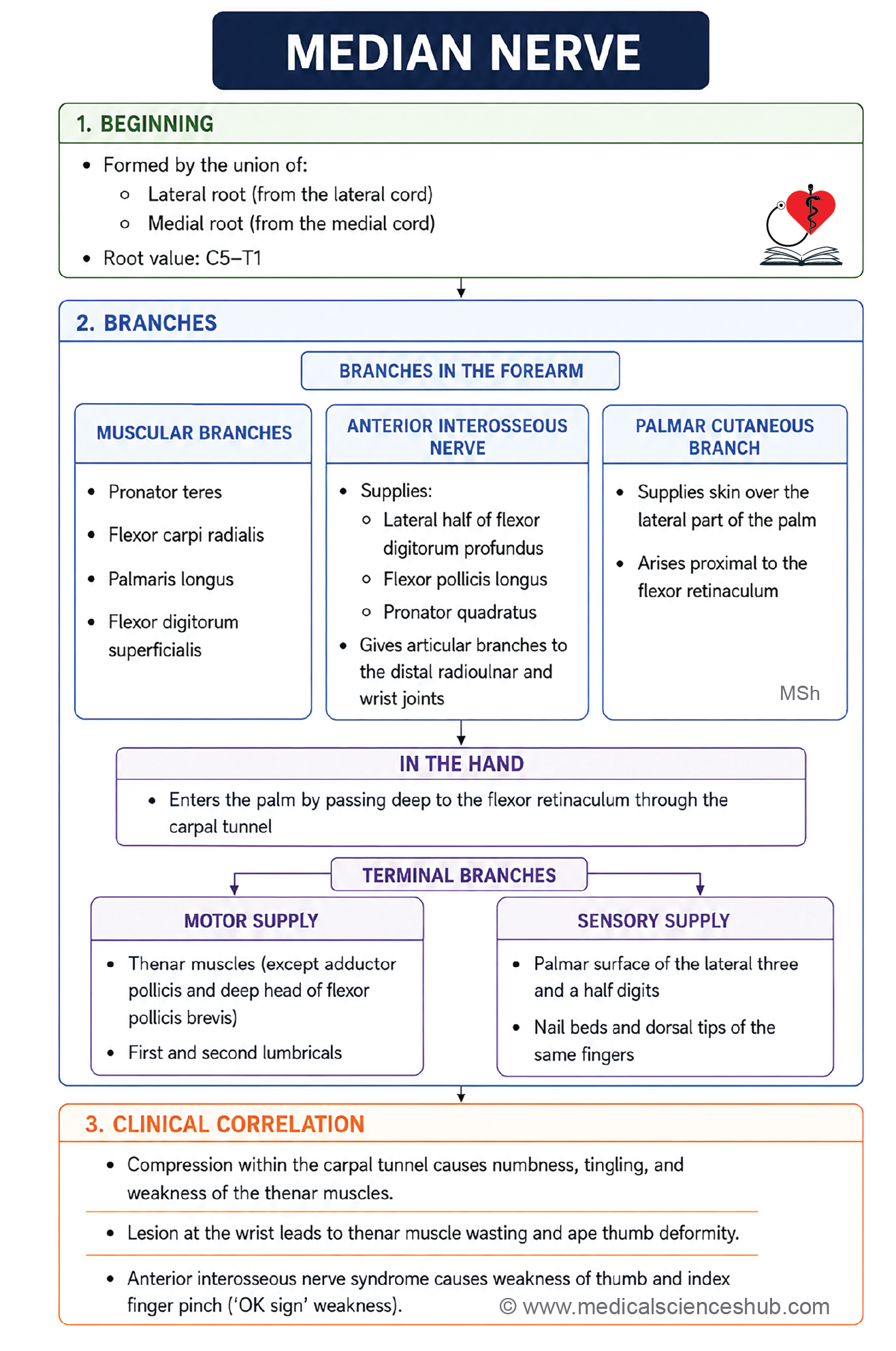
- The median nerve is formed by the union of the medial and lateral roots from the medial and lateral cords of the brachial plexus in the axilla.
Course and main relations
In the Axilla
- The median nerve is formed in the lower part of the axilla.
- It crosses in front of the axillary artery and then continues anterior to the brachial artery, moving from its lateral side to its medial side.
In the Cubital Fossa
- The median nerve lies medial to the brachial artery.
In the Forearm
- The nerve enters the forearm by passing between the two heads of the pronator teres.
- At this point, the deep head of the pronator teres separates the median nerve from the ulnar artery.
- It then passes deep to the fibrous arch of the flexor digitorum superficialis.
- About 5 cm above the flexor retinaculum, the nerve becomes superficial and appears lateral to the tendons of the flexor digitorum superficialis.
At the Wrist
- The median nerve passes deep to the flexor retinaculum through the carpal tunnel to enter the palm.
In the Palm
- The nerve lies lateral to the thenar muscles.
- It divides into terminal branches that supply the thenar muscles, the first and second lumbricals, and the lateral three and a half digits.
Branches
In the Axilla
- The median nerve gives no branches.
In the Arm
- It gives a muscular branch to the pronator teres just above the elbow.
In the Cubital Fossa
- It gives muscular branches to:
- Flexor carpi radialis
- Palmaris longus
- Flexor digitorum superficialis
In the Forearm
- It gives the anterior interosseous nerve, which supplies:
- Flexor pollicis longus
- Lateral half of the flexor digitorum profundus
- Pronator quadratus
- Inferior radioulnar joint
- Wrist joint
- It also gives a palmar cutaneous branch, which supplies the skin over the thenar eminence.
In the Palm
- The recurrent muscular branch supplies the thenar muscles:
- Abductor pollicis brevis
- Opponens pollicis
- Superficial head of flexor pollicis brevis
- It also supplies the first and second lumbricals.
- Its digital branches supply the skin of the lateral three and a half fingers, including the nail beds.
Key Description
- The median nerve crosses the brachial artery from its lateral side to its medial side in the arm.
- The anterior interosseous nerve arises from the median nerve in the upper part of the forearm as the median nerve passes between the two heads of the pronator teres.
- The anterior interosseous nerve descends on the anterior surface of the interosseous membrane between the flexor pollicis longus and flexor digitorum profundus, accompanied by the anterior interosseous artery.
- It supplies the flexor pollicis longus, the lateral half of the flexor digitorum profundus, the pronator quadratus, and the wrist and inferior radioulnar joints.
- The anterior interosseous nerve enters the pronator quadratus from its deep surface.
- The palmar cutaneous branch passes superficial to the flexor retinaculum. Therefore, the skin over the thenar eminence is usually spared in carpal tunnel syndrome.
Important Questions
- Write a short note on pronator teres muscle.
- List the branches of ulnar artery.
- Write a short note on radial artery.
📝 Test Your Knowledge – Practice MCQs
Attempt the chapter MCQ quiz and assess your understanding of key concepts.
Click to see: Medical Disclaimer and Copyright Notice