
Competencies
- AN12.3: Identify and describe flexor retinaculum with its attachments.
- AN12.4: Explain anatomical basis of carpal tunnel syndrome.
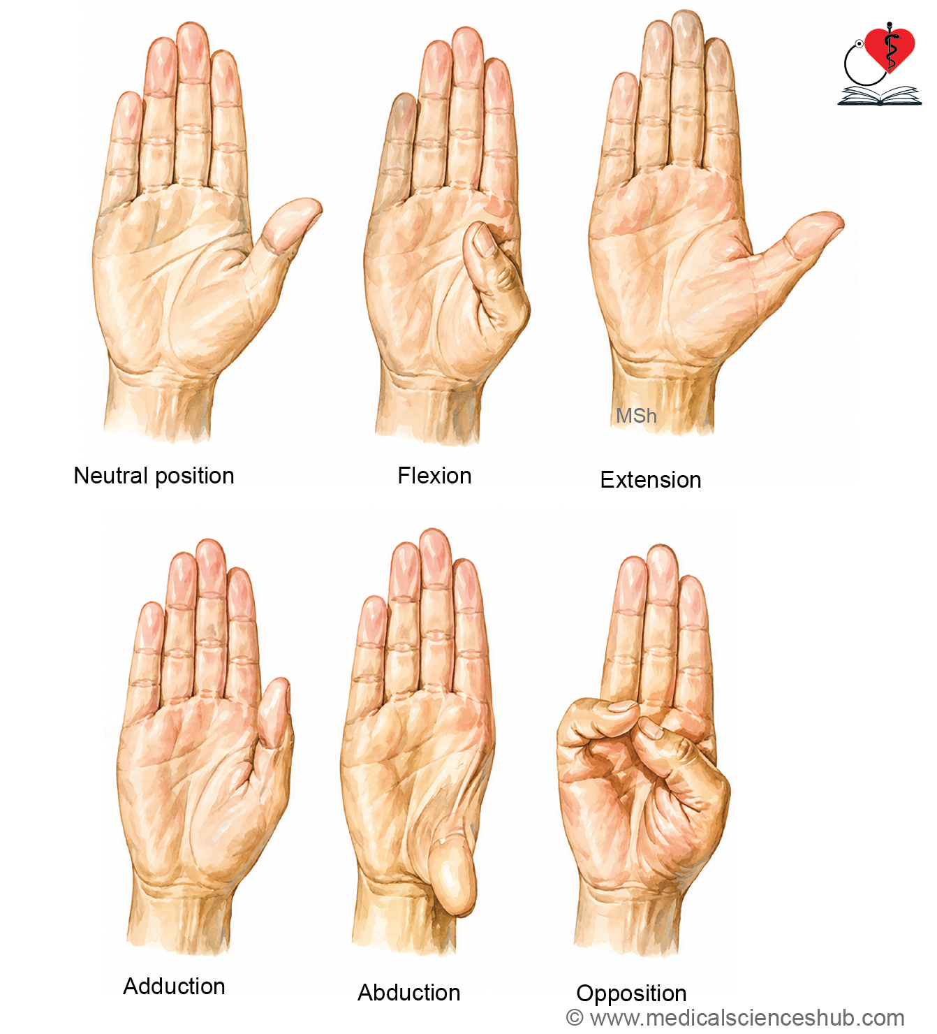
- AN12.5: Identify and describe small muscles of hand. Also describe movements of thumb and muscles involved.
- AN12.6: Describe and demonstrate movements of thumb and muscles involved.
- AN12.7: Identify and describe course and branches of important blood vessels and nerves in hand.
- AN12.8: Describe anatomical basis of claw hand.
- AN12.9: Identify and describe fibrous flexor sheaths, ulnar bursa, radial bursa, and digital synovial sheaths.
- AN12.10: Explain infection of fascial spaces of palm.
Introduction
- The hand is the distal (terminal) part of the upper limb.
- It is divided into:
- Palm: the anterior (front) surface.
- Dorsum: the posterior (back) surface.
- Five fingers: thumb, index, middle, ring, and little finger, arranged from lateral to medial.
- The skeleton of the hand is formed by:
- 8 carpal bones (wrist bones)
- 5 metacarpal bones (bones of the palm)
- 14 phalanges (finger bones)
- The wrist (carpus) is the region between the forearm and the hand. It is made up of eight carpal bones arranged in two rows.
- The skin of the palm is thick and specially adapted for a firm grip. Histologically, it is thick skin, characterized by the presence of a stratum lucidum and a very thick stratum corneum in the epidermis.
Flexion Creases of Hand
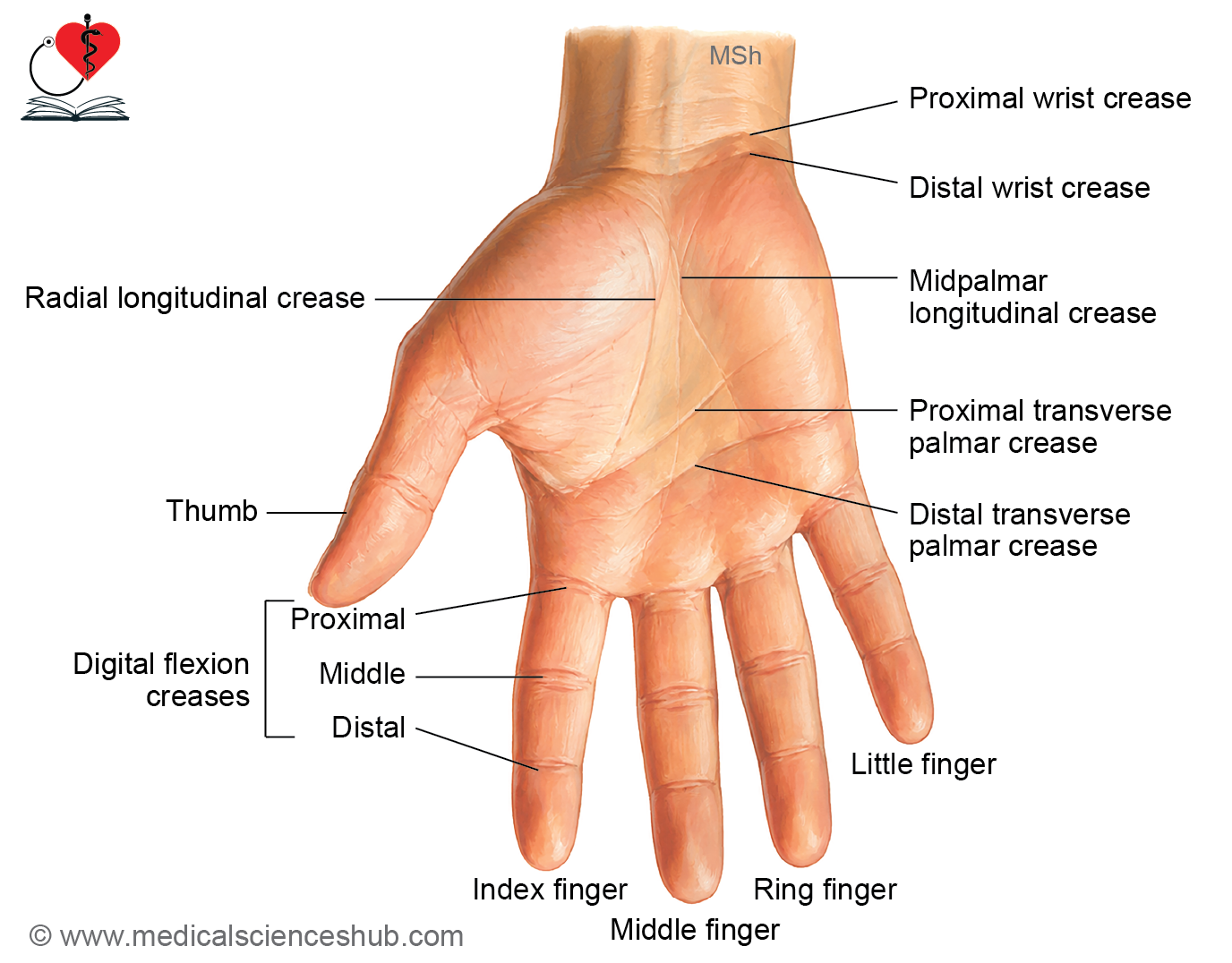
The skin of the hand shows several flexion creases (skin folds) that develop at sites of repeated movement of the joints.
Flexion creases of wrist
There are two transverse wrist creases:
- Proximal transverse wrist crease
- A short transverse crease.
- Lies opposite the wrist joint.
- Distal transverse wrist crease
- A complete transverse crease.
- Corresponds to the proximal border of the flexor retinaculum.
Creases of palm
Although the palm has many skin creases, four are consistently present:
- Radial longitudinal crease
- Separates the thenar eminence from the rest of the palm.
- Commonly called the lifeline in palmistry.
- Midpalmar longitudinal crease
- Separates the hypothenar eminence from the central part of the palm.
- Known as the line of fate in palmistry.
- Proximal transverse palmar crease
- Extends medially from the distal end of the radial longitudinal crease.
- Distal transverse palmar crease
- Extends medially from the interdigital cleft between the index and middle fingers.
Digital flexion creases
Each finger has three flexion creases:
- Proximal digital flexion crease
- Located at the base of the finger.
- Lies about 2 cm distal to the metacarpophalangeal (MP) joint.
- Middle digital flexion crease
- Corresponds to the proximal interphalangeal (PIP) joint.
- Distal digital flexion crease
- Corresponds to the distal interphalangeal (DIP) joint.
Superficial Fascia of Palm
- The superficial fascia of the palm is made up of dense fibrous bands and subcutaneous fat.
- Fibrous septa extend from the skin to the deep fascia, firmly anchoring the skin.
Features of the Superficial Fascia
1. Fat Cushions
- Fibrous septa divide the subcutaneous fat into small compartments.
- These fat-filled compartments act as cushions, protecting the palm during gripping and grasping.
2. Superficial Transverse Metacarpal (Natatory) Ligament
- Formed by condensations of the superficial fascia.
- Extends across the bases of the fingers.
- Protects the digital nerves and vessels.
- Helps prevent excessive separation of adjacent fingers.
3. Palmaris Brevis Muscle
- A thin subcutaneous muscle located in the superficial fascia over the hypothenar eminence.
- Consists of transversely arranged muscle fibers.
- It wrinkles the skin on the medial side of the palm and improves grip.
CLINICAL ANATOMY
- Palmaris brevis is an example of the panniculus carnosus, a subcutaneous muscle.
- It is identified as a thin sheet of transverse muscle fibers lying in the superficial fascia over the hypothenar eminence.
- The skin of the palm is difficult to separate from the deep fascia because fibrous septa firmly anchor it to the underlying palmar aponeurosis. This provides stability during forceful hand movements.
Deep Fascia of Palm
- Deep fascia of hand forms the following modifications:
- Flexor retinaculum
- Palmar aponeurosis
- Fibrous flexor sheaths in fingers
Flexor Retinaculum
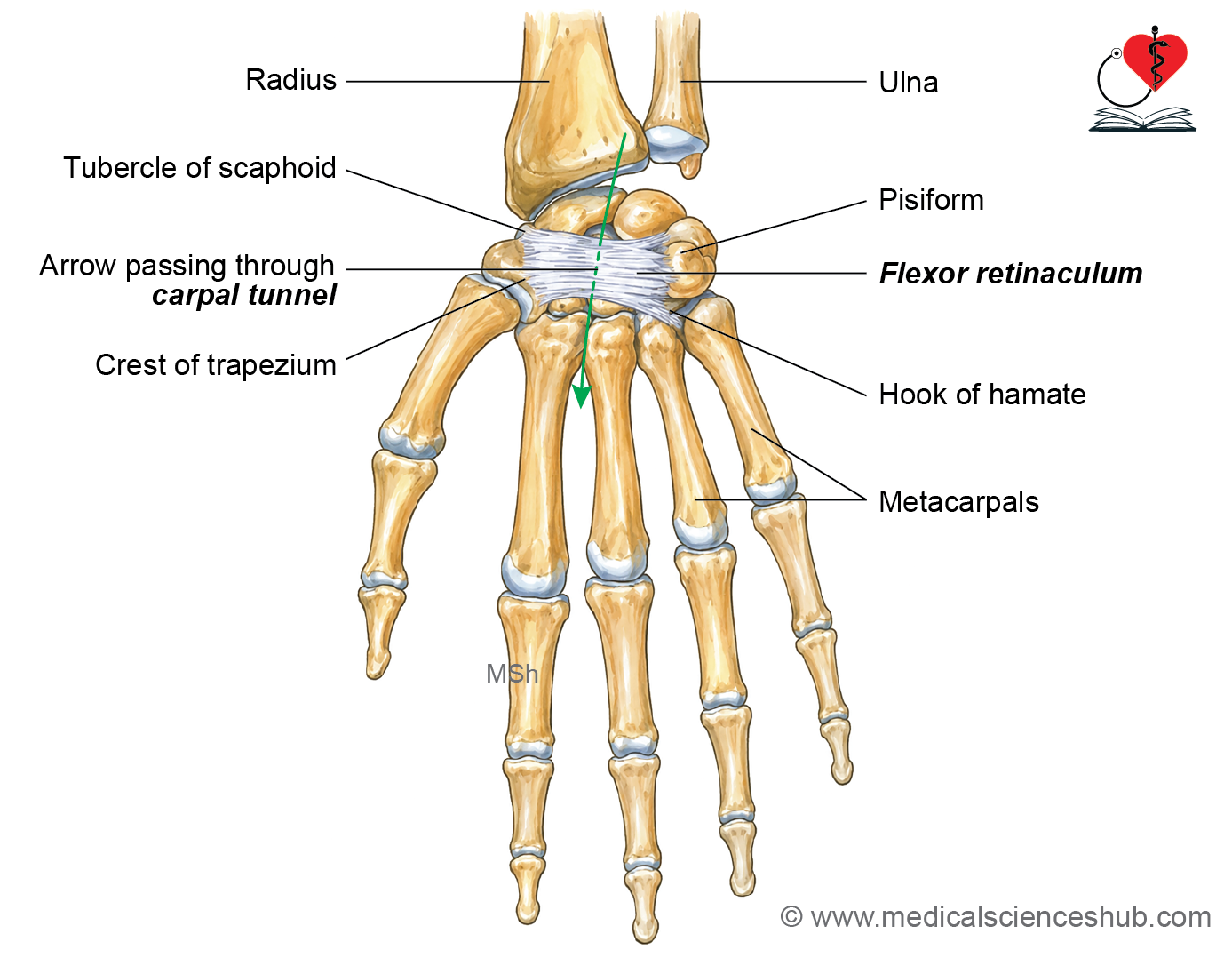
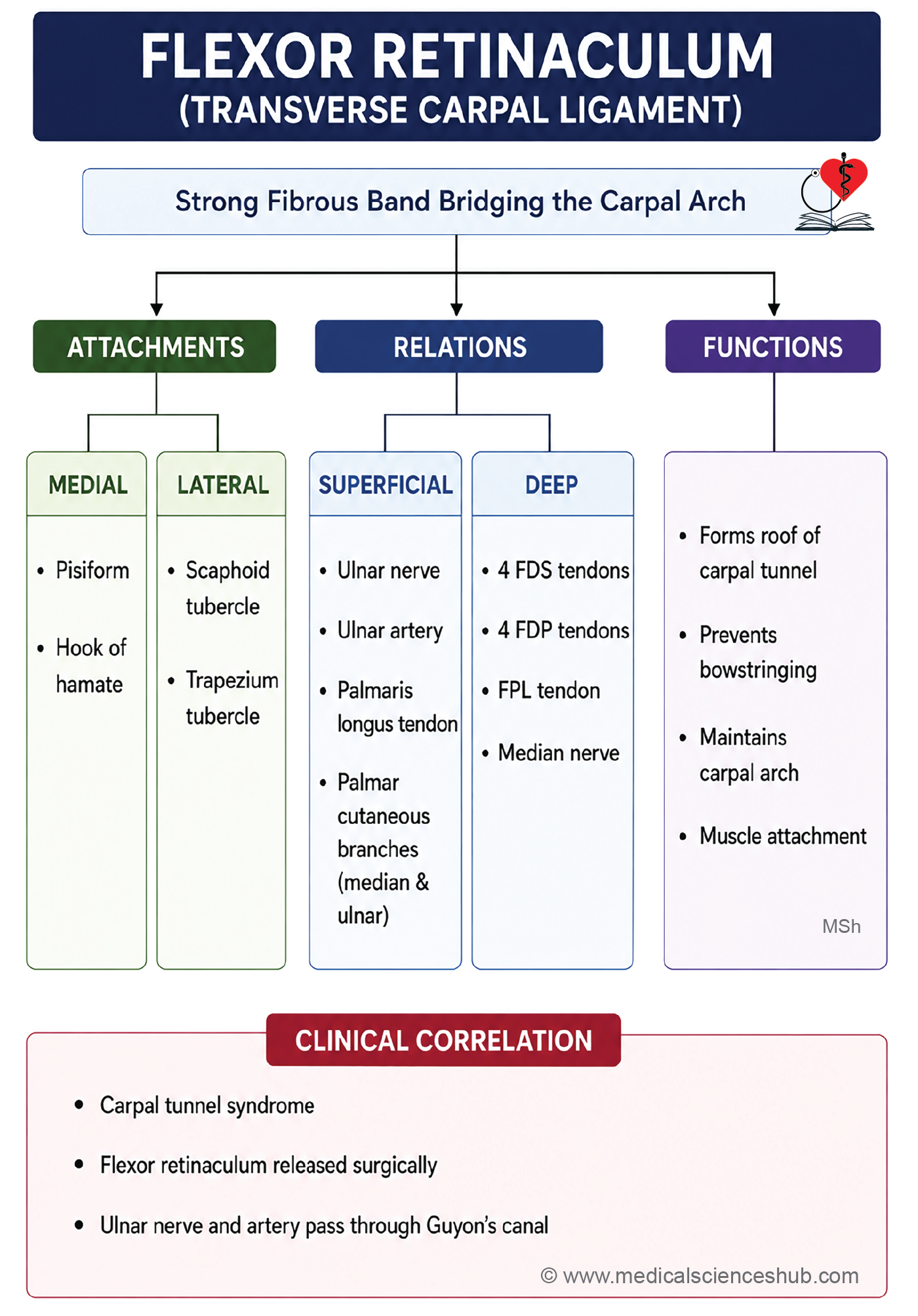
- The flexor retinaculum is a strong, thickened band of deep fascia that bridges the concavity of the carpal bones on the anterior aspect of the wrist.
- It is also known as the transverse carpal ligament.
- It converts the carpal groove into the carpal tunnel, through which the flexor tendons and median nerve enter the hand.
Attachments
Medial attachment
- Pisiform
- Hook of hamate
Lateral attachment
- Tubercle of scaphoid
- Crest of trapezium
Dimension
- Length: Approximately 2.5 cm
- Width: Approximately 2.5 cm
Tunnels formed by flexor retinaculum
- Tunnel for the Ulnar Nerve and Ulnar Vessels (Guyon’s Canal)
- Medially, the flexor retinaculum gives off a superficial slip, known as the volar carpal ligament.
- This ligament is attached to the pisiform.
- The ulnar nerve and ulnar vessels pass between the volar carpal ligament and the flexor retinaculum, forming Guyon’s canal.
- Tunnel for the Tendon of Flexor Carpi Radialis
- Laterally, the flexor retinaculum gives off a deep slip attached to the medial lip of the groove on the trapezium.
- The tendon of flexor carpi radialis (FCR) passes through this fibro-osseous tunnel to enter the palm.
- The FCR tendon does not pass through the carpal tunnel.
Carpal tunnel
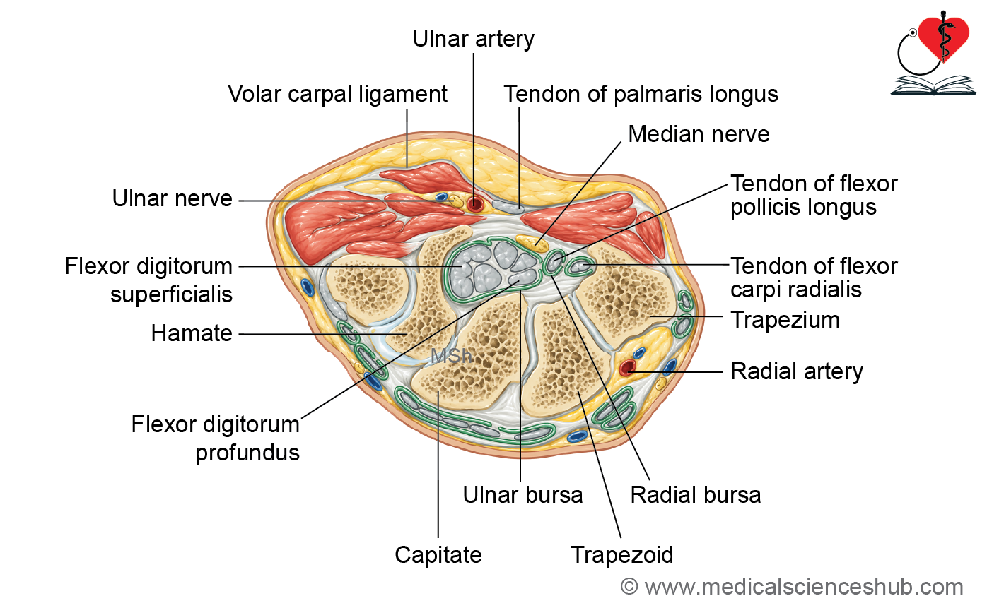
The carpal tunnel is a narrow osseofibrous passage at the wrist.
- Anterior boundary (roof): Flexor retinaculum
- Posterior boundary (floor): Concavity of the carpal bones
It provides passage for the long flexor tendons of the fingers and thumb and the median nerve from the forearm into the hand.
Relations
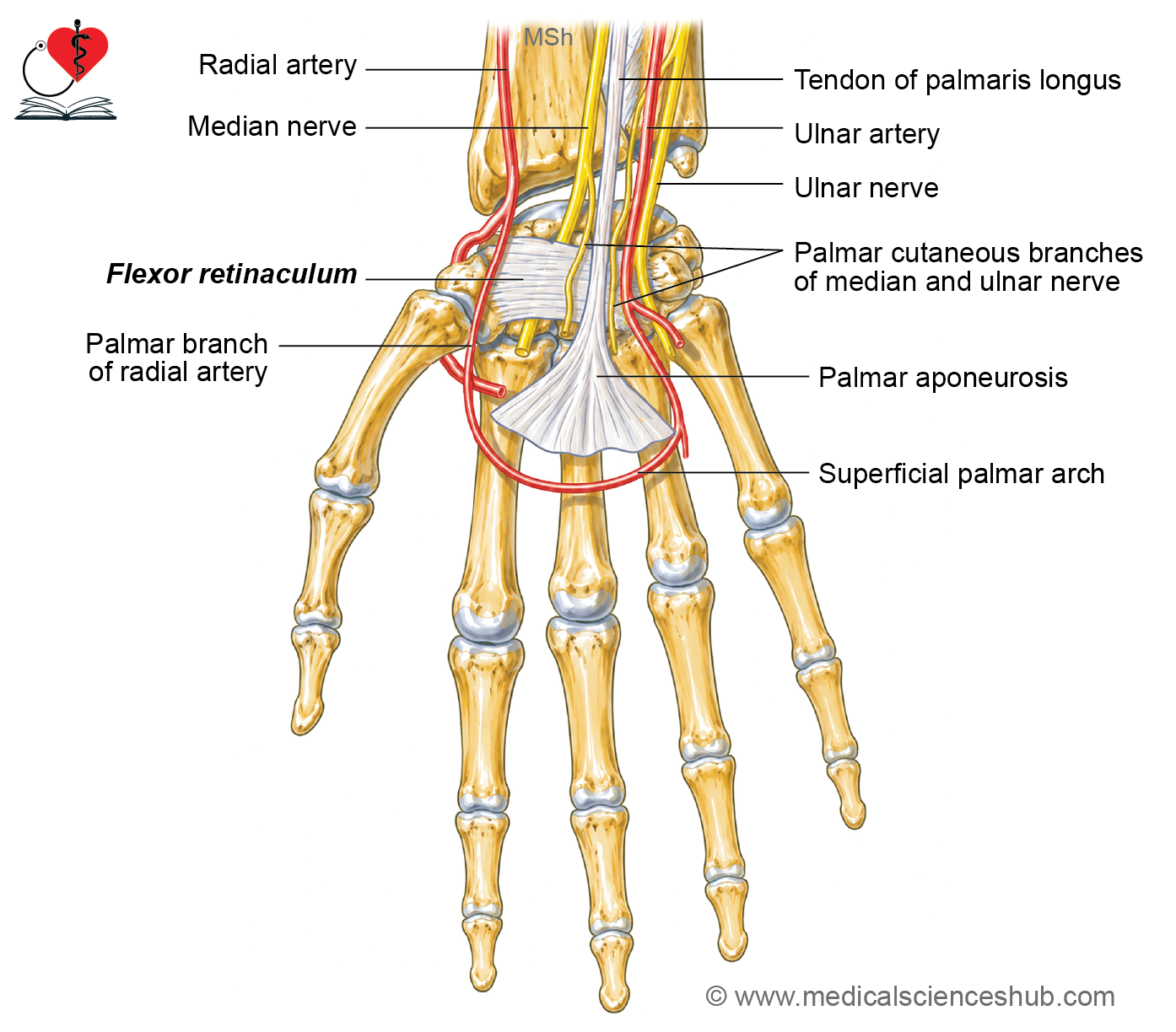
Structures Passing Superficial to the Flexor Retinaculum
(From medial to lateral)
- Ulnar nerve
- Ulnar artery
- Palmar cutaneous branch of the ulnar nerve
- Tendon of palmaris longus
- Palmar cutaneous branch of the median nerve
Note: The palmar cutaneous branch of the median nerve passes superficial to the flexor retinaculum and therefore is not affected in carpal tunnel syndrome.
Structures Passing Through the Carpal Tunnel
The carpal tunnel contains the following structures:
- Median nerve
- Four tendons of flexor digitorum superficialis (FDS)
- Four tendons of flexor digitorum profundus (FDP)
- Tendon of flexor pollicis longus (FPL)
- Ulnar bursa (common flexor synovial sheath)
- Radial bursa (synovial sheath of FPL tendon)
Palmar Aponeurosis
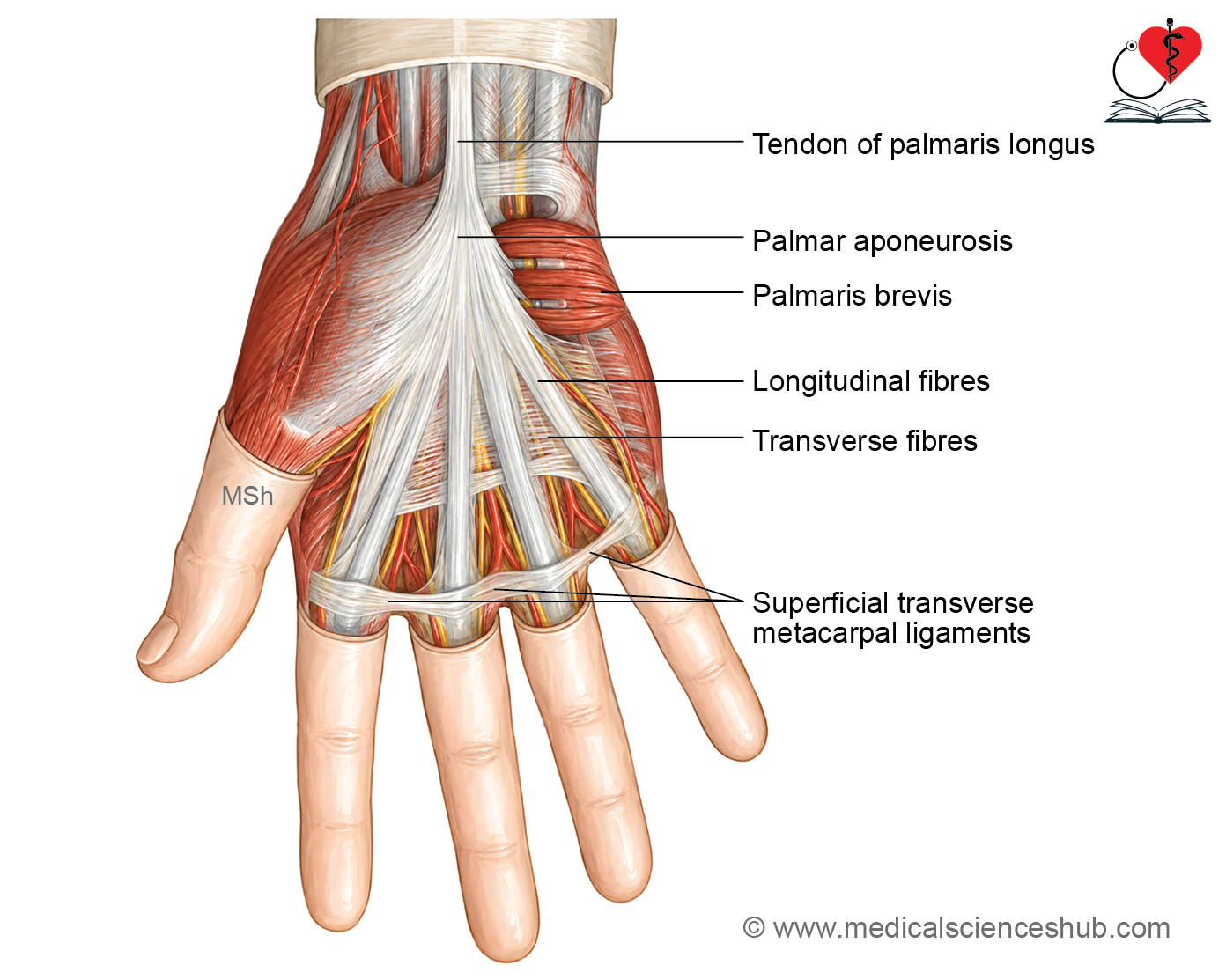
- The palmar aponeurosis is the thickened central part of the deep fascia of the palm.
- It is triangular in shape, with the apex directed proximally (towards the wrist) and the base directed distally (towards the fingers).
- It is considered a degenerated tendon of the palmaris longus muscle.
Features
1. Apex
- Directed towards the wrist.
- Blends with the flexor retinaculum.
- Receives the tendon of the palmaris longus muscle (when present).
2. Base
- Directed towards the fingers.
- Opposite the heads of the metacarpal bones, it divides into four digital slips, one for each of the medial four fingers (index, middle, ring, and little fingers).
Each digital slip further divides into:
- Superficial longitudinal fibers
- Blend with the dermis of the skin.
- Deep longitudinal fibers
- Split into medial and lateral slips.
- Become continuous with the fibrous flexor sheath of the corresponding finger.
3. Medial Border
- Continuous with the deep fascia covering the hypothenar muscles.
- Gives origin to the palmaris brevis muscle.
4. Lateral Border
- Continuous with the deep fascia covering the thenar muscles.
Functions
- Protects the underlying tendons, nerves, and blood vessels during gripping.
- Firmly anchors the skin of the palm to the deep fascia, preventing it from sliding.
- Improves the efficiency of grip and grasp by stabilizing the palmar skin.
CLINICAL INTEGRATION
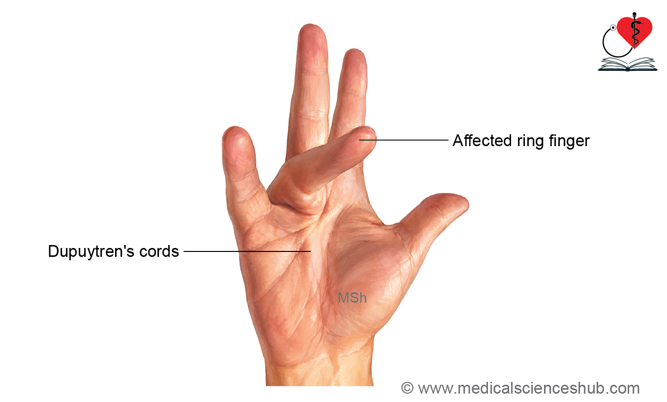
Dupuytren’s contracture is a condition characterized by progressive fibrosis, thickening, and shortening of the medial part of the palmar aponeurosis. It is also known as Viking disease.
Cause
- The exact cause is usually unknown (idiopathic).
- It may have a genetic predisposition and is more common in people of Northern European descent.
- Infection is not a common cause.
Clinical Features
- Progressive thickening and shortening of the palmar aponeurosis.
- Formation of fibrous nodules and cords in the palm.
- Permanent flexion deformity of the:
- Metacarpophalangeal (MCP) joints
- Proximal interphalangeal (PIP) joints
- The ring finger is most commonly affected, followed by the little finger.
- The distal interphalangeal (DIP) joints are usually not involved.
Treatment
- Conservative treatment
- Steroid injections (selected cases)
- Collagenase injections to dissolve fibrous cords
- Surgical treatment
- Fasciotomy (division of the fibrous cords)
- Fasciectomy (excision of the diseased palmar fascia) in more advanced cases
Palmar Septa
The palmar septa are fibrous partitions that extend from the palmar aponeurosis to the metacarpal bones. They divide the palm into compartments and provide support to the deeper structures.
Types of Palmar Septa
- Medial palmar septum
- Extends from the medial border of the palmar aponeurosis to the 5th metacarpal.
- Intermediate palmar septum
- Extends from the medial border of the palmar aponeurosis to the 3rd metacarpal.
- Separates the midpalmar space from the thenar space.
- Lateral palmar septum
- Extends from the lateral border of the palmar aponeurosis to the 1st metacarpal.
Practical Anatomy
- The palmar septa firmly attach the palmar aponeurosis to the metacarpal bones, making it difficult to separate the palmar aponeurosis from the deeper structures during dissection.
- Fibers of the palmar aponeurosis merge with the dermis in the web spaces, making it difficult to trace and clean the aponeurosis during practical dissection.
Transverse Metacarpal Ligaments
Superficial Transverse Metacarpal Ligaments (Natatory Ligaments)
- Formed by condensations of the superficial fascia.
- Located immediately beneath the skin in the web spaces between the fingers.
- Help maintain the web spaces and limit excessive separation of adjacent fingers.
Deep Transverse Metacarpal Ligaments
- Strong fibrous bands connecting the heads of the 2nd, 3rd, 4th, and 5th metacarpal bones.
- Run across the palmar aspect of the metacarpal heads.
- Help maintain the transverse arch of the hand and stabilize the metacarpals during gripping.
Fibrous Flexor Sheaths of Fingers
- The fibrous flexor sheath is a strong fibrous tunnel formed by the deep fascia on the anterior (palmar) aspect of each finger.
- It encloses the long flexor tendons and their digital synovial sheath, keeping the tendons closely applied to the phalanges during finger movements.
Extent
Extends from the head of the metacarpal bone to the base of the distal phalanx.
Attachment
- The sheath arches over the flexor tendons and is attached to the sides of the phalanges, forming an osseofibrous tunnel.
Proximally
- Fuses with the palmar aponeurosis.
- Provides an opening for the entry of the long flexor tendons.
Distally
- Closed and attached to the base of the distal phalanx.
Thus, the fibrous sheath forms a blind osseofibrous tunnel, open only proximally, through which pass:
- Tendons of flexor digitorum superficialis (FDS)
- Tendons of flexor digitorum profundus (FDP)
- Tendon of flexor pollicis longus (FPL) (thumb)
- Their digital synovial sheaths
- The sheath is thin over the interphalangeal joints, allowing free flexion and extension of the fingers.
Pattern of fibers of fibrous flexor sheath
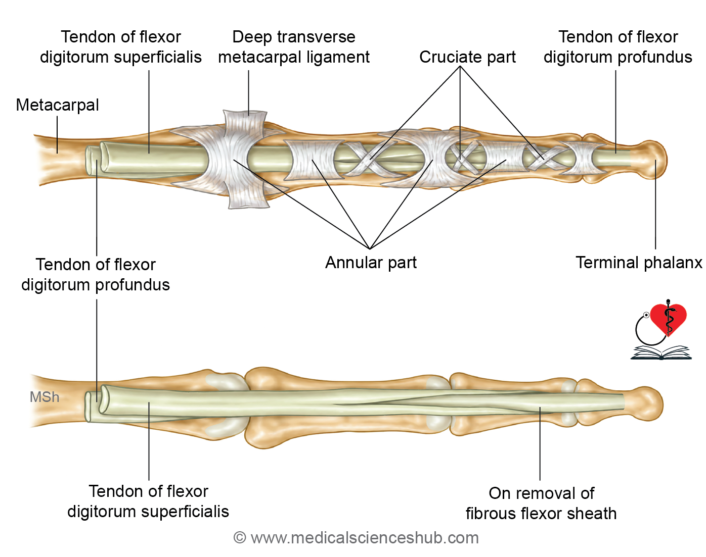
The fibrous flexor sheath consists of two types of reinforcing fibers:
- Annular (transverse) fibers
- Form strong circular bands around the tendons.
- Prevent bowstringing of the tendons during flexion.
- Cruciate fibers
- Cross each other in an X-shaped pattern.
- Provide flexibility while maintaining the tendons close to the bones.
Together, these fibers give the sheath strength and flexibility.
Function
- Holds the FDS, FDP, and FPL tendons close to the phalanges.
- Prevents bowstringing of the flexor tendons during finger flexion.
- Provides a smooth osseofibrous tunnel for efficient tendon movement.
- Improves the mechanical efficiency of finger flexion.
CLINICAL INTEGRATION
Trigger finger (stenosing tenosynovitis)
- Trigger finger is caused by localized thickening of a long flexor tendon, usually at the level of the fibrous flexor sheath.
- The thickened tendon cannot glide smoothly through the fibrous flexor sheath during finger movements.
- As a result, the finger locks in a flexed position and cannot be extended easily. Extension often occurs suddenly with a painful “snap” or “trigger” action.
- Treatment
- Local corticosteroid injection into the tendon sheath.
- Surgical release (excision or division) of the constricting part of the fibrous flexor sheath if conservative treatment fails.
Synovial Sheaths of Long Flexor Tendons
- The long flexor tendons pass through osseofibrous tunnels, such as the carpal tunnel and the fibrous flexor sheaths of the fingers.
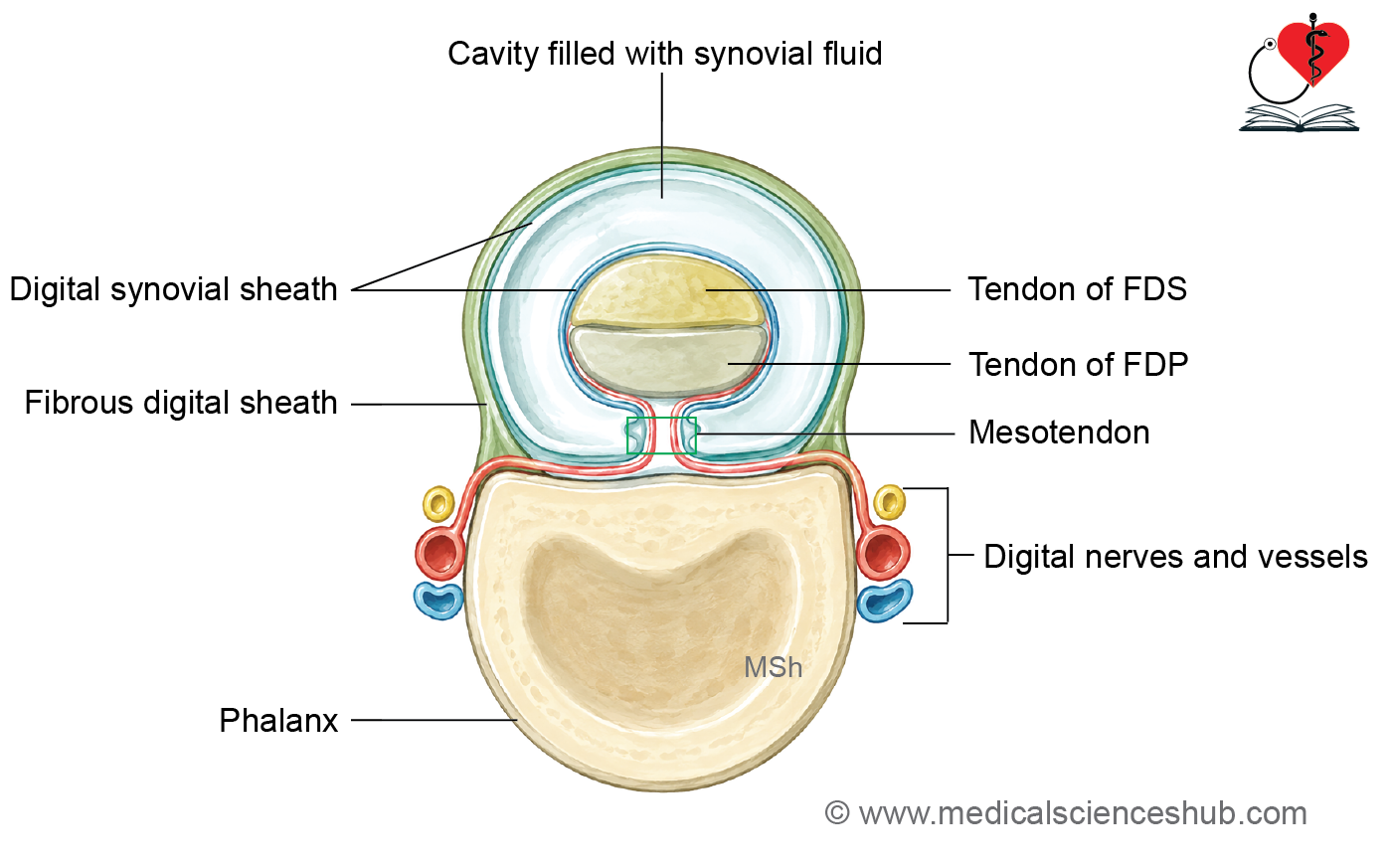
- To reduce friction and ensure smooth tendon movement, they are enclosed by double-layered synovial sheaths containing synovial fluid.
Mesotendon
- The mesotendon is a fold of synovial membrane that carries blood vessels to the tendon.
Types of Synovial Sheaths
- Ulnar bursa
- Radial bursa
- Digital synovial sheaths
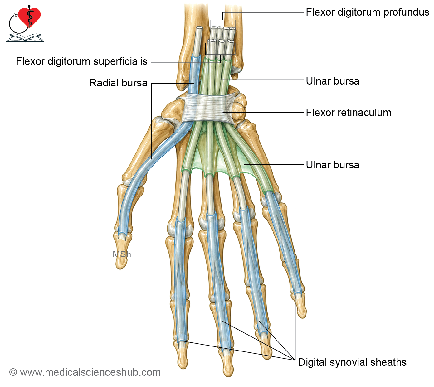
Ulnar bursa
- The ulnar bursa is the synovial sheath surrounding the long flexor tendons of the four medial fingers.
- It encloses:
- Four tendons of flexor digitorum superficialis (FDS)
- Four tendons of flexor digitorum profundus (FDP)
Extent
- Proximal: Extends about 5 cm proximal to the flexor retinaculum into the forearm.
- Distal: Reaches the middle of the metacarpal shafts.
Continuity
- Distally, it is continuous with the digital synovial sheath of the little finger.
Radial bursa
- The radial bursa is the synovial sheath surrounding the tendon of flexor pollicis longus (FPL).
Extent
- Proximal: Extends about 5 cm proximal to the flexor retinaculum into the forearm.
- Distal: Continues as the digital synovial sheath of the thumb.
Digital synovial sheath
- These synovial sheaths surround the long flexor tendons as they pass beneath the fibrous flexor sheaths of the fingers.
Extent
- Proximal: Up to the heads of the metacarpals.
- Distal: Up to the bases of the distal phalanges.
Continuity
- Thumb: Digital synovial sheath is continuous with the radial bursa.
- Little finger: Digital synovial sheath is continuous with the ulnar bursa.
- Index, middle, and ring fingers: Their digital synovial sheaths are usually separate from the ulnar bursa.
Function
- Facilitate smooth, friction-free movement of the long flexor tendons within the osseofibrous tunnels during finger and thumb movements.
CLINICAL INTEGRATION
Tenosynovitis of Flexor Tendon Sheaths
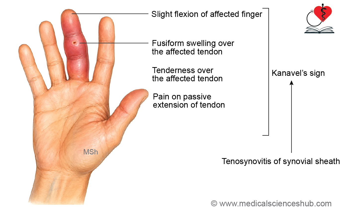
- Flexor tenosynovitis is an infection of the synovial sheath surrounding the flexor tendon, usually caused by a penetrating injury to the finger.
- The infection leads to accumulation of pus within the synovial sheath, resulting in pain, swelling, and restricted finger movements.
- It is a surgical emergency, as delayed treatment may lead to tendon necrosis, stiffness, and loss of finger function.
Kanavel’s Signs
Kanavel’s signs are the four cardinal clinical features used to diagnose pyogenic flexor tenosynovitis.
They include:
- Slight flexion of the affected finger (finger is held in a flexed position).
- Fusiform (spindle-shaped) swelling of the affected finger.
- Pain on passive extension of the finger.
- Tenderness along the course of the flexor tendon sheath.
Space of Parona
- The space of Parona is a potential space in the distal forearm.
- It lies between the pronator quadratus muscle posteriorly and the overlying long flexor tendons anteriorly.
- The overlying tendons include:
- Flexor digitorum superficialis (FDS)
- Flexor digitorum profundus (FDP)
- Flexor pollicis longus (FPL)
- Flexor carpi radialis (FCR)
- Flexor carpi ulnaris (FCU)
- It is also known as the subtendinous space of the wrist.
Communication
- The space of Parona communicates with the midpalmar space through the region deep to the flexor retinaculum.
Clinical Importance
- Infection can spread to the space of Parona from:
- Ulnar bursa, usually following infection of the little finger.
- Radial bursa, usually following infection of the thumb.
- Midpalmar space of the hand.
- Because of these communications, infections of the thumb or little finger may spread into the forearm through the space of Parona, producing a deep forearm abscess that requires prompt treatment.
Vincula Longa and Vincula Brevia
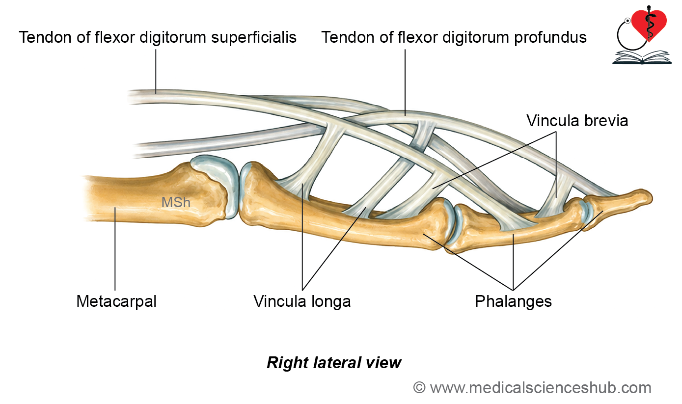
- Vincula are slender connective tissue bands that connect the long flexor tendons to the phalanges.
- They help anchor the tendons while allowing them to move freely within their synovial sheaths.
Types of Vincula
- Vincula longa
- Long, slender fibrous bands.
- Extend between the flexor tendons and the phalanges.
- Vincula brevia
- Short, triangular fibrous bands.
- Attach the flexor tendons more closely to the phalanges.
Functions
- Provide a pathway for blood vessels to reach and nourish the flexor tendons.
- Help maintain the vascular supply of the tendons while permitting smooth tendon movement within the synovial sheath.
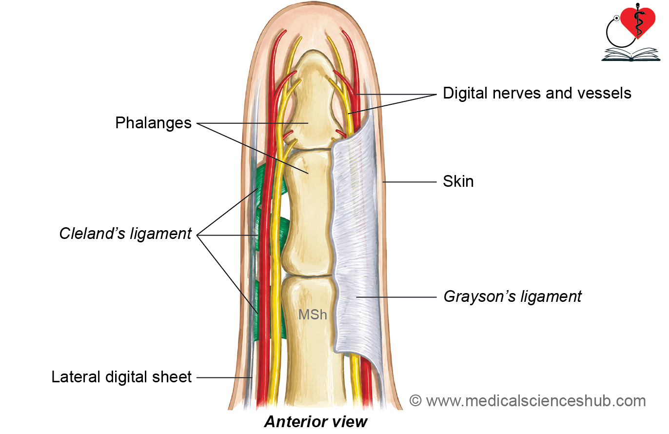
Cleland and Grayson ligament
- Cleland’s ligament: It extends from sides of the phalanges to the skin. This ligament is situated dorsal to the neurovascular bundle of the digit (digital artery and nerve).
- Grayson’s ligament: It extends from sides of the phalanges to the skin, but it is situated ventral to the neurovascular bundle of digit (digital artery and nerve).
- These ligaments prevent longitudinal translation of the skin during rotational movements of digits and they help to stabilize the neurovascular bundles.
Intrinsic Muscles of Hand
- The intrinsic muscles of the hand are small muscles located entirely within the hand.
- They are responsible for fine, skilled movements of the fingers and thumb and enhance the grip strength of the hand.
- The palm is traversed by the long flexor tendons, around which these muscles are arranged.
- There are 20 intrinsic muscles in the hand.
Classification of Intrinsic Muscles
1. Thenar Muscles (3)
These muscles act on the thumb.
- Abductor pollicis brevis
- Flexor pollicis brevis
- Opponens pollicis
2. Adductor of the Thumb (1)
- Adductor pollicis
3. Hypothenar Muscles (4)
These muscles act on the little finger.
- Palmaris brevis
- Abductor digiti minimi
- Flexor digiti minimi brevis
- Opponens digiti minimi
4. Lumbricals (4)
- Four lumbrical muscles associated with the tendons of flexor digitorum profundus.
5. Interossei (8)
Located between the metacarpal bones.
- Four palmar interossei
- Four dorsal interossei
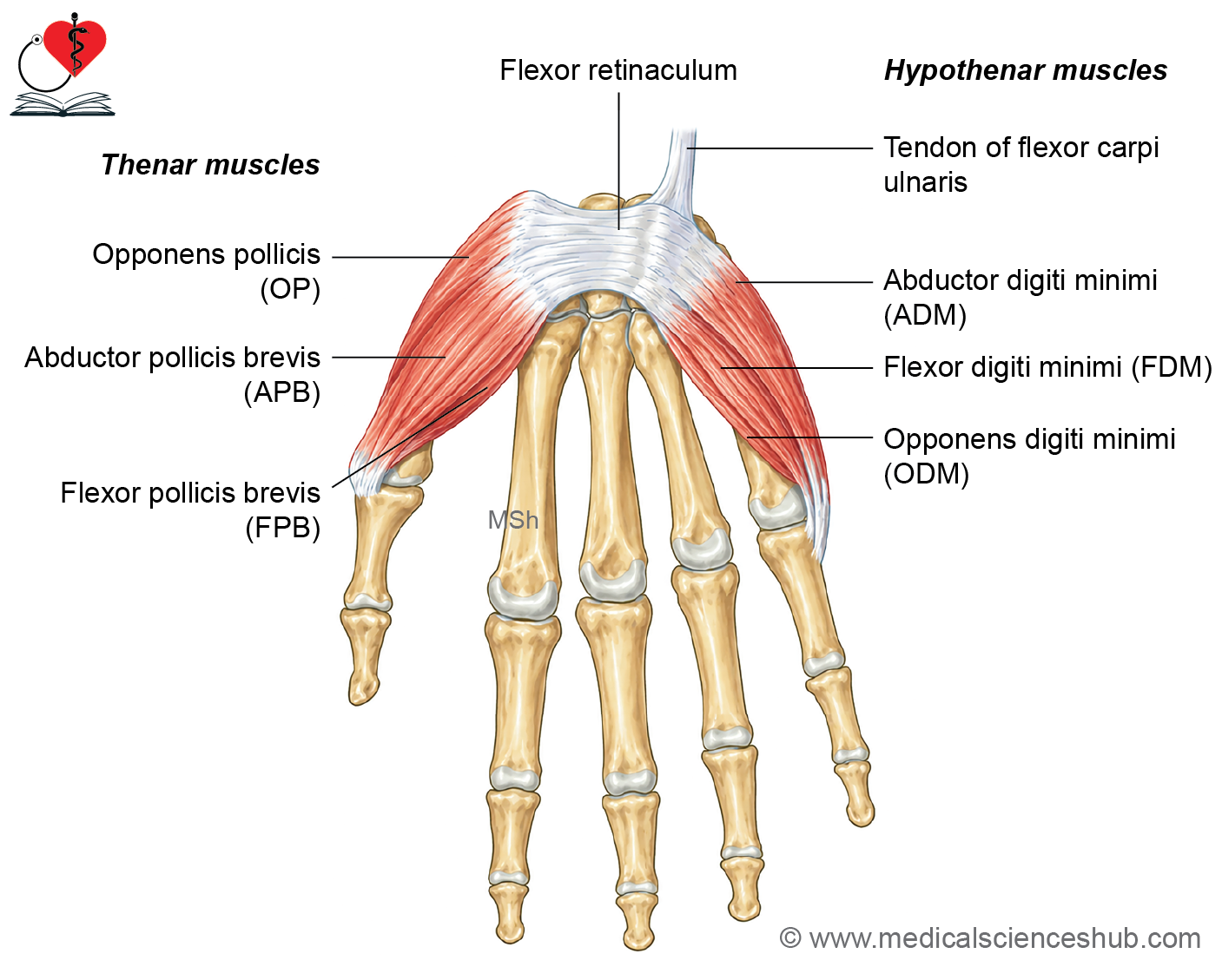
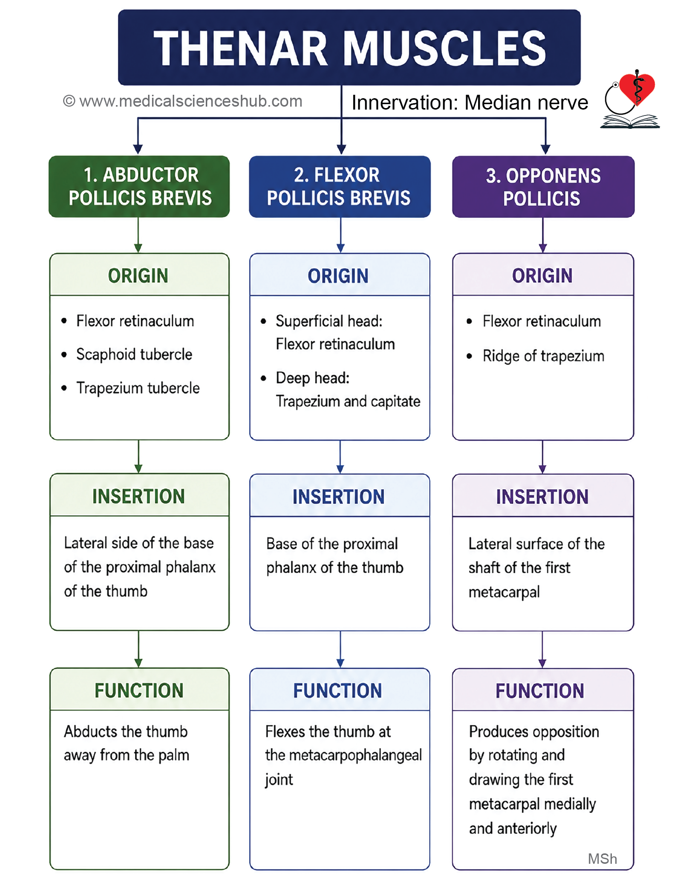
Muscles of Thenar Eminence
- The thenar eminence is the muscular prominence at the base of the thumb.
- It is formed by three intrinsic muscles:
- Abductor pollicis brevis
- Flexor pollicis brevis
- Opponens pollicis
Note: The origin, insertion, nerve supply, and actions of these muscles are described separately.
Nerve Supply
- All muscles of the thenar eminence are supplied by the recurrent (thenar) branch of the median nerve.
- Exception: The deep head of flexor pollicis brevis is usually supplied by the deep branch of the ulnar nerve.
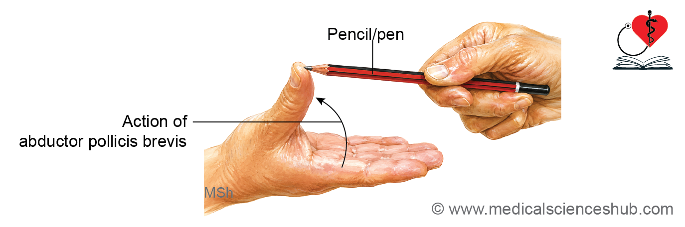
Abductor Pollicis Brevis
Origin
- Tubercle of scaphoid
- Crest of trapezium
- Flexor retinaculum
Insertion
- Lateral side of the base of the proximal phalanx of the thumb
Innervation
- Recurrent (motor) branch of the median nerve (C8, T1)
Actions
- Abducts the thumb at the carpometacarpal and metacarpophalangeal joints.
- Assists in opposition of the thumb.
- Helps stabilize the thumb during gripping and pinching.
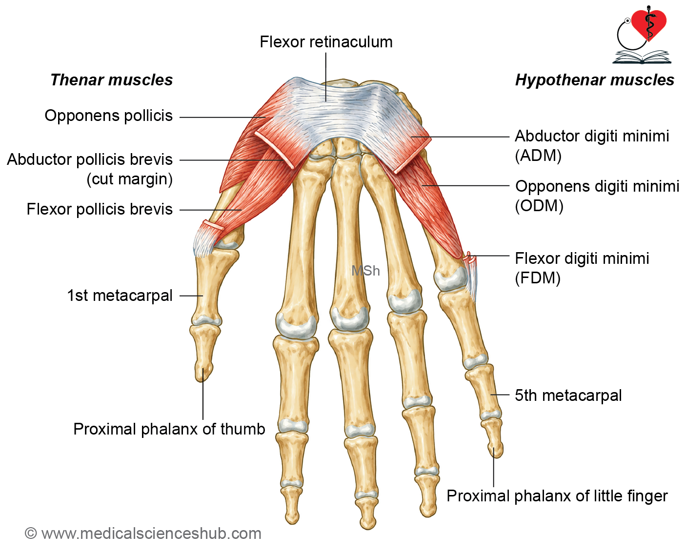
Flexor Pollicis Brevis
Origin
- Crest of trapezium
- Flexor retinaculum
- Capitate bone (deep head)
Insertion
- Lateral side of the base of the proximal phalanx of the thumb
Innervation
- Superficial head: Recurrent branch of the median nerve (C8, T1)
- Deep head: Deep branch of the ulnar nerve (C8, T1)
Actions
- Flexes the thumb at the metacarpophalangeal joint.
- Assists in flexion at the carpometacarpal joint.
- Helps in opposition and gripping movements.
Opponens Pollicis
Origin
- Crest of trapezium
- Flexor retinaculum
Insertion
- Lateral half of the palmar surface of the shaft of the first metacarpal
Innervation
- Recurrent (motor) branch of the median nerve (C8, T1)
Actions
- Opposes the thumb by drawing the first metacarpal medially and rotating it across the palm.
- Brings the thumb into contact with the fingertips.
- Plays a key role in precision grip and fine manipulative movements.
Adductor of Thumb
Adductor Pollicis
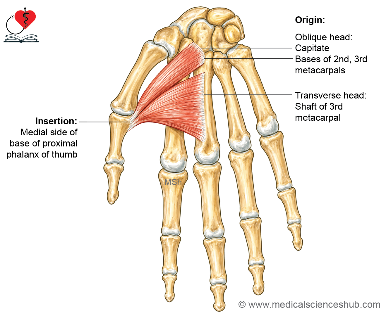
- Adductor pollicis is a fan-shaped intrinsic muscle of the hand.
- It has two heads:
- Oblique head
- Transverse head
- The muscle lies deep to the tendons of flexor digitorum superficialis (FDS), flexor digitorum profundus (FDP), and the lumbricals.
- During dissection, the long flexor tendons and lumbricals must be retracted or reflected to expose the adductor pollicis.
Origin
Oblique Head
- Capitate bone.
- Bases of the second and third metacarpals.
Transverse Head
- Anterior surface of the shaft of the third metacarpal.
Direction of Fibres
- Oblique head: Fibres pass downward and laterally.
- Transverse head: Fibres pass laterally to join the oblique head.
Insertion
Medial side of the base of the proximal phalanx of the thumb.
Innervation
Deep branch of the ulnar nerve (C8, T1).
Actions
- Adducts the thumb towards the palm.
- Assists in power grip and pinch grip by bringing the thumb towards the index finger.
CLINICAL aNATOMY
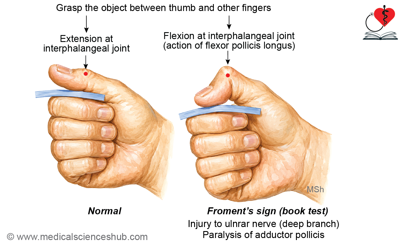
Froment’s Sign
Procedure
- Ask the patient to hold a sheet of paper or a book between the thumb and the lateral side of the index finger while the examiner attempts to pull it away.
Interpretation
- Normal: The thumb remains straight at the interphalangeal (IP) joint, indicating a functioning adductor pollicis.
- Positive Froment’s sign: The IP joint of the thumb flexes while gripping the paper due to compensatory action of the flexor pollicis longus, indicating paralysis or weakness of the adductor pollicis from ulnar nerve injury.
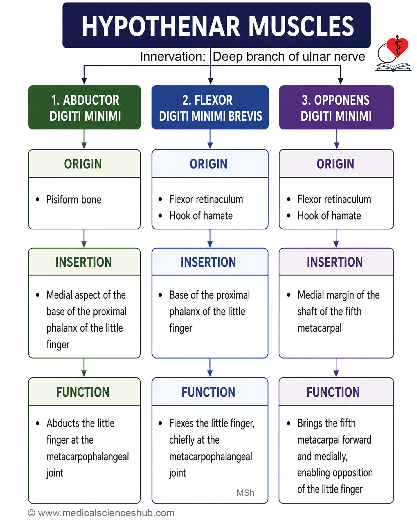
Muscles of Hypothenar Eminence
The hypothenar eminence contains three intrinsic muscles that control the movements of the little finger:
- Abductor digiti minimi
- Flexor digiti minimi
- Opponens digiti minimi
Abductor Digiti Minimi
Origin
- Pisiform bone
- Tendon of flexor carpi ulnaris
Insertion
- Medial side of the base of the proximal phalanx of the little finger
Innervation
- Deep branch of the ulnar nerve (C8, T1)
Actions
- Abducts the little finger at the metacarpophalangeal joint.
- Assists in flexion of the metacarpophalangeal joint.
- Helps stabilize the little finger during gripping.
Flexor Digiti Minimi Brevis
Origin
- Hook of hamate
- Flexor retinaculum
Insertion
- Medial side of the base of the proximal phalanx of the little finger
Innervation
- Deep branch of the ulnar nerve (C8, T1)
Actions
- Flexes the little finger at the metacarpophalangeal joint.
- Assists in cupping the palm during grasping.
- Supports fine movements of the little finger.
Opponens Digiti Minimi
Origin
- Hook of hamate
- Flexor retinaculum
Insertion
- Medial border of the shaft of the fifth metacarpal
Innervation
- Deep branch of the ulnar nerve (C8, T1)
Actions
- Draws the fifth metacarpal anteriorly and rotates it slightly.
- Opposes the little finger towards the thumb.
- Deepens the hollow of the palm (cupping of the palm).
- Assists in power grip by increasing the mobility of the fifth metacarpal.
Palmaris Brevis Muscle
Origin
- Medial part of the palmar aponeurosis
- Flexor retinaculum
Insertion
- Skin and subcutaneous tissue along the medial border of the palm
Innervation
- Superficial branch of the ulnar nerve (C8, T1)
Actions
- Wrinkles the skin over the hypothenar eminence.
- Deepens the hollow of the palm.
- Improves grip by increasing friction between the palm and the object being held.
- Helps protect the ulnar neurovascular structures in the hypothenar region during forceful gripping.
CLINICAL aNATOMY
Palmaris brevis is the only muscle in the hand supplied by the superficial branch of the ulnar nerve. All other intrinsic muscles supplied by the ulnar nerve receive motor fibers from its deep branch.
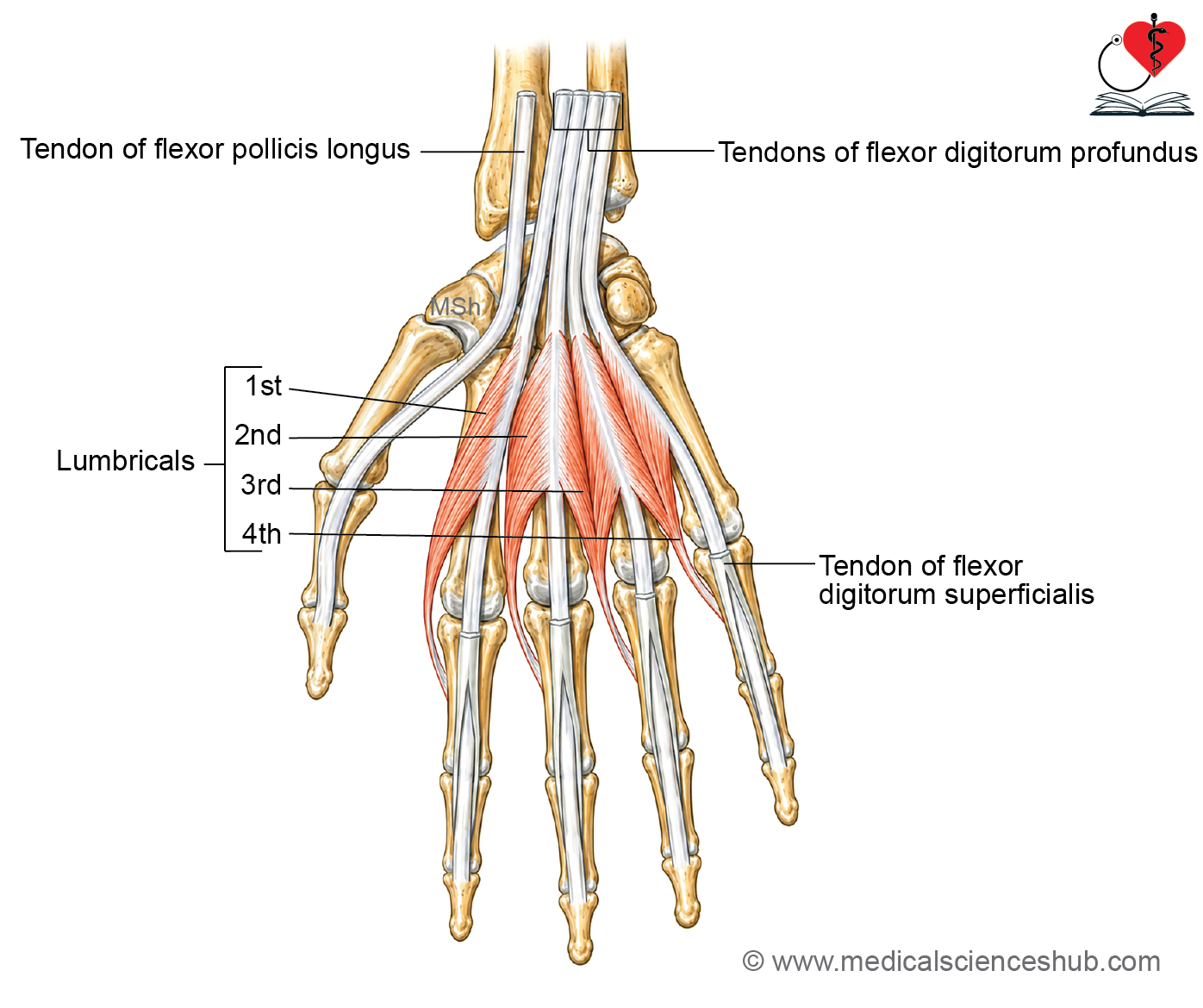
Lumbricals
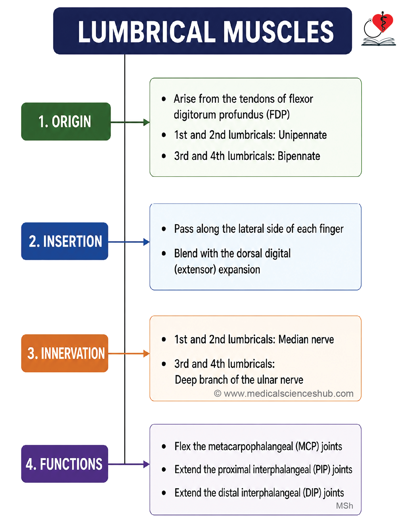
- Lumbricals are four small intrinsic muscles of the hand.
- They arise from the tendons of flexor digitorum profundus (FDP).
- They are numbered 1st to 4th from lateral (thumb side) to medial (little finger side).
- The 1st and 2nd lumbricals are unipennate, while the 3rd and 4th are bipennate.
- The term lumbrical means “worm-like” in Latin because of their slender appearance.
Origin
| Lumbrical | Origin |
| 1st | Lateral side of FDP tendon to the index finger |
| 2nd | Lateral side of FDP tendon to the middle finger |
| 3rd | Adjacent sides of FDP tendons to the middle and ring fingers |
| 4th | Adjacent sides of FDP tendons to the ring and little fingers |
Insertion
- Each tendon passes along the lateral side of the corresponding metacarpophalangeal (MCP) joint.
- Inserts into the lateral side of the extensor expansion (dorsal digital expansion) of the corresponding finger.
Innervation
| Lumbricals | Nerve Supply |
| 1st and 2nd | Median nerve |
| 3rd and 4th | Deep branch of ulnar nerve |
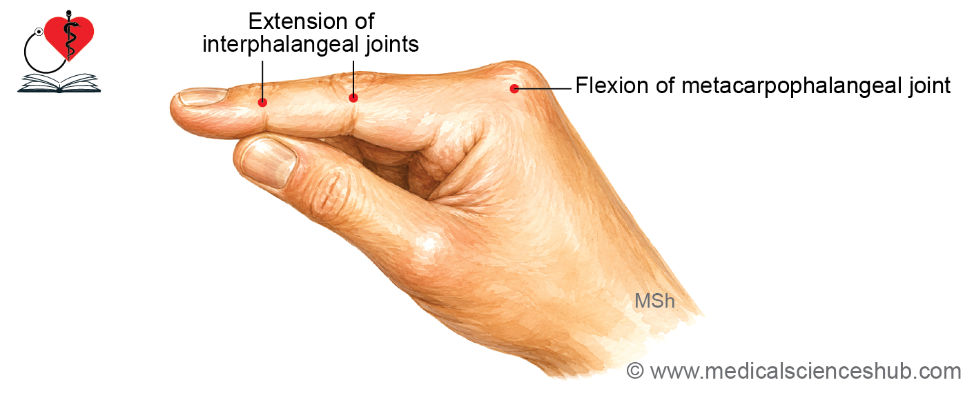
Actions
- Flex the metacarpophalangeal (MCP) joints.
- Extend both the proximal and distal interphalangeal (PIP and DIP) joints through the extensor expansion.
- Produce the characteristic “writing position” or “Z-movement” of the fingers.
CLINICAL aNATOMY
Clinical Testing
- Ask the patient to extend the interphalangeal joints against resistance while the examiner keeps the MCP joints hyperextended.
- Lumbricals are usually tested together with the interossei (lumbrical-interossei complex) because they cannot be tested independently.
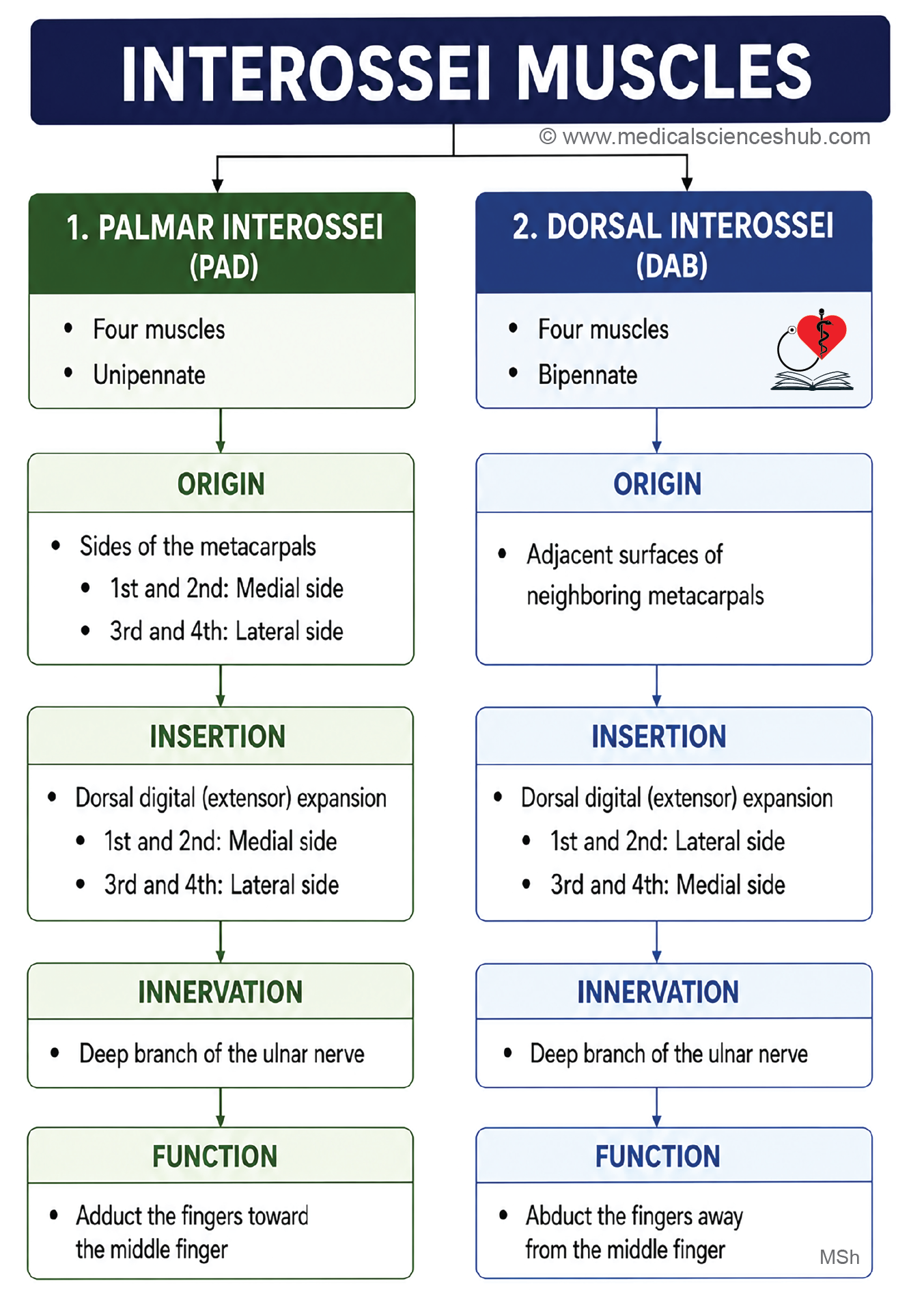
Interossei
- The interossei are intrinsic muscles of the hand located in the spaces between the metacarpal bones.
- There are eight interossei muscles in total:
- Four palmar interossei
- Four dorsal interossei
Palmar Interossei
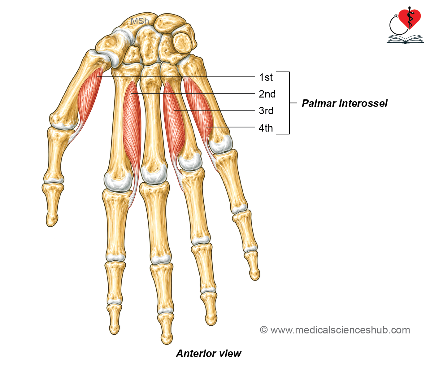
- The palmar (volar) interossei consist of four small unipennate muscles.
- They are numbered 1st to 4th from the lateral (thumb) side to the medial (little finger) side.
- The 1st palmar interosseous muscle may be absent in some individuals.
Origin
Each palmar interosseous muscle arises from a single metacarpal bone.
- 1st: Medial side of the base of the 1st metacarpal
- 2nd: Medial side of the shaft of the 2nd metacarpal
- 3rd: Lateral side of the shaft of the 4th metacarpal
- 4th: Lateral side of the shaft of the 5th metacarpal
The muscle fibers form a small fusiform belly that continues as a tendon.
Insertion
The tendons insert into the extensor expansion (dorsal digital expansion) of the corresponding digits.
- 1st: Medial side of the thumb
- 2nd: Medial side of the index finger
- 3rd: Lateral side of the ring finger
- 4th: Lateral side of the little finger
Innervation
All palmar interossei are supplied by the deep branch of the ulnar nerve.
Actions
The primary function of the palmar interossei is adduction of the fingers, bringing the fingers toward the middle finger, which serves as the reference axis of the hand.
CLINICAL aNATOMY
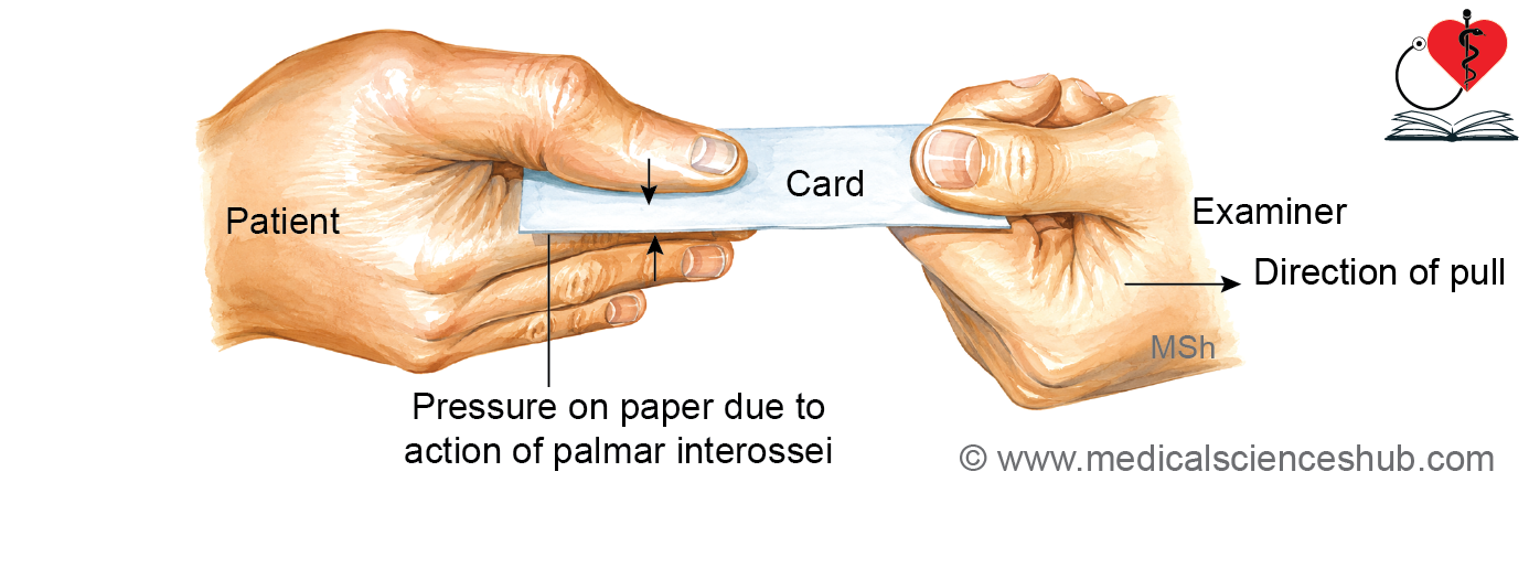
Clinical testing: Egawa’s paper (card) test
- Ask the patient to hold a piece of paper between two adjacent fingers while the examiner attempts to pull it away.
- In ulnar nerve palsy, weakness or paralysis of the palmar interossei results in an inability to hold the paper firmly because finger adduction is impaired.
Dorsal Interossei
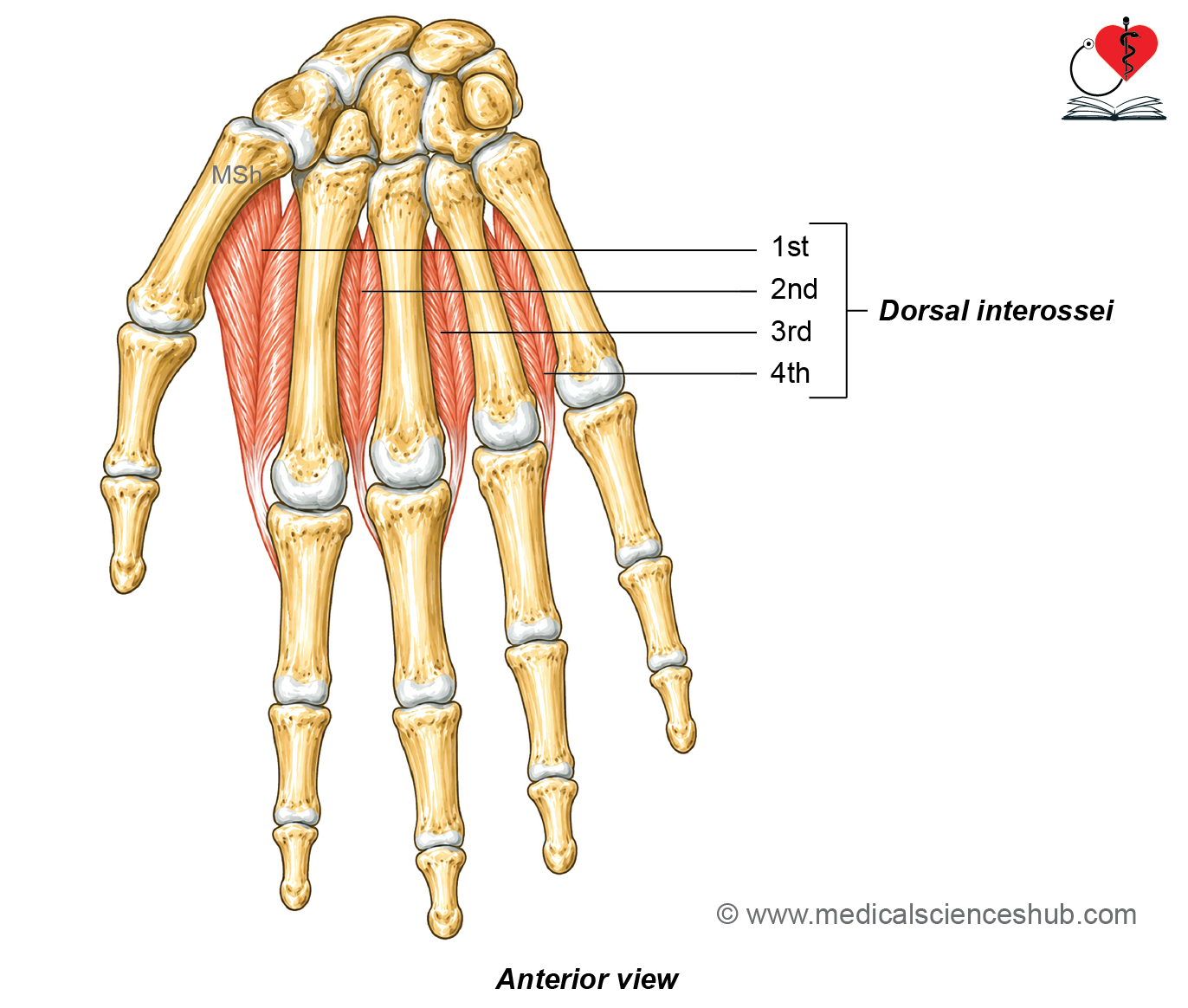
- The dorsal interossei are four intrinsic muscles located in the spaces between the metacarpal bones on the dorsum of the hand.
- They are bipennate muscles and are numbered 1st to 4th from the lateral (thumb) side to the medial (little finger) side.
Origin
Each dorsal interosseous muscle arises from the adjacent surfaces of two metacarpal bones.
- 1st: Adjacent sides of the shafts of the 1st and 2nd metacarpals
- 2nd: Adjacent sides of the shafts of the 2nd and 3rd metacarpals
- 3rd: Adjacent sides of the shafts of the 3rd and 4th metacarpals
- 4th: Adjacent sides of the shafts of the 4th and 5th metacarpals
The muscle fibers converge to form a tendon.
Insertion
he tendons insert into the extensor expansion (dorsal digital expansion) of the corresponding fingers.
- 1st: Lateral side of the index finger
- 2nd: Lateral side of the middle finger
- 3rd: Medial side of the middle finger
- 4th: Medial side of the ring finger
Innervation
All dorsal interossei are supplied by the deep branch of the ulnar nerve.
Actions
- The primary action of the dorsal interossei is abduction of the fingers, moving the fingers away from the middle finger, which serves as the reference axis of the hand.
- They also assist the lumbricals in flexing the metacarpophalangeal (MCP) joints and extending the interphalangeal (IP) joints through the extensor expansion.
CLINICAL aNATOMY
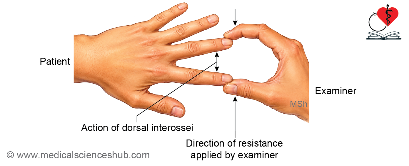
Clinical testing
- Ask the patient to abduct the fingers against resistance.
- Weakness or paralysis of the dorsal interossei, usually due to ulnar nerve injury, results in loss of finger abduction.
Arteries of Hand
- In the palm, two arteries enter and they from palmar arch arteries.
- These are ulnar artery and radial artery.
Ulnar Artery
The ulnar artery enters the palm together with the ulnar nerve by passing superficial to the flexor retinaculum.
After entering the hand, it divides into two terminal branches:
- Superficial branch (larger): Continues across the palm to form the superficial palmar arch.
- Deep branch (smaller): Passes between the abductor digiti minimi and flexor digiti minimi muscles, then pierces the opponens digiti minimi. It joins the radial artery to complete the deep palmar arch.
Radial Artery
Course
In the anatomical snuffbox
- The radial artery passes across the posterior aspect of the wrist through the anatomical snuffbox.
- It lies deep to the tendons of abductor pollicis longus, extensor pollicis brevis, and extensor pollicis longus.
In the hand
- The artery enters the palm by passing between the two heads of the first dorsal interosseous muscle.
- It then runs between the oblique and transverse heads of the adductor pollicis and continues as the deep palmar arch.
Branches
On the dorsum of the hand
- First dorsal metacarpal artery: Arises just before the radial artery passes between the two heads of the first dorsal interosseous muscle. It divides into two branches that supply the adjacent sides of the thumb and index finger.
In the palm
- Princeps pollicis artery: Passes deep to the oblique head of the adductor pollicis and superficial to the first palmar interosseous muscle. It divides into two branches that supply the palmar surface of the thumb.
- Radialis indicis artery: Descends between the first dorsal interosseous muscle and the transverse head of the adductor pollicis. It supplies the lateral side of the index finger.
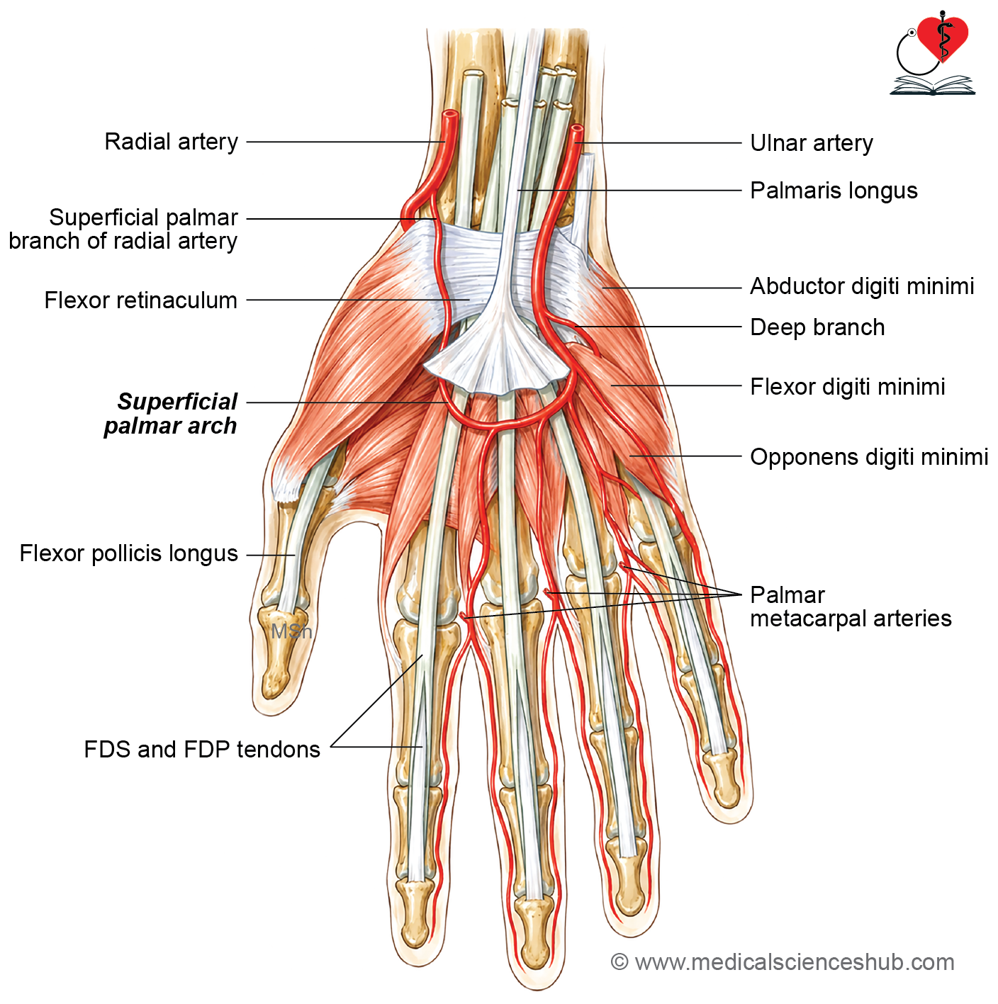
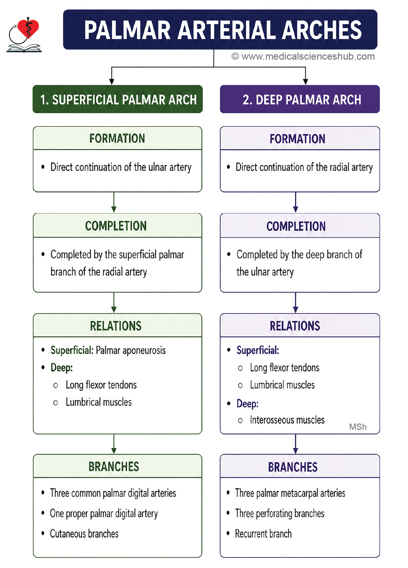
Superficial Palmar Arch
- The superficial palmar arch is the main arterial arch of the palm. It lies deep to the palmar aponeurosis and is also known as the superficial volar arch, ulnar arch, or arcus volaris superficialis.
Formation
- The superficial palmar arch is formed mainly by the superficial branch of the ulnar artery. Viva
- It begins over the muscles of the hypothenar eminence, just distal to the flexor retinaculum.
Course
- The arch passes laterally across the palm with its convexity directed downward.
- Its highest point of convexity is located at the level of the lower border of a fully extended thumb.
Completion
The arch is completed by the superficial palmar branch of the radial artery.
Relations
Superficial
- Palmar aponeurosis
- Palmaris brevis
Deep
- Flexor digiti minimi
- Tendons of flexor digitorum superficialis (FDS)
- Tendons of flexor digitorum profundus (FDP)
- Lumbrical muscles
Branches
- Three common palmar digital arteries that supply the adjacent sides of the medial 3½ digits.
- One proper palmar digital artery to the medial side of the little finger.
- Cutaneous branches to the skin of the palm.
Common Palmar Digital Arteries
- Three common palmar digital arteries arise from the convexity of the superficial palmar arch.
- They descend over the lumbrical muscles toward the fingers.
- Each artery is reinforced by a corresponding palmar metacarpal artery from the deep palmar arch.
- Near the webs of the fingers, each common palmar digital artery divides into two proper palmar digital arteries that supply the adjacent sides of the fingers.
Applied Anatomy
The superficial palmar arch lies deep to the palmar aponeurosis, an important feature for anatomical identification during dissection and practical examinations.
Deep Palmar Arch
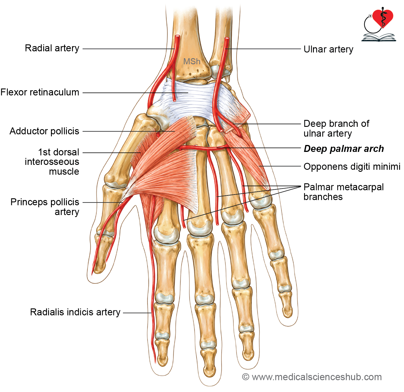
The deep palmar arch is an arterial arcade located in the palm, deep to the long flexor tendons of the fingers. It is also known as the deep volar arch.
Formation
- The deep palmar arch is formed mainly by the continuation of the radial artery after it enters the palm by passing between the transverse and oblique heads of the adductor pollicis muscle.
- It is completed on the medial side by the deep palmar branch of the ulnar artery.
Course
- The arch passes medially across the palm with its convexity directed distally (downward).
- It lies on the anterior surfaces of the metacarpal bones and the interossei muscles.
Completion
- Deep palmar arch is completed medially by deep palmar branch of ulnar artery at the base of 5th metacarpal bone.
Relations
Superficial
- Tendons of flexor digitorum superficialis (FDS)
- Tendons of flexor digitorum profundus (FDP)
- Lumbrical muscles
Deep
- Metacarpal bones
- Interossei muscles
Branches
- Three palmar metacarpal arteries
- Three perforating arteries
- Recurrent branch to the superficial palmar arch
Clinical Anatomy
- The deep palmar arch is situated deep to the tendons of FDS, FDP, and the lumbrical muscles, an important feature for anatomical identification during dissection.
- It lies approximately 1 cm proximal to the superficial palmar arch.
- The three palmar metacarpal arteries unite with the common palmar digital arteries, contributing to the arterial supply of the fingers.
- The three perforating arteries pass through the interosseous spaces to anastomose with the dorsal metacarpal arteries, establishing communication between the palmar and dorsal arterial systems.
Nerves in Palm
- In the palm of hand, there are two nerves: Ulnar nerve, and median nerve.
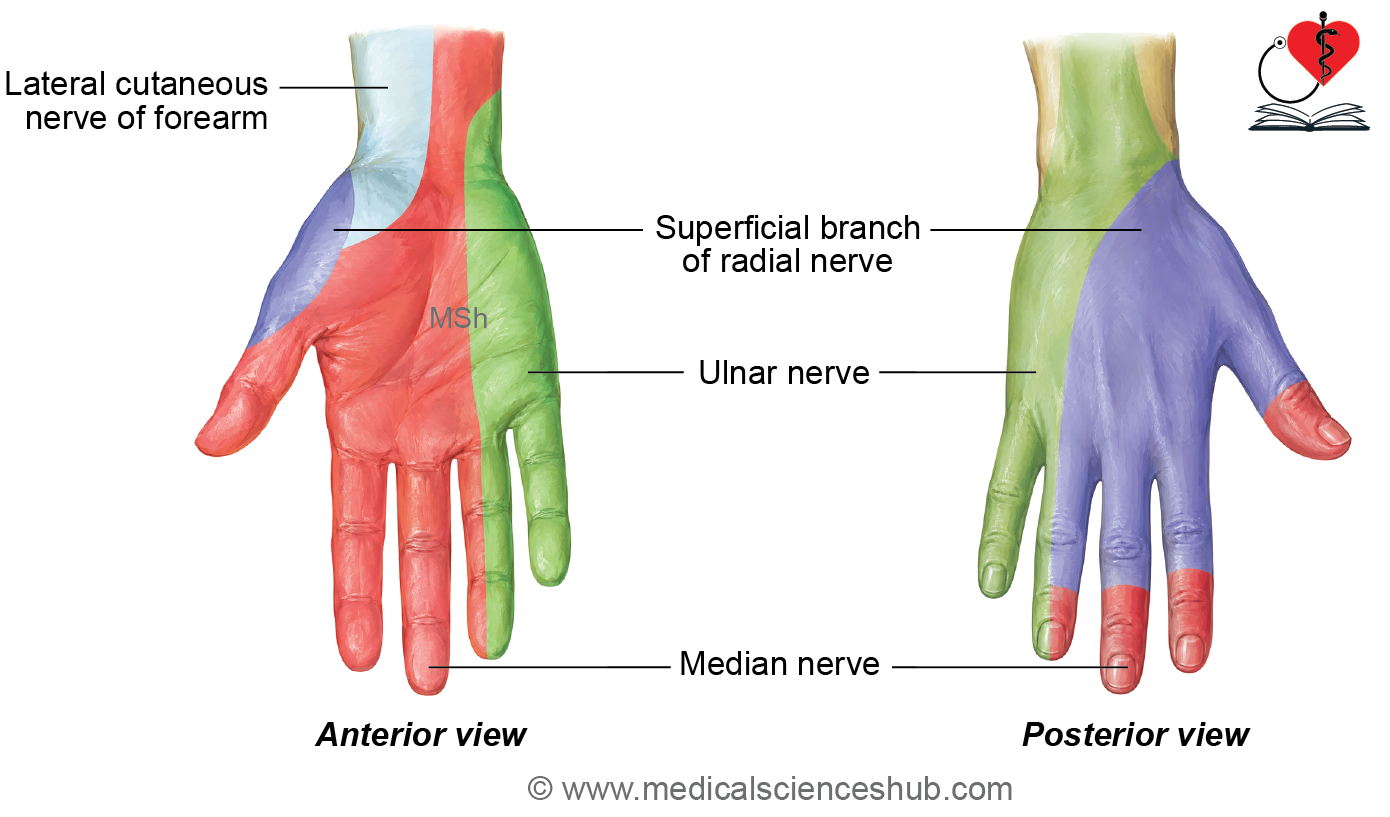
- They supply 20 intrinsic muscles and skin of the palm except skin over thenar and hypothenar eminence that is supplied by branches of median and ulnar nerve given in the forearm.
Ulnar Nerve
The ulnar nerve is the principal motor nerve of the hand, supplying the majority of its intrinsic muscles. Functionally, it is the homologue of the lateral plantar nerve of the foot. Because it innervates the muscles responsible for precise and coordinated finger movements, it is often referred to as the “musician’s nerve.”
Course
- The ulnar nerve enters the palm by passing superficial to the flexor retinaculum and deep to the palmar (volar) carpal ligament.
- At the wrist, it lies between the pisiform bone and the ulnar artery, an important anatomical landmark for identification.
Termination
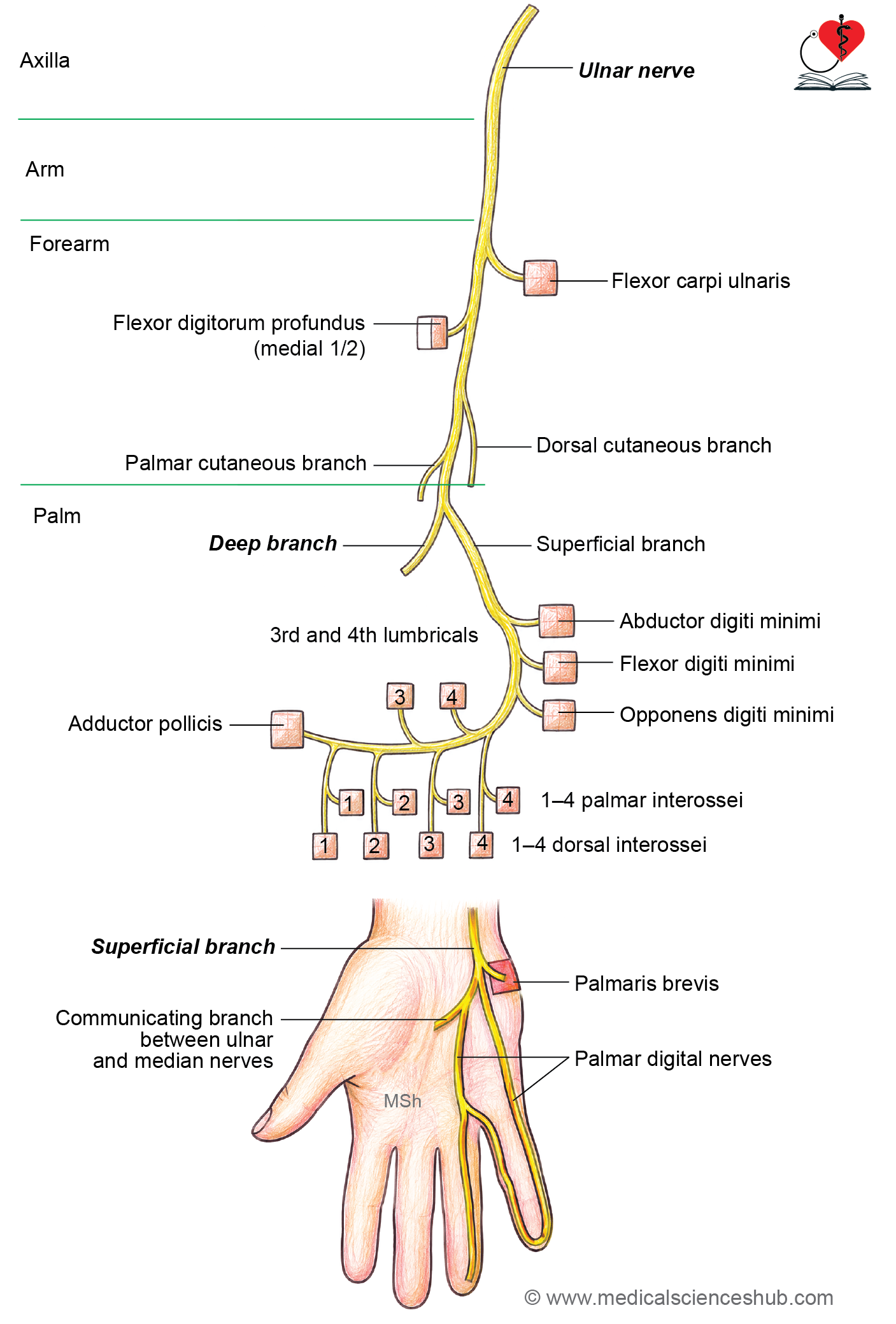
At the distal border of the flexor retinaculum, the ulnar nerve divides into two terminal branches:
- Superficial branch
- Deep branch
Branches and distribution
Superficial Branch of Ulnar Nerve
Muscular Branch
- Supplies the palmaris brevis muscle.
Cutaneous Branches
- Provides sensory innervation to the medial 1½ digits through two palmar digital branches:
- Medial palmar digital nerve to the medial side of the little finger.
- Lateral palmar digital nerve, which divides to supply the adjacent sides of the ring and little fingers.
Deep Branch of Ulnar Nerve
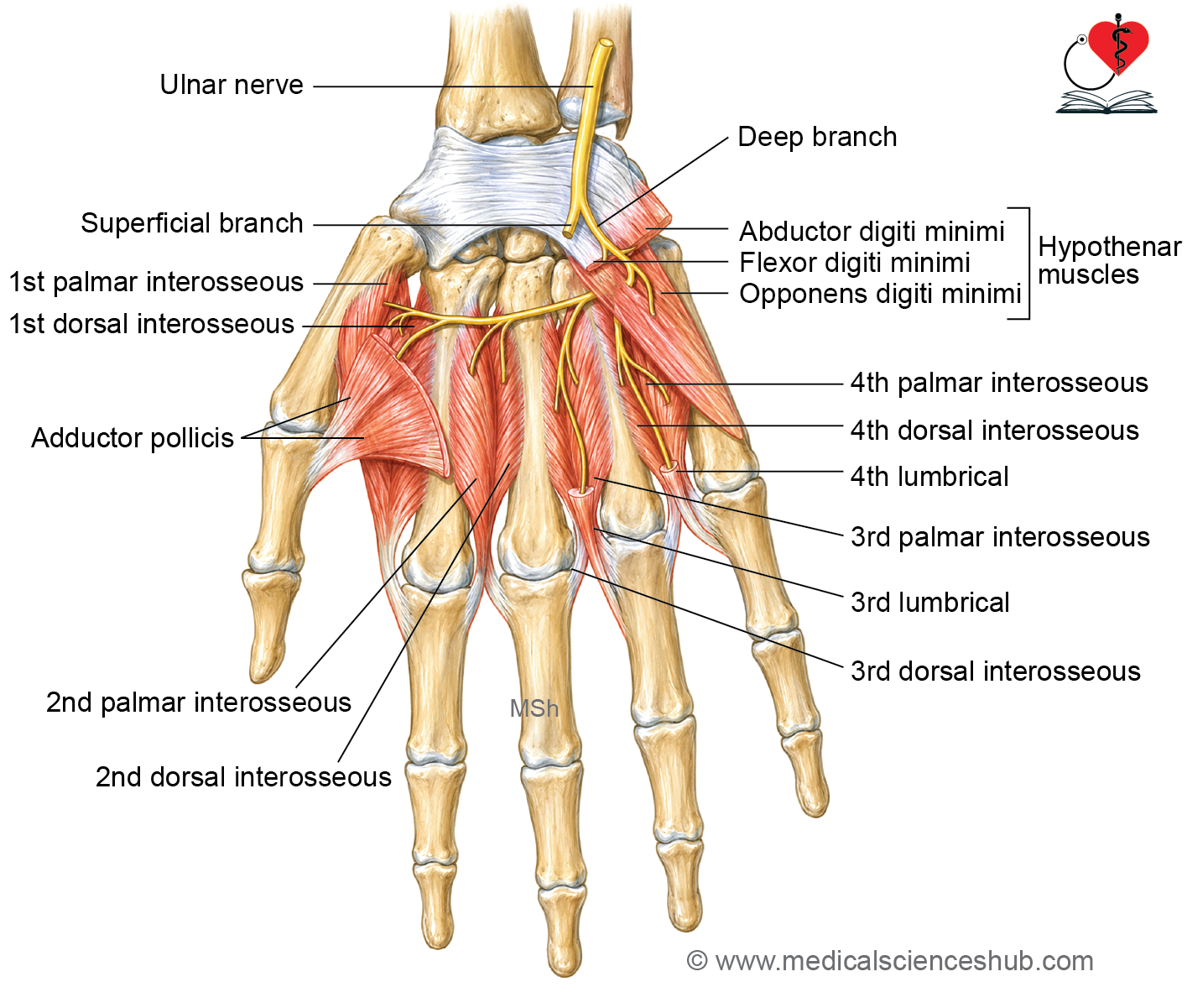
Muscular Branches
Supplies:
- Hypothenar muscles
- Abductor digiti minimi
- Flexor digiti minimi brevis
- Opponens digiti minimi
- Four palmar interossei
- Four dorsal interossei
- Third and fourth lumbricals
- Adductor pollicis
Articular Branches
- Supply the:
- Intercarpal joints
- Carpometacarpal joints
- Intermetacarpal joints
Clinical Anatomy
Handlebar Neuropathy
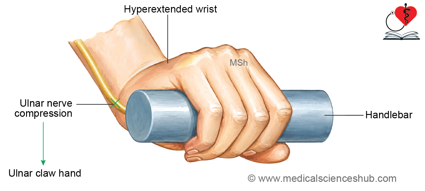
- Handlebar neuropathy is a compressive injury of the ulnar nerve at the wrist, commonly seen in cyclists and motorcyclists.
- It results from prolonged pressure over Guyon’s canal, particularly when riding with the wrist in hyperextension. Compression is usually produced against the hook of the hamate.
- Clinical features include weakness of the intrinsic hand muscles and may progress to an ulnar claw hand.
Ulnar Nerve Injury
The ulnar nerve is commonly injured at two clinically important sites:
- Behind the medial epicondyle of the humerus at the elbow.
- At the wrist, where it passes through Guyon’s canal.
Effects of Ulnar Nerve Injury at the Wrist
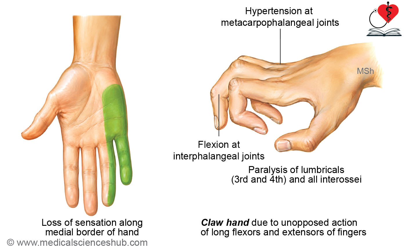
A lesion at the wrist produces ulnar claw hand, also known as spinster’s claw deformity, because the intrinsic muscles of the hand supplied by the ulnar nerve are affected while the forearm flexor muscles remain intact.
Motor Deficits
- Paralysis of the third and fourth lumbricals
- Hyperextension at the metacarpophalangeal (MCP) joints.
- Flexion at the interphalangeal (IP) joints of the ring and little fingers.
- Inability to fully extend the ring and little fingers.
- Paralysis of the palmar interossei
- Loss of finger adduction.
- Paralysis of the dorsal interossei
- Loss of finger abduction.
Sensory Deficits
- Loss of sensation over the medial one and a half fingers.
Autonomic Changes
- Interruption of sympathetic fibers results in vasomotor disturbances, producing dry skin over the medial one and a half fingers.
Effects of Ulnar Nerve Injury at the Elbow
Injury to the ulnar nerve behind the medial epicondyle affects both the intrinsic muscles of the hand and the ulnar-innervated muscles of the forearm.
Motor Deficits
- Flexor carpi ulnaris paralysis
- Weakness of wrist flexion.
- Loss of wrist adduction (ulnar deviation).
- Paralysis of the medial half of flexor digitorum profundus
- Inability to flex the distal interphalangeal (DIP) joints of the ring and little fingers.
- Paralysis of the intrinsic hand muscles supplied by the ulnar nerve produces:
- Ulnar claw hand.
- Loss of finger abduction and adduction.
- Weakness of fine movements of the fingers.
Sensory Deficits
- Loss of sensation over the medial one and a half fingers.
- Additional sensory loss over the hypothenar eminence, as the lesion is proximal to the origin of the palmar cutaneous branch.
Clinical Note
- Although clawing occurs after lesions at both the wrist and elbow, the deformity is typically more pronounced in wrist lesions because the medial half of the flexor digitorum profundus remains functional, producing greater flexion of the distal interphalangeal joints. This phenomenon is known as the ulnar paradox.
Median Nerve
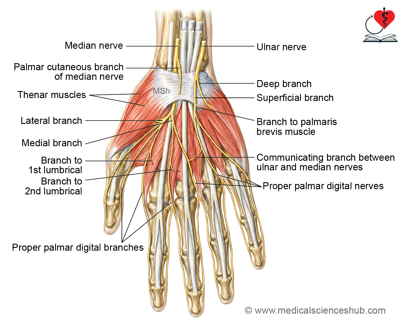
The median nerve is formed by the union of the medial and lateral roots of the brachial plexus in the lower part of the axilla. It is the principal nerve supplying the anterior compartment of the forearm and several muscles of the hand.
Root value: C5–T1
Course and relations
- In the arm, the median nerve descends alongside the brachial artery to the cubital fossa.
- At the elbow, it passes between the humeral and ulnar heads of the pronator teres muscle.
- It then travels through the anterior compartment of the forearm and enters the hand by passing deep to the flexor retinaculum through the carpal tunnel.
Relations in the Carpal Tunnel
Within the carpal tunnel, the median nerve:
- Lies deep to the flexor retinaculum.
- Is positioned lateral to the tendons of flexor digitorum superficialis (FDS).
- Lies anterior to the radial bursa and the tendon of flexor pollicis longus (FPL).
Termination
After entering the palm, the median nerve divides into:
- Medial branch
- Lateral branch
Branches
Medial Branch
The medial branch gives rise to two common palmar digital nerves.
1. Lateral Common Palmar Digital Nerve
- Supplies the adjacent sides of the index and middle fingers.
- Provides a motor branch to the second lumbrical.
2. Medial Common Palmar Digital Nerve
- Supplies the adjacent sides of the middle and ring fingers.
Lateral Branch
The lateral branch gives:
Muscular Branches
- To the thenar muscles:
- Abductor pollicis brevis
- Opponens pollicis
- Superficial head of flexor pollicis brevis
Digital Branches
Three proper palmar digital nerves supply:
- Lateral side of the thumb.
- Medial side of the thumb.
- Lateral side of the index finger.
Clinical Anatomy
Carpal tunnel syndrome
Carpal tunnel syndrome (CTS) is the most common entrapment neuropathy of the upper limb. It results from compression of the median nerve within the carpal tunnel, an osteofibrous canal formed by the carpal bones posteriorly and the flexor retinaculum anteriorly.
Clinical Features
Motor Deficits
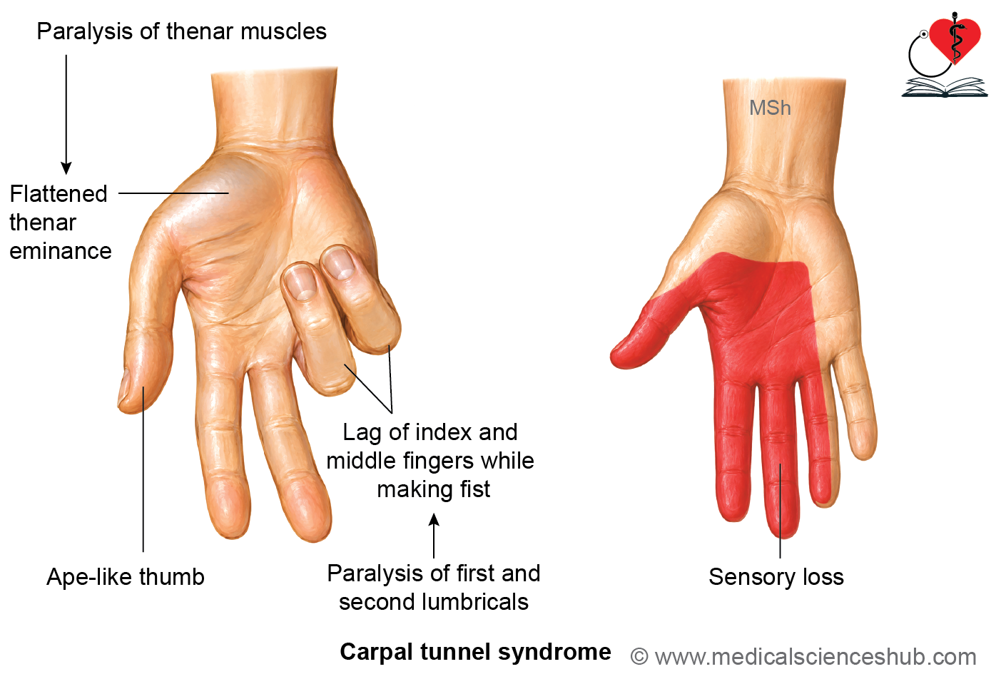
- Paralysis or weakness of the thenar muscles results in an ape thumb deformity, characterized by:
- Loss of thumb abduction.
- Loss of thumb opposition.
- Weakness of thumb flexion.
- The thumb lies in an extended and adducted position.
- Paralysis of the first and second lumbricals causes difficulty flexing the metacarpophalangeal joints while extending the interphalangeal joints of the index and middle fingers. During fist formation, these fingers show a characteristic lag.
Sensory Deficits
- Numbness, tingling, or loss of sensation over the lateral three and a half digits, including the nail beds of these fingers.
Autonomic Changes
- Compression of sympathetic fibers may produce vasomotor disturbances, leading to dry skin over the lateral three and a half fingers.
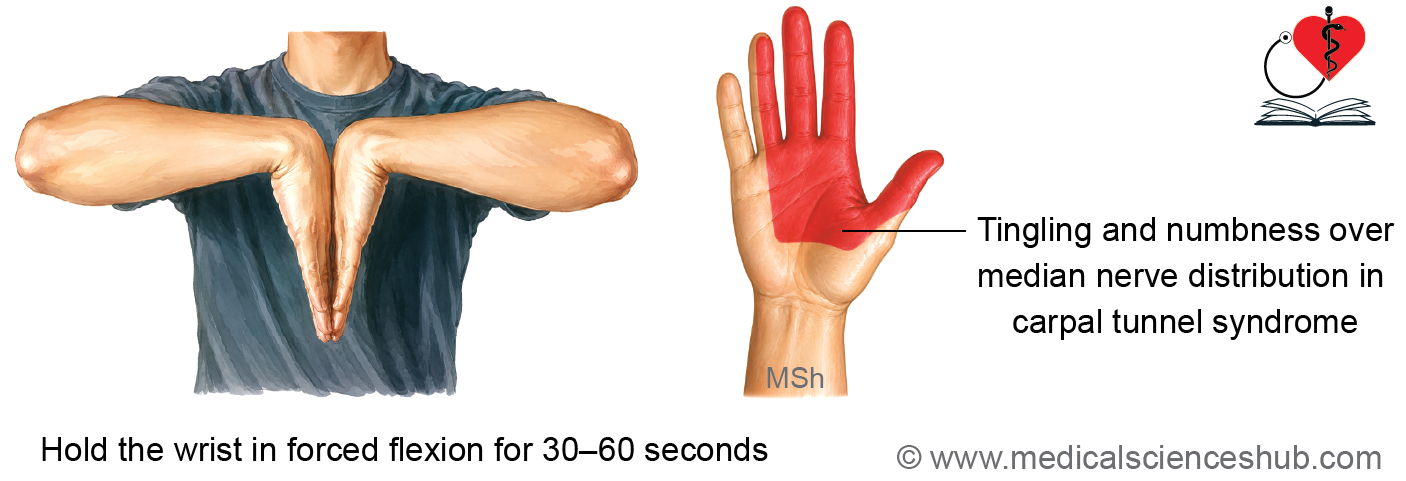
Phalen’s Test (Phalen’s Maneuver)
- The patient maintains maximal wrist flexion for approximately 60 seconds.
- Reproduction of numbness, tingling, or pain in the lateral three and a half fingers indicates median nerve compression within the carpal tunnel.
Durkan’s Test (Carpal Compression Test)
- Direct pressure is applied over the carpal tunnel for about 30 seconds.
- Provocation of pain or paresthesia in the median nerve distribution suggests carpal tunnel syndrome.
Causes
Common causes and risk factors include:
- Idiopathic (most common)
- Repetitive or work-related hand activities
- Obesity
- Pregnancy
- Rheumatoid arthritis
- Hypothyroidism
- Other inflammatory conditions affecting the wrist
Treatment
Management depends on the severity of symptoms and includes:
- Activity modification and physiotherapy
- Wrist splinting, particularly in a neutral position, to reduce median nerve compression
- Surgical carpal tunnel release, involving division of the flexor retinaculum, in patients with persistent or severe symptoms or progressive nerve dysfunction.
Fascial Spaces of Hand
The fascial spaces of the hand are potential spaces formed by the arrangement of the palmar fascia and its septa. Under normal conditions, these spaces contain loose areolar tissue and fat. They become clinically significant when they are distended by the accumulation of pus, blood, or inflammatory fluid, serving as pathways for the spread of infection.
Palmar Fascial Spaces
The principal fascial spaces in the palm are:
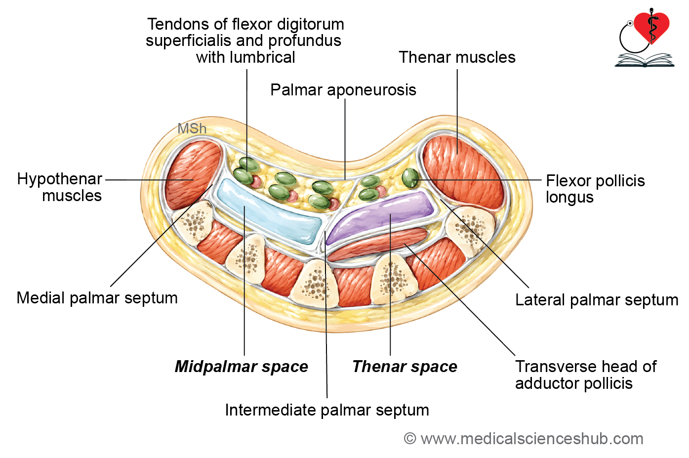
- Midpalmar space
- Thenar space
- Pulp spaces of the fingers
Dorsal Fascial Spaces
The dorsal aspect of the hand contains:
- Dorsal subcutaneous space
- Dorsal subaponeurotic space
Forearm Fascial Space
- Space of Parona: A potential fascial space located in the distal forearm that communicates with the deep fascial spaces of the hand and may act as a route for the proximal spread of infection.
Midpalmar Space
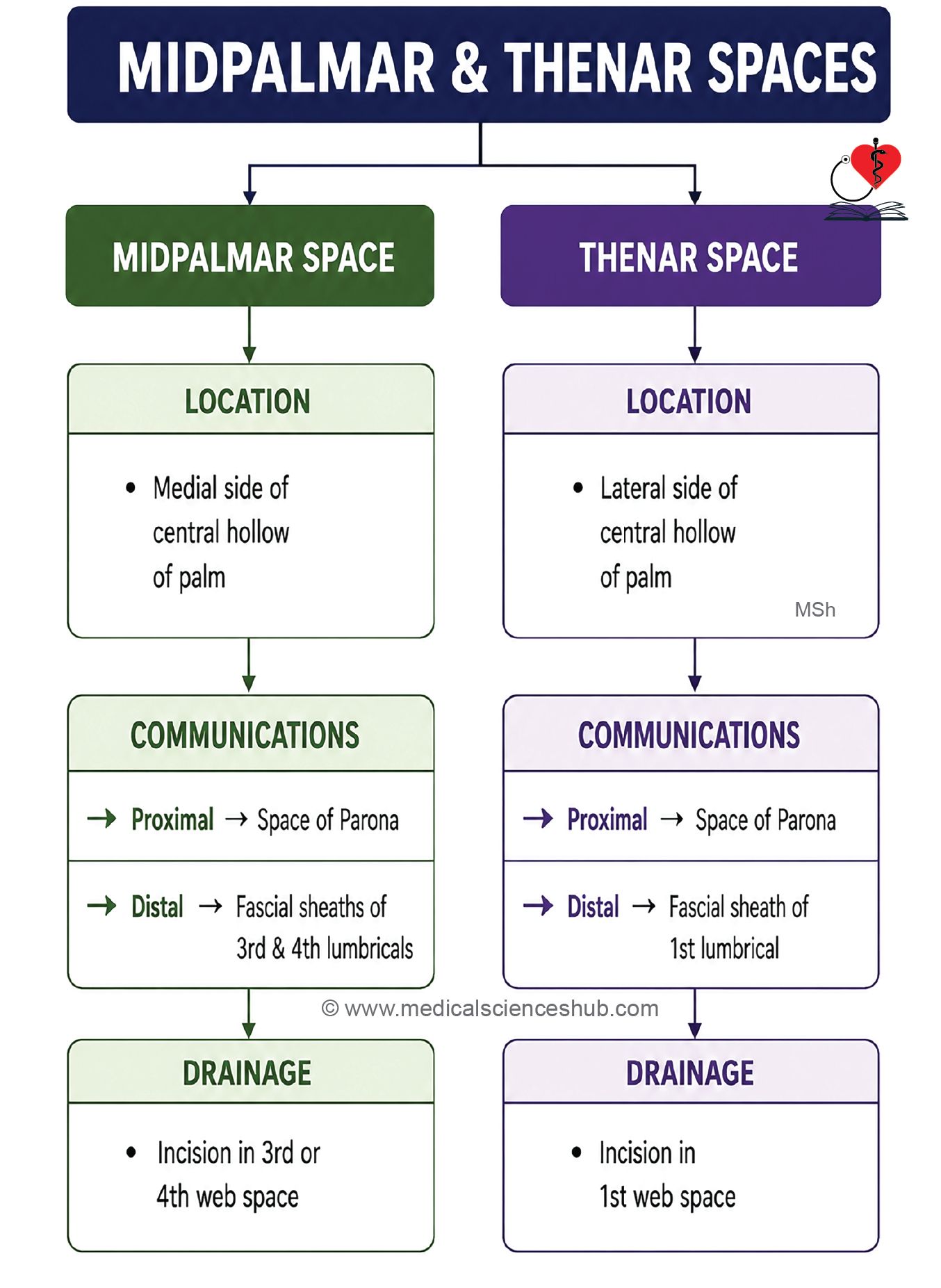
The midpalmar space is a potential fascial space located in the medial part of the palm. Under normal conditions, it contains loose areolar tissue, but it becomes clinically important when infected, as it may fill with pus and form a deep palmar abscess.
Shape
- Triangular
Extent
- Proximal: Extends to the distal margin of the flexor retinaculum.
- Distal: Reaches the distal palmar crease.
ommunications
- Proximally: Communicates with the space of Parona in the distal forearm.
- Distally: Continues with the fascial sheaths surrounding the third and fourth lumbricals, providing a pathway for the spread of infection into the fingers.
Boundaries
Anterior
- Palmar aponeurosis
- Tendons of flexor digitorum superficialis (FDS) and flexor digitorum profundus (FDP) to the third, fourth, and fifth fingers
- Second, third, and fourth lumbrical muscles
Posterior
- Interossei muscles
- Third, fourth, and fifth metacarpal bones
Medial
- Medial palmar septum
Lateral
- Intermediate palmar septum
Clinical Importance
- Infection within the midpalmar space results in a midpalmar abscess, which may spread proximally into the space of Parona or distally along the lumbrical canals.
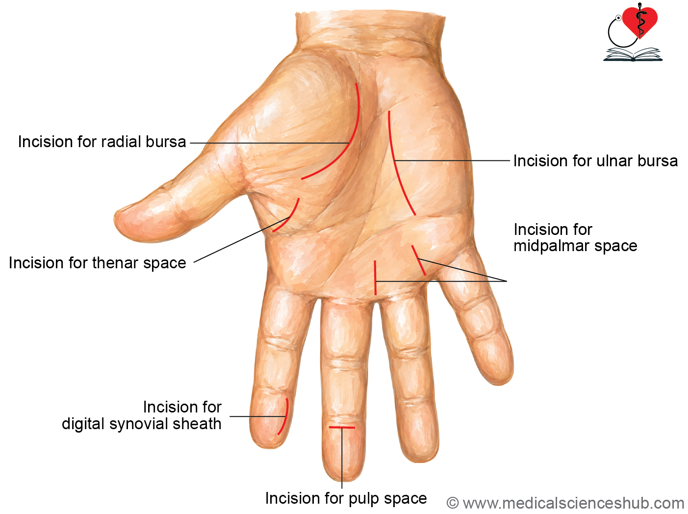
- Surgical drainage is commonly performed through an incision in the third or fourth web space, which provides effective drainage while minimizing injury to important neurovascular structures.
Thenar Space
The thenar space is a potential fascial space situated beneath the lateral half of the hollow of the palm. Normally filled with loose areolar tissue, it becomes clinically important when infected, leading to the formation of a thenar space abscess.
Shape
- Triangular
Extent
- Proximal: Extends to the distal margin of the flexor retinaculum.
- Distal: Reaches the proximal transverse palmar crease.
Communications
- Proximally: Communicates with the space of Parona in the distal forearm.
- Distally: Continues with the fascial sheath surrounding the first lumbrical, providing a route for the spread of infection.
Boundaries
Anterior
- Thenar muscles
- Tendons of flexor digitorum superficialis (FDS) and flexor digitorum profundus (FDP) to the index finger
- First lumbrical muscle
- Palmar aponeurosis
- Tendon of flexor pollicis longus (FPL)
Posterior
- Transverse head of the adductor pollicis muscle
Medial
- Intermediate palmar septum
Lateral
- Lateral palmar septum
Clinical Importance
- Infection of the thenar space produces a thenar space abscess, which may spread proximally into the space of Parona or distally along the first lumbrical sheath.
- Surgical drainage is usually performed through an incision in the first web space, providing adequate drainage while preserving the important neurovascular structures of the thumb and index finger.
Pulp Space of Fingers
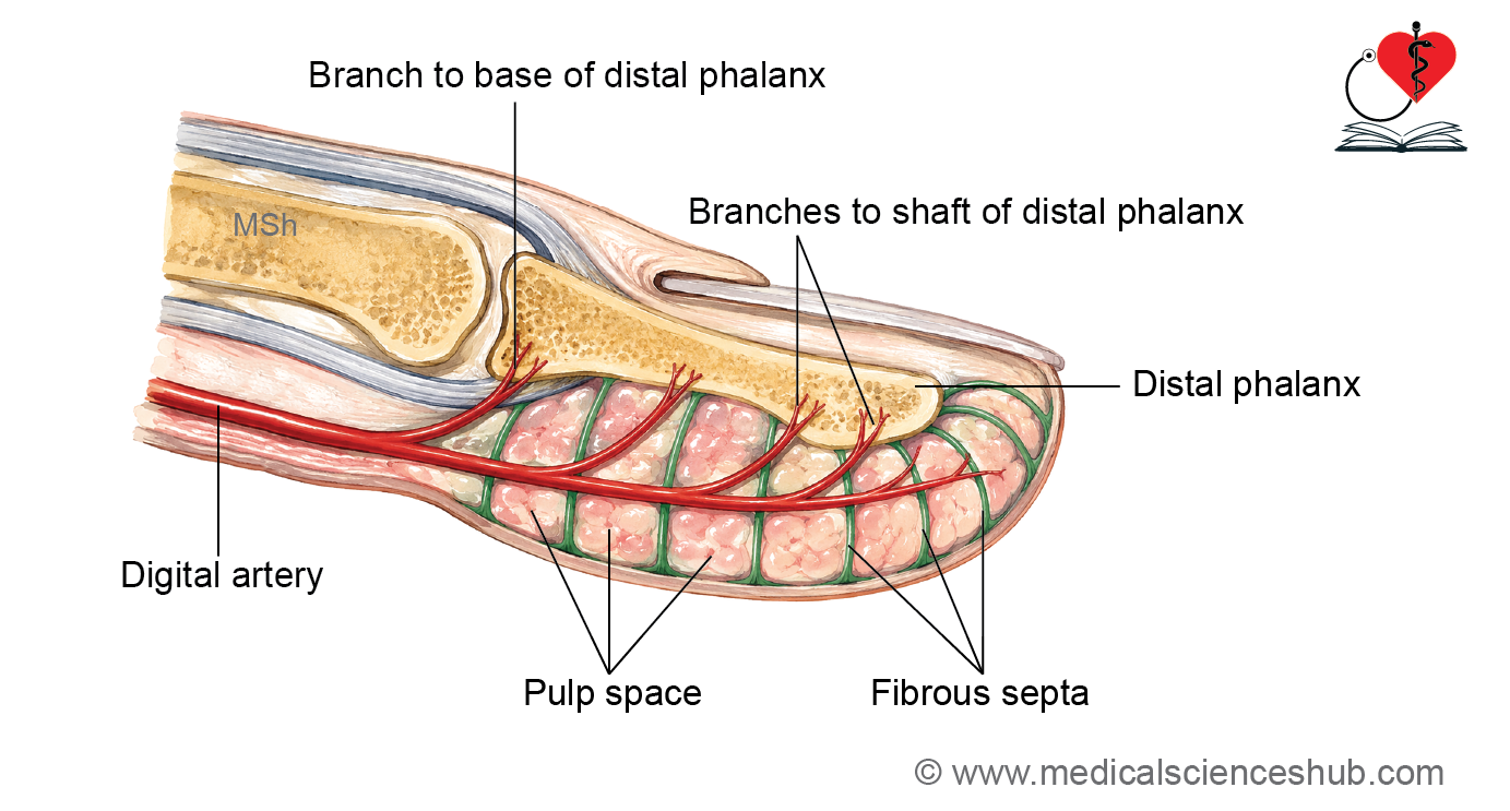
The pulp spaces are small subcutaneous fascial compartments located on the palmar aspect of the terminal phalanges of the fingers and thumb. They contain fibro-fatty tissue that provides cushioning and enhances grip and tactile sensation.
Boundaries
Superficial
- Skin
- Superficial fascia
Deep
- Distal phalanx covered by its periosteum
Internal Arrangement
- Numerous fibrous septa extend from the skin to the periosteum of the distal phalanx.
- These septa divide the pulp into multiple small compartments (loculi), firmly anchoring the skin to the underlying bone and preventing excessive movement of the fingertip.
Contents
- Digital artery
- Fibrofatty (subcutaneous) tissue
Clinical Importance
- Infection of a pulp space results in a felon (whitlow), a localized abscess of the fingertip.
- Because the pulp is divided into multiple loculi by fibrous septa, the infection is associated with severe throbbing pain due to increased tissue pressure.
- If left untreated, the infection may spread to the distal phalanx, causing osteomyelitis.
- Prompt surgical drainage is often required to relieve pressure, preserve blood supply, and prevent complications.
CLINICAL INTEGRATION
Infection of the Pulp Space
Infection within the pulp space leads to the formation of pus in a confined compartment. Because the pulp space is divided by dense fibrous septa and cannot expand, pressure rises rapidly, producing severe pain and compromising the blood supply to the distal phalanx.
Consequences
- Increased tissue pressure may compress the vessels supplying the distal phalanx.
- Prolonged vascular compromise can result in avascular necrosis of the distal four-fifths of the terminal phalanx.
Whitlow (Felon)
- A whitlow (felon) is a localized abscess of the pulp space of the fingertip.
- It is characterized by:
- Severe throbbing pain
- Swelling and tenderness of the fingertip
- Tense pulp due to the non-distensible nature of the compartment
Surgical Drainage
- Drainage is performed through a small lateral incision on the fingertip.
- A lateral approach allows decompression of the multiple loculi while minimizing injury to the digital nerves, arteries, and the highly sensitive pulp skin.
Important Questions
- Write a short note on flexor retinaculum of hand.
- List the structures passing superficial to flexor retinaculum.
- Write a short note on palmar aponeurosis.
- List the intrinsic muscles of hand.
- Write a short note on thenar muscles.
- Write a short note on palmaris brevis muscle.
- Write a short note on interossei of hand.
- Write a short note on superficial palmar arch.
- Write a short note on deep palmer arch.
- Write a short note on cutaneous innervations of palm.
- Write a short note on ulnar nerve.
- Write a short note on claw hand (ulnar claw hand).
- Write a short note on median nerve in hand.
- Write a short note on carpal tunnel syndrome.
- Write a short note on ape thumb deformity.
- Write a short note on midpalmar space.
- Write a short note on thenar space.
📝 Test Your Knowledge – Practice MCQs
Attempt the chapter MCQ quiz and assess your understanding of key concepts.
Click to see: Medical Disclaimer and Copyright Notice