
Competencies
- AN13.3: Identify and describe the type, articular surfaces, capsule, synovial membrane, ligaments, relations, movements, blood and nerve supply of …… and first carpometacarpal joint.
- AN13.4: Describe sternoclavicular joint, acromioclavicular joint, carpometacarpal joints and metacarpophalangeal joint.
Introduction
The joints of the hand are highly specialized to provide both stability and remarkable mobility, enabling precise and coordinated movements. These adaptations allow humans to perform a wide range of skilled activities such as writing, drawing, sewing, handling surgical instruments, using tools, and manipulating small objects with great accuracy.
The articulations of the hand are classified into the following groups:
- Intercarpal joints – Joints between adjacent carpal bones.
- Midcarpal joint – Joint between the proximal and distal rows of carpal bones.
- Carpometacarpal joints – Joints between the distal row of carpal bones and the bases of the metacarpals.
- Intermetacarpal joints – Joints between the bases of adjacent metacarpal bones.
- Metacarpophalangeal (MCP) joints – Joints between the heads of the metacarpals and the bases of the proximal phalanges.
- Interphalangeal (IP) joints – Joints between adjacent phalanges of the fingers and thumb.
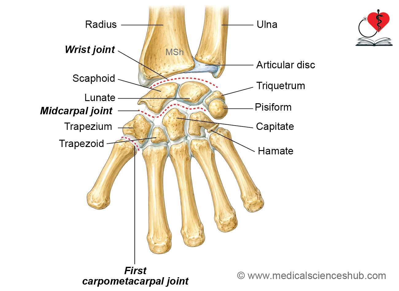
Intercarpal joints
The intercarpal joints are the articulations between adjacent carpal bones. These joints contribute to the flexibility and stability of the wrist.
Arrangement of the Carpal Bones
The eight carpal bones are arranged in two rows.
Proximal row
- Scaphoid
- Lunate
- Triquetrum
- Pisiform
Distal row
- Trapezium
- Trapezoid
- Capitate
- Hamate
Type
- The intercarpal joints are plane synovial joints.
Pisiotriquetral joint
The pisotriquetral joint is formed between the pisiform and the triquetrum.
Type
- Plane synovial joint
Midcarpal joint
The midcarpal joint is situated between the proximal and distal rows of the carpal bones.
Type
- Compound ellipsoid synovial joint
Articular Surfaces
- Formed between the distal surfaces of the scaphoid, lunate, and triquetrum (pisiform excluded) and the proximal surfaces of the trapezium, trapezoid, capitate, and hamate.
Carpometacarpal joints
The carpometacarpal joints are formed between the distal row of carpal bones and the bases of the metacarpals.
Type
- Second to fifth CMC joints: Plane synovial joints
- First CMC joint (thumb): Saddle synovial joint
Intermetacarpal joints
The intermetacarpal joints are formed between the adjacent bases of the second to fifth metacarpals.
Type
- Plane synovial joints
First Carpometacarpal Joint
The first carpometacarpal joint is the articulation between the trapezium and the base of the first metacarpal. It is the most mobile carpometacarpal joint and is responsible for the remarkable mobility of the thumb.
Type
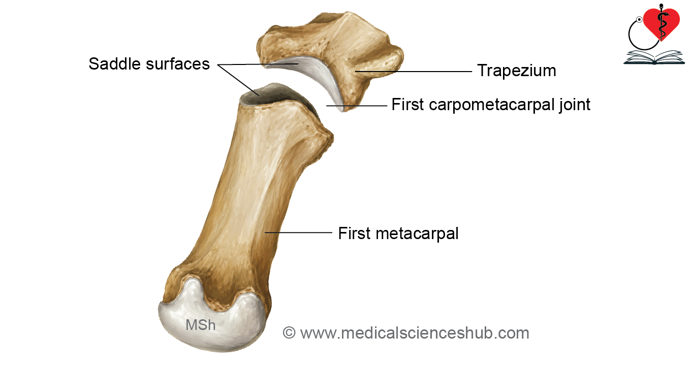
- Saddle (sellar) variety of synovial joint
The unique saddle-shaped articular surfaces allow movement in two planes as well as axial rotation, making the thumb highly versatile.
Articular surface
Proximal Articular Surface
- Inferior surface of the trapezium
Distal Articular Surface
- Superior surface of the base of the first metacarpal
Both articular surfaces are reciprocally concavoconvex, with each surface being concave in one direction and convex in the other. This reciprocal arrangement is characteristic of a saddle joint and permits a wide range of movements.
Ligaments
Capsular Ligament
- A strong fibrous capsule surrounds the joint.
- Attached to the margins of the articular surfaces of the trapezium and first metacarpal.
- The inner surface is lined by the synovial membrane.
Lateral Ligament
- A broad ligament extending from the lateral surface of the trapezium to the lateral aspect of the base of the first metacarpal.
- Reinforces the lateral side of the joint.
Anterior and Posterior Ligaments
- Oblique fibrous bands extending from the anterior and posterior surfaces of the trapezium to the medial side of the base of the first metacarpal.
- These ligaments strengthen the anterior and posterior aspects of the joint.
Relations
Anterior
- Muscles of the thenar eminence
Posterior
- Tendons of:
- Extensor pollicis longus (EPL)
- Extensor pollicis brevis (EPB)
Medial
- Radial artery
- First dorsal interosseous muscle
Lateral
- Tendon of abductor pollicis longus (APL)
Blood and Nerve Supply
Blood Supply
- Supplied mainly by branches of the radial artery.
Nerve Supply
- Articular branches arise primarily from the median nerve.
Movements
The first carpometacarpal joint permits movements in multiple planes because of its saddle-shaped configuration.
The first metacarpal is positioned anteriorly and rotated medially by approximately 90° relative to the other metacarpals. Consequently, movements of the thumb occur at right angles to those of the fingers.
Flexion
- Movement of the thumb across the plane of the palm toward the little finger.
Extension
- Movement of the thumb away from the palm in the plane of the palm.
Abduction
- Movement of the thumb away from the palm in a plane perpendicular to the plane of the palm.
Adduction
- Movement of the thumb back toward the plane of the palm from the abducted position.
Opposition
- A complex movement in which the thumb is brought across the palm so that the pulp of the thumb contacts the pulp of one or more fingers.
- This movement combines abduction, flexion, and medial rotation of the first metacarpal.
- Opposition is the anatomical basis of the human hand’s precision grip and fine manual dexterity.
Circumduction
- A circular movement produced by the sequential combination of flexion, extension, abduction, and adduction.
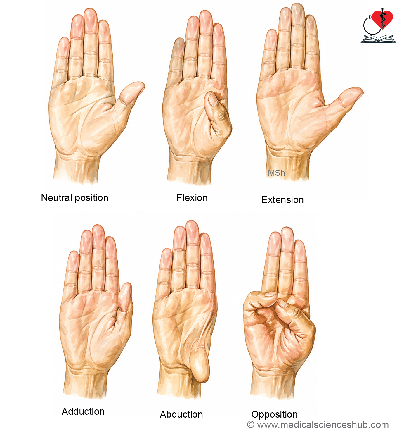
Table 15.1: Movements of thumb and responsible muscle
| Movement | Principal Muscles |
|---|---|
| Flexion | Flexor pollicis longus, Flexor pollicis brevis, Opponens pollicis |
| Extension | Extensor pollicis longus, Extensor pollicis brevis |
| Adduction | Adductor pollicis |
| Abduction | Abductor pollicis longus, Abductor pollicis brevis |
| Opposition | Opponens pollicis (chief muscle) |
CLINICAL INTEGRATION
Osteoarthritis of the First Carpometacarpal Joint
- The first carpometacarpal (CMC) joint is one of the most common sites of osteoarthritis, particularly in postmenopausal women.
- Osteoarthritis is a degenerative joint disorder characterized by progressive wear of the articular cartilage with advancing age.
- Patients commonly present with:
- Pain at the base of the thumb
- Stiffness
- Tenderness
- Reduced grip and pinch strength
- Difficulty performing activities such as writing, opening jars, turning keys, or buttoning clothes
- In advanced cases, deformity and instability of the thumb may develop.
Rheumatoid Arthritis
- Rheumatoid arthritis is a chronic autoimmune inflammatory disease that primarily affects synovial joints.
- It is characterized by:
- Pain
- Swelling
- Warmth
- Morning stiffness
- Progressive joint destruction and deformity if untreated
- The disease commonly involves:
- Metacarpophalangeal joints
- Proximal interphalangeal joints
- Wrist joints
- Long-standing disease may also affect the first carpometacarpal joint, leading to pain and impairment of thumb movements.
Metacarpophalangeal Joints
The metacarpophalangeal (MCP) joints are formed by the articulation of the heads of the metacarpal bones with the bases of the proximal phalanges. These joints provide both stability and mobility, allowing the fingers to perform powerful grasping as well as fine, precise movements.
Type
- Ellipsoid (condyloid) variety of synovial joints
Being biaxial joints, they permit movement in two planes.
Articular Surfaces
Proximal (Superior) Articular Surface
- Rounded head of the metacarpal
Distal (Inferior) Articular Surface
- Shallow base of the proximal phalanx
The convex metacarpal head articulates with the concave base of the proximal phalanx.
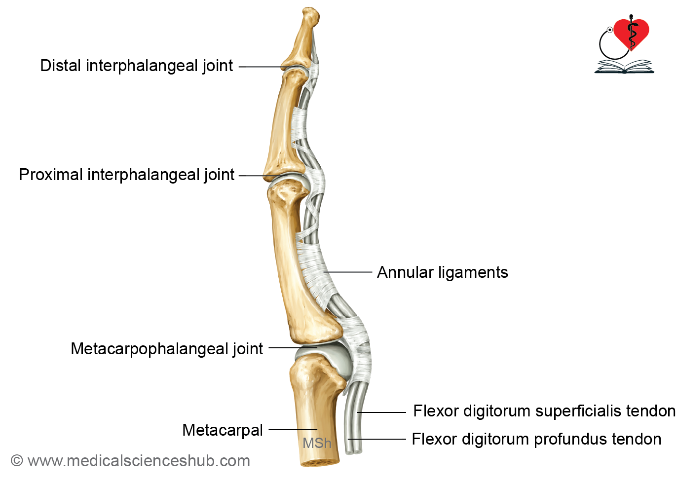
Ligaments
Capsular Ligament
- A fibrous capsule surrounds each metacarpophalangeal joint.
- It is attached to the margins of the articular surfaces and lined internally by a synovial membrane.
Palmar (Volar) Ligament
- A strong fibrocartilaginous plate firmly attached to the base of the proximal phalanx.
- Reinforces the palmar aspect of the joint.
- Prevents hyperextension and increases joint stability.
Collateral Ligaments
Each joint is strengthened on either side by:
- Medial collateral ligament
- Lateral collateral ligament
These are strong, cord-like fibrous bands that stabilize the joint during flexion and gripping.
Movements
The metacarpophalangeal joints permit the following movements:
- Flexion
- Extension
- Abduction
- Adduction
- Circumduction
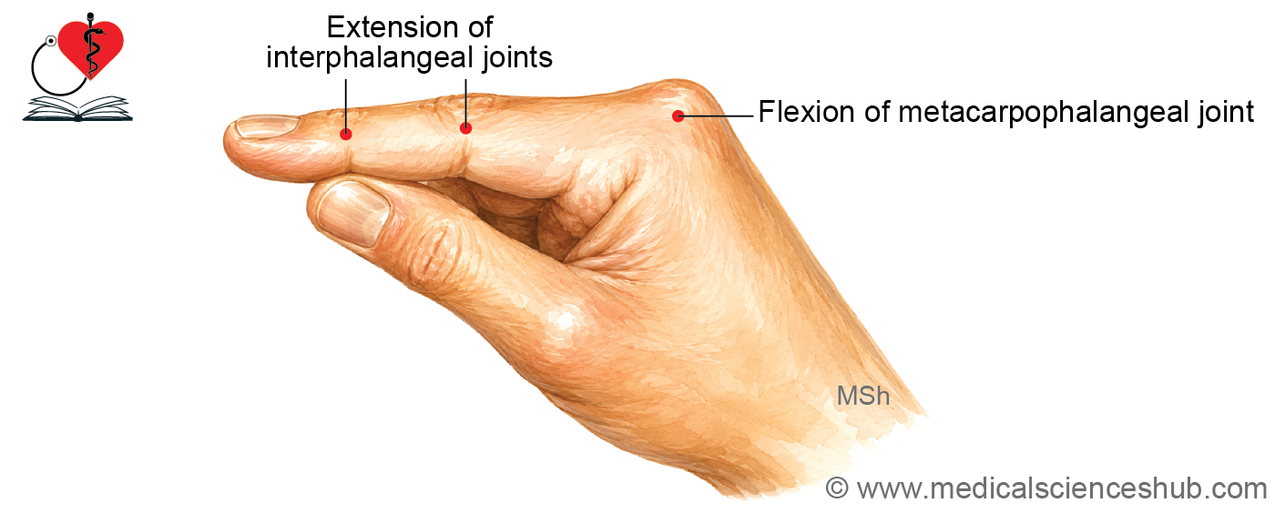
Table 15.2: Movements at metacarpophalangeal (MP) joint and responsible muscles
| Movement | 1st MP joint | 2nd–5th MP joints |
|---|---|---|
| Flexion | Flexor pollicis longus Flexor pollicis brevis | Interossei Lumbricals |
| Extension | Extensor pollicis longus Extensor pollicis brevis | Extensors of fingers |
| Adduction | Adductor pollicis | Palmar interossei |
| Abduction | Abductor pollicis | Dorsal interossei |
Interphalangeal Joints
The interphalangeal (IP) joints are the articulations between adjacent phalanges of the digits.
- The index, middle, ring, and little fingers each have:
- Proximal interphalangeal (PIP) joint
- Distal interphalangeal (DIP) joint
- The thumb has only one interphalangeal (IP) joint between its proximal and distal phalanges.
Type
- Hinge variety of synovial joints
These are uniaxial joints that permit movement in a single plane.
Ligaments
Each interphalangeal joint is reinforced by:
- Fibrous capsule
- Palmar (volar) fibrocartilaginous ligament (volar plate)
- Medial collateral ligament
- Lateral collateral ligament
These structures provide stability while preventing excessive lateral movement and hyperextension.
Movements
As hinge joints, the interphalangeal joints permit only:
- Flexion
- Extension
Muscles Producing the Movements
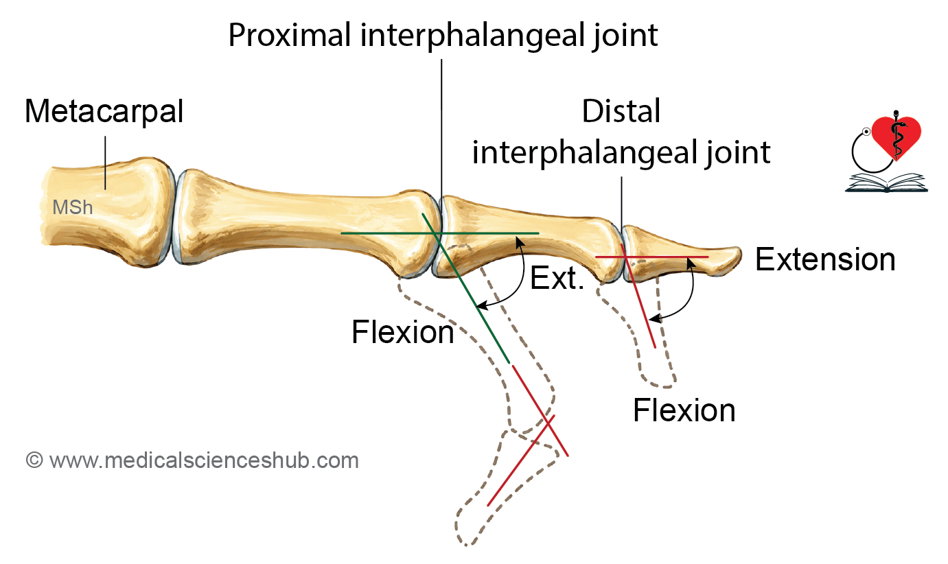
Table 15.3: Movements of interphalangeal joints and responsible Muscles
| Movement | Thumb (IP Joint) | Index to Little Fingers |
|---|---|---|
| Flexion | Flexor pollicis longus | Proximal IP (PIP): Flexor digitorum superficialis and Flexor digitorum profundus Distal IP (DIP): Flexor digitorum profundus |
| Extension | Extensor pollicis longus | Extensor digitorum, assisted by the lumbricals and interossei |
Important Questions
- Write a short note on 1st carpometacarpal joint.
- Write a short note on metacarpophalangeal joint.
📝 Test Your Knowledge – Practice MCQs
Attempt the chapter MCQ quiz and assess your understanding of key concepts.
Click to see: Medical Disclaimer and Copyright Notice