
Competencies
- AN7.2: List components of nervous tissue and their functions.
- AN7.3: Describe parts of a neuron and classify them based on number of neurites, size and function.
- AN7.7: Describe various types of synapses.
- AN68.1: Describe and identify multipolar and unipolar neurons, ganglia and peripheral nerves.
- AN68.2: Describe the structure-function correlation of neuron.
- AN68.3: Describe the ultrastructure of nervous tissue.
Introduction
- The nervous system functions as a highly coordinated control network, maintaining harmony within the body. It continuously interacts with other organ systems, receiving signals and regulating their activities in return.
- Structurally, it consists of neurons with their processes, supported by neuroglial cells and protected by the meninges.
- Functionally, it detects sensory inputs from both internal and external environments, processes and interprets this information, and generates appropriate responses.
- It governs voluntary and involuntary actions, stores experiences, and applies past information to guide future behavior. Additionally, it forms the basis of intelligence, memory, learning, thought, and creative activity.
Divisions of Nervous System
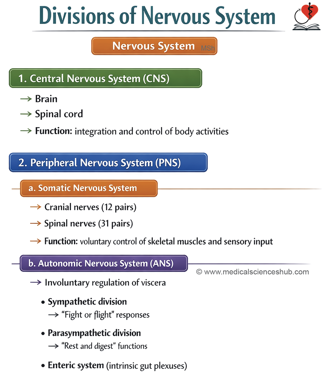
The nervous system is classified on anatomical and functional bases.
Anatomical or Structural Divisions
- The nervous system has two main components.
- The central nervous system (CNS)
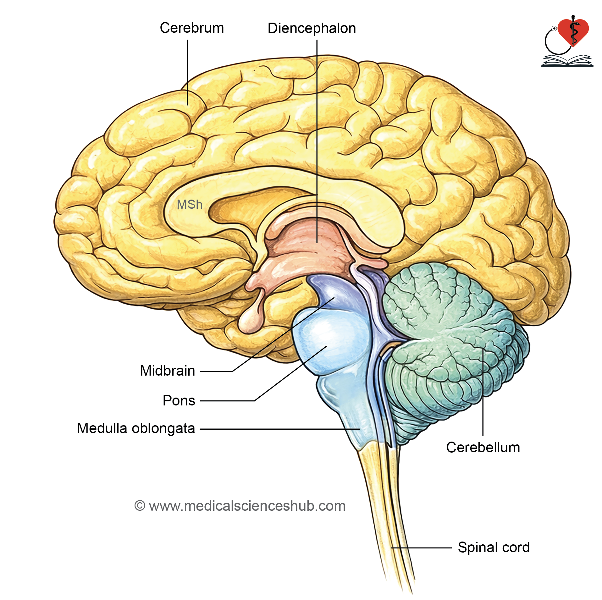
- It includes the brain and spinal cord.
- It receives, integrates, and processes information and coordinates appropriate responses.
- It is responsible for higher functions such as memory, learning, and emotions.
- The peripheral nervous system (PNS) consists of nerves and ganglia located outside the CNS.
- It includes 12 pairs of cranial nerves and 31 pairs of spinal nerves.
- It also contains dorsal root ganglia, cranial nerve ganglia, and autonomic ganglia.
- The PNS transmits sensory information to the CNS and carries motor commands to muscles, glands, and viscera.
Functional (Physiological) Divisions
- Functionally, the nervous system is divided into afferent and efferent components.
- The afferent division carries sensory input from receptors to the CNS.
- The efferent division transmits motor output from the CNS to effector organs. The efferent division is further divided into the somatic and autonomic nervous systems.
- The somatic nervous system controls voluntary activity of skeletal muscles.
- The autonomic nervous system regulates involuntary functions of smooth muscle, cardiac muscle, glands, and blood vessels.
Enteric Nervous System
- The enteric nervous system (ENS) is a network of neurons and ganglia present within the wall of the gastrointestinal tract.
- It functions as a part of the autonomic nervous system and regulates gut motility and secretion.
- The ENS has two major plexuses.
- The myenteric (Auerbach’s) plexus is located between the inner circular and outer longitudinal layers of the muscularis externa and primarily controls intestinal movements.
- The submucosal (Meissner’s) plexus lies in the submucosa and regulates glandular secretion and local blood flow.
Functions of Enteric nervous system
- The enteric nervous system (ENS) detects mechanical and chemical changes within the intestine.
- It receives sensory input from intestinal distension and the chemical composition of luminal contents.
- It regulates peristalsis by coordinating contraction of intestinal smooth muscle.
- It controls intestinal secretions by acting on glands and epithelial cells.
- It also influences local blood flow and nutrient absorption in the intestinal wall.
Cellular Organization of Nervous System
The nervous system is composed of two main cell types:
- Neurons, which are specialized excitable cells responsible for generating and transmitting nerve impulses.
- Supporting cells, which are non-excitable and include neuroglial cells, ependymal cells, Schwann cells, and satellite cells, all of which provide structural support, protection, and maintenance of the neural environment.
Neurons
- Neurons are the structural and functional units of the nervous system.
- They receive, integrate, and transmit information to other neurons or effector cells.
Structure of Neuron
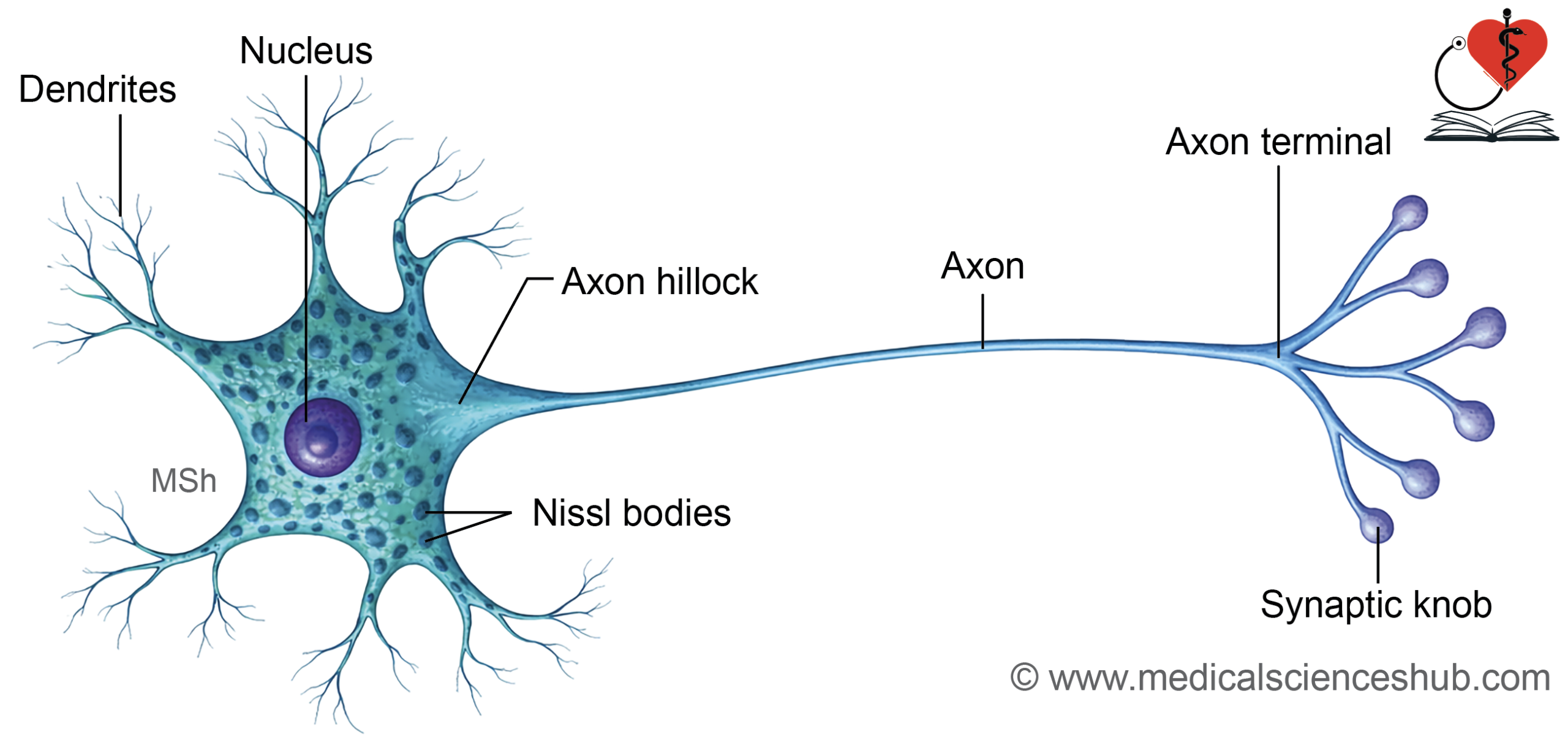
- Each neuron consists of a cell body (soma), dendrites, an axon, and synaptic terminals.
Cell body or soma
- The soma contains a large, vesicular nucleus with a prominent nucleolus.
- Most neurons have a centrally placed nucleus, while some (e.g., in sympathetic ganglia) show an eccentric nucleus.
- In females, a Barr body may be seen beneath the nuclear membrane.
- Mature neurons are amitotic and do not divide.
- The cytoplasm contains Nissl bodies, mitochondria, rough endoplasmic reticulum, and Golgi apparatus.
- Nissl granules are basophilic structures involved in protein synthesis.· They are present in the soma and dendrites but are absent in the axon and axon hillock.
Neurites
Neurites are processes arising from the soma and include dendrites and the axon.
Dendrites
- Dendrites are multiple, short, tapering processes that receive incoming signals.
- Their branching forms a dendritic tree.
- They contain most organelles, including Nissl bodies, but lack a well-defined Golgi apparatus.
- Some neurons show dendritic spines, which increase surface area for synaptic input.
Axon
- The axon is a single, long process with a uniform diameter that conducts impulses away from the soma.
- Its cytoplasm is called axoplasm, and its membrane is the axolemma.
- The axon hillock is the origin of the axon and lacks Nissl bodies.
- The initial segment (preaxon) extends from the hillock to the beginning of myelination.
- Axons may give collateral branches, and terminal branches are called telodendrites.
- Terminal enlargements, known as synaptic boutons, form junctions with target cells.
- In skeletal muscle, the terminal forms a motor end plate.
Axonal transport:
- Axonal transport is the system of transport of substances within the axon.
- Axonal transport moves materials within the axon.
- Anterograde transport carries substances away from the soma.
- Retrograde transport carries substances toward the soma.
- For example, neurotransmitters such as dopamine are transported along axons, and substances taken up at terminals can return to the cell body.
CLINICAL NEUROANATOMY
- The rabies virus is transmitted through the saliva of infected animals during a bite. After entry, it reaches nerve endings and enters axons at the site of injury. It travels toward the central nervous system by retrograde axonal transport.
- The herpes zoster virus remains dormant in sensory ganglia. On reactivation, it moves from neuronal cell bodies to the skin by anterograde axonal transport, producing characteristic lesions.
Classification of Neurons
Neurons are classified according to the number of their processes, their size, and their functional properties.
Classification based on Number of Processes
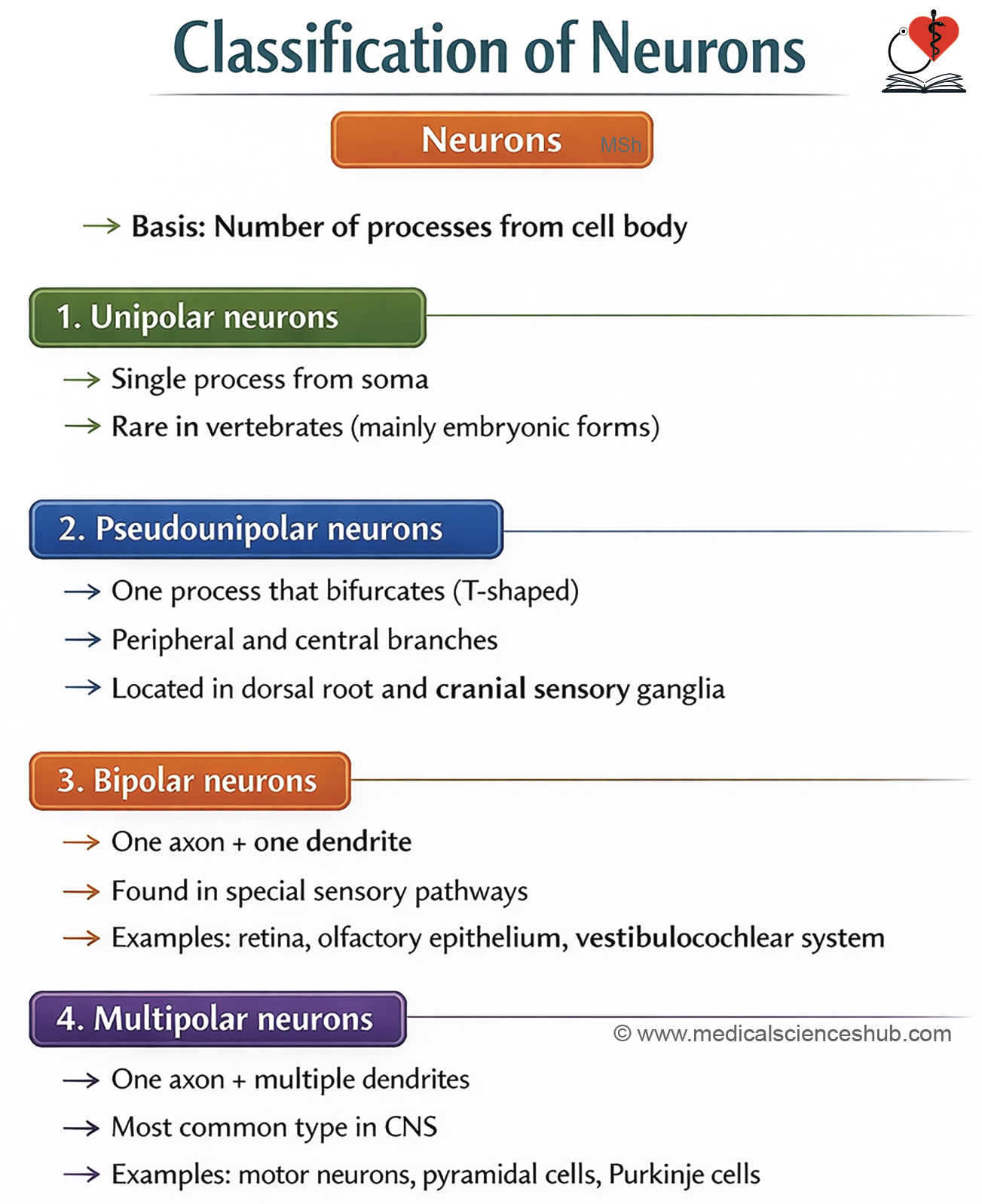
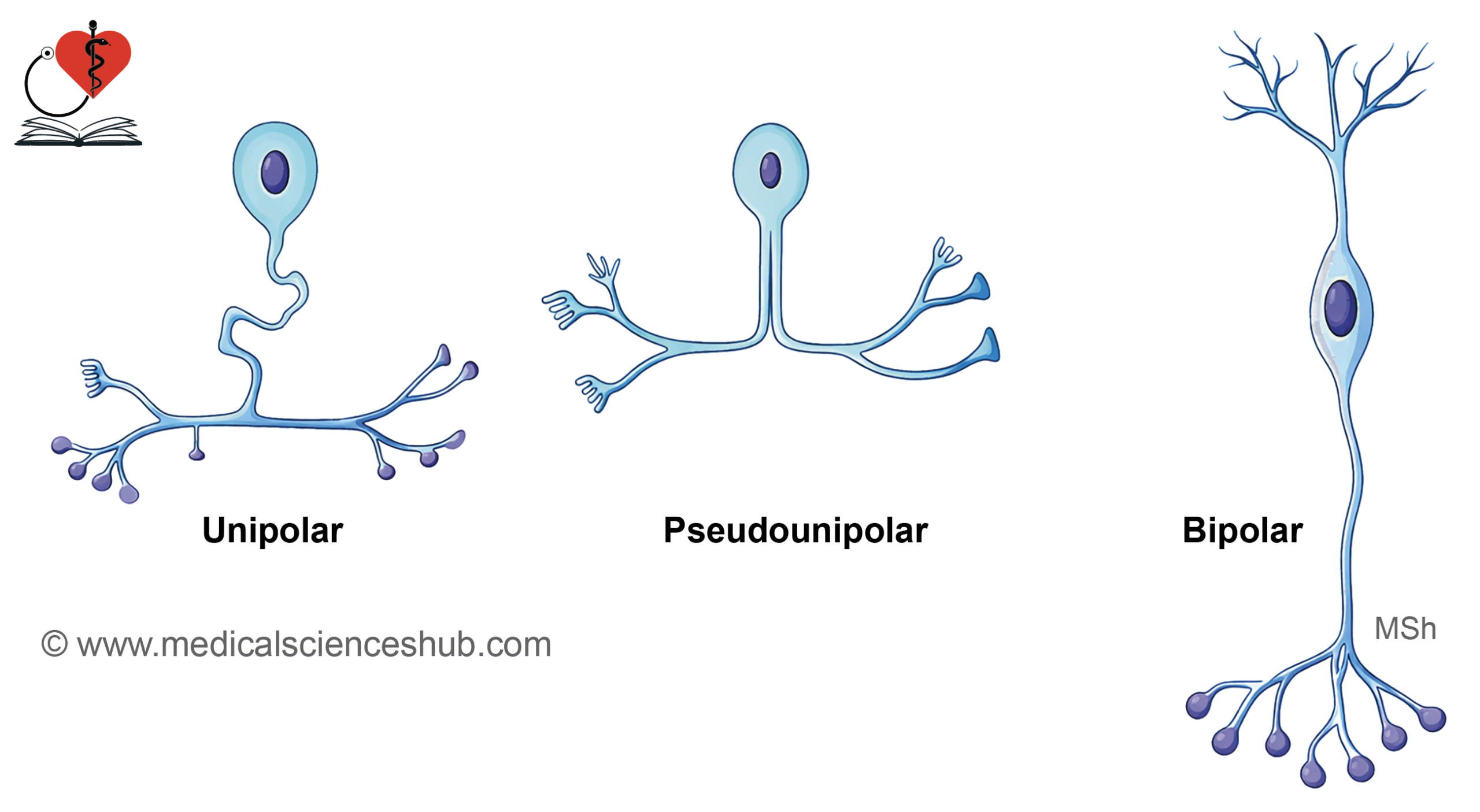
Neurons can be structurally categorized according to the number of processes arising from the cell body as unipolar, pseudounipolar, bipolar, and multipolar. In current understanding, unipolar and pseudounipolar types are often grouped together.
- Unipolar neurons possess a single process, typically resembling a dendrite. They are seen in the mesencephalic nucleus of the trigeminal nerve and in certain stages of embryonic development.
- Pseudounipolar neurons have one process that bifurcates into peripheral and central branches. These are characteristic of neurons in dorsal root ganglia and other sensory ganglia.
- Bipolar neurons exhibit two processes—one axon and one dendrite—arising from opposite poles of the cell body. They are found in the retina, olfactory epithelium, and vestibular and spiral ganglia.
- Multipolar neurons contain one axon and multiple dendrites. Common examples include pyramidal, Purkinje, and stellate neurons.
Classification based on length of axon
Neurons are also categorized according to the length of their axon into Golgi type I and Golgi type II.
Golgi type I neurons
These neurons possess a single, long axon that may extend over considerable distances, sometimes exceeding a meter. Typical examples include motor neurons of the spinal cord, pyramidal neurons of the cerebrum, and Purkinje cells of the cerebellum.
Golgi type II neurons
These neurons have short axons, which may be minimal or even absent. They are mainly involved in local circuit functions. Examples include stellate cells and granule cells of the cerebellar cortex.
Physiological classification of neurons
According to function, neurons are grouped into sensory, interneurons, and motor types.
- Sensory (afferent) neurons
These neurons detect stimuli such as pain, touch, and temperature, and convey this information to the central nervous system. - Interneurons
They form connections between sensory and motor neurons. Located within the central nervous system, they are responsible for processing and integrating incoming signals. They constitute the majority of neurons in the body. - Motor (efferent) neurons
These transmit impulses from the central nervous system to muscles, glands, and viscera. They are further divided into:
A. Somatic motor neurons
They innervate skeletal muscles.
- Upper motor neurons: Situated in the motor cortex; their axons project to cranial nerve nuclei or anterior horn cells of the spinal cord.
- Lower motor neurons: Located in cranial nerve motor nuclei and anterior horn cells; they directly supply skeletal muscles.
B. Visceral (autonomic) motor neurons
They control smooth muscle, cardiac muscle, and glands.
- Preganglionic neurons: Found in autonomic nuclei of cranial nerves (e.g., Edinger–Westphal, salivatory, lacrimal, and dorsal motor nucleus of vagus) and in the lateral horn of the spinal cord.
- Postganglionic neurons: Located in autonomic ganglia and relay impulses to target organs.
Neuroglia
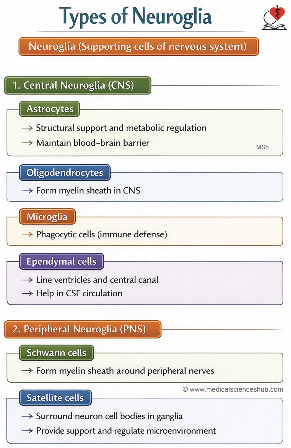
Neuroglial cells provide structural and functional support to neurons. They contribute to insulation and play a key role in forming the blood–brain barrier. Unlike neurons, they are non-excitable and retain the ability to divide.
They are broadly classified into:
- Central neuroglia (within the central nervous system)
- Astrocytes
- Oligodendrocytes
- Microglia
- Ependymal cells
- Peripheral neuroglia (within the peripheral nervous system)
- Schwann cells
- Satellite cells
Central Neuroglia
- Neuroglia are the supporting cells of the central nervous system.
- The main types include:
- Astrocytes
- Oligodendrocytes
- Microglia
- Ependymal cells
Astrocytes
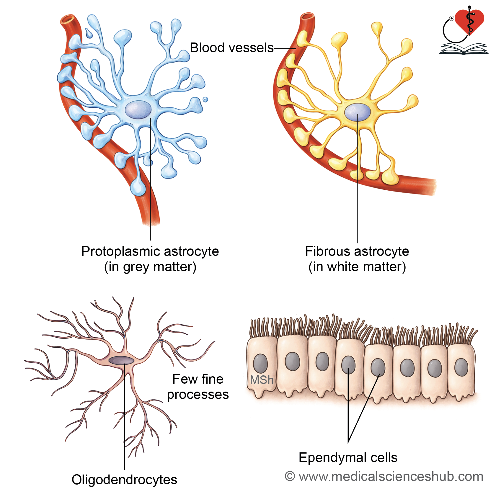
- Astrocytes are large, stellate cells with numerous branching processes.
Classification
- Fibrous astrocytes
- Located mainly in white matter
- Possess fewer, long, slender processes
- Terminal ends of processes expand to form vascular end feet
- Protoplasmic astrocytes
- Predominantly found in grey matter
- Have abundant cytoplasm with many short, thick processes
- Contribute significantly to the formation of the blood–brain barrier
Histological Identification
- Both types express glial fibrillary acidic protein (GFAP)
- They can be demonstrated using immunohistochemical staining with anti-GFAP antibodies
Functions of Astrocytes
- Provide structural and metabolic support to neurons and capillaries
- Regulate the extracellular environment by controlling ions, metabolites, and neurotransmitters
- Serve as an energy reserve by storing glycogen
- Participate in blood–brain barrier formation by surrounding capillaries with their end feet
- Form the glia limitans, a thin layer beneath the pia mater and ependyma, acting as a boundary between the CNS tissue and cerebrospinal fluid
Oligodendrocytes
- Oligodendrocytes are small, round neuroglial cells with only a few cytoplasmic extensions (“oligo” means few).
- A single oligodendrocyte can form myelin around segments of several nearby axons.
- Node of Ranvier: These are small interruptions between successive myelin segments along an axon.
- Function: They are responsible for forming and maintaining the myelin sheath in the central nervous system (brain and spinal cord).
Microglia
- Microglia are the smallest glial cells in the central nervous system, comprising roughly 5% of the total glial population. They act as resident phagocytes and are part of the mononuclear phagocyte system.
- Origin: They arise from granulocyte–monocyte progenitor cells in the bone marrow and reach the CNS via the bloodstream.
- Structure: These cells are small with elongated nuclei and possess short, irregular, branched processes. Their cytoplasm is rich in lysosomes, inclusions, and vesicles.
Functions:
- Microglia act as phagocytes, clearing:
- Dead and damaged cells
- Cellular debris
- Infectious agents such as bacteria
- Abnormal or neoplastic cells
- They are actively involved in inflammatory responses and contribute to various degenerative disorders of the CNS.
Ependymal Cells
Ependymal cells form a lining over the fluid-filled spaces of the brain and spinal cord. They are arranged in a single layer and appear cuboidal to columnar in shape.
They develop from the neural tube during early embryonic life.
Types of ependymal cells:
- Ependymocytes: These cells line the ventricles of the brain and the central canal of the spinal cord.
- Choroid epithelial cells: These cells are present in the choroid plexus and are responsible for producing cerebrospinal fluid (CSF).
- Tanycytes: These are specialized cells located in the floor of the third ventricle. They help monitor and respond to changes in metabolite levels, especially glucose.
Ependymal cells have tight junctions near their apical surface. Their apical surface shows cilia and microvilli, while the basal surface has many infoldings.
Functions:
- Choroid epithelial cells produce CSF in the choroid plexus.
- Ependymocytes contribute to the barrier between CSF and brain tissue.
- Tanycytes help regulate and monitor metabolite levels in the brain.
Peripheral Neuroglia
• These are present in the PNS. These are of two types:
1. Schwann cells
2. Satellite cells
Schwann Cells (Neurolemmocytes)
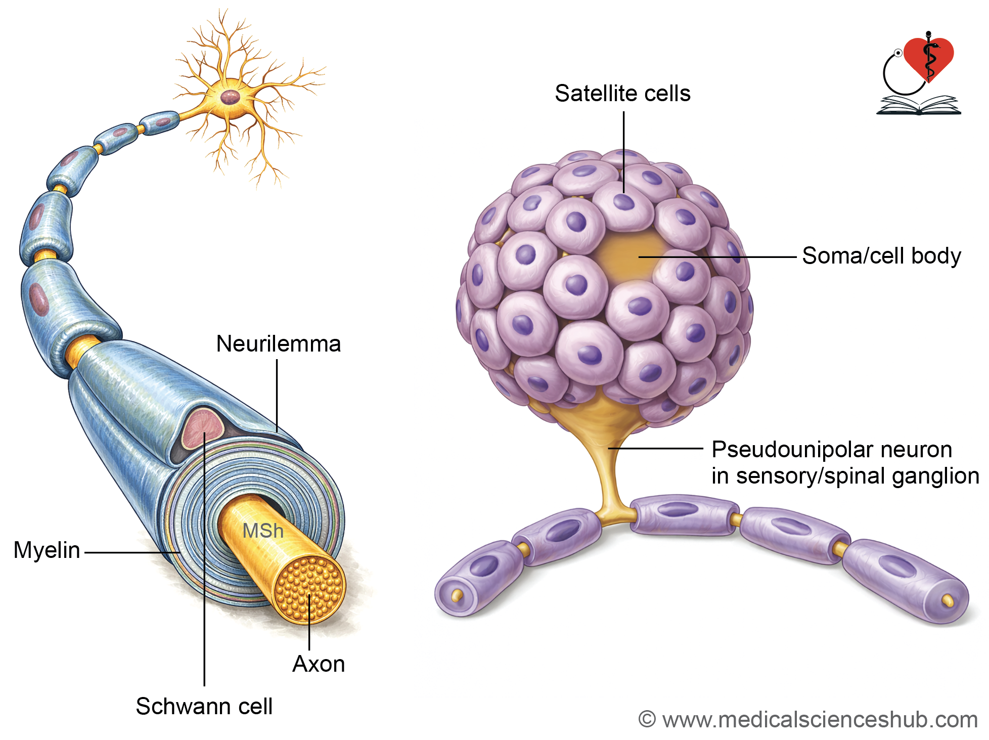
- Schwann cells are found only in the peripheral nervous system (PNS). They are flattened cells with a flattened nucleus and a relatively large amount of cytoplasm. These cells develop from neural crest cells.
- Schwann cells form the myelin sheath around axons in the PNS. Along a myelinated nerve fiber, gaps are present between adjacent Schwann cells. These gaps are called nodes of Ranvier, where the axon is not covered by myelin.
• Functions:
- Schwann cells produce myelin, which helps in rapid transmission of nerve impulses by saltatory conduction.
- They also surround and support non-myelinated nerve fibers.
- Schwann cells play an important role in the repair and regeneration of damaged axons in the PNS.
Satellite Cells
- Satellite cells are found in peripheral ganglia, including both sensory and autonomic ganglia. They are small, cuboidal cells arranged in a single layer around the cell body of neurons.
- In sensory ganglia, satellite cells completely surround the neuron and form a capsule. This capsule allows the nerve fibers to pass through. In autonomic ganglia, the covering formed by satellite cells is incomplete because synapses are present in these ganglia.
Functions:
- They protect and support the neurons in peripheral ganglia.
- They help maintain a stable environment around neurons and assist in exchange of nutrients and waste.
- They provide insulation to the neuronal cell body.
Table 1.2: Glial cells
| Cell Type | Origin | Location | Functions |
|---|---|---|---|
| Central Neuroglia | |||
| Fibrous astrocytes | Neural tube | White matter of CNS | Provide structural support and help maintain the blood–brain barrier |
| Protoplasmic astrocytes | Neural tube | Grey matter of CNS | Participate in repair processes and form glial scar after injury |
| Oligodendrocytes | Neural tube | Predominantly in white matter of CNS | Produce myelin sheath around CNS axons |
| Microglia | Mesoderm (bone marrow lineage) | Throughout CNS | Act as phagocytic cells for immune defense |
| Ependymal cells | Neural tube | Line ventricles of brain and central canal of spinal cord; form choroid plexus | Contribute to cerebrospinal fluid production and form the brain–CSF interface |
| Peripheral Neuroglia | |||
| Schwann cells | Neural crest cells | Peripheral nerves | Form myelin sheath around peripheral axons; support regeneration of damaged nerves |
| Satellite cells | Neural crest cells | Sensory and autonomic ganglia | Provide support, insulation, and regulate the microenvironment of neuronal cell bodies |
Synapse
- A nerve impulse is an electrical signal that travels along a neuron. This signal is passed from one neuron to another at a specialized junction called a synapse.
- A synapse is a specific site of contact between two or more neurons. It can also occur between a neuron and a muscle cell or a gland cell.
- At a synapse, the impulse is transmitted across a small gap from one cell to another. Therefore, conduction occurs by contact (contiguity) and not by direct continuity.
Classification of Synapse
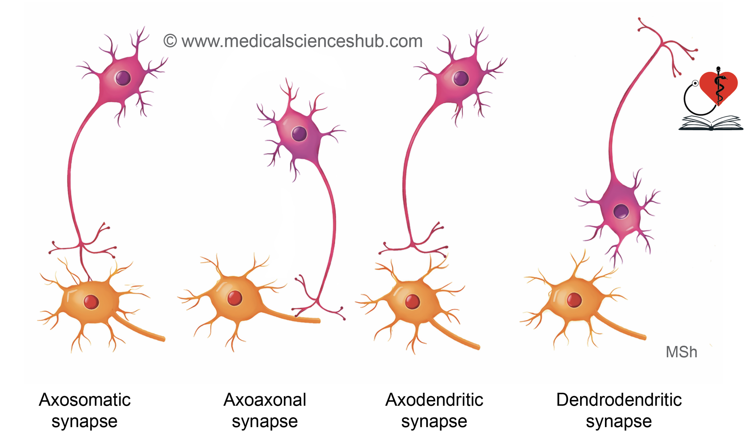
Structural Classification of Synapse
Synapses can be classified based on the parts of neurons that come in contact with each other:
- Axodendritic synapse: The axon of one neuron connects with the dendrite of another neuron.
- Axosomatic synapse: The axon of one neuron connects with the cell body (soma) of another neuron.
- Axoaxonic synapse: The connection occurs between the axons of two neurons.
- Dendroaxonic synapse: The dendrite of one neuron connects with the axon of another neuron.
- Dendrodendritic synapse: The connection occurs between the dendrites of two neurons.
- Somatosomatic synapse: The cell bodies of two neurons are in contact.
- Somatodendritic synapse: The cell body of one neuron connects with the dendrite of another neuron.
Chemical and Electric Synapses
Synapses can also be classified based on how nerve impulses are transmitted:
- Chemical synapse: In this type, the nerve impulse is transmitted by releasing chemical substances called neurotransmitters from one neuron to another.
- Electrical synapse: In this type, the electrical signal passes directly from one cell to another through gap junctions without the use of neurotransmitters.
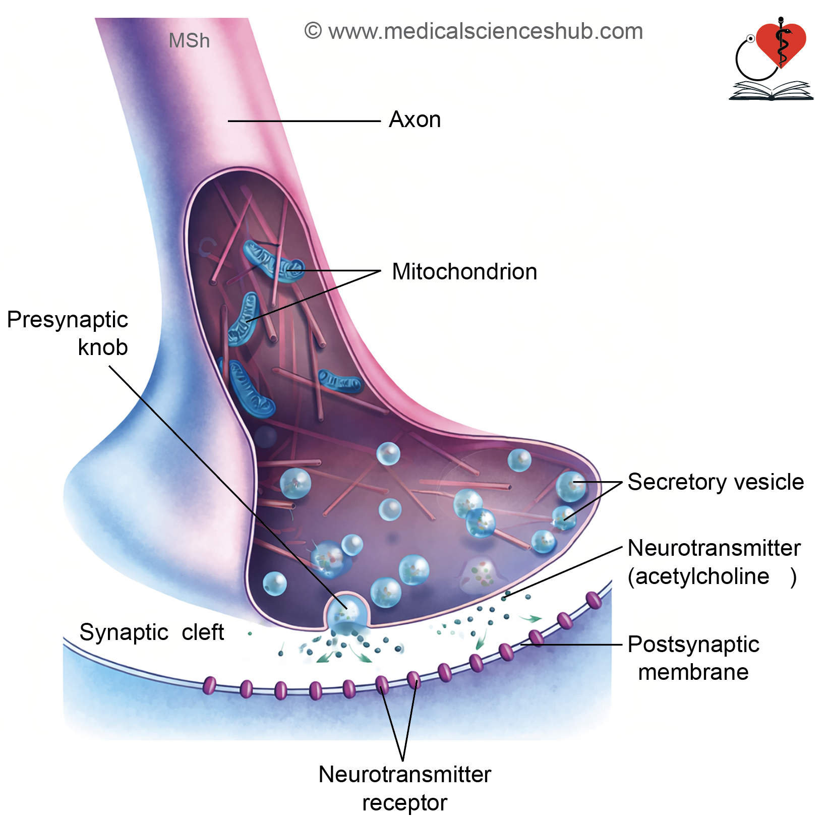
Structure of Chemical Synapse
Chemical synapses are the most common type of synapse. In these synapses, nerve impulses are transmitted by the release of chemical substances called neurotransmitters.
Parts of a chemical synapse
A chemical synapse has three main parts:
- Presynaptic knob
This is the terminal end of the sending neuron. It contains mitochondria and many synaptic vesicles. These vesicles store neurotransmitters such as acetylcholine, adrenaline, noradrenaline, and dopamine. The vesicles are formed from the Golgi apparatus or smooth endoplasmic reticulum, and some are recycled by endocytosis. When an impulse arrives, these vesicles release neurotransmitters by exocytosis. - Synaptic cleft
This is a small gap (about 20–30 nm wide) between the presynaptic and postsynaptic cells. Neurotransmitters diffuse across this gap. - Postsynaptic membrane
This is the receiving surface of the next cell. It is thickened and contains specific receptor proteins. These receptors bind to neurotransmitters and help continue the transmission of the nerve impulse.
Properties of Synapses
- Multiple connections of a neuron
A single neuron can form synapses with one or many other neurons (up to about 5500). For example, bipolar cells in the retina connect with a single ganglion cell, which helps maintain precise vision. - Feed-forward inhibition
In this type of circuit, one excitatory neuron sends signals to two neurons. At the same time, it also sends a branch to an inhibitory interneuron. This interneuron suppresses one of the target neurons. As a result, one pathway is activated while the other is inhibited. This mechanism helps in coordinated actions, such as activating an agonist muscle and relaxing its antagonist. - Feedback inhibition
This mechanism prevents excessive activity in neurons. A good example is the Renshaw cell in the anterior horn of the spinal cord. It receives a branch from a motor neuron and then inhibits the same neuron. This control helps avoid overactivity of muscles, ensures smooth and coordinated movements, and regulates reflexes like the knee jerk. - Synaptic delay
Synaptic delay is the time taken for an impulse to pass from one neuron to another. It includes the release of neurotransmitter, its movement across the synaptic cleft, and its binding to receptors. The average delay is about 0.5 milliseconds. This delay allows the postsynaptic neuron to process signals, including summation or inhibition of inputs.
CLINICAL NEUROANATOMY
- In tetanus, the bacterium Clostridium tetani releases a toxin at the site of a wound. This toxin blocks the function of Renshaw cells in the spinal cord, which normally inhibit motor neurons. When this inhibition is lost, motor neurons become overactive. As a result, muscles remain continuously contracted, leading to stiffness and painful spasms.
NEUROTRANSMITTERS
Neurotransmitters are chemical messengers released by neurons at synapses. They help in transmitting signals and regulating many body functions.
Major neurotransmitters and their locations:
- Acetylcholine – Neuromuscular junction, parasympathetic neurons, some sympathetic neurons, basal ganglia
- Noradrenaline – Sympathetic postganglionic neurons, locus coeruleus
- Adrenaline – Adrenal medulla
- Dopamine – Substantia nigra, corpus striatum, limbic system
- Serotonin – Raphe nuclei of brainstem
- Histamine – Hypothalamus
Based on function:
- Excitatory neurotransmitters: Glutamate, aspartate
- Inhibitory neurotransmitters: GABA, glycine
Other important neurotransmitters:
- Nitric oxide – Causes smooth muscle relaxation in autonomic pathways
- Neuropeptides – Found in hypothalamus and pituitary gland
- Substance P – Present in spinal and trigeminal ganglia (pain transmission)
- Vasoactive intestinal peptide (VIP) – Found in cerebral cortex and hypothalamus
- Endorphins and enkephalins – Involved in pain relief and emotional responses
Important Questions
- Write a short note on classification of neurons.
- Write a short note on synapse.
- Write a short note on neurotransmitters.
📝 Test Your Knowledge – Practice MCQs
Attempt the chapter MCQ quiz and assess your understanding of key concepts.
Click to see: Medical Disclaimer and Copyright Notice