
Competencies
- AN68.1: Describe and identify multipolar and unipolar neurons, ganglia and peripheral nerves.
- AN68.3: Describe the ultrastructure of nervous tissue.
- AN7.8 Describe differences between sympathetic and spinal ganglia
Introduction
- Discover the fascinating world of peripheral nerves and ganglia, which connect the brain and spinal cord to the rest of the body. Learn how these structures transmit sensory information and coordinate motor and autonomic functions essential for everyday life.
Peripheral Nerve
- The PNS connects body organs with the central nervous system (CNS).
- It enables transmission of sensory input and motor output.
Components
- Somatic nerves control voluntary activities.
- Autonomic nerves regulate involuntary functions and include sympathetic and parasympathetic divisions.
Constituents
- The PNS consists of:
- 12 pairs of cranial nerves
- 31 pairs of spinal nerves
- Autonomic (visceral) nerves
Types of Nerve Fibers
- Somatic afferent fibers carry sensory signals from skin, muscles, bones, and joints to the CNS.
- Somatic efferent fibers transmit motor impulses to skeletal muscles.
- Visceral afferent fibers convey sensory information from organs and blood vessels.
- Visceral efferent fibers supply glands, smooth muscle, and cardiac muscle.
- In the CNS, bundles of nerve fibers are termed tracts.
Structures of Peripheral Nerve
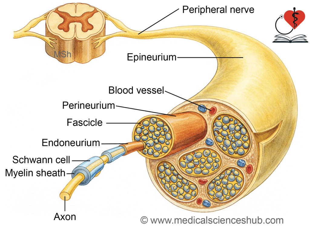
- A peripheral nerve is a bundle of multiple nerve fibers enclosed by connective tissue.
- The term nerve fiber refers to a neuronal process, not a connective tissue fiber.
Types of Nerve Fibers
- Motor (efferent) fibers are axons that transmit impulses from the CNS to muscles or glands.
- Their cell bodies are located in the grey matter of the spinal cord and brainstem.
- Sensory (afferent) fibers carry impulses toward the CNS from peripheral structures.
- Their cell bodies lie in sensory ganglia.
Ganglion
- A ganglion is a cluster of nerve cell bodies situated outside the CNS.
- Examples include the dorsal root ganglion and autonomic ganglia.
Basic Structure of Peripheral Nerve
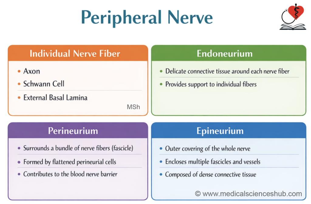
- A nerve fiber, along with Schwann cells and a basal lamina, forms the structural unit of a peripheral nerve.
Connective Tissue Coverings
- Peripheral nerves have three layers:
1. Endoneurium
- Each nerve fiber is enclosed by delicate loose connective tissue.
- It contains collagen fibers and a few supporting cells.
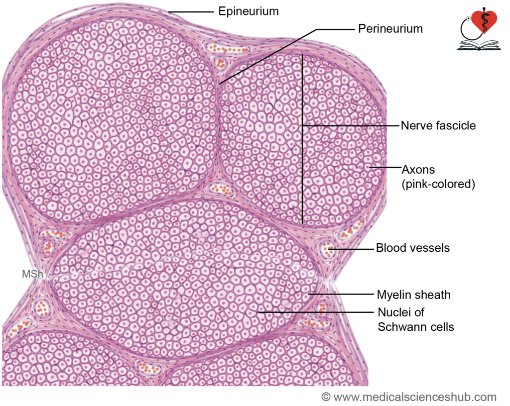
- Most nuclei seen in sections belong to Schwann cells.
2. Perineurium
- Surrounds a bundle of fibers (fascicle).
- Formed by flattened cells arranged in layers with tight junctions.
- Creates a protective blood–nerve barrier.
3. Epineurium
- Encloses multiple fascicles forming the whole nerve.
- Composed of dense connective tissue with blood vessels.
- In H&E staining, myelin appears clear, while osmium tetroxide stains it black.
Myelin Sheath
- Myelin is a specialized insulating covering that surrounds the axons of certain nerve fibers.
- It is formed by oligodendrocytes in the central nervous system (CNS).
- In the peripheral nervous system (PNS), myelin is produced by Schwann cells.
- The primary function of myelin is to insulate and protect axons.
- It significantly enhances the speed of nerve impulse conduction along the axon.
Formation of myelin sheath
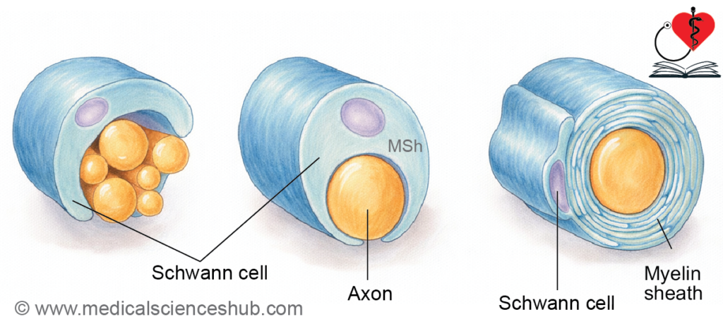
- During myelination, the axon initially lies within a groove on a Schwann cell.
- The Schwann cell membrane differentiates into three regions:
- Abaxonal membrane, which faces the external environment.
- Adaxonal (periaxonal) membrane, which is in direct contact with the axon.
- Mesaxon, a double-layered fold connecting the two membranes.
- The mesaxon elongates and wraps concentrically around the axon in a spiral manner.
- As wrapping progresses, most of the cytoplasm is displaced, forming a compact lipid-rich myelin sheath.
- A thin outer layer of Schwann cell cytoplasm remains, known as the neurilemma (neurilemmal sheath).
Composition of myelin
- Myelin is mainly composed of lipids (about 80%), along with proteins and water.
- Schmidt–Lanterman clefts are small cytoplasmic pockets retained within the layers of the myelin sheath.
- These clefts represent areas where cytoplasm is not completely expelled during myelin formation.
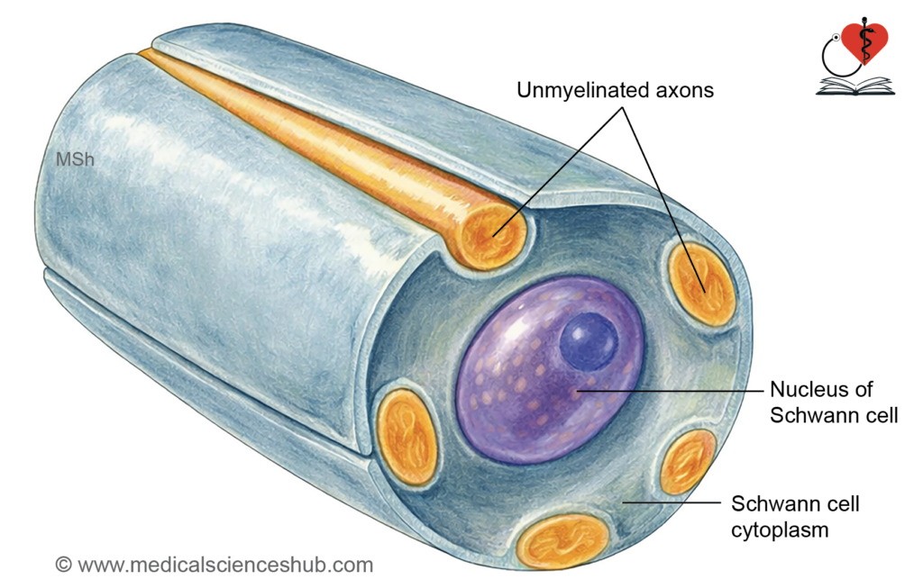
- Non-Myelinated Fibers: In the PNS, non-myelinated axons are also enclosed by Schwann cells. A single Schwann cell can envelop multiple non-myelinated axons, without forming a true myelin sheath.
Functions of myelin sheath
- The myelin sheath provides protection and structural support to axons.
- It acts as an insulator, preventing current loss.
- It enhances impulse transmission via saltatory conduction.
- The neurilemma is essential for nerve regeneration.
TABLE 2.1: The difference between myelinated and unmyelinated fibers
| Feature | Myelinated Fibers | Unmyelinated Fibers |
|---|---|---|
| Myelin sheath | It is present, formed by Schwann cells (PNS) or oligodendrocytes (CNS). | It is absent; axons are only partially enclosed by Schwann cells. |
| Nodes of Ranvier | They are present as gaps between myelin segments. | They are absent, forming a continuous membrane. |
| Conduction speed | Impulse conduction is fast due to saltatory conduction. | Impulse conduction is slow and continuous. |
| Diameter | Fibers have a larger diameter (1–20 μm). | Fibers are smaller (0.2–1.5 μm). |
| Energy efficiency | They are more efficient, as depolarization occurs only at nodes. | They are less efficient, as the entire membrane depolarizes. |
| Location | Found in motor and sensory pathways. | Found in autonomic fibers and some pain/temperature pathways. |
| Impulse propagation | Impulses jump between nodes, increasing speed. | Impulses travel along the entire length, reducing speed. |
Clinical Neuroanatomy
Enuresis (bed-wetting)
- Enuresis is the involuntary passage of urine, commonly seen in early childhood.
- It is typically observed during the first 3–6 years of life.
- This occurs until full myelination of corticospinal tract fibers that regulate bladder control is achieved.
- Embryological Basis: Myelination begins before birth and continues for several years after birth. Therefore, enuresis is considered physiological (normal) in infants and young children.
Classification of Nerve Fibers
- Nerve fibers are categorized according to their diameter and degree of myelination.
Group A Fibers
- These are highly myelinated fibers.
- They have a large diameter and conduct impulses rapidly.
Group B Fibers
- These fibers are thinly myelinated.
- Their diameter is up to about 3 μm.
- Conduction is slower than Group A fibers.
Group C Fibers
- These are non-myelinated fibers.
- Their diameter ranges from approximately 0.2–1.5 μm.
- They conduct impulses slowly.
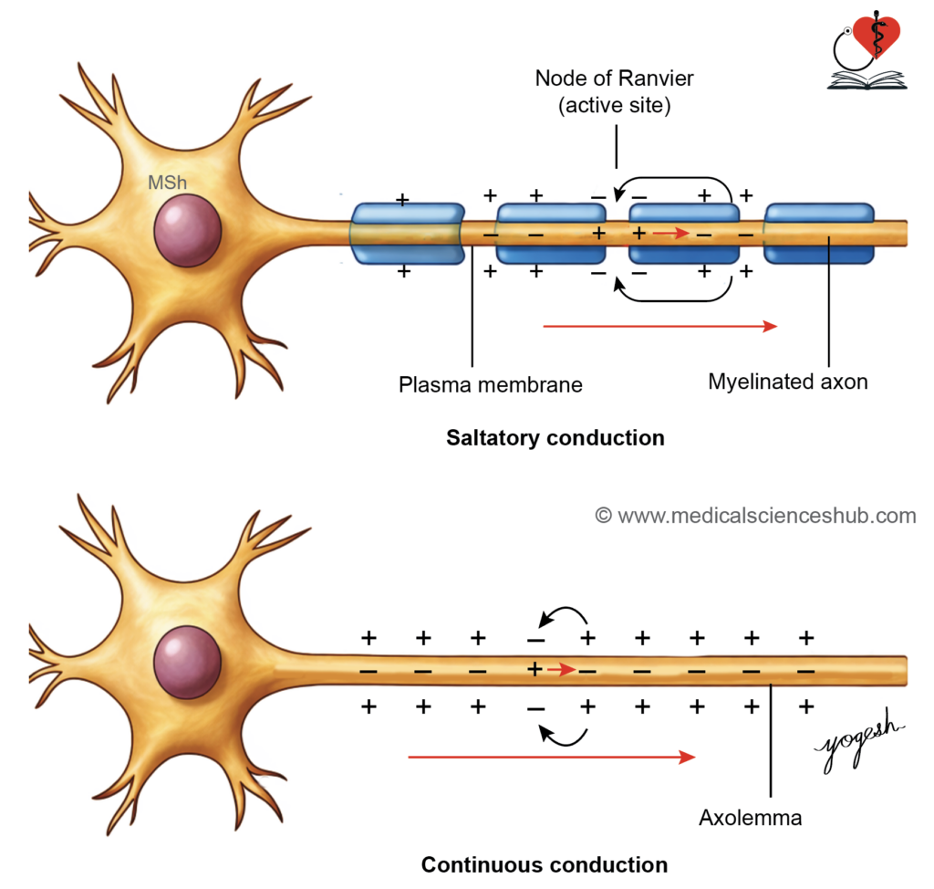
TABLE 2.2: Classification of peripheral nerve fibres
| Group & Type | Example/Function | Diameter | Myelin | Velocity (m/s) |
|---|---|---|---|---|
| Group A (Afferent) | ||||
| Type Ia | Muscle spindle (annulospiral endings) | 20 µm | Present | 80–120 |
| Type Ib | Golgi tendon organ | 20 µm | Present | 80–120 |
| Type II | Muscle spindle (flower-spray endings) | 12 µm | Present | 33–75 |
| Type III | Cutaneous receptors (touch, pressure) | 5 µm | Thin | ~30 |
| Type IV | Free nerve endings (pain, temperature) | — | Absent | — |
| Group A (Efferent) | ||||
| Alpha (α) | Extrafusal muscle fibres | 20 µm | Present | ~120 |
| Beta (β) | Extrafusal + spindle fibres | 12 µm | Present | ~70 |
| Gamma (γ) | Intrafusal muscle fibres | 8 µm | Present | ~24 |
| Group B | Preganglionic autonomic fibres | 5 µm | Present | ~15 |
| Group C | Postganglionic autonomic fibres | 1.5 µm | Absent | ~2 |
Nerve Injuries
- The rate of nerve regeneration is about 3.5–4.5 mm/day, and adequate nutrition supports faster recovery.
Classification (Based on Severity)
- Neurotmesis: Both axon and myelin sheath are completely damaged.
- Axonotmesis: The axon is damaged, but the myelin sheath remains intact.
- Neuropraxia: Both axon and myelin sheath are preserved, with temporary functional loss.
Regeneration
- Regeneration occurs in peripheral nerves in neuropraxia and axonotmesis.
- No regeneration occurs in neurotmesis due to complete structural disruption.
Nerve Degeneration and Regeneration
- Injury to an axon with its myelin sheath leads to degeneration followed by regeneration.
Degeneration of Nerve Fiber
- It refers to structural changes occurring after nerve injury.
- Changes involve both the nerve fiber and the cell body.
Changes in Nerve Fiber
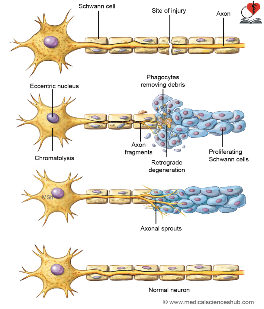
- Wallerian degeneration: The distal segment degenerates, with breakdown of axon and myelin.
- The proximal segment degenerates up to the nearest node of Ranvier.
Changes in Cell Body (Retrograde Changes)
- These occur due to loss of neural activity.
- Chromatolysis: Nissl bodies disintegrate.
- The cell body swells, and the nucleus shifts eccentrically.
- These changes are more marked when the injury is closer to the cell body.
Regeneration of Nerve Fiber
- Regeneration is slow and usually begins about 2 weeks after injury.
Steps in Regeneration
- Debris removal: Macrophages clear degenerated material by phagocytosis.
- Schwann cell proliferation: Schwann cells multiply and form a guiding tube (bands of Büngner).
- Axonal sprouting: Multiple sprouts arise from the proximal end.
- Axonal elongation: One sprout grows along the Schwann cell pathway to reach the target.
- Remyelination: Schwann cells reform the myelin sheath, restoring function.
Factors Necessary for Satisfactory Regeneration
- The endoneurial sheath must remain intact to guide regenerating axons.
- The gap between proximal and distal stumps should be minimal (only a few millimeters).
- There should be no infection at the injury site, as it impairs healing.
- Adequate levels of nerve growth factors are essential to promote axonal growth.
- Proper physiotherapy is required to maintain muscle function and support recovery.
Clinical Neuroanatomy
Neuroma
- A neuroma is a benign growth arising after nerve injury.
- It develops when there is a large gap between the cut ends of a nerve.
- The gap becomes filled with fibrous tissue containing regenerating axonal sprouts.
- These disorganized nerve fibers form a nodular mass.
- Neuromas are typically painful due to abnormal nerve regeneration.
Ganglia
- A ganglion is a collection of neuronal cell bodies located outside the central nervous system.
Types of Ganglia
- Sensory ganglia: Associated with afferent neurons that carry sensory information.
- Autonomic ganglia: Associated with the autonomic nervous system and involved in visceral functions.
Sensory Ganglia
Sensory ganglia are located just outside the CNS and transmit impulses toward the CNS.
- Examples:
- Dorsal root ganglia of spinal nerves.
- Sensory ganglia of cranial nerves: trigeminal, facial, vestibulocochlear, glossopharyngeal, and vagus.
Structure of Sensory Ganglia
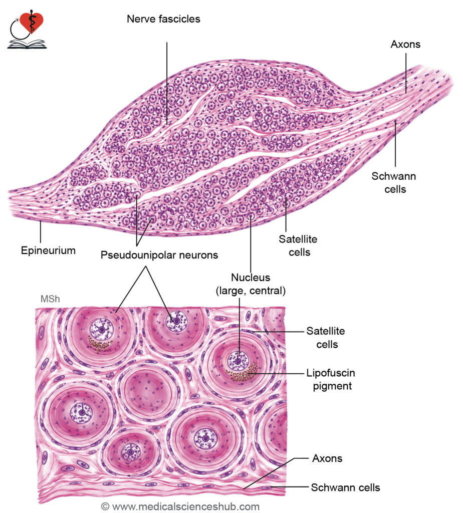
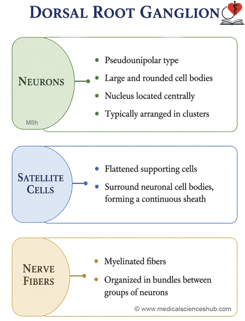
- They contain mainly pseudounipolar neurons (except vestibulocochlear ganglia, which are bipolar).
- Each neuron has:
- A peripheral process carrying input from receptors.
- A central process transmitting impulses to the CNS.
- Neuronal cell bodies are large, arranged in groups, often at the periphery.
- Nuclei are large, vesicular, and centrally placed.
- Groups of neurons are separated by bundles of myelinated nerve fibers.
Supporting Cells
- Each neuron is surrounded by satellite (capsular) cells, forming a complete capsule.
- These cells are flattened (low cuboidal).
- The ganglion is enclosed by a thin connective tissue capsule.
Autonomic Ganglia
- Autonomic ganglia supply smooth muscle and glands.
- The autonomic pathway involves two neurons:
- Preganglionic neuron: Located in the CNS; its axon reaches the ganglion.
- Postganglionic neuron: Located in the ganglion; its axon supplies target organs.
Examples
- Sympathetic: Sympathetic trunk, paravertebral ganglia (celiac, superior and inferior mesenteric, aorticorenal), and adrenal medulla.
Parasympathetic:Ciliary, submandibular, otic, pterygopalatine, and terminal ganglia near organs.
Structure of Autonomic Ganglia
- They are smaller than sensory ganglia.
- Contain multipolar postganglionic neurons scattered throughout.
- Neurons have large, pale, eccentrically placed nuclei with prominent nucleoli.
- Cells are separated by bundles of mainly nonmyelinated fibers.
Supporting Cells
- Satellite cells are few and do not form a complete capsule due to multiple neuronal processes and synapses.
- The ganglion is enclosed by a connective tissue capsule.
- Nissl granules are prominent, and silver stains highlight neuronal processes.
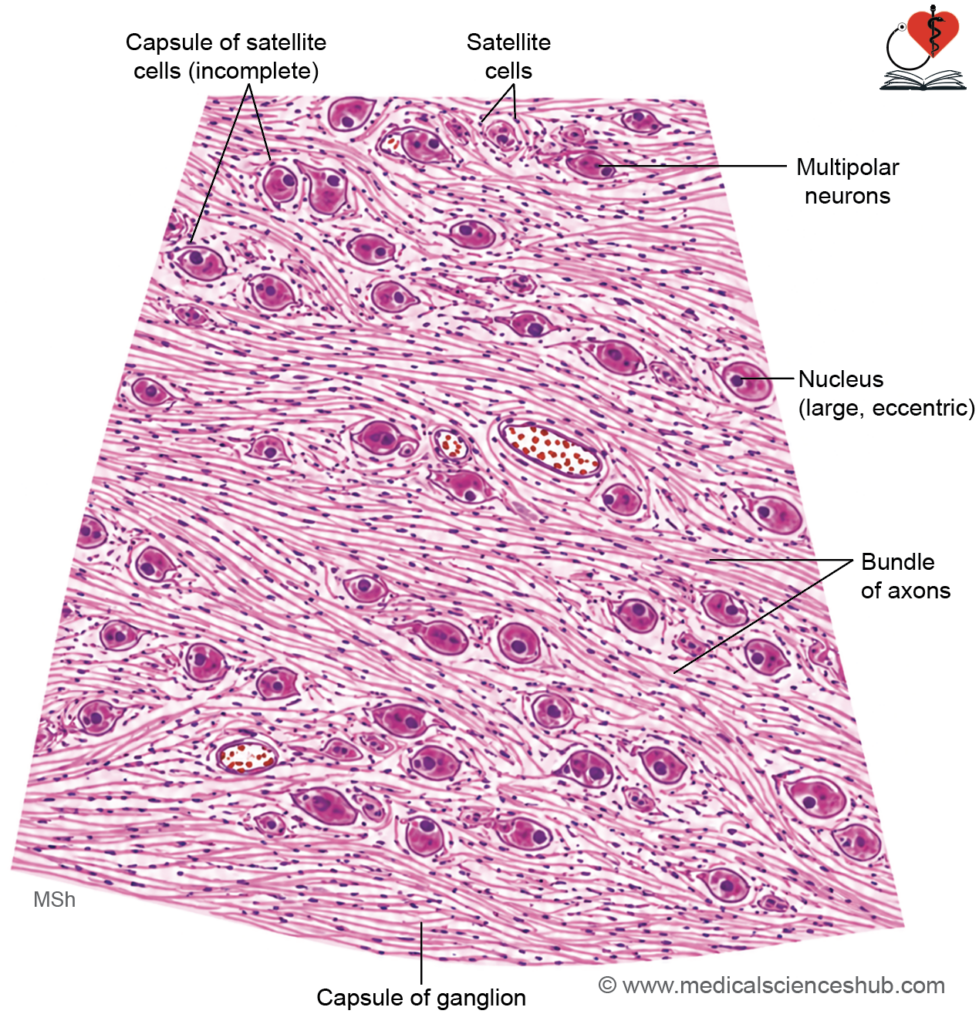
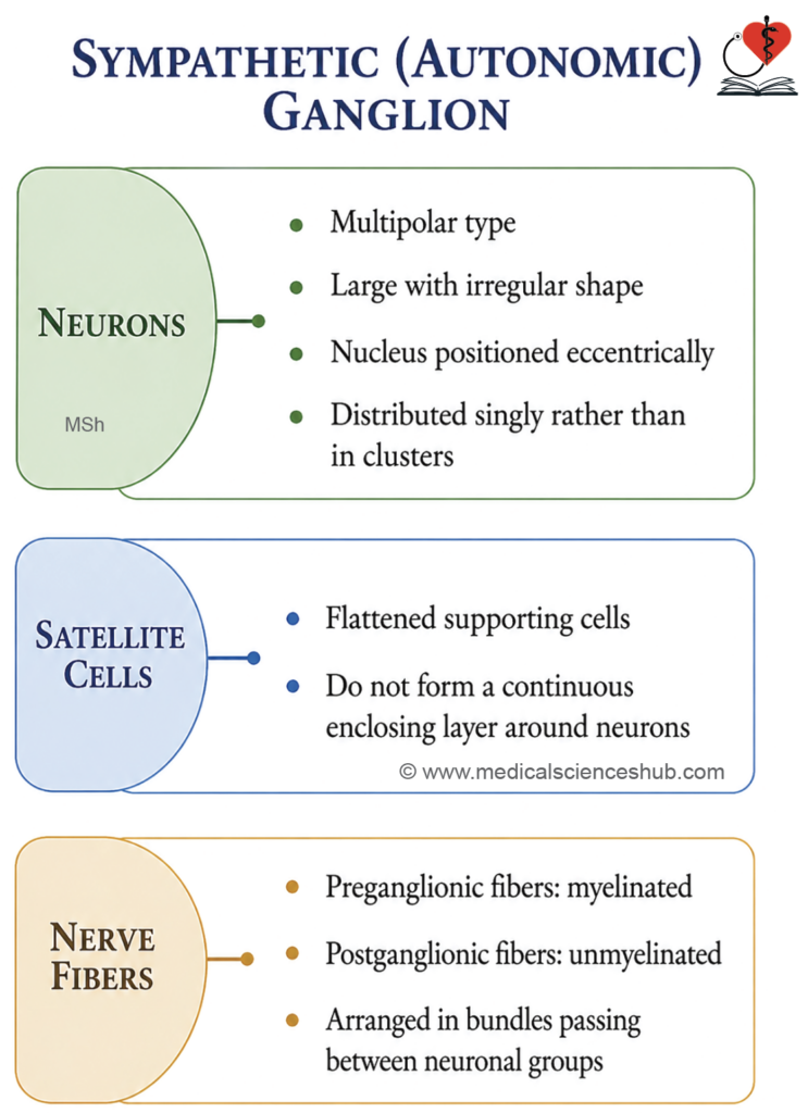
TABLE 2.3: Differences between sensory and autonomic ganglia
| Feature | Sensory (Dorsal Root) Ganglion | Autonomic (Sympathetic) Ganglion |
|---|---|---|
| Neuron type | Contains pseudounipolar neurons. | Contains multipolar neurons. |
| Cell body shape | Neurons are large and spherical. | Neurons are small and irregular. |
| Function | Composed of sensory neurons. | Contains postganglionic autonomic neurons. |
| Nucleus | Centrally placed, large and vesicular. | Eccentrically placed, large and vesicular. |
| Satellite cells | Form a complete sheath around neurons. | Incomplete covering by few satellite cells. |
| Arrangement | Neurons are arranged in clusters. | Neurons are scattered. |
| Nerve fibers | Fibers form bundles separating clusters. | Fibers run between scattered neurons. |
Important Questions
- Write a shot note on the histological structure of a peripheral nerve in transverse section (TS).
- Describe the microscopic features of a sensory (dorsal root) ganglion.
- Enumerate the key differences between dorsal root ganglion and sympathetic ganglion.
- Describe the microscopic features of an autonomic (sympathetic) ganglion.
📝 Test Your Knowledge – Practice MCQs
Attempt the chapter MCQ quiz and assess your understanding of key concepts.
Click to see: Medical Disclaimer and Copyright Notice