
Competencies
- AN57.4: Enumerate ascending and descending tracts at midthoracic level of spinal cord.
Introduction
- Tracts are bundles of nerve fibers in the central nervous system sharing common origin, course, termination, and function.
- Synonyms include fasciculi and lemnisci (ribbon-like bundles).
Classification of Tracts
• The tracts of the spinal cord are classified into three types:
1. Ascending
2. Descending
3. Intersegmental.
Locations of Tracts in spinal cord
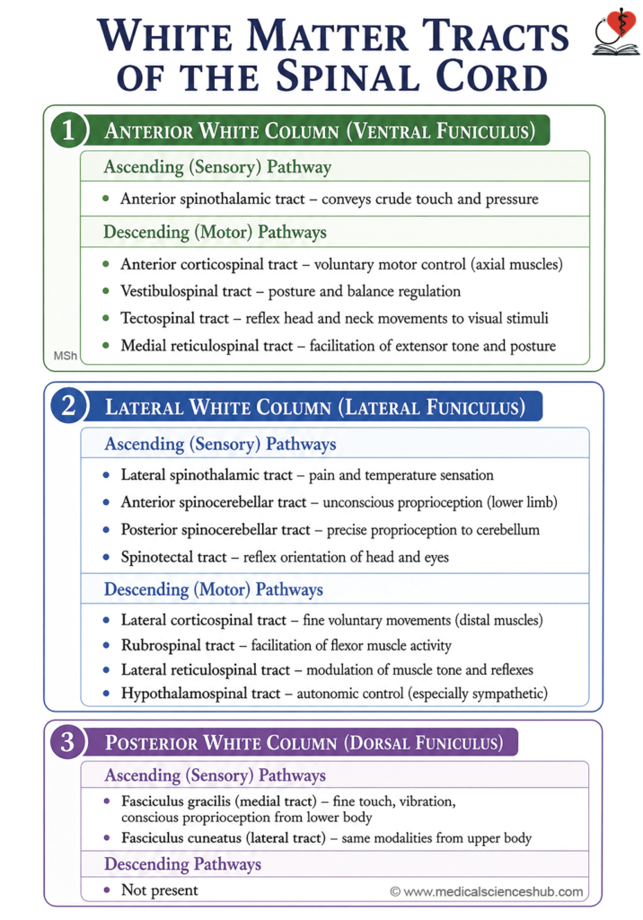
- The spinal cord tracts are organised within the white matter and are strategically positioned: motor (descending) pathways occupy the anterior and lateral regions, while sensory (ascending) pathways are located predominantly in the posterior region. This anatomical arrangement is clinically significant for localising the level and nature of neurological injury or disease.
- Anterior white column contains the ascending anterior spinothalamic tract and the descending anterior corticospinal, vestibulospinal, tectospinal, and medial rubrospinal tracts.
- Lateral white column contains the ascending lateral spinothalamic, anterior spinocerebellar, posterior spinocerebellar, and spinotectal tracts, and the descending lateral corticospinal, rubrospinal, lateral reticulospinal, and hypothalamospinal tracts. · Posterior white column contains ascending tracts only: the fasciculus gracilis (tract of Goll) and the fasciculus cuneatus (tract of Burdach).
Ascending Tracts
- Ascending tracts transmit sensory information from peripheral receptors to higher centres of the central nervous system (CNS), conveying: pain and temperature; fine touch and conscious proprioception; and unconscious proprioception.
- Proprioception refers to the perception of body position and movement. It operates at two levels:
- Conscious proprioception enables voluntary awareness and deliberate adjustment of body position.
- Unconscious proprioception contributes to postural balance during rest and movement, and participates in reflex activity.
General Arrangements of Sensory Pathway
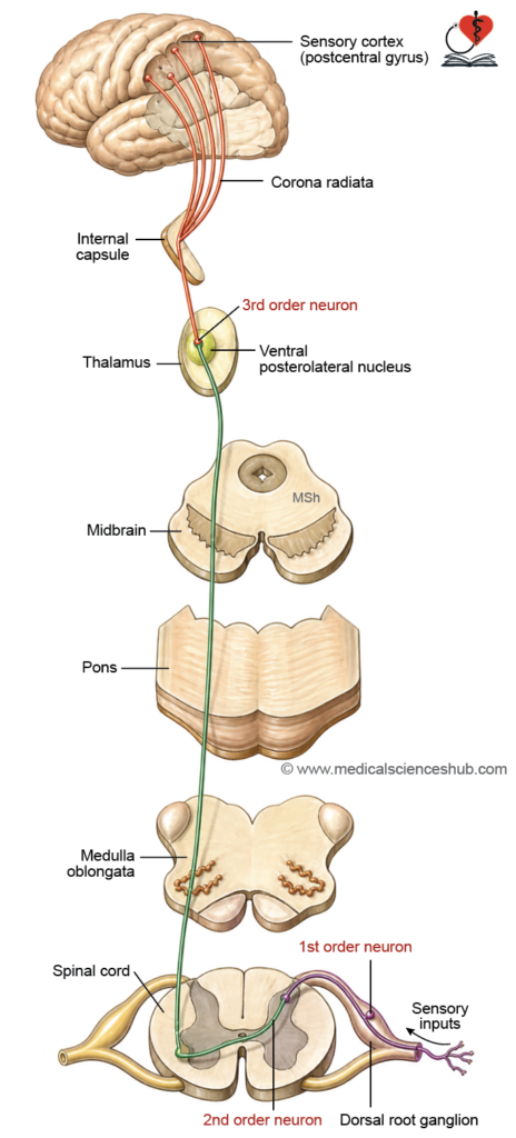
- Sensory pathways connect peripheral receptors to the cerebral cortex through a relay of three sequentially arranged neurons.
- First-order neurons are located in the dorsal root ganglia of spinal nerves and in the ganglia of cranial nerves. They are pseudounipolar neurons possessing two processes: a peripheral process that receives signals from receptors, and a central process that transmits those signals into the CNS to synapse with second-order neurons.
- Second-order neurons are located within the grey matter of the spinal cord and brainstem. The axons of most second-order neurons decussate (cross to the contralateral side) before ascending. Notably, some first-order neurons also synapse directly with anterior horn cells to mediate spinal reflex arcs.
- Third-order neurons are located in the ventral posterolateral (VPL) nucleus of the thalamus and project their axons to the primary somatosensory cortex.
Lateral Spinothalamic Tract
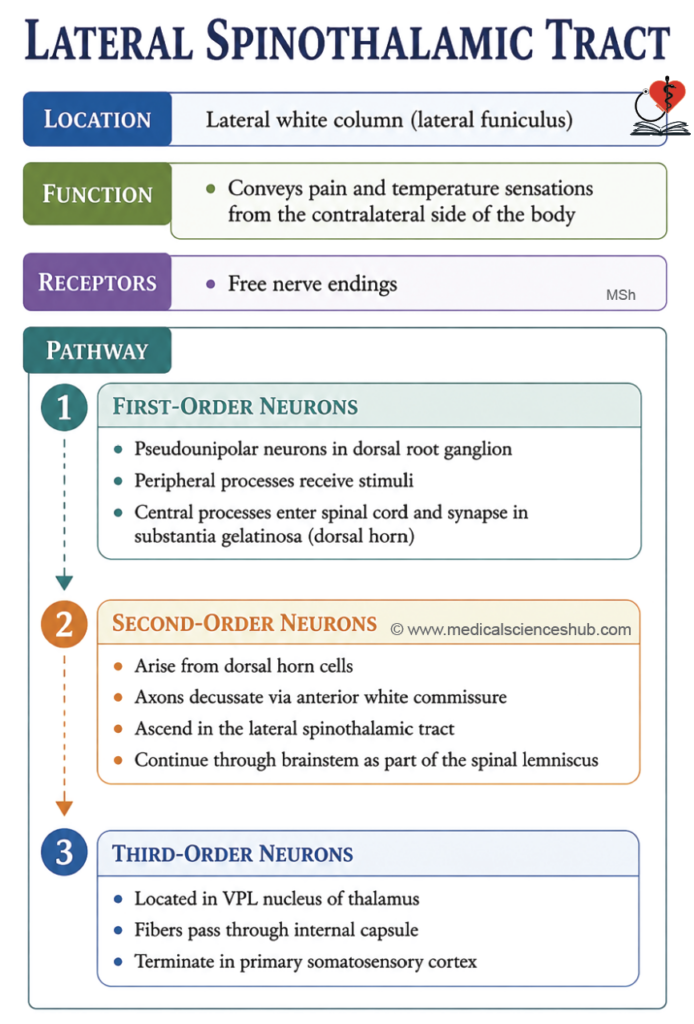
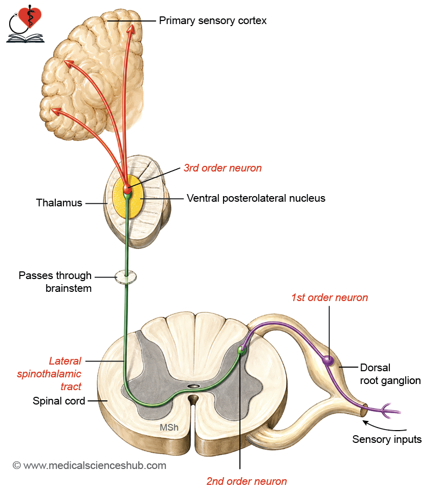
- The lateral spinothalamic tract transmits pain and temperature sensations from the contralateral side of the body to the brain. It plays a critical protective role by detecting potentially harmful stimuli and initiating appropriate responses.
- Location: Lateral white column of the spinal cord.
- Receptors: Free nerve endings detect both pain and temperature stimuli.
Composition/Pathway
- First-order neurons are pseudounipolar neurons located in the dorsal root ganglia. They possess:
- A peripheral process that conducts pain and temperature signals via the spinal nerve and its dorsal root.
- A central process that enters the spinal cord through the lateral division of the dorsal root, ascends one to two segments within the dorsolateral tract of Lissauer (at the tip of the dorsal horn), and synapses with neurons of the substantia gelatinosa in the posterior horn.
- Second-order neurons originate in the dorsal horn. Their axons decussate through the anterior white commissure to the opposite side and ascend as the lateral spinothalamic tract in the contralateral lateral white column. Within the brainstem, these fibres continue as the spinal lemniscus and terminate in the ventral posterolateral (VPL) nucleus of the thalamus.
- Third-order neurons are located in the VPL nucleus of the thalamus. Their axons project to the primary somatosensory cortex via the posterior limb of the internal capsule.
CLINICAL NEUROANATOMY
- Unilateral tract lesion produces contralateral loss of pain and temperature sensation, beginning approximately one to two spinal segments below the site of injury. This offset occurs because first-order fibres ascend briefly in Lissauer’s tract before crossing.
- Cordotomy is a surgical procedure in which the lateral spinothalamic tract is intentionally sectioned to relieve intractable pain. It is anatomically feasible because pain-conducting fibres are located on the lateral aspect of the lateral white column.
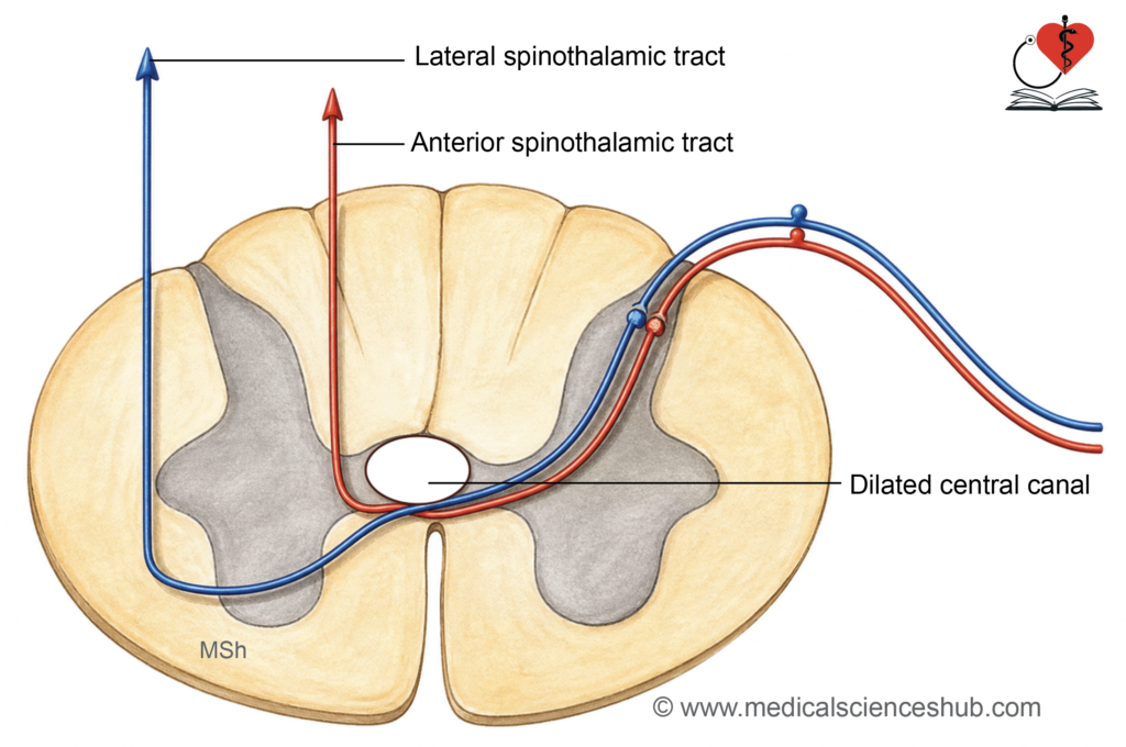
- Syringomyelia involves pathological expansion of the central canal, which compresses the decussating spinothalamic fibres within the anterior white commissure. This produces a bilateral, segmental loss of pain and temperature sensation at the affected spinal levels, while fine touch and proprioception are preserved — a pattern termed dissociated sensory loss.
Pathway for pain and temperature from head region:
- Pain and temperature sensations from the head are conveyed by branches of the trigeminal nerve (cranial nerve V).
- First-order neurons reside in the trigeminal (semilunar) ganglion. Their peripheral processes form the three branches of the trigeminal nerve. Their central processes enter the pons and descend as the spinal tract of the trigeminal nerve into the medulla and upper cervical spinal cord. Fibres are somatotopically arranged from anterior to posterior as: ophthalmic, maxillary, and mandibular.
- Second-order neurons are located in the nucleus of the spinal tract of the trigeminal nerve. Their axons cross to the opposite side at the level of this nucleus and ascend through the brainstem as the trigeminothalamic tract, terminating in the thalamus.
- Third-order neurons are located in the ventral posteromedial (VPM) nucleus of the thalamus and project their axons to the postcentral gyrus of the cerebral hemisphere, where conscious sensory perception occurs.
Anterior Spinothalamic Tract
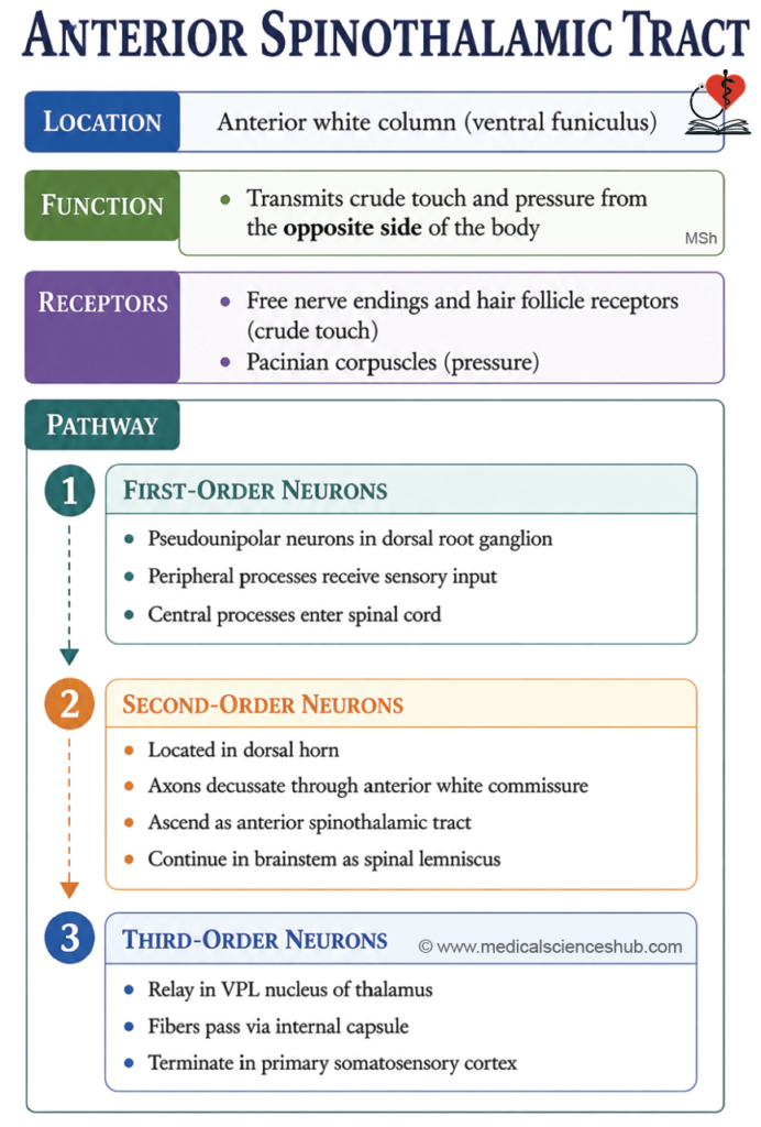
- The anterior spinothalamic tract transmits crude touch and pressure sensations from the contralateral side of the body to the brain.
- Location: Anterior white column of the spinal cord.
- Receptors:
- Hair follicle receptors and free nerve endings detect crude touch.
- Pacinian corpuscles detect pressure.
Pathway
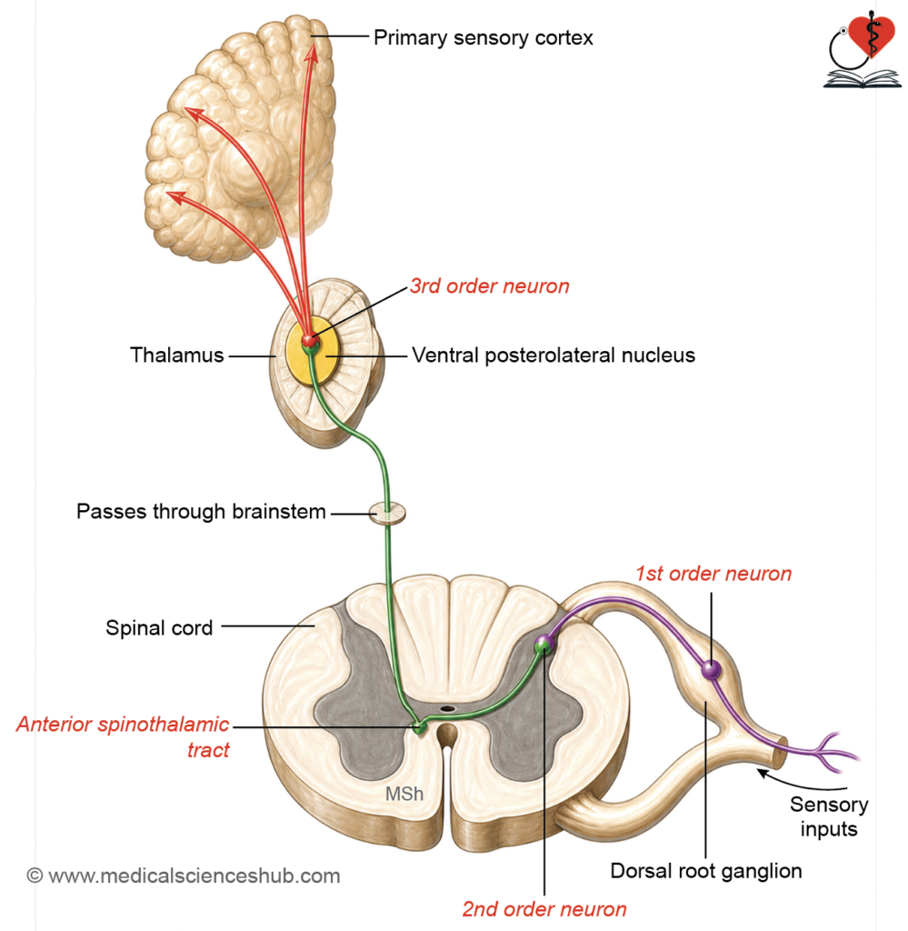
- First-order neurons are pseudounipolar neurons located in the dorsal root ganglia of spinal nerves. They possess:
- A peripheral process that conducts sensory signals via the spinal nerve and dorsal root.
- A central process that is heavily myelinated, enters the spinal cord through the medial division of the dorsal root, ascends one to two segments in the dorsolateral tract of Lissauer, and synapses with neurons of the substantia gelatinosa in the dorsal horn.
- Second-order neurons arise from the dorsal horn (substantia gelatinosa), and their axons decussate through the anterior white commissure to reach the contralateral anterior white column. They ascend as the anterior spinothalamic tract, positioned just anterior to the anterior horn, and continue as the spinal lemniscus through the brainstem, terminating in the ventral posterolateral (VPL) nucleus of the thalamus.
- Third-order neurons are located in the VPL nucleus of the thalamus and project their axons to the primary somatosensory cortex via the posterior limb of the internal capsule.
CLINICAL NEUROANATOMY
- Lesion of the anterior spinothalamic tract produces only a mild reduction in crude touch perception, because fine touch, vibration, and pressure sensations are simultaneously conveyed by the dorsal columns (fasciculus gracilis and fasciculus cuneatus), which remain intact.
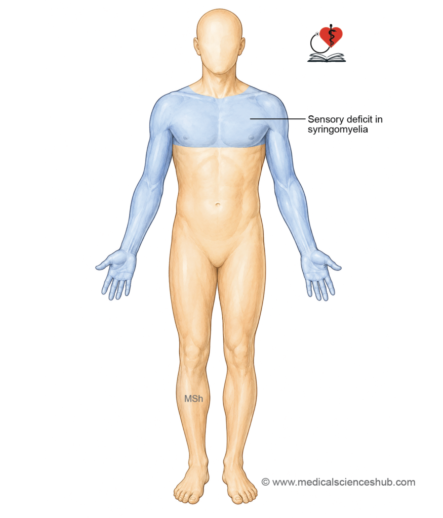
- · Dissociated sensory loss refers to selective impairment of pain and temperature sensation with preservation of touch. This pattern is characteristic of syringomyelia, in which expansion of the central canal compresses decussating fibres within the anterior white commissure. The fibres affected include:
- Crossing fibres of the lateral spinothalamic tract — resulting in loss of pain and temperature sensation.
- Crossing fibres of the anterior spinothalamic tract — resulting in minimal or no loss of touch, as this modality is simultaneously carried by the uncrossed dorsal column tracts.
- “Cape” or “jacket” pattern of sensory loss in syringomyelia results from compression of the crossing lateral spinothalamic fibres at the level of the central canal expansion, typically in the cervical region. The resulting loss of pain and temperature sensation affects the corresponding dermatomes in a cape-like distribution, and may extend slightly above or below the lesion depending on the extent of the syrinx.
Pain Receptors and Fibre Types
- Nociceptors are high-threshold free nerve endings distributed across the external body surface, internal tissues, and viscera. They respond to potentially damaging stimuli and transmit pain signals centrally.
- Thinly myelinated (Aδ) fibres conduct impulses rapidly and carry sharp, well-localised, short-duration pain, such as that produced by a pinprick.
- Unmyelinated (type C) fibres conduct slowly and transmit dull, persistent, and poorly localised pain, such as the aching discomfort associated with sustained tissue stretch or deep injury.
Tract of Goll (Fasciculus Gracilis) and Tract of Burdach (Fasciculus Cuneatus)
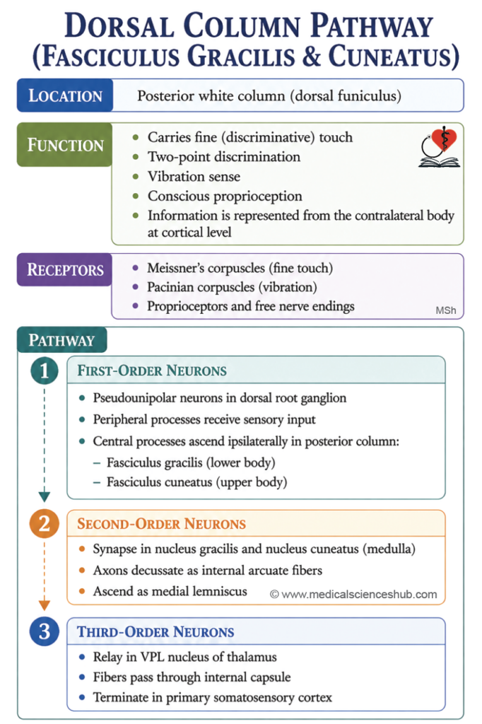
- These two tracts, collectively forming the dorsal columns, transmit fine touch, vibration, and conscious proprioception from the contralateral side of the body, enabling precise tactile discrimination and accurate perception of limb position.
- Location: Posterior white column of the spinal cord.
- Functions: They carry the following sensations:
- Fine touch and two-point discrimination — the ability to distinguish two simultaneously applied stimuli as separate points. Spatial resolution is finest at the fingertips (approximately 2–3 mm) and coarser at the dorsum of the hand (approximately 3 cm).
- Vibration sense.
- Conscious proprioception, encompassing joint position sense, stereognosis (the ability to identify objects by touch alone without visual input), and conscious kinaesthesia (awareness of body part movement derived from sensory inputs from muscles and tendons).
Receptors
- Meissner’s corpuscles — fine touch.
- Pacinian corpuscles — vibration.
- Muscle spindles and Golgi tendon organs — proprioceptive input (note: these primarily contribute to unconscious proprioception via the spinocerebellar tracts).
Pathway
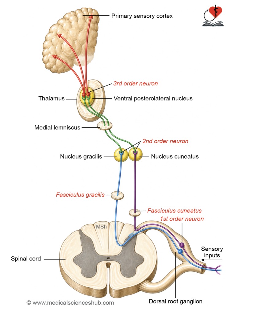
- First-order neurons are pseudounipolar neurons in the dorsal root ganglia of spinal nerves. Their peripheral processes carry sensory signals through the spinal nerve and dorsal root, while their heavily myelinated central processes enter the spinal cord and ascend ipsilaterally in the posterior white column as follows:
- Fasciculus gracilis — carries fibres from the coccygeal, sacral, lumbar, and lower thoracic spinal levels (T7 and below), occupying the medial aspect of the posterior column.
- Fasciculus cuneatus — carries fibres from upper thoracic and cervical levels (T6 and above), occupying the lateral aspect.
- The somatotopic arrangement from medial to lateral is: sacral, lumbar, thoracic, and cervical fibres.
- The fasciculus gracilis terminates at the nucleus gracilis and the fasciculus cuneatus at the nucleus cuneatus, both located on the posterior surface of the medulla oblongata.
- Second-order neurons arise from the nuclei gracilis and cuneatus. Their axons curve ventromedially as internal arcuate fibres, decussate in the medulla, and ascend contralaterally as the medial lemniscus through the brainstem — traversing the pons and midbrain — before terminating in the ventral posterolateral (VPL) nucleus of the thalamus.
- Third-order neurons are located in the VPL nucleus of the thalamus and project via the posterior limb of the internal capsule to the primary somatosensory cortex.
CLINICAL NEUROANATOMY
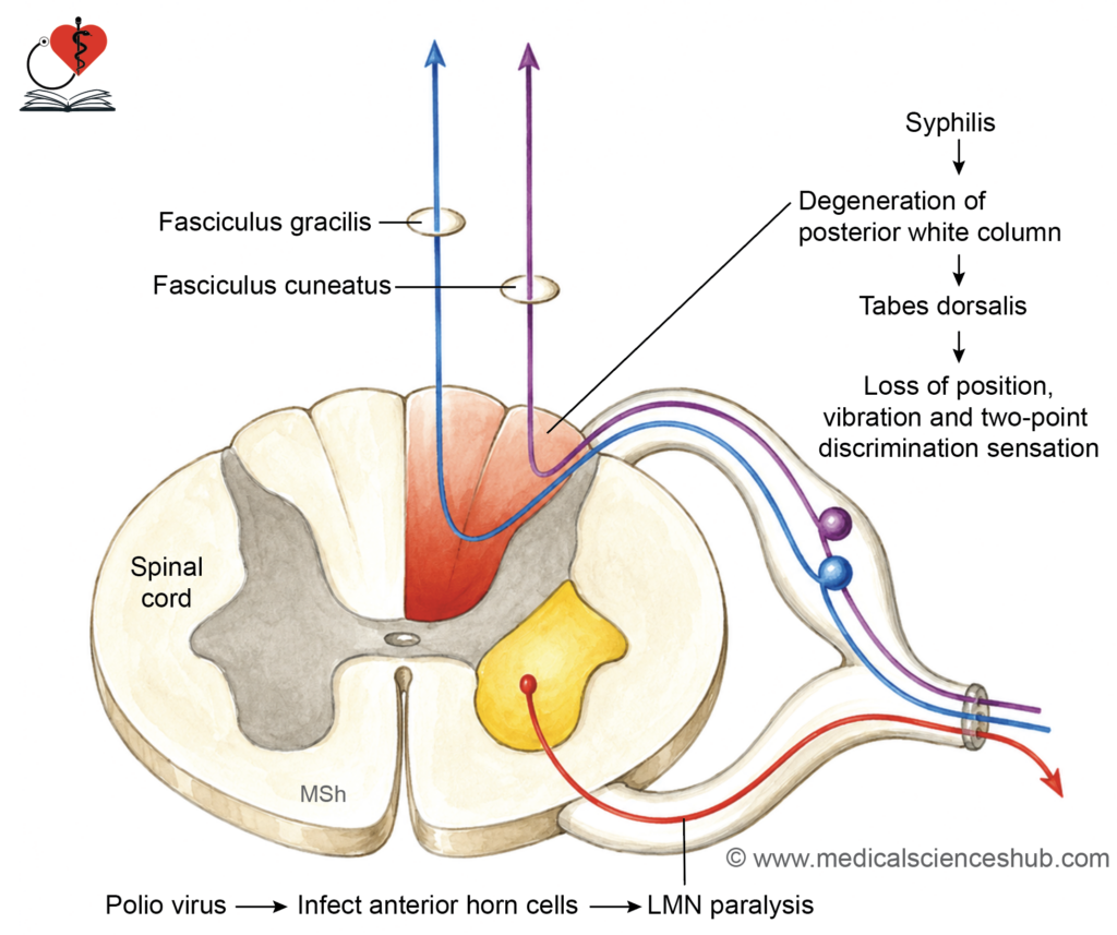
- Unilateral dorsal column lesion produces ipsilateral loss of fine touch, vibration, and position sense at and below the level of the lesion — in contrast to spinothalamic tract lesions, which cause contralateral deficits.
- Tabes dorsalis is a late manifestation of neurosyphilis characterised by progressive degeneration of the posterior white columns, resulting in loss of position sense, vibration, and two-point discrimination.
- Romberg’s sign is elicited by asking the patient to stand with feet together and eyes closed. A positive result — indicated by loss of balance — suggests impaired position sense due to dorsal column damage (as in tabes dorsalis) or peripheral neuropathy affecting the dorsal roots, both of which eliminate the proprioceptive input required for postural stability in the absence of visual compensation.
Spinocerebellar Tracts
- The spinocerebellar tracts transmit unconscious proprioceptive signals from muscles, tendons, and joints to the cerebellum, enabling the cerebellum to coordinate posture, balance, and smooth, precise movement without conscious awareness.
- Receptors: Muscle spindles (detecting muscle stretch) and Golgi tendon organs (detecting tendon tension) serve as the primary peripheral receptors for unconscious proprioception.
Pathway
The spinocerebellar tracts include:
- Dorsal spinocerebellar tract
- Ventral spinocerebellar tract
- Cuneocerebellar tract
- Rostral spinocerebellar tract
Posterior (dorsal) Spinocerebellar Tract
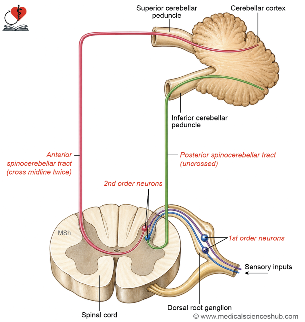
- First-order neurons are pseudounipolar neurons of the dorsal root ganglia whose central processes enter the spinal cord and synapse in Clarke’s column (nucleus dorsalis), located at spinal levels C8–L2.
- Second-order neurons arise from Clarke’s column and their axons ascend ipsilaterally in the dorsolateral part of the lateral white column. They pass through the medulla oblongata, enter the cerebellum via the inferior cerebellar peduncle, and terminate in the ipsilateral cerebellar cortex (vermis).
Anterior (Ventral) Spinocerebellar Tract
- First-order neurons follow a course similar to those of the dorsal spinocerebellar tract.
- Second-order neurons are located in laminae V–VII of the lumbosacral spinal cord. Their axons predominantly decussate and ascend in the anterolateral part of the lateral white column. They continue to the midbrain, enter the cerebellum via the superior cerebellar peduncle, and decussate again within the cerebellum, ultimately terminating in the ipsilateral cerebellar cortex (vermis). The double decussation ensures that the cerebellum receives information from the ipsilateral side of the body.
Cuneocerebellar Tract
- First-order neurons are located in the dorsal root ganglia at spinal levels C2–T5. Their central processes ascend in the fasciculus cuneatus and terminate in the accessory (external) cuneate nucleus in the medulla — a structure functionally analogous to Clarke’s column, which is absent above the C8 level.
- Second-order neurons arise from the accessory cuneate nucleus, and their axons enter the cerebellum via the inferior cerebellar peduncle, terminating in the ipsilateral cerebellar cortex.
- This tract carries unconscious proprioceptive information from the neck, upper limb, and upper trunk — regions not served by Clarke’s column.
Rostral Spinocerebellar Tract
- First-order neurons are dorsal horn cells at levels C4–C8 that convey unconscious proprioceptive information from the head and upper limb, synapsing in lamina VII of the dorsal horn.
- Second-order neurons in lamina VII (C4–C8) give rise to axons that ascend alongside the ventral spinocerebellar tract and enter the cerebellum primarily via the inferior cerebellar peduncle (with a smaller contribution through the superior cerebellar peduncle), terminating in the cerebellar cortex.
Table 5.1: Spinocerebellar tracts
| Tract | First-order neuron | Second-order neuron | Termination | Functions |
|---|---|---|---|---|
| Posterior (dorsal) spinocerebellar tract (T1–L2/L3) | Cells of dorsal root ganglion | Clarke’s nucleus (nucleus dorsalis) | Ipsilateral cerebellar cortex (vermis/intermediate zone) via inferior cerebellar peduncle | Unconscious proprioception from ipsilateral trunk and lower limb; posture and movement coordination |
| Anterior (ventral) spinocerebellar tract (L2–S3) | Cells of dorsal root ganglion | Spinal border cells (laminae V–VII) | Cerebellar cortex (vermis); fibers cross twice, functionally ipsilateral | Conveys integrated spinal activity for coordination of lower limb movements |
| Cuneocerebellar tract (above T6) | Dorsal root ganglion | Accessory cuneate nucleus | Ipsilateral cerebellar cortex via inferior cerebellar peduncle | Unconscious proprioception from ipsilateral upper limb and neck |
| Rostral spinocerebellar tract (cervical) | Dorsal root ganglion | Cervical spinal interneurons (dorsal horn) | Cerebellum via superior/inferior cerebellar peduncles; largely uncrossed | Upper limb proprioception and interneuronal activity for coordination |
Proprioception from Head
- Conscious proprioceptive signals from the head are conveyed by the trigeminal nerve (cranial nerve V), with first-order neurons located in the mesencephalic nucleus of the trigeminal nerve — uniquely, the only primary sensory nucleus situated within the brainstem itself.
- These neurons synapse with second-order neurons in the reticular formation, whose axons ascend via the trigeminothalamic tract to the ventral posteromedial (VPM) nucleus of the thalamus.
- Third-order thalamic neurons project through the internal capsule to the primary somatosensory cortex. A subset of fibres from the mesencephalic nucleus projects to the insular cortex, conveying unconscious proprioceptive information from the head.
Descending Tracts
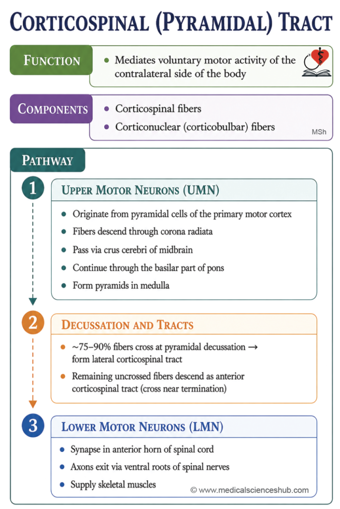
Corticospinal (Pyramidal) Tract
- The corticospinal tract is the principal descending motor pathway, transmitting voluntary motor commands from the cerebral cortex to the spinal cord. It is named after the pyramids of the medulla oblongata, through which its fibres pass.
- It enables precise, skilled, and purposeful movements, with particular importance for fine motor control of the distal limb muscles.
Components
The tract has two components:
- Corticospinal fibres — terminate on anterior horn cells of the spinal cord.
- Corticonuclear (corticobulbar) fibres — terminate on motor nuclei of cranial nerves within the brainstem.
Pathway
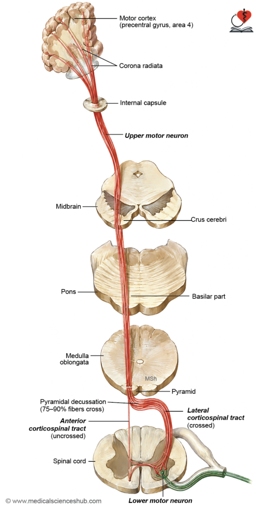
- Upper motor neurons (UMNs) originate primarily from Betz cells (giant pyramidal neurons) in the primary motor cortex (area 4) located in the precentral gyrus, with additional contributions from the premotor cortex (area 6). Their axons initially radiate as the corona radiata, then converge and descend through the genu and posterior limb of the internal capsule.
- In the midbrain: Fibres pass through the middle three-fifths of the crus cerebri.
- In the pons: Fibres are dispersed into small bundles by the pontine nuclei and transversely oriented pontocerebellar fibres within the basilar pons.
- In the medulla: Fibres reconverge to form the compact pyramids. At the junction of the medulla and spinal cord, 75–90% of fibres decussate at the pyramidal decussation and descend as the lateral corticospinal tract in the contralateral lateral white column. The remaining fibres continue ipsilaterally as the anterior (ventral) corticospinal tract in the anterior white column, crossing in the anterior white commissure at their segmental level of termination.
- Somatotopic organisation: In the internal capsule, fibres are arranged from anterior to posterior as: head, upper limb, trunk, and lower limb. In the midbrain, this arrangement shifts to medial to lateral in the same sequence.
- Approximately 55% of corticospinal fibres terminate at cervical levels (primarily controlling upper limb movement), 20% at thoracic levels, and 25% at lumbosacral levels.
- Lower motor neurons (LMNs) reside in the anterior horn of the spinal cord and in the cranial nerve motor nuclei of the brainstem. Most UMN-to-LMN transmission is indirect, mediated via interneurons (only approximately 5% of UMN axons synapse directly on LMNs). LMN axons exit via the ventral roots of spinal nerves or motor cranial nerves to innervate skeletal muscle, producing contraction.
- Corticonuclear fibres travel medial to the corticospinal fibres within the brainstem, decussating to terminate on contralateral cranial nerve motor nuclei via interneurons. Some fibres project bilaterally. Relevant motor nuclei include those of cranial nerves III, IV, V, VI, VII, IX/X (nucleus ambiguus), and XII.
- Functional distinction: The lateral corticospinal tract governs rapid, skilled voluntary movements of distal muscles, particularly of the hand. The anterior corticospinal tract controls axial and proximal limb musculature.
Table 5.2: Descending tracts
| Tract | First-order neuron | Second-order neuron | Termination | Functions |
|---|---|---|---|---|
| Dorsal spinocerebellar tract (T1–L2/L3) | Neurons in dorsal root ganglia | Clarke’s column (nucleus dorsalis) | Ipsilateral cerebellar cortex (vermis/paravermis) via inferior cerebellar peduncle | Conveys unconscious proprioceptive input from trunk and lower limb; essential for posture and coordinated movement |
| Ventral spinocerebellar tract (L2–S3) | Neurons in dorsal root ganglia | Spinal border cells (laminae V–VII) | Cerebellar cortex via superior cerebellar peduncle; fibers decussate twice | Transmits integrated spinal interneuronal activity for motor coordination of lower limbs |
| Cuneocerebellar tract (≥ T6) | Dorsal root ganglia | Accessory cuneate nucleus | Ipsilateral cerebellar cortex via inferior cerebellar peduncle | Carries unconscious proprioception from upper limb and neck |
| Rostral spinocerebellar tract (cervical segments) | Dorsal root ganglia | Cervical interneurons (dorsal horn) | Cerebellum via superior and inferior cerebellar peduncles; predominantly uncrossed | Conveys upper limb proprioceptive and segmental activity for coordination |
CLINICAL NEUROANATOMY
Upper Motor Neuron (UMN) Lesions
- UMN lesions produce spastic paralysis (hypertonia), because the inhibitory control of pyramidal fibres over LMNs is lost, leaving them subject to facilitatory input from extrapyramidal pathways.
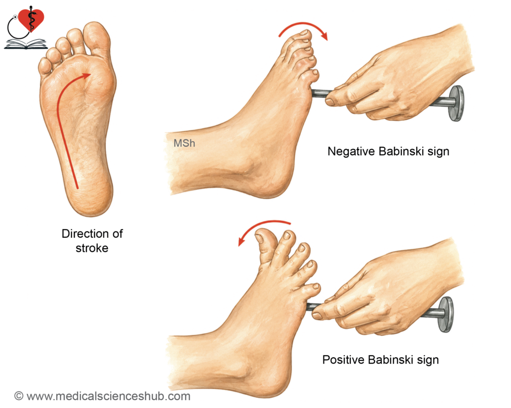
- Positive Babinski sign: Stroking the lateral plantar surface of the foot with a blunt object elicits extension (dorsiflexion) of the great toe with fanning of the remaining toes. In neurologically intact adults, the normal response is plantar flexion. An extensor response is physiologically normal in infants up to approximately 12 months due to incomplete corticospinal myelination; in older individuals it indicates a UMN lesion.
- Additional features include absence of superficial reflexes (abdominal and cremasteric) and muscle clonus — a rhythmic, oscillating stretch reflex elicited, for example, by sudden downward displacement of the patella, producing repetitive rhythmic contractions of the quadriceps rather than a single response.
- Localisation of UMN lesions:
- Motor cortex or internal capsule — contralateral hemiplegia with contralateral lower facial and tongue weakness.
- Midbrain — contralateral hemiplegia with ipsilateral CN III palsy.
- Pons — Raymond’s syndrome (contralateral hemiplegia with ipsilateral CN VI palsy) or Millard–Gubler syndrome (contralateral hemiplegia with ipsilateral CN VII palsy).
- Medulla — crossed hypoglossal paralysis (contralateral hemiplegia with ipsilateral CN XII palsy).
- Spinal cord — Brown-Séquard syndrome (ipsilateral motor deficit with contralateral sensory loss).
Lower Motor Neuron (LMN) Lesions
- LMN lesions may involve the anterior horn cells, ventral roots, or motor cranial nerve nuclei, producing:
- Flaccid paralysis with loss of muscle tone.
- Muscle atrophy due to denervation.
- Loss of all reflexes (both deep tendon and superficial).
- Absence of Babinski sign.
- Spasticity denotes a velocity-dependent increase in muscle tone associated with UMN lesions. Flaccidity refers to reduced or absent muscle tone resulting from LMN lesions or disruption of the reflex arc.
TABLE 5.3: Differences between upper and lower motor neuron Paralysis
| Feature | Upper motor neuron (UMN) lesion | Lower motor neuron (LMN) lesion |
|---|---|---|
| Site of lesion | Lesion above anterior horn cells or cranial nerve motor nuclei (corticospinal/corticobulbar pathways) | Lesion involving anterior horn cells, cranial nerve motor nuclei, or peripheral motor nerves |
| Type of paralysis | Spastic paralysis | Flaccid paralysis |
| Muscle tone | Increased (hypertonia) | Decreased (hypotonia) |
| Distribution of weakness | Typically widespread and affects groups of muscles | Usually localized to muscles supplied by the affected neuron |
| Deep tendon reflexes | Exaggerated (hyperreflexia) | Reduced or absent (hyporeflexia/areflexia) |
| Superficial reflexes | Absent | Absent in the affected segment |
| Plantar response | Extensor (Babinski positive) | Flexor or absent |
| Muscle wasting | Mild and late (disuse atrophy) | Marked and early (denervation atrophy) |
| Clonus | Present | Absent |
Extrapyramidal Tracts
- The extrapyramidal system encompasses all descending motor pathways to the spinal cord other than the corticospinal tract. These tracts regulate posture, balance, muscle tone, and the smooth coordination of movement, and mediate subconscious motor activities such as arm swinging during walking.
- Functionally, these tracts serve as relay pathways, conveying modulatory signals from various brain regions to the spinal cord’s anterior horn cells.
Tectospinal Tract
- Function: Mediates reflex orientation of the eyes, head, and upper trunk in response to visual, auditory, and vestibular stimuli.
- Physiological basis: Visual association cortex (areas 18 and 19) → corticotectal tract → superior colliculus of the midbrain → tectospinal tract → anterior horn cells → coordinated eye and upper trunk movement.
- Pathway:
- Location: Anterior white column of the spinal cord.
- Fibres originate from the superior colliculus of the midbrain, decussate between the periaqueductal grey matter and the red nucleus via the dorsal tegmental decussation, and descend in the medial anterior white column.
- Fibres terminate on interneurons in laminae VI–VIII of the anterior horn, restricted to cervical and upper thoracic segments.
Rubrospinal Tract
- Function: Facilitates flexor muscle tone and inhibits extensor muscle tone in the upper limb, contributing to the control of hand and digit movements.
- Physiological basis: Sensorimotor cortex → corticorubral tract → red nucleus (midbrain) → rubrospinal tract → anterior horn cells of the spinal cord (and a parallel rubrobulbar projection to cranial nerve motor nuclei).
- Pathway:
- Location: Lateral white column of the spinal cord.
- Fibres arise from the red nucleus in the midbrain tegmentum, decussate immediately via the ventral tegmental decussation, and descend as a compact bundle in the lateral white column.
- Fibres terminate on anterior horn cells in laminae V–VII.
Reticulospinal Tract
- Function: Modulates the motor activity of axial and proximal limb muscles, contributing to postural control and limb orientation. Also influences autonomic functions via lateral horn cell connections.
- Physiological basis: Sensorimotor cortex → bilateral corticoreticular projections → brainstem reticular formation → two distinct tracts:
- Pontine reticular nuclei → medial (pontine) reticulospinal tract.
- Medullary reticular nuclei → lateral (medullary) reticulospinal tract.
Pathway:
- Medial reticulospinal tract arises from the pontine reticular nuclei, descends ipsilaterally in the anterior white column throughout the entire length of the spinal cord, and terminates on anterior horn cells. It facilitates extensor muscle activity and inhibits flexor muscle activity.
- Lateral reticulospinal tract arises from the medullary reticular nuclei, descends bilaterally in the lateral white column, and terminates on anterior horn cells. It inhibits extensor muscles and facilitates flexor muscles. Collateral projections to the lateral horn mediate sympathetic effects including increased heart rate, pupillary dilatation, and sweating.
Vestibulospinal Tract
Function:
- Lateral vestibulospinal tract — maintains posture and balance by activating antigravity muscles.
- Medial vestibulospinal tract — coordinates head movements to maintain stable gaze fixation.
- Physiological basis: The vestibular nuclear complex, located in the floor of the fourth ventricle at the pontomedullary junction, receives sensory input regarding head position and movement from the vestibular apparatus via the vestibular division of the vestibulocochlear nerve (CN VIII), and integrating input from the cerebellum.
- Pathway:
- Lateral vestibulospinal tract arises from the lateral vestibular nucleus (Deiters’ nucleus), descends ipsilaterally in the anterior white column throughout the entire spinal cord, and terminates on anterior horn cells. It activates axial and proximal limb extensor (antigravity) muscles while inhibiting limb flexors, thereby maintaining upright posture in response to vestibular input.
- Medial vestibulospinal tract arises from the medial vestibular nucleus and descends via two routes:
- Ipsilaterally within the medial longitudinal fasciculus (MLF) in the brainstem, projecting to motor nuclei of cranial nerves III, IV, and VI to coordinate eye movements.
- Ipsilaterally in the anterior white column of the spinal cord, reaching only the upper thoracic segments, where it terminates on anterior horn cells governing cervical musculature to mediate head and neck movements during gaze stabilisation.
CLINICAL NEUROANATOMY
Brown-Séquard Syndrome (Spinal Cord Hemisection)
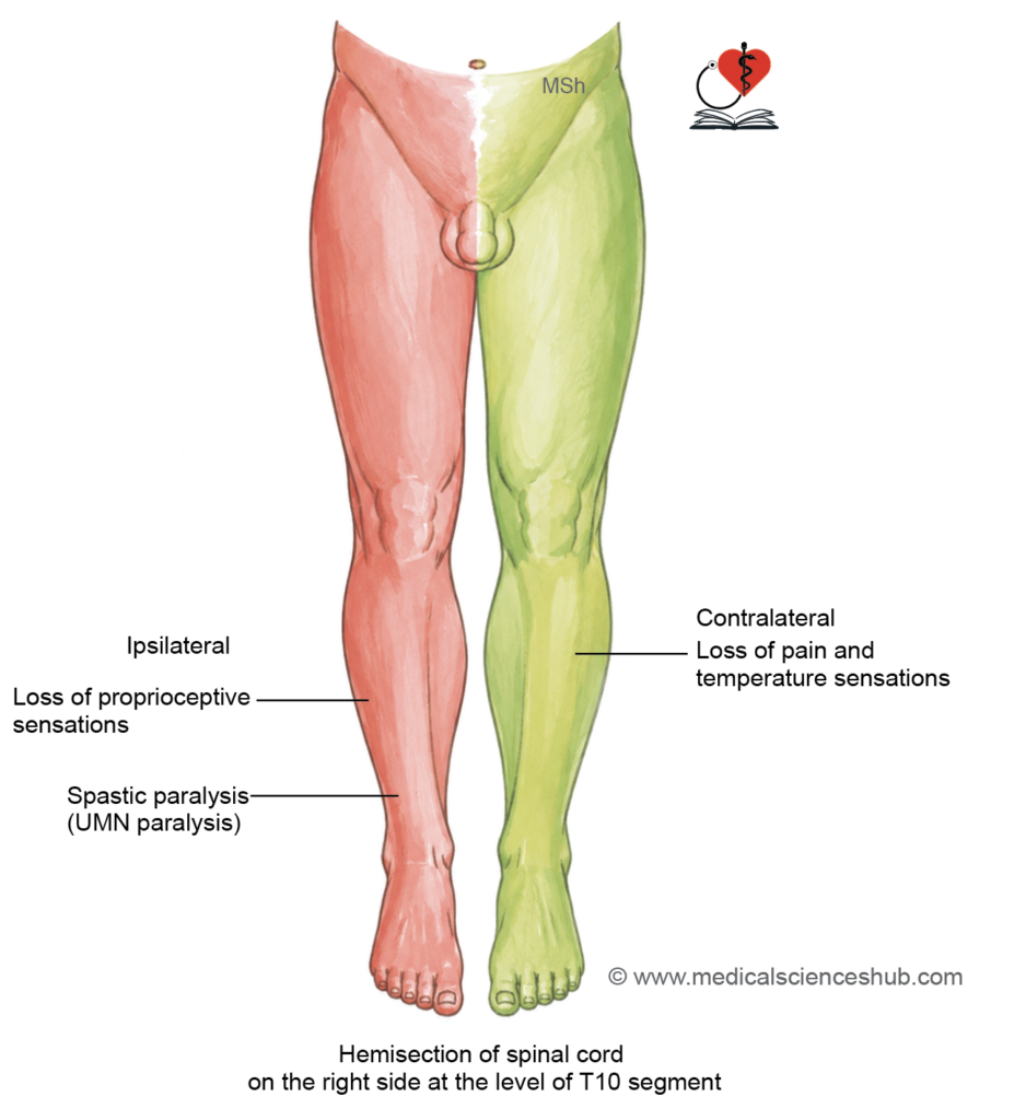
- Brown-Séquard syndrome results from hemisection of the spinal cord, producing a characteristic pattern of ipsilateral motor deficits and contralateral sensory loss below the level of injury.
Ipsilateral Effects (Same Side as Lesion)
- Spastic (UMN) paralysis below the level of the lesion, due to interruption of the lateral corticospinal tract. Hemisection at the upper cervical level produces hemiplegia (paralysis of both ipsilateral limbs); hemisection at thoracic levels produces monoplegia (paralysis of one lower limb).
- Flaccid (LMN) paralysis at the exact level of the lesion, due to direct damage to anterior horn cells and the ventral root of the spinal nerve at that segment.
- Loss of fine touch, vibration, and conscious proprioception below the lesion, due to damage to the ipsilateral fasciculus gracilis and fasciculus cuneatus (dorsal columns).
- Segmental anaesthesia (loss of all sensation) at the level of the lesion itself, due to local destruction of the dorsal root entry zone.
Contralateral Effects (Opposite Side)
- Loss of pain and temperature sensation below the level of the lesion, due to damage to the lateral spinothalamic tract, whose fibres have already decussated at or near their level of entry.
Complete Spinal Cord Transection
- Severe spinal cord injury may result in complete transection, producing deficits that evolve through two distinct clinical phases.
Phase 1 — Spinal Shock (first 3 weeks)
- Immediately following transection, spinal shock develops and typically persists for approximately three weeks. It is characterised by:
- Flaccid paralysis of all muscles below the level of injury.
- Complete loss of all reflexes (both superficial and deep tendon) below the lesion.
- Urinary retention, resulting from failure of the detrusor muscle to contract and inability of the internal urethral sphincter to relax.
- Faecal retention, due to loss of coordinated relaxation of the external anal sphincter.
- Overflow (retention) incontinence, in which urine accumulates until intravesical pressure overcomes sphincter resistance, resulting in involuntary leakage.
Phase 2 — Recovery Phase (after 3 weeks)
- As spinal shock resolves, reflex activity below the lesion gradually returns. This phase is characterised by:
- Spastic (UMN) paralysis below the level of injury, with the extent depending on lesion level:
- Above C5 — respiratory failure due to paralysis of the phrenic nerve and loss of diaphragmatic function.
- C2–T1 — quadriplegia (paralysis of all four limbs).
- Below T1 — paraplegia (paralysis of both lower limbs).
- Automatic bladder and bowel emptying, characterised by involuntary reflex evacuation triggered by sufficient visceral distension, as higher voluntary control is permanently lost.
- Spastic (UMN) paralysis below the level of injury, with the extent depending on lesion level:
Important Questions
- Describe the lateral spinothalamic tract responsible for transmission of pain and temperature sensations.
- Describe the pathway and functions of the corticospinal tract.
- Write a short note on lower motor neuron (LMN) paralysis.
- What are the differences between upper motor neuron (UMN) and LMN paralysis?
- Explain Brown–Séquard syndrome with its clinical basis.
- Enumerate the neurological deficits following hemisection of the spinal cord.
- Write a concise note on spinal shock and its stages.
📝 Test Your Knowledge – Practice MCQs
Attempt the chapter MCQ quiz and assess your understanding of key concepts.
Click to see: Medical Disclaimer and Copyright Notice