
Competencies
- AN62.5: Describe boundaries, parts, gross relations, major nuclei, and connections of dorsal thalamus, hypothalamus, epithalamus, metathalamus, and subthalamus.
Introduction
- The diencephalon contains the thalamus and its associated regions—the metathalamus, epithalamus, and subthalamus. Together, these structures integrate and relay sensory, motor, limbic, and autonomic information, playing essential roles in consciousness, emotion, movement, and regulation of body functions.
Diencephalon
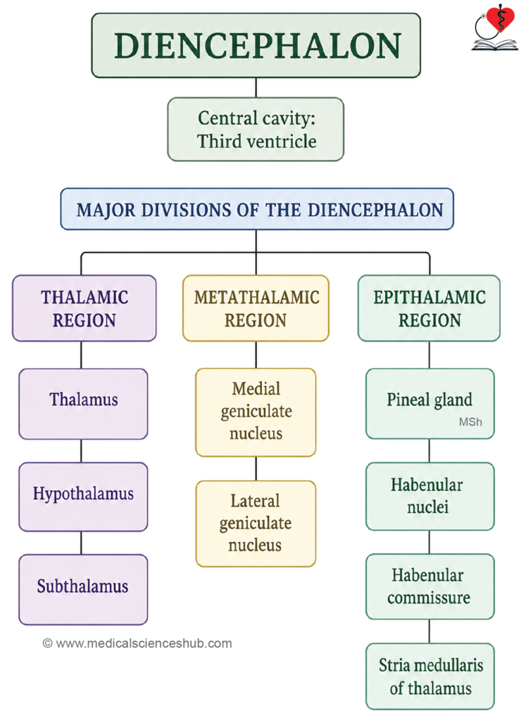
- The diencephalon is a subdivision of the brain situated between the cerebral hemispheres superiorly and the midbrain inferiorly, forming a critical relay and integrative hub of the central nervous system.
- Its internal cavity is the third ventricle, a midline cerebrospinal fluid-filled space flanked by the paired components on either side.
- The diencephalon comprises five paired divisions:
- Thalamus — the principal relay station for sensory and motor signals traveling to and from the cerebral cortex.
- Hypothalamus — regulates autonomic functions, endocrine activity, homeostasis, and behavioral drives including hunger, thirst, and circadian rhythms.
- Metathalamus — consists of two paired nuclei serving as relay centers for special senses:
- The medial geniculate body relays auditory information to the auditory cortex.
- The lateral geniculate body relays visual information to the primary visual cortex.
- Subthalamus — involved in the modulation of motor activity, particularly in connection with the basal ganglia circuitry.
- Epithalamus — the most dorsal component, comprising:
- The pineal body (pineal gland), an endocrine structure that secretes melatonin and plays a role in regulating circadian rhythms.
- The habenular nuclei and habenular commissure, which are involved in limbic and olfactory connections.
- The posterior commissure, a white matter tract crossing the midline near the junction of the diencephalon and midbrain.
- The stria medullaris thalami, a fiber bundle connecting the septal nuclei, hypothalamus, and basal forebrain to the habenular nuclei.
Thalamus
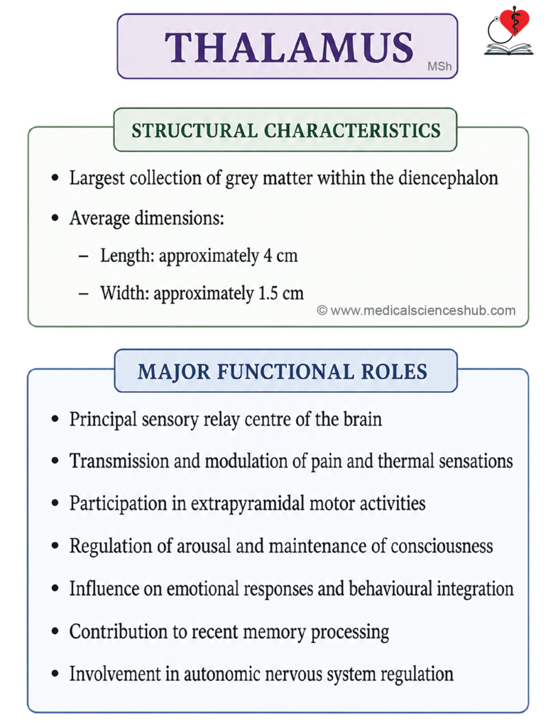
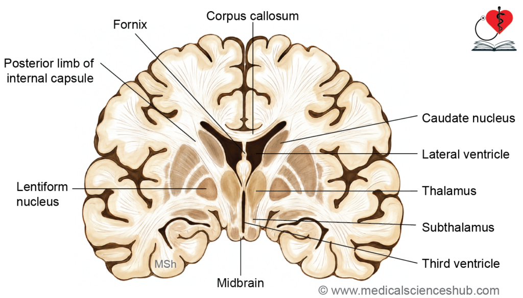
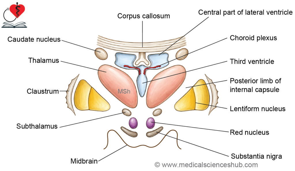
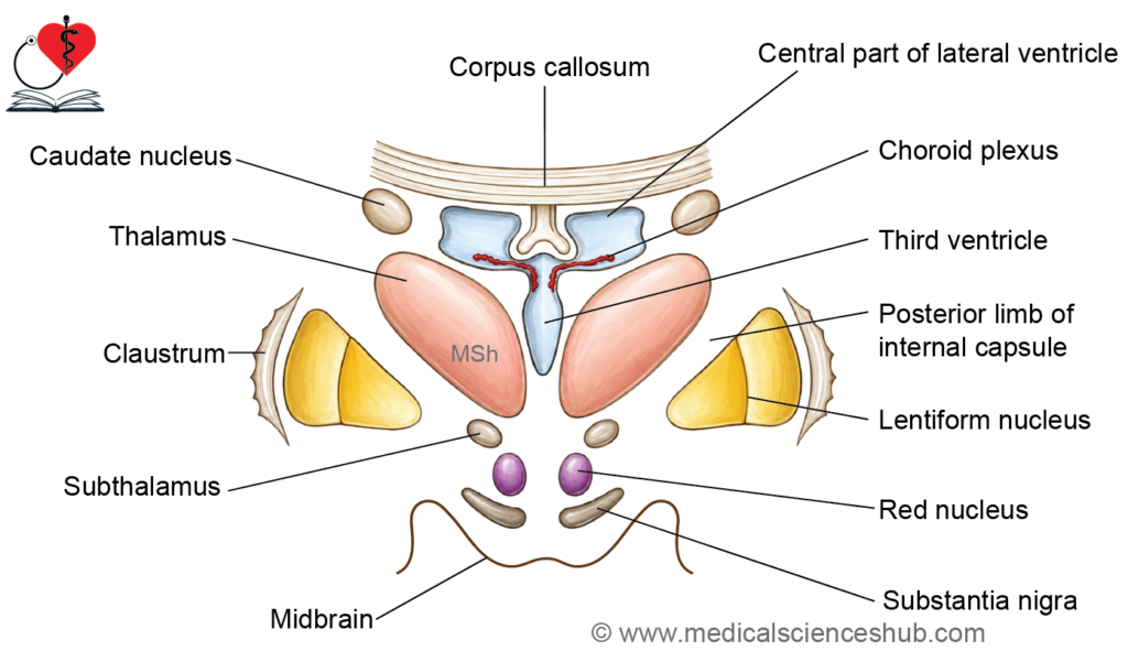
- The thalamus (from the Greek thalamos, meaning “chamber”) is a large, ovoid mass of grey matter located on either side of the third ventricle, positioned superior to the midbrain.
- Dimensions:
- Anteroposterior length: approximately 4 cm
- Width and vertical diameter: approximately 1.5 cm each
- The long axis of the thalamus is oriented obliquely, directed posteriorly and laterally.
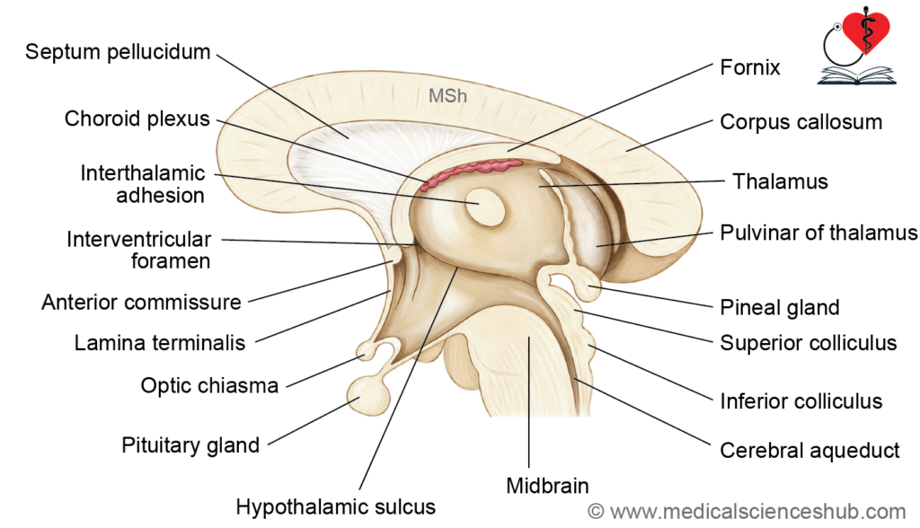
- The interthalamic adhesion (also called the massa intermedia) is a narrow band of grey matter that bridges the two thalami across the midline through the cavity of the third ventricle. It is present in the majority of individuals but is not a true commissure, as it contains few if any crossing nerve fibers.
Functions of Thalamus
- Sensory relay: The thalamus serves as the principal relay station for nearly all sensory modalities ascending to the cerebral cortex, with the notable exception of olfaction, which projects directly to the cortex via the olfactory bulb.
- Pain and temperature perception: The thalamus mediates crude awareness of pain and temperature, including basic localization and intensity. Precise discrimination and conscious interpretation of these sensations, however, are carried out at the level of the somatosensory cortex.
- Motor coordination: As a key node in extrapyramidal circuitry, the thalamus receives processed motor signals from the cerebellum and basal ganglia and relays them to the motor cortex, contributing to the coordination and refinement of voluntary movement.
- Consciousness and arousal: The thalamus plays an important role in maintaining wakefulness and alertness by modulating activity within the ascending reticular activating system (ARAS), which regulates the transition between sleep and conscious states.
- Limbic functions: As a component of the limbic system, the thalamus contributes to emotional behavior and the consolidation of recent memory, primarily through its connections with the hippocampus, cingulate gyrus, and prefrontal cortex.
- Autonomic regulation: The thalamus participates in visceral and autonomic control indirectly, largely through its bidirectional connections with the hypothalamus.
- Emotional regulation and mood: By maintaining reciprocal communication between the limbic system and the cerebral cortex, the thalamus helps modulate mood, emotional reactivity, and affective stability.
- Influence on higher cortical functions: Through extensive thalamocortical projections, the thalamus indirectly shapes personality, executive function, and cognitive processing, serving as a critical gateway between subcortical structures and association cortices.
External Features of Thalamus
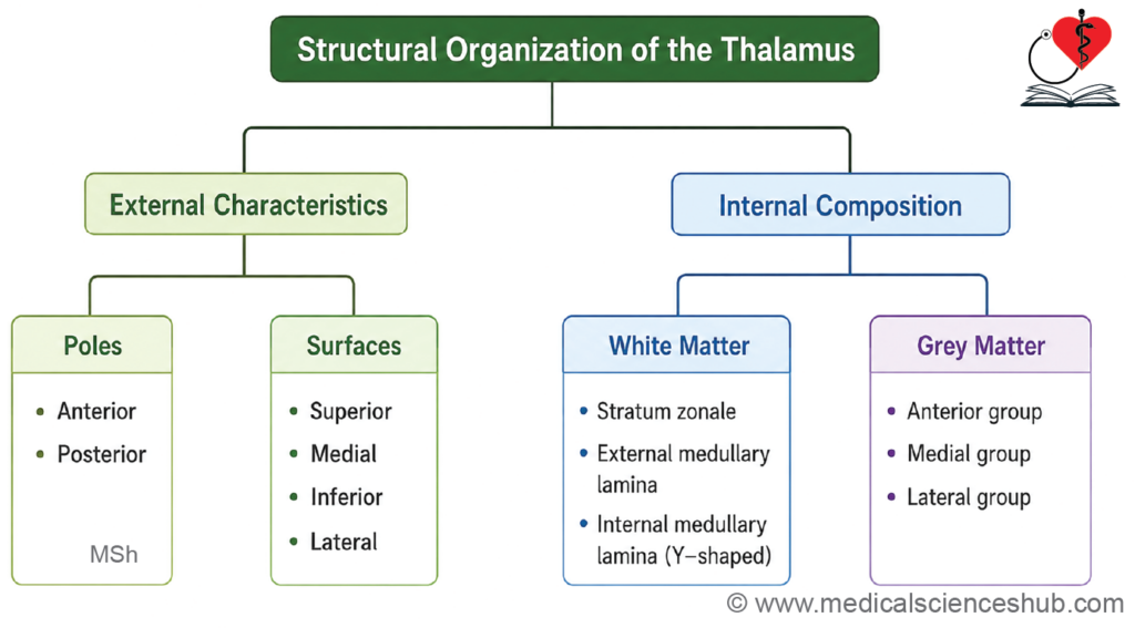
Each thalamus has two ends (anterior and posterior) and four surfaces (superior, inferior, medial, and lateral).
Ends of the Thalamus
- Anterior end: The anterior end is narrow and rounded, positioned close to the midline. It forms a small projection known as the anterior tubercle of the thalamus and contributes to the posterior boundary of the interventricular foramen (foramen of Monro), through which the lateral ventricle communicates with the third ventricle.
- Posterior end: The posterior end is broad and expanded, forming the pulvinar, which overhangs the superior colliculus and its brachium in the midbrain. The pulvinar is functionally associated with the integration of visual, auditory, and somatosensory information.
Surfaces of the Thalamus
- Superior surface: The superior surface is convex and covered by a thin layer of white matter called the stratum zonale. Its lateral part forms the floor of the lateral ventricle, while the medial part is related to the tela choroidea of the third ventricle, which separates it from the overlying body of the fornix.
- Medial surface: The medial surface forms the greater part of the lateral wall of the third ventricle and is lined by ependymal cells. It typically bears the interthalamic adhesion, a band of grey matter connecting the two thalami across the midline.
- Inferior surface: The inferior surface is continuous anteriorly with the subthalamus. Posteriorly, it becomes free and contributes to the inferior aspect of the pulvinar.
- Lateral surface: The lateral surface is flat and is closely applied to the internal capsule, forming its medial boundary. This surface serves as a major conduit for thalamocortical and corticothalamic fibers connecting the thalamus with wide areas of the cerebral cortex.
Internal Structure of Thalamus
The thalamus is composed predominantly of grey matter, with a relatively small but structurally significant component of white matter arranged as distinct laminae.
White Matter of the Thalamus
The white matter of the thalamus is organised into three thin laminae:
- The stratum zonale covers the superior surface of the thalamus as a thin superficial sheet of white matter.
- The external medullary lamina is a white matter layer applied to the lateral surface, separating the thalamus proper from the thalamic reticular nucleus.
- The internal medullary lamina is a vertically oriented sheet of white matter situated within the substance of the thalamus. It has a Y-shaped configuration, with the stem running posteriorly and the two limbs diverging anterosuperiorly to partially encircle the anterior nuclear group.
Grey Matter and Thalamic Nuclei
- The internal medullary lamina divides the thalamic grey matter into three main regions:
- The anterior part, enclosed between the two diverging limbs of the “Y.”
- The medial part, located medial to the stem of the “Y.”
- The lateral part, located lateral to the stem of the “Y.”
- Each region contains discrete clusters of neurons referred to collectively as the thalamic nuclei. Anatomically, the thalamus is further divided into a dorsal tier (positioned superiorly) and a ventral tier (positioned inferiorly).
- The principal groups of thalamic nuclei include the anterior, medial, lateral, intralaminar, and reticular nuclei, each with distinct afferent and efferent connections and specific functional roles in sensory relay, motor coordination, limbic processing, and cortical activation.
Location of Thalamic Nuclei
Anterior Thalamic Nuclei
- The anterior thalamic nuclei are enclosed within the fork formed by the bifurcation of the Y-shaped internal medullary lamina, effectively nestled between its two diverging anterior limbs.
Medial Thalamic Nuclei
- These nuclei lie medial to the internal medullary lamina. In humans, this group is represented principally by the dorsomedial nucleus, which has extensive connections with the prefrontal cortex and limbic structures.
Lateral Thalamic Nuclei
- The lateral nuclei lie lateral to the internal medullary lamina and are subdivided into two tiers:
- Dorsal tier: Comprises the lateral dorsal (LD), lateral posterior (LP), and pulvinar nuclei, arranged from anterior to posterior along the superior aspect of the thalamus.
- Ventral tier: Comprises the ventral anterior (VA), ventral lateral (VL), and ventral posterior (VP) nuclei, arranged sequentially from anterior to posterior.
- The ventral anterior (VA) nucleus occupies the anterior pole of the ventral tier, positioned between the external and internal medullary laminae, extending from the reticular nucleus anteriorly to the ventral lateral nucleus posteriorly.
- The ventral lateral (VL) nucleus lies between the VA nucleus anteriorly and the ventral posterior (VP) nucleus posteriorly, bounded superiorly by the lateral dorsal nucleus and inferiorly by the subthalamus.
- The ventral posterior (VP) nucleus is situated posterior to the VL nucleus and anterior to the pulvinar, inferior to the lateral posterior nucleus. It is subdivided into:
- Ventral posterior medial (VPM) nucleus — relays sensory information from the head and face via the trigeminal system.
- Ventral posterior lateral (VPL) nucleus — relays somatosensory input from the trunk and limbs via the medial lemniscus and spinothalamic tract.
Geniculate Bodies (Metathalamus)
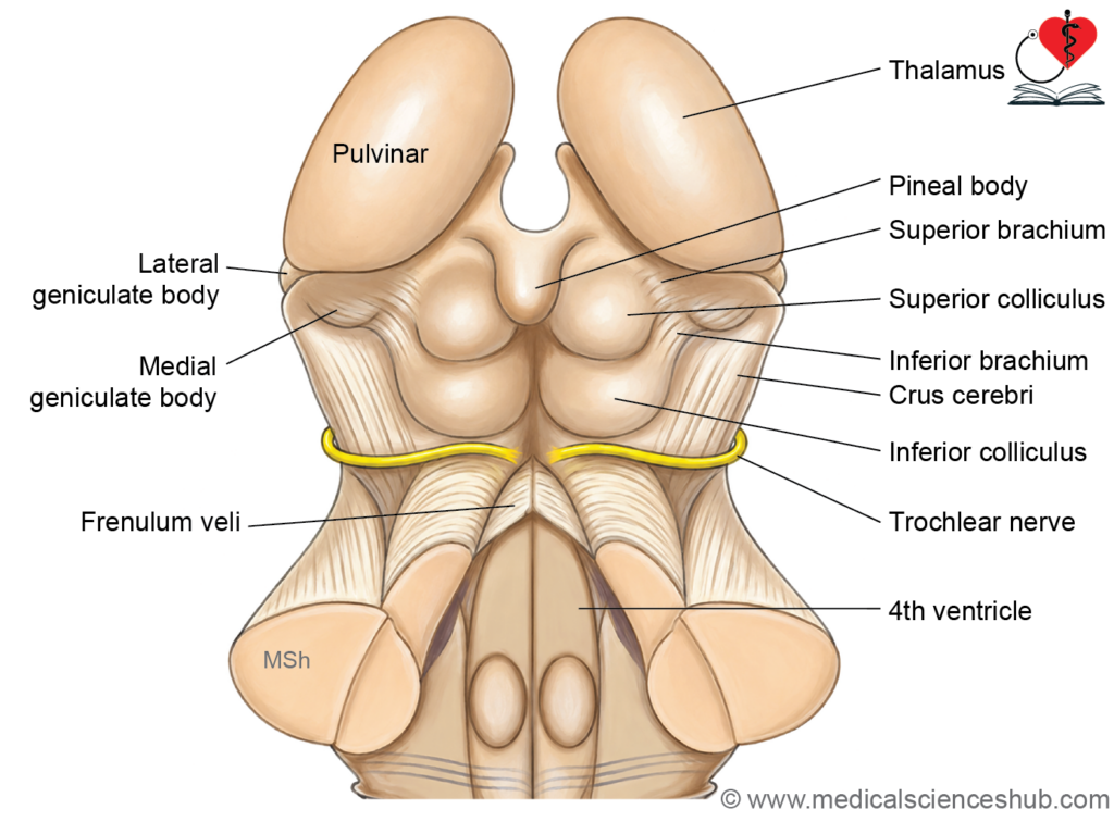
- The medial geniculate body (MGB) is a rounded elevation on the posterior inferior surface of the thalamus, separated from the pulvinar by the superior brachium. It serves as the principal auditory relay nucleus.
- The lateral geniculate body (LGB) is a small ovoid projection on the inferolateral aspect of the pulvinar, positioned anterior and lateral to the MGB. It functions as the primary relay for visual information en route to the occipital cortex.
Dorsal Tier Nuclei
- The lateral dorsal (LD) nucleus occupies the anterior portion of the dorsal (superior) thalamus and is functionally associated with the limbic system.
- The lateral posterior (LP) nucleus lies in the middle portion of the dorsal thalamus, interposed between the LD nucleus anteriorly and the pulvinar posteriorly.
- The pulvinar is the largest and most posterior projection of the thalamus, overhanging the superior colliculus of the midbrain. It integrates visual, auditory, and somatosensory information and projects extensively to association cortices.
Intralaminar Nuclei
- The intralaminar nuclei are embedded within the substance of the internal medullary lamina. They receive input from the ascending reticular activating system and project diffusely to the cortex, contributing to arousal and consciousness.
Midline Nuclei
- The midline nuclei are located deep to the ependymal lining of the third ventricular wall. They include the paratenial nucleus, nucleus reuniens (situated within the interthalamic adhesion), rhomboid nucleus, and paraventricular nucleus. These nuclei are associated with limbic and visceral functions.
Reticular Nucleus
- The thalamic reticular nucleus forms a curved lamina of GABAergic neurons that wraps around the lateral surface and anterior end of the thalamus. It is separated from the thalamus proper by the external medullary lamina and functions as a modulatory gateway, regulating thalamocortical communication without itself projecting to the cortex.
Nonspecific Thalamic Nuclei
- The nonspecific thalamic nuclei do not receive direct input from ascending sensory tracts. Instead, they are interconnected with other diencephalic structures and with cortical association areas of the frontal and parietal lobes. This group includes the anterior, dorsomedial, lateral dorsal, and lateral posterior nuclei, as well as the pulvinar.
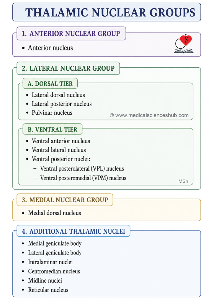
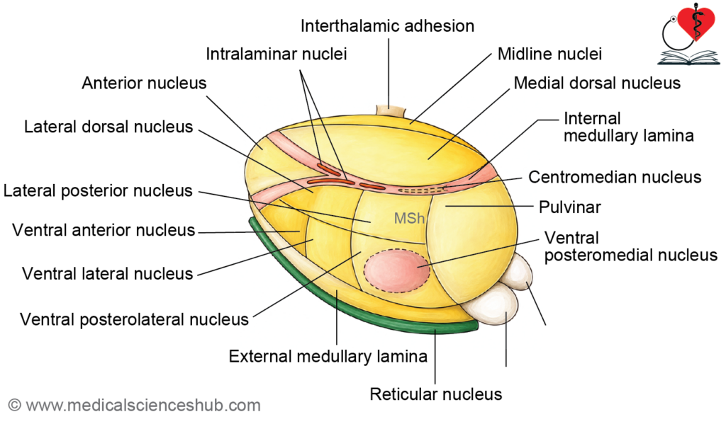
Table 10.1: Thalamic nuclei
| Part | Nuclei | |
|---|---|---|
| Anterior part | Anterior nucleus | |
| Medial part | Medial dorsal nucleus (only one in humans) | |
| Lateral part | 1. Dorsal tier nuclei | Lateral dorsal nucleus Pulvinar Lateral posterior nucleus |
| 2. Ventral tier nuclei | Ventral anterior nucleus Ventral posterior nucleus: Ventral posterolateral (VPL) and ventral posteromedial (VPM) Ventral lateral nucleus |
|
| Other nuclei | Medial geniculate body (MGB) Lateral geniculate body (LGB) Intralaminar nuclei Centromedian nucleus (CM) Midline nuclei Reticular nucleus |
Table 10.2: Connections and functions of thalamic nuclei
| Nucleus | Afferent input | Efferent output | Functions |
|---|---|---|---|
| Anterior nucleus | Mammillary body via mammillothalamic tract | Cingulate gyrus | Integral part of the Papez circuit (limbic system); contributes to emotional behavior and consolidation of recent memory. |
| Mediodorsal nucleus | Hypothalamus, amygdala, other thalamic nuclei | Prefrontal cortex | Integrates visceral and somatic information; shapes personality, mood, and affective experience. |
| Lateral dorsal group (LD, LP, pulvinar) | Superior colliculus, pretectal area | Cingulate gyrus, parahippocampal gyrus, parietal, occipital, and temporal association cortices | Participates in limbic circuitry; supports higher-order sensory analysis by the parieto-occipito-temporal association cortex. |
| VPM | Trigeminal lemniscus; solitariothalamic tract | Postcentral gyrus (primary somatosensory cortex) | Relay station for pain, temperature, touch, and proprioception from the head and face; also relays taste and visceral afferents. |
| VPL | Medial lemniscus; spinal lemniscus (spinothalamic tract) | Postcentral gyrus | Relay station for pain, temperature, touch, and proprioception from the trunk and limbs (entire body excluding head and face). |
| Ventral anterior (VA) | Globus pallidus via subthalamic fasciculus and ansa lenticularis | Premotor cortex | Relays striatal (basal ganglia) output to the cortex; contributes to modulation of voluntary motor activity. |
| Ventral lateral (VL) | Dentate nucleus of cerebellum (dentatorubrothalamic tract); red nucleus | Primary motor and premotor cortices | Principal relay for cerebellar output to the motor cortex; coordinates initiation of voluntary movement. Key DBS target in movement disorders. |
| Intralaminar nuclei (centromedian and parafascicular) | Ascending reticular formation; spinothalamic tract | Other thalamic nuclei; corpus striatum | Mediates general awareness of painful stimuli; contributes to sensorimotor integration and maintenance of cortical arousal. |
| Midline nuclei | Reticular formation | Hippocampus; amygdala | Integrated into the limbic system; involved in memory processing and mediating arousal responses. |
| Thalamic reticular nucleus | Collaterals of corticothalamic and thalamocortical fibers | Other thalamic relay nuclei (GABAergic; no direct cortical projection) | Modulates thalamocortical communication via inhibitory feedback; acts as a gateway regulating the flow of sensory information to the cortex. |
Blood Supply of Thalamus
Arterial Supply
- The thalamus receives its arterial supply from branches of three main vessels:
- The posterior communicating artery, a branch of the internal carotid artery that forms part of the circle of Willis, supplies the anterior and anterolateral thalamic territories.
- The posterior cerebral artery (PCA), the principal source of thalamic blood supply, contributes multiple perforating branches — including the thalamoperforating and thalamogeniculate arteries — that supply the paramedian, inferolateral, and posterior thalamic regions.
- The posterior choroidal arteries, arising from the PCA, supply the pulvinar and the posterior thalamus, as well as contributing to the choroid plexus of the third ventricle.
Venous Drainage
- Venous blood from the thalamus drains primarily via two sets of veins:
- The thalamostriate veins drain the superolateral thalamus and the adjacent striatum, running in the groove between the thalamus and the caudate nucleus.
- The choroidal veins of the third ventricle drain the medial and inferior thalamic regions.
- Both the thalamostriate and choroidal veins converge to form the internal cerebral vein on each side. The two internal cerebral veins subsequently unite to form the great cerebral vein (vein of Galen), which drains into the straight sinus.
CLINICAL NEUROANATOMY
Thalamic syndrome
Thalamic syndrome arises from a lesion — most commonly vascular — involving the thalamogeniculate branch of the posterior cerebral artery, which supplies the posterolateral thalamus. Disruption of thalamic sensory processing produces a characteristic combination of contralateral sensory, motor, and emotional disturbances.
Clinical Features
- Hyperesthesia: Sensory stimuli of ordinary intensity are perceived as abnormally exaggerated or unpleasant, reflecting a lowered threshold and heightened central processing of afferent input.
- Spontaneous thalamic pain: Severe, unprovoked pain arises on the side of the body contralateral to the lesion, typically described as burning, stabbing, or aching. It occurs without any identifiable peripheral stimulus and is often refractory to conventional analgesics.
- Emotional instability: Lesions disrupting thalamolimbic connections may produce pseudobulbar-type affective lability, characterized by episodes of involuntary laughter or crying disproportionate to the emotional context.
- Thalamic phantom phenomenon: The patient loses proprioceptive awareness of the affected limb when visual feedback is removed, occasionally accompanied by a perception that the limb is absent or displaced from its normal position.
- Thalamic hand: A characteristic postural abnormality develops in the contralateral upper limb, consisting of forearm pronation, wrist flexion, flexion at the metacarpophalangeal joints, and extension at the interphalangeal joints. This stereotyped posture is attributed to disruption of thalamocortical motor pathways.
Thalamic Pain Syndrome (Déjerine–Roussy Syndrome)
- This syndrome typically follows an ischaemic or haemorrhagic stroke affecting the posterolateral thalamus via the thalamogeniculate artery. In the acute phase, the patient experiences contralateral hemisensory loss or hypesthesia. Over subsequent weeks to months, this sensory deficit evolves into persistent, severe central neuropathic pain in the same distribution.
- The pain is characteristically burning or lancinating in quality and is readily provoked by light touch, cold, or emotional stress — stimuli that would ordinarily be innocuous. It is largely resistant to standard analgesics because it originates from pathological reorganization of central sensory processing rather than peripheral nociceptive input.
Sensory Disturbances in Thalamic Syndrome
- Allodynia: Pain is evoked by stimuli that are not normally noxious, such as light cutaneous contact, clothing, or a gentle breeze. This reflects aberrant central sensitization in which non-nociceptive afferent signals are misinterpreted as painful.
- Hyperpathia: There is an exaggerated and often prolonged pain response to stimuli that are mildly noxious. The perceived intensity far exceeds what the initiating stimulus would normally produce, and the sensation may persist or radiate beyond the point of contact.
- Dysesthesia: Spontaneous or stimulus-evoked abnormal sensations — such as burning, tingling, or a crawling feeling — occur in the absence of adequate peripheral stimulation. These sensations are consistently unpleasant and reflect disordered somatosensory cortical integration resulting from disrupted thalamic relay function.
Metathalamus
It consists of
- Medial geniculate body
- Lateral geniculate body
Medial Geniculate Body (MGB)
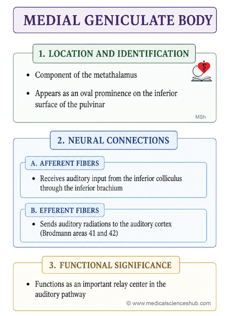
- The medial geniculate body (MGB) is an oval elevation located on the inferior aspect of the pulvinar of the thalamus, positioned lateral to the superior colliculus. It forms part of the metathalamus and is connected to the inferior colliculus of the midbrain via the inferior brachium.
- The name geniculate derives from the Latin genu (meaning “knee”), reflecting the characteristic knee-shaped cellular arrangement observed within the nucleus.
Connections
- Afferent input: The MGB receives ascending auditory signals primarily from the inferior colliculus via the inferior brachium, along with some direct fibers from the lateral lemniscus.
- Efferent output: The MGB projects via the auditory radiation to the primary auditory cortex in the superior temporal gyrus (Brodmann areas 41 and 42).
Functions
- Auditory relay: The MGB serves as the principal subcortical relay station in the ascending auditory pathway, transmitting processed sound signals from the brainstem — including the cochlear nuclei and inferior colliculus — to the auditory cortex for conscious perception and interpretation.
- Sound localization: By integrating binaural inputs, the MGB contributes to determining the spatial origin of auditory stimuli, which is essential for directional hearing and environmental awareness.
- Frequency and tone discrimination: The MGB participates in the tonotopic processing of sound, enabling discrimination of different frequencies and tonal patterns underlying speech recognition, music perception, and identification of environmental sounds.
- Auditory attention and filtering: The MGB contributes to selective auditory attention by helping to filter relevant acoustic signals from competing background noise, a process that also involves descending corticothalamic modulation.
CLINICAL NEUROANATOMY
- Lesions of the MGB typically result in central auditory processing dysfunction rather than complete deafness, since auditory input reaches both hemispheres. Clinical manifestations may include partial hearing loss, impaired sound localization, difficulty recognizing speech or complex auditory patterns, and auditory processing disorder. These deficits can significantly affect communication and daily functioning.
Lateral Geniculate Body
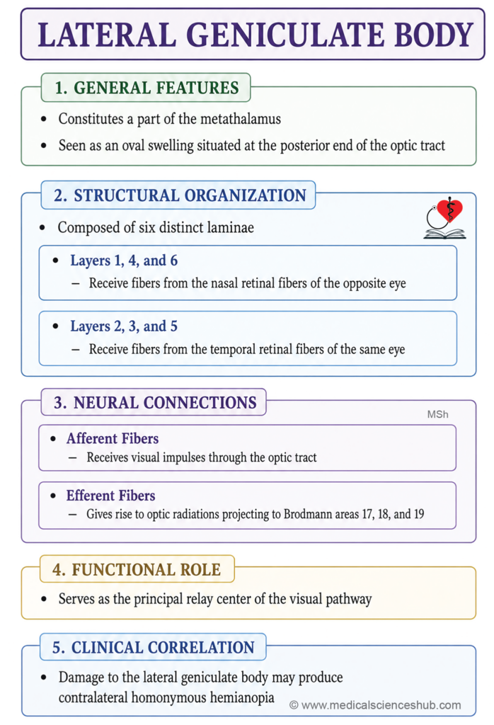
The lateral geniculate body (LGB) is an oval prominence situated at the posterior end of the optic tract, on the inferolateral aspect of the pulvinar and anterolateral to the medial geniculate body. It forms part of the metathalamus and serves as the principal thalamic relay nucleus in the visual pathway.
Structure
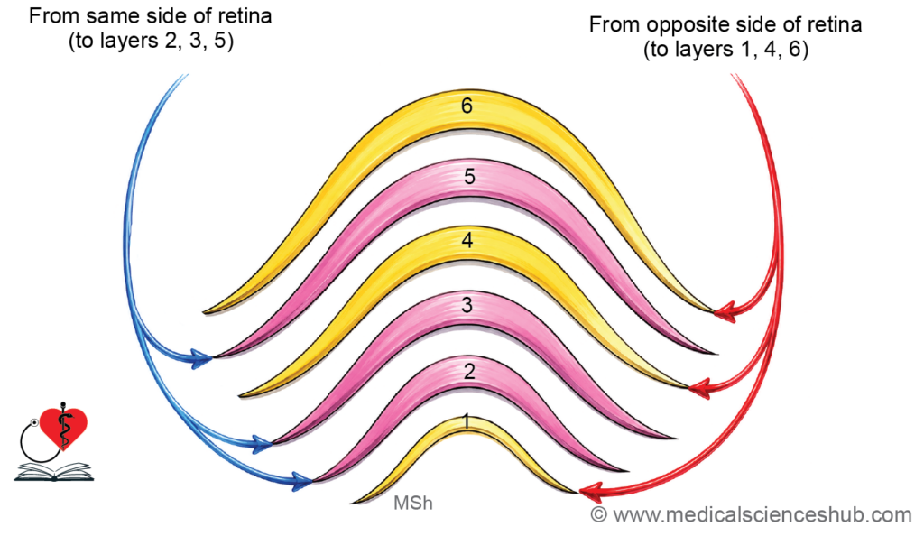
- The LGB is organized into six distinct laminae (layers 1–6), numbered sequentially from ventral to dorsal, separated by interlaminar zones of nerve fibers.
- This laminar arrangement maintains a precise retinotopic map of the contralateral visual field:
- Layers 1, 4, and 6 receive crossed fibers originating from the nasal hemiretina of the contralateral eye.
- Layers 2, 3, and 5 receive uncrossed fibers from the temporal hemiretina of the ipsilateral eye.
- Functionally, layers 1 and 2 are termed magnocellular layers, processing motion and contrast, while layers 3–6 are parvocellular layers, handling fine detail, color, and form perception.
Connections
- Afferent input: The LGB receives retinal ganglion cell axons via the optic tract.
- Efferent output: The LGB projects via the optic radiation (geniculocalcarine tract), which passes through the retrolentiform part of the internal capsule to reach the primary visual cortex (Brodmann area 17) in the occipital lobe, with additional projections to visual association areas (areas 18 and 19).
Functions
- Visual relay: The LGB transmits processed retinal signals from retinal ganglion cells to the primary visual cortex, enabling conscious perception of images, shapes, colors, and motion.
- Detail and pattern processing: Through its parvocellular layers, the LGB contributes to the discrimination of fine spatial detail, edges, textures, and patterns — functions essential for object recognition and reading.
- Color and contrast perception: Parvocellular neurons of the LGB encode chromatic and luminance contrast information, supporting accurate interpretation of the visual environment.
- Visual attention and filtering: The LGB, modulated by descending corticogeniculate projections, participates in selective visual attention by amplifying task-relevant signals and suppressing irrelevant visual input, thereby supporting spatial awareness and visuomotor coordination.
CLINICAL NEUROANATOMY
- A lesion of the LGB interrupts the relay of visual information from both eyes for the contralateral visual hemifield, producing contralateral homonymous hemianopia — loss of the same half of the visual field in both eyes — without affecting the pupillary light reflex, since the latter is mediated by the pretectal nucleus rather than the LGB.
- Additional manifestations may include impaired motion perception, reduced color discrimination, and difficulty recognizing objects, depending on the extent and precise location of the lesion.
Epithalamus
The epithalamus consists of:
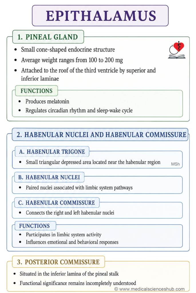
- Pineal body
- Habenular nucleus and commissure
- Posterior commissure.
Pineal Gland (Epiphysis Cerebri)
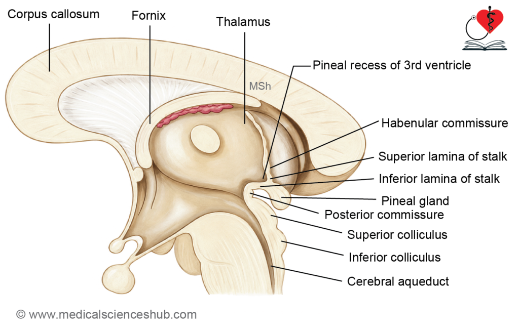
- The pineal gland is a small, pine cone-shaped midline structure located between the two superior colliculi of the midbrain, inferior to the splenium of the corpus callosum.
- Weight: approximately 100–200 mg.
Gross Anatomy
- The pineal gland is connected to the diencephalon by a stalk that divides into two laminae:
- The inferior lamina is continuous with the posterior commissure.
- The superior lamina is continuous with the habenular commissure.
- The pineal recess is a small diverticulum of the third ventricle that projects between the two laminae of the pineal stalk, bringing the ventricular cavity into close proximity with the gland.
- Sympathetic innervation is provided by postganglionic fibers (historically termed nervus conarii) arising from the superior cervical sympathetic ganglion, which regulate melatonin secretion in response to ambient light levels via a retinohypothalamic relay.
Microstructure
- The pineal gland is composed of two principal cell types:
- Pinealocytes are the predominant secretory cells responsible for synthesizing and releasing melatonin.
- Interstitial (glial) cells are less numerous and provide structural support to the pinealocytes.
- Corpora arenacea (brain sand) are calcified concretions that develop progressively within the pineal gland with advancing age. Their presence is a normal finding and serves as a useful radiological landmark for identifying the midline on plain skull radiographs and CT imaging.
Functions
- Melatonin secretion: Pinealocytes synthesize melatonin from serotonin in a light-dependent manner. Melatonin release is greatest during darkness and suppressed by light, thereby entraining the body’s circadian rhythm (biological clock) to the external light–dark cycle.
- Gonadotropin inhibition: Melatonin exerts an inhibitory influence on the hypothalamic–pituitary–gonadal axis by suppressing the release of gonadotropin-releasing hormone (GnRH) from the hypothalamus, thereby modulating reproductive endocrine activity.
CLINICAL NEUROANATOMY
- Pineal tumors (pinealomas): Space-occupying lesions of the pineal gland may compress the cerebral aqueduct, causing obstructive hydrocephalus, or impinge on the pretectal area, producing Parinaud’s syndrome (loss of voluntary upward gaze). Tumors occurring in children may disrupt melatonin-mediated gonadotropin inhibition, leading to precocious puberty.
- Physiological calcification of the pineal gland is common from the second decade onward and is of no clinical significance in itself, though marked displacement of the calcified gland on imaging may indicate a midline space-occupying lesion.
Habenular Nucleus and Habenular Commissure
- The habenular trigone (from the Latin habenula, meaning “little rein”) is a small, depressed triangular area on the dorsomedial surface of the posterior thalamus. It is bounded:
- Superiorly by the stria medullaris thalami and the superior lamina of the pineal stalk
- Inferiorly by the superior colliculus of the midbrain
- Laterally by the pulvinar of the thalamus
Habenular Nucleus
- The habenular nucleus lies within the habenular trigone and forms a component of the epithalamus. It is functionally integrated into the limbic system, serving as a critical relay between the forebrain — including the septal nuclei, hypothalamus, and basal ganglia — and the monoaminergic nuclei of the midbrain, particularly the raphe nuclei and the ventral tegmental area.
Habenular Commissure
- The habenular commissure consists of nerve fibers that interconnect the habenular nuclei of both sides across the midline. These fibers are carried within the superior lamina of the pineal stalk.
Functions
- As a component of the limbic system, the habenular nucleus mediates primitive visceral and behavioral responses to basic emotional drives, including pain, stress, and reward.
- It plays a modulatory role in motivation, mood regulation, and reward processing by influencing dopaminergic and serotonergic signaling in the midbrain. Dysregulation of habenular activity has been implicated in depression, anhedonia, and addiction disorders.
CLINICAL NEUROANATOMY
- Lesions or pathological hyperactivity of the lateral habenular nucleus — which exerts inhibitory control over midbrain reward circuitry — have been associated with major depressive disorder and treatment-resistant depression, making the habenula a focus of interest in the neuroscience of mood disorders and a potential target for deep brain stimulation (DBS).
Posterior Commissure
- The posterior commissure is a transversely oriented bundle of white matter fibers that crosses the midline within the inferior lamina of the pineal stalk, at the junction of the diencephalon and the dorsal midbrain.
Composition
- The posterior commissure carries fibers from several sources, including the medial longitudinal fasciculus (MLF), the interstitial nucleus of Cajal, the nucleus of Darkschewitsch, and various adjacent pretectal and tectal nuclei. This diverse fiber composition reflects its role as an integrative commissural pathway rather than a simple decussation of a single tract.
Functions
- The posterior commissure interconnects corresponding pretectal and midbrain nuclei of both sides, and is particularly important for:
- Coordination of conjugate eye movements, especially vertical gaze, through its connections with the interstitial nucleus of Cajal and the rostral interstitial nucleus of the MLF.
- Consensual pupillary light reflex, by linking the pretectal olivary nuclei of both sides, enabling the contralateral pupillary constriction that occurs when light is directed into one eye.
Clinical Relevance
- Lesions involving the posterior commissure and the surrounding dorsal midbrain (pretectal) region produce the characteristic features of dorsal midbrain syndrome (Parinaud’s syndrome), which includes:
- Paralysis of voluntary upward gaze
- Loss or impairment of the pupillary light reflex with preservation of the near response (light-near dissociation)
- Convergence-retraction nystagmus on attempted upward gaze
- These findings are commonly associated with pineal region tumors, hydrocephalus causing dorsal midbrain compression, or midbrain infarction.
Subthalamus
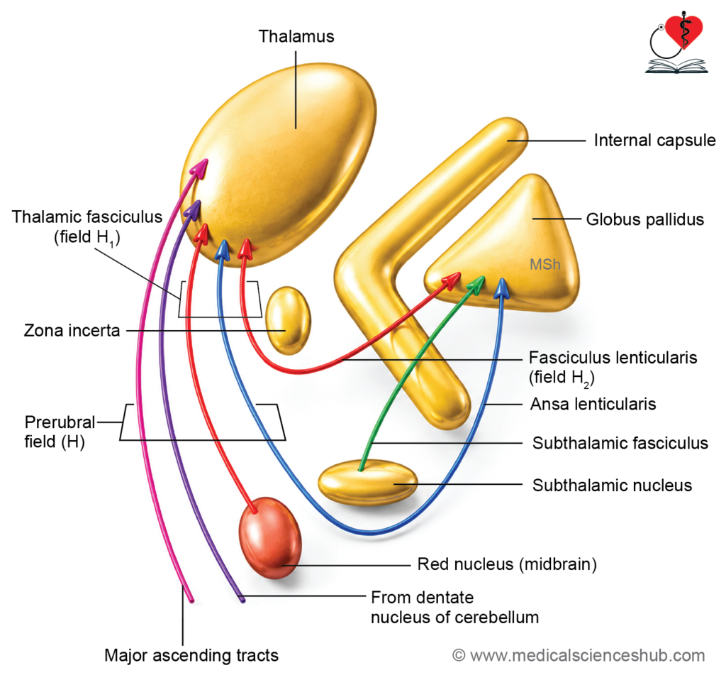
- The subthalamus is a diencephalic region situated inferior to the thalamus, medial to the internal capsule, and superior to the midbrain tegmentum. It functions as a critical node in the basal ganglia–thalamocortical circuit, modulating and refining voluntary motor activity.
Components of the Subthalamus
- Subthalamic nucleus: A lens-shaped (biconvex) grey matter nucleus lying on the medial aspect of the internal capsule, immediately superior to the substantia nigra. It is a key excitatory nucleus within the basal ganglia circuitry, exerting glutamatergic drive on the globus pallidus internus. A lesion of the subthalamic nucleus — most commonly due to a small vascular infarct — produces hemiballismus, characterized by sudden, violent, involuntary flinging movements of the contralateral limbs, resulting from loss of inhibitory control over thalamic motor circuits.
- Zona incerta: A thin lamina of grey matter interposed between the subthalamic nucleus and the thalamus, representing a rostral continuation of the brainstem reticular formation. It integrates multimodal sensory and motor signals and contributes to arousal, attentional gating, and movement coordination.
- Fasciculus lenticularis (lenticular fasciculus — Field H2 of Forel): A fiber bundle arising from the globus pallidus internus that traverses the posterior limb of the internal capsule to reach the subthalamic region. It constitutes part of the pallidofugal pathway, contributing to the regulation of smooth voluntary movement.
- Ansa lenticularis: A curved fiber bundle originating from the globus pallidus internus that arcs around the ventral border of the internal capsule before ascending to the ventral anterior and ventral lateral thalamic nuclei. It carries inhibitory GABAergic pallidothalamic projections and plays a central role in modulating thalamocortical motor output.
- Fasciculus subthalamicus: A bidirectional fiber tract connecting the globus pallidus and the subthalamic nucleus, forming the structural basis of the indirect pathway of the basal ganglia. It maintains the excitatory–inhibitory balance necessary for controlled voluntary movement; its disruption contributes to involuntary movements such as hemiballismus.
- Thalamic fasciculus (Field H1 of Forel): A fiber bundle that runs from the prerubral field to the ventral anterior and ventral lateral nuclei of the thalamus. It represents the merged pathway of the ansa lenticularis and fasciculus lenticularis, along with cerebellar efferents from the dentatorubrothalamic tract, forming the principal conduit for subcortical motor signals reaching the thalamus.
- Prerubral field (Field H of Forel): Located anterior to the red nucleus in the subthalamic region, this area serves as a convergence zone for pallidothalamic fibers from the globus pallidus. It plays an integrative role in coordinating and relaying motor signals within the basal ganglia–thalamic axis.
- Cranial extensions of the red nucleus and substantia nigra: The rostral poles of the red nucleus and substantia nigra extend into the subthalamic region, contributing dopaminergic and rubrospinal modulatory influences on basal ganglia and motor thalamic circuits.
Clinical Relevance
The subthalamic nucleus is the primary surgical target for deep brain stimulation (DBS) in the management of Parkinson’s disease, where high-frequency stimulation of this nucleus reduces the pathological overactivity of the indirect pathway, alleviating rigidity, tremor, and bradykinesia.
Stria Medullaris Thalami
- The stria medullaris thalami is a discrete bundle of white matter fibers running along the junction of the medial and superior surfaces of the thalamus, deep to the taenia thalami — the line of ependymal attachment of the roof of the third ventricle.
Connections
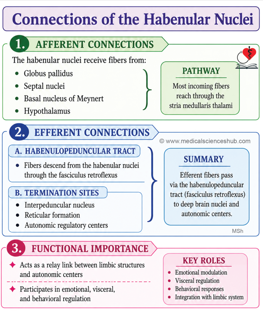
- The stria medullaris thalami carries fibers from several forebrain structures to the habenular nucleus of the epithalamus:
- Septal nuclei — conveying limbic and reward-related signals
- Basal nucleus of Meynert — the principal source of cholinergic innervation to the cerebral cortex, linking forebrain cholinergic circuitry to the habenula
- Hypothalamus — transmitting autonomic and neuroendocrine modulatory input
Functional Significance
- By linking the septal nuclei, basal forebrain, and hypothalamus with the habenular nucleus, the stria medullaris thalami forms an important component of the limbic system, serving as a conduit through which emotional, motivational, and autonomic signals are relayed to the habenulo-interpeduncular pathway — also known as the fasciculus retroflexus — and onward to the monoaminergic nuclei of the midbrain, including the raphe nuclei and the ventral tegmental area.
- It thus contributes to the regulation of mood, motivation, reward processing, and visceral autonomic responses driven by basic emotional states.
Important Questions
- Write a short note on medial geniculate body.
- Write a short note on lateral geniculate body.
- List the components of epithalamus.
📝 Test Your Knowledge – Practice MCQs
Attempt the chapter MCQ quiz and assess your understanding of key concepts.
Click to see: Medical Disclaimer and Copyright Notice