
Competency
- AN62.5: Describe boundaries, parts, gross relations, major nuclei, and connections of hypothalamus, …
Introduction
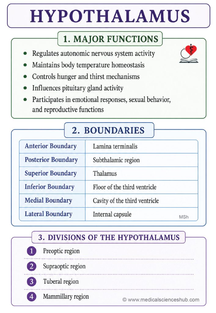
- The hypothalamus is a component of the diencephalon, situated inferior to the thalamus — a positional relationship reflected in its name. Despite weighing only approximately 4 grams (less than 1% of total brain weight), it serves as one of the most functionally significant structures in the central nervous system.
- It acts as the principal integrative center linking the nervous system and the endocrine system, coordinating a broad range of visceral, autonomic, and neuroendocrine functions essential for maintaining homeostasis.
- The hypothalamus regulates body temperature, fluid and electrolyte balance, feeding behavior, circadian rhythms, reproductive function, and the stress response, largely through its control of the pituitary gland (hypophysis) and the autonomic nervous system.
- Lesions of the hypothalamus can produce a wide spectrum of clinical disorders, including diabetes insipidus (due to loss of antidiuretic hormone secretion), hypothalamic obesity, central hyperthermia or poikilothermia, disrupted sleep–wake cycles, and panhypopituitarism, depending on the specific nuclei and pathways affected.
Functions of Hypothalamus
- Autonomic regulation: The hypothalamus serves as the highest subcortical center for autonomic nervous system control, modulating heart rate, arterial blood pressure, gastrointestinal motility, and respiratory activity. It integrates visceral responses to both internal physiological changes and external stressors, coordinating sympathetic and parasympathetic outflow through descending projections to the brainstem and spinal cord.
- Thermoregulation: The hypothalamus functions as the central thermostat of the body. Thermosensitive neurons — particularly within the anterior hypothalamic and preoptic areas — detect deviations in core body temperature and initiate appropriate corrective responses: sweating and cutaneous vasodilation to dissipate heat, or shivering and vasoconstriction to conserve it, maintaining core temperature within a narrow physiological range.
- Regulation of food and water intake: Discrete hypothalamic nuclei govern appetite and thirst. The lateral hypothalamic area promotes feeding behavior, while the ventromedial nucleus signals satiety. Osmoreceptors within the hypothalamus detect changes in plasma osmolality, stimulating thirst and modulating antidiuretic hormone (ADH) secretion to regulate water balance.
- Control of pituitary gland function: The hypothalamus regulates both lobes of the pituitary gland (hypophysis). It secretes releasing and inhibiting hormones into the hypophyseal portal system to control anterior pituitary hormone output — including growth hormone, cortisol, thyroid hormones, and gonadotropins. It also directly synthesizes oxytocin and ADH (vasopressin), which are transported to and released from the posterior pituitary.
- Emotional behavior, sexual function, and reproduction: As a core component of the limbic system, the hypothalamus modulates emotional responses including fear, aggression, and pleasure, largely through its connections with the amygdala and prefrontal cortex. It also regulates sexual drive and reproductive behavior by controlling gonadotropin-releasing hormone (GnRH) secretion, which governs the hypothalamic–pituitary–gonadal axis.
- Circadian rhythm regulation: The suprachiasmatic nucleus (SCN) of the hypothalamus functions as the primary biological clock, receiving direct photic input from the retina via the retinohypothalamic tract. It synchronizes the sleep–wake cycle and other circadian rhythms — including hormonal secretion patterns and metabolic activity — to the external light–dark environment.
Boundaries of Hypothalamus
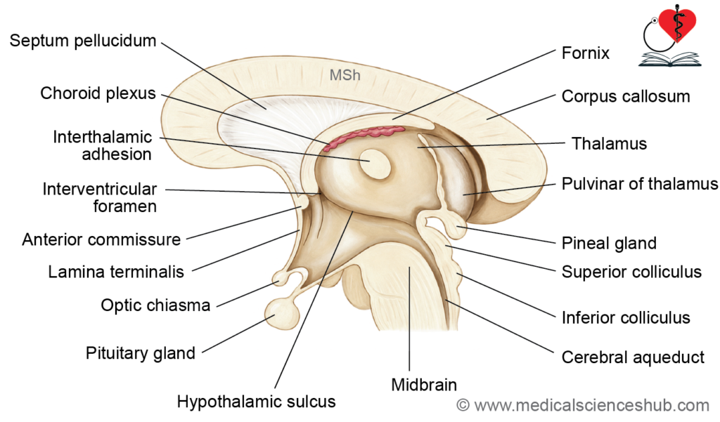
- Anteriorly: The hypothalamus is bounded by the lamina terminalis, a thin membrane stretching from the optic chiasma to the anterior commissure, representing the rostral limit of the diencephalon.
- Posteriorly: It is continuous with the subthalamus, with no sharp macroscopic boundary demarcating the two regions.
- Superiorly: The hypothalamus is separated from the thalamus by the hypothalamic sulcus, a shallow groove on the medial wall of the third ventricle that extends from the interventricular foramen (foramen of Monro) anteriorly to the cerebral aqueduct posteriorly.
- Inferiorly: The inferior boundary is formed by the floor of the third ventricle, which includes the tuber cinereum, mammillary bodies, and the infundibulum (pituitary stalk).
- Medially: The hypothalamus borders the cavity of the third ventricle, whose lateral walls it partially forms.
- Laterally: It is bounded by the posterior limb of the internal capsule and the subthalamus, which separate it from the adjacent basal ganglia and thalamic structures.
- The hypothalamic sulcus is a particularly important anatomical landmark, as it defines the boundary between the hypothalamus inferiorly and the thalamus superiorly along the entire length of the lateral wall of the third ventricle.
Parts of Hypothalamus
The hypothalamus is classically divided into four anatomical regions based on their location and associated nuclei.
- Preoptic Region
The preoptic region is situated anterior to the optic chiasma and extends up to the anterior commissure. It mainly contains the preoptic nucleus, which is involved in regulation of body temperature and reproductive functions. - Supraoptic Region
The supraoptic region lies superior to the optic chiasma. It contains the supraoptic, paraventricular, and anterior nuclei. These nuclei play important roles in neuroendocrine regulation, fluid balance, and autonomic control. The supraoptic and paraventricular nuclei are also associated with the production of antidiuretic hormone (ADH) and oxytocin. - Tuberal Region
The tuberal region forms the central part of the hypothalamus. It includes the tuber cinereum, arcuate nucleus, ventromedial nucleus, and dorsomedial nucleus. This region is mainly concerned with regulation of feeding behavior, metabolism, endocrine activity, and energy balance. - Mammillary Region
The mammillary region forms the posterior part of the hypothalamus. It includes the mammillary bodies and the posterior nucleus. These structures are involved in memory pathways, emotional responses, and autonomic functions.
Hypothalamic Nuclei and Connections
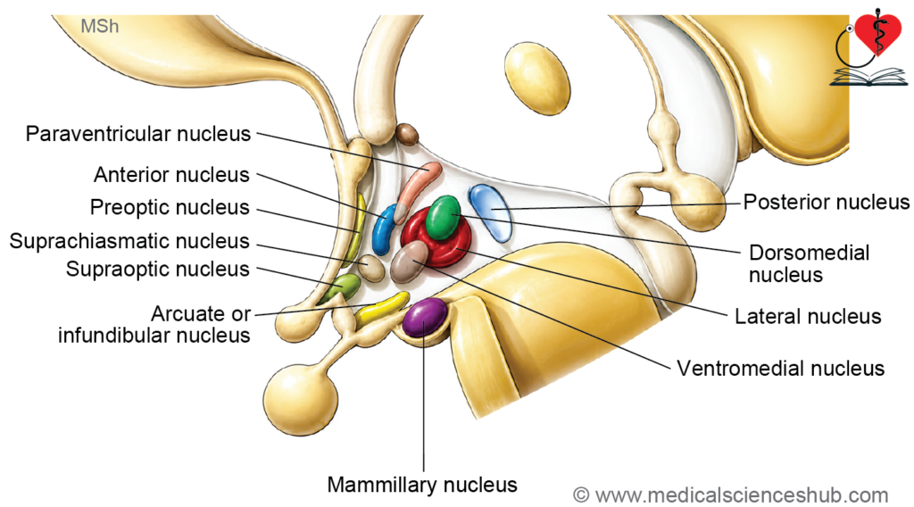
The nuclei of the hypothalamus are arranged in distinct groups and are generally named according to their anatomical location. The major hypothalamic nuclei include the preoptic, supraoptic, paraventricular, anterior, arcuate, ventromedial, dorsomedial, posterior, mammillary, and lateral nuclei.
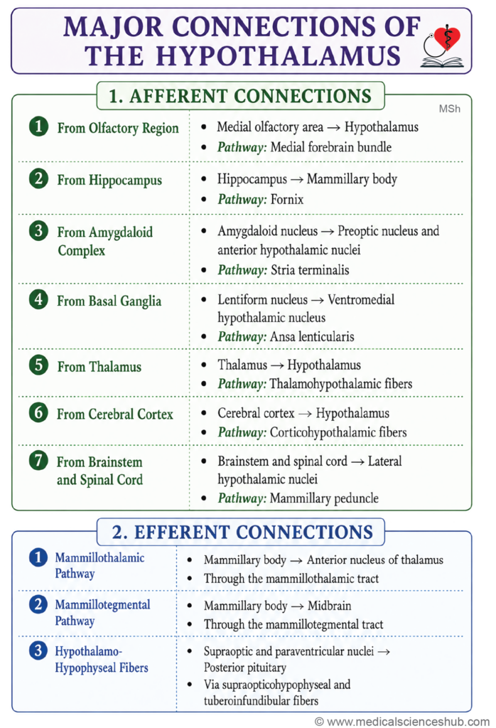
Afferent Fibers for Hypothalamus
The hypothalamus receives afferent fibers from different regions of the brain and spinal cord. These connections help integrate autonomic, endocrine, emotional, and behavioral functions.
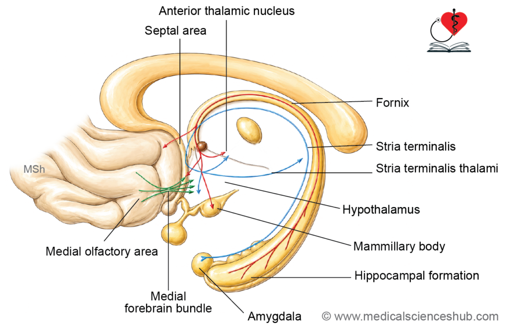
- Medial Forebrain Bundle: The medial forebrain bundle connects the olfactory region, including the olfactory bulb and anterior perforated substance, with the preoptic and lateral hypothalamic nuclei. It forms an important communication pathway between the hypothalamus, limbic system, and brainstem. This bundle is involved in regulation of emotion, motivation, reward mechanisms, and autonomic activities. Damage to this pathway may result in emotional disturbances, reduced motivation, and behavioral changes.
- Fornix: The fornix is a curved bundle of white matter fibers that connects the hippocampus with the mammillary bodies and other parts of the limbic system. It is an important pathway related to memory formation and recall. The fornix also participates in emotional and cognitive functions through its connections with the hypothalamus. Lesions of the fornix may produce memory impairment and confusion.
- Stria Terminalis: The stria terminalis connects the amygdaloid body with the preoptic and anterior hypothalamic nuclei. It carries fibers associated with emotional and autonomic responses, particularly those related to fear, stress, and anxiety. Injury to this tract may alter emotional behavior and stress responses.
- Ansa Lenticularis (Pallidohypothalamic Fibers): The ansa lenticularis consists of fibers arising from the globus pallidus of the basal ganglia and projecting to the ventromedial hypothalamic nucleus and related regions. These fibers help coordinate motor activity with autonomic and behavioral responses. Disturbance of this pathway may contribute to abnormal involuntary movements and motor dysfunction.
- Thalamohypothalamic Fibers: These fibers connect the thalamus with the hypothalamus. They help integrate sensory information with autonomic and endocrine activities, thereby contributing to maintenance of homeostasis.
- Corticohypothalamic Fibers: Corticohypothalamic fibers arise mainly from the limbic cortex and prefrontal cortex and terminate in the hypothalamus. Through these connections, higher cortical centers influence autonomic, endocrine, and emotional functions, especially during stress and behavioral responses.
Mammillary Peduncle: The mammillary peduncle connects the mammillary bodies with the brainstem and related pathways. It participates in circuits concerned with memory and emotional regulation. Lesions involving the mammillary connections may lead to memory disturbances, as seen in conditions such as Wernicke–Korsakoff syndrome.
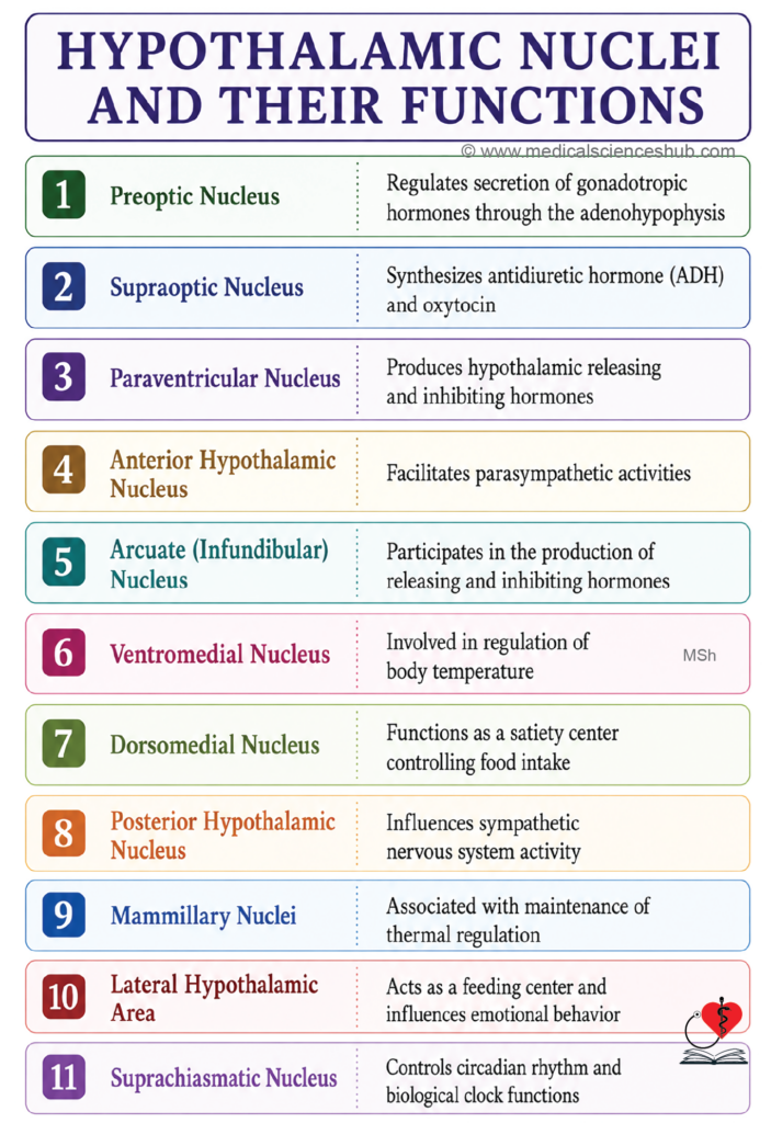
Table 11.1: Hypothalamic nucleus and their functions
| Nucleus | Region | Principal Functions |
|---|---|---|
| Preoptic nucleus | Preoptic region | Regulates pulsatile secretion of gonadotropin-releasing hormone (GnRH), thereby influencing reproductive function and gonadotropin release from the anterior pituitary. Also participates in thermoregulation and initiation of sleep. |
| Supraoptic nucleus | Supraoptic region | Synthesizes antidiuretic hormone (ADH/vasopressin) and oxytocin, which are transported to the posterior pituitary. ADH regulates water balance and plasma osmolarity, while oxytocin is involved in uterine contraction and milk ejection. |
| Paraventricular nucleus | Supraoptic region | Produces ADH and oxytocin and also secretes releasing hormones such as CRH and TRH into the hypophyseal portal system. Plays an important role in autonomic and neuroendocrine regulation. |
| Anterior nucleus | Supraoptic region | Regulates parasympathetic activity and promotes heat loss through sweating and cutaneous vasodilatation, thereby helping maintain normal body temperature. |
| Arcuate (infundibular) nucleus | Tuberal region | Secretes releasing and inhibitory hormones, including GHRH, dopamine, somatostatin, and GnRH, which regulate anterior pituitary function related to growth, metabolism, and reproduction. |
| Ventromedial nucleus | Tuberal region | Functions as the satiety center. Lesions may lead to excessive eating and obesity. Also influences emotional behavior and defensive responses. |
| Dorsomedial nucleus | Tuberal region | Modulates feeding behavior, emotional responses, autonomic activity, and certain cardiovascular and gastrointestinal functions. |
| Lateral hypothalamic area | Tuberal region | Acts as the feeding center and stimulates appetite through orexin-containing neurons. Lesions may cause reduced food intake and weight loss. |
| Posterior nucleus | Mammillary region | Regulates sympathetic activity and promotes heat conservation through vasoconstriction and shivering. Lesions may impair temperature regulation. |
| Mammillary body | Mammillary region | Receives fibers from the hippocampus through the fornix and projects to the anterior thalamic nucleus via the mammillothalamic tract. It is involved in memory and emotional processing as part of the Papez circuit. |
| Suprachiasmatic nucleus | Supraoptic region | Functions as the principal biological clock of the body. It receives retinal input and regulates circadian rhythms, including the sleep–wake cycle and hormonal rhythms. |
Efferent Fibers of Hypothalamus
The hypothalamus sends efferent fibers to different parts of the brain, brainstem, spinal cord, and pituitary gland. These pathways help regulate autonomic, endocrine, emotional, and behavioral functions.
- Mammillothalamic Tract
The mammillothalamic tract connects the mammillary bodies with the anterior nuclei of the thalamus. It forms an important part of the limbic system and is involved in memory processing and emotional integration. Damage to this tract may result in memory disturbances, disorientation, and impairment of learning functions. - Mammillotegmental Tract
The mammillotegmental tract extends from the mammillary bodies to the reticular formation of the midbrain tegmentum. This pathway influences autonomic activity, arousal, and emotional responses. Lesions may produce disturbances in autonomic regulation and altered levels of alertness. - Descending Fibers to Brainstem and Spinal Cord
Descending hypothalamic fibers project to autonomic centers in the brainstem and spinal cord. Through these connections, the hypothalamus regulates cardiovascular activity, body temperature, gastrointestinal functions, and other visceral activities. - Hypothalamo-hypophyseal Fibers
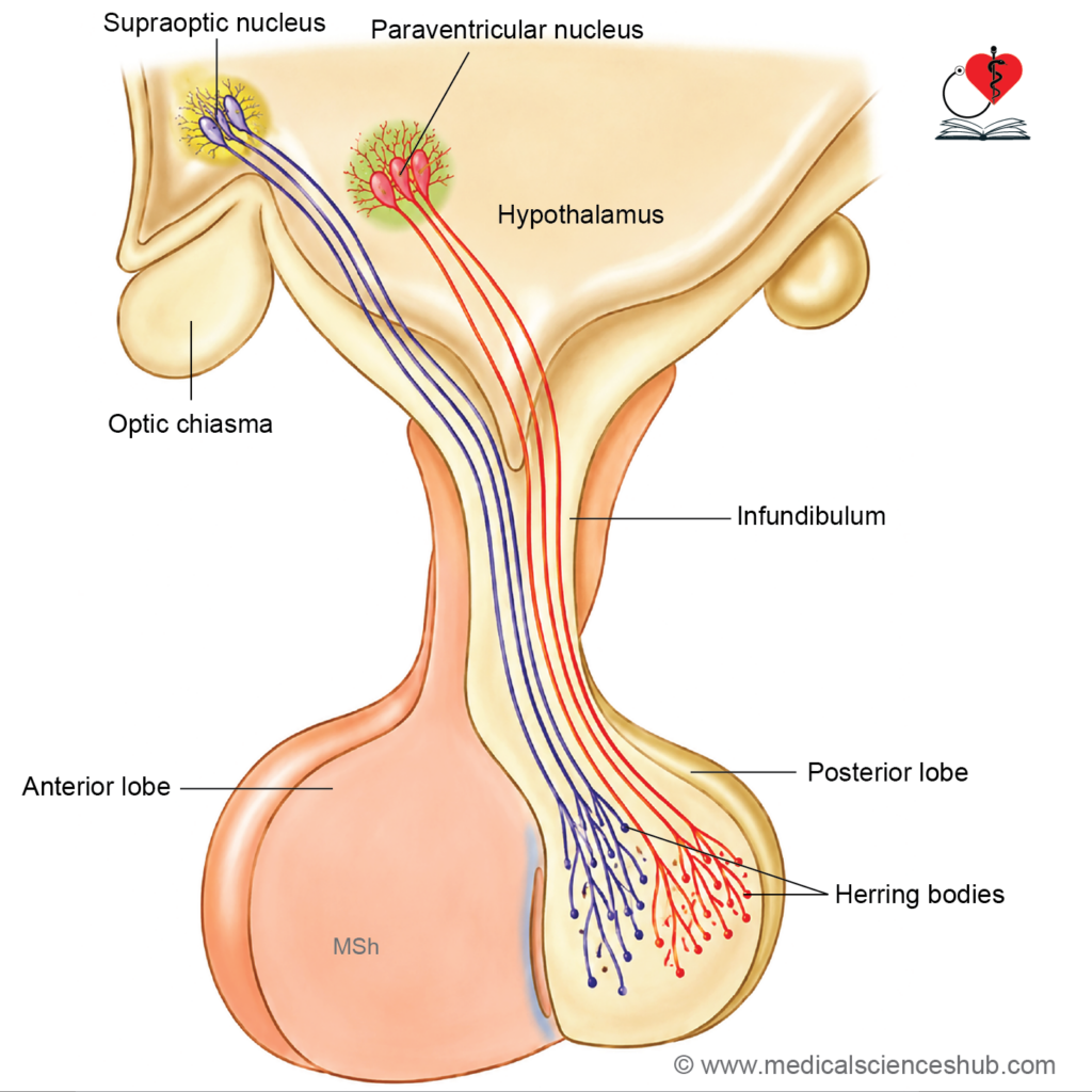
These fibers connect the hypothalamus with the pituitary gland and are functionally divided into two groups:- Supraopticohypophyseal fibers arise mainly from the supraoptic and paraventricular nuclei and pass to the posterior pituitary. They transport antidiuretic hormone (ADH) and oxytocin, which are synthesized in the hypothalamus and released from the neurohypophysis.
- Tuberoinfundibular fibers arise chiefly from the arcuate nucleus and related hypothalamic regions. They terminate in the median eminence and regulate hormone secretion from the anterior pituitary through releasing and inhibitory hormones.
Damage to these pathways may produce endocrine disturbances such as diabetes insipidus, reproductive disorders, impaired lactation, and metabolic imbalance.
Hypothalamo-hypophyseal Portal System
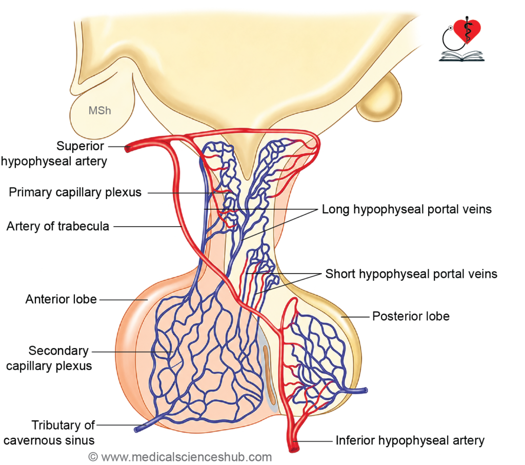
The pituitary gland receives its blood supply mainly from branches of the internal carotid artery. The superior hypophyseal arteries supply the median eminence, tuber cinereum, and infundibular stalk, whereas the inferior hypophyseal arteries primarily supply the posterior pituitary.
In the median eminence and upper part of the infundibulum, the superior hypophyseal arteries form a network of primary capillaries. These capillaries unite to form the hypophyseal portal veins, which descend along the pituitary stalk to the anterior pituitary. Within the adenohypophysis, the portal veins divide again into a network of secondary capillaries. This portal circulation allows hypothalamic releasing and inhibitory hormones to reach the anterior pituitary directly and regulate its secretory activity.
Venous blood from the pituitary gland drains into the cavernous sinus through hypophyseal veins. The supraoptic and paraventricular nuclei of the hypothalamus synthesize antidiuretic hormone (ADH) and oxytocin. These hormones are transported along hypothalamo-hypophyseal fibers and stored in the posterior pituitary before their release into the bloodstream.
CLINICAL NEUROANATOMY
Hypothalamic disorders
Lesions of the hypothalamus — arising from vascular infarction, trauma, neoplasms, or infections — disrupt its broad regulatory functions, producing a characteristic constellation of neuroendocrine, autonomic, and behavioral disturbances.
- Diabetes insipidus: Destruction of the supraoptic or paraventricular nuclei, or damage to the hypothalamo-hypophyseal tract, reduces or abolishes ADH (vasopressin) synthesis. The renal collecting ducts consequently fail to reabsorb water, resulting in the excretion of large volumes of dilute urine.
- Polydipsia and polyuria: ADH deficiency leads to continuous renal water loss (polyuria), which stimulates compensatory excessive fluid intake (polydipsia). If the thirst mechanism is also impaired, the patient may be unable to compensate adequately, resulting in clinically significant dehydration and hypernatremia.
- Hypothalamic obesity or aphagia: Lesions of the ventromedial nucleus — the primary satiety center — remove the normal inhibitory control over feeding, producing hyperphagia (polyphagia) and progressive obesity. Conversely, damage to the lateral hypothalamic area (the feeding center) may abolish appetite entirely, causing aphagia and severe weight loss. Both presentations are possible depending on the site of the lesion.
- Somnolence: Hypothalamic injury, particularly involving the posterior hypothalamus and its connections with the ascending reticular activating system, disrupts the regulation of the sleep–wake cycle, manifesting as excessive daytime somnolence or prolonged hypersomnia.
- Thermoregulatory failure (poikilothermia): Loss of hypothalamic thermoregulatory control renders the patient unable to maintain a stable core temperature, a condition termed poikilothermia. Lesions of the anterior hypothalamus typically impair heat dissipation, predisposing to hyperthermia, while posterior lesions may impair heat conservation.
- Loss of libido and reproductive dysfunction: Disruption of GnRH-secreting neurons within the hypothalamus reduces gonadotropin release from the anterior pituitary, leading to hypogonadism, diminished libido, and impaired reproductive function.
- Emotional and behavioral disturbances: Hypothalamic lesions can profoundly alter emotional regulation, producing aggression, pathological rage, or — in other cases — marked apathy and emotional blunting, depending on which nuclei and their limbic connections are affected.
Important Questions
- Describe the anatomical divisions of the hypothalamus and mention the nuclei present in each region.
- Explain the afferent and efferent connections of the hypothalamus with suitable examples.
- Describe the hypothalamo-hypophyseal portal system and discuss its functional importance.
- Write a note on the major hypothalamic nuclei and their functions in regulation of autonomic and endocrine activities.
- Discuss the role of the hypothalamus in maintenance of homeostasis, including regulation of temperature, hunger, thirst, and circadian rhythm.
📝 Test Your Knowledge – Practice MCQs
Attempt the chapter MCQ quiz and assess your understanding of key concepts.
Click to see: Medical Disclaimer and Copyright Notice