
Competencies
- AN62.2: Describe and demonstrate surfaces, sulci, gyri, poles, and functional areas of cerebral hemisphere.
- AN64.1: Describe and identify the microanatomical features of spinal cord, cerebellum, and cerebrum
Introduction
- The cerebrum is the largest division of the brain, composed of two mirror-image halves known as the right and left cerebral hemispheres.
- The two hemispheres are anatomically separated by a deep groove called the longitudinal cerebral fissure, which runs along the midline of the brain.
- Despite this separation, the hemispheres remain functionally connected through a broad band of nerve fibers known as the corpus callosum, which facilitates communication between the two sides.
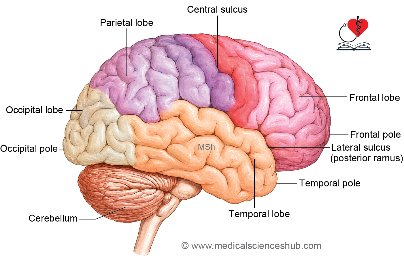
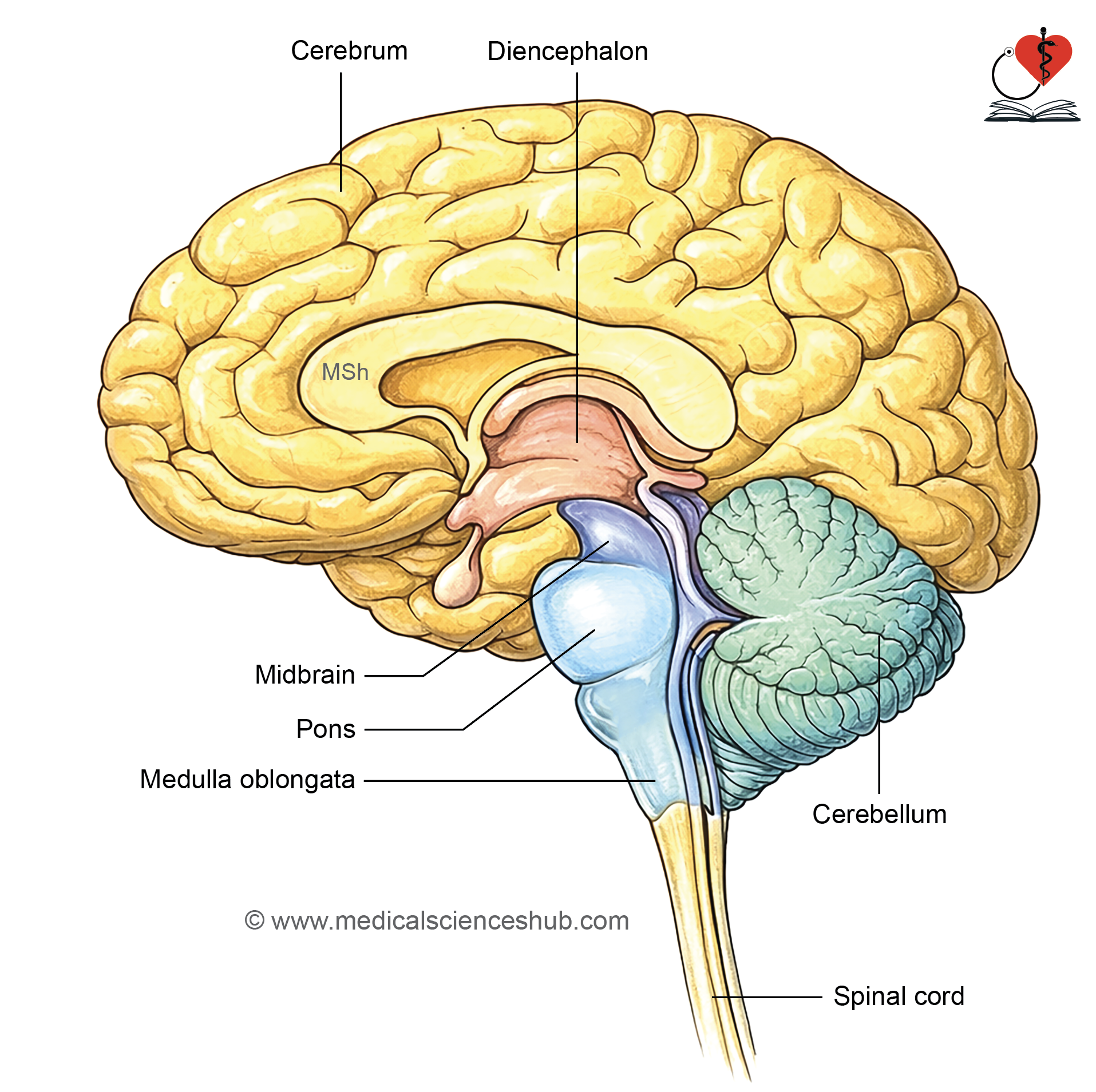
Components of Cerebral Hemisphere
Each cerebral hemisphere consists of four distinct structural components:
- Cerebral cortex — The outermost layer of the hemisphere, composed of grey matter (neuronal cell bodies), responsible for higher cognitive and sensory functions.
- White matter — The inner region beneath the cortex, consisting of myelinated nerve fibers that transmit signals between cortical areas and other brain regions.
- Basal nuclei (basal ganglia) — Clusters of grey matter embedded deep within the white matter, primarily involved in the coordination and regulation of voluntary movement.
- Lateral ventricle — A fluid-filled cavity located within each hemisphere, forming part of the ventricular system and containing cerebrospinal fluid (CSF).
External Features of the Cerebral Hemisphere
The surface of each cerebral hemisphere is characteristically folded, displaying:
- Gyri — Elevated, ridge-like convolutions of the cortical surface.
- Sulci — Grooves or furrows that separate adjacent gyri.
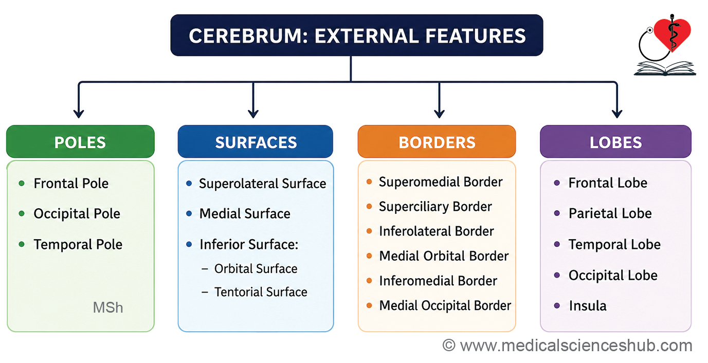
Poles of Cerebral Hemisphere
Each cerebral hemisphere presents three poles marking its extreme projecting points:
- Frontal pole — The anterior-most tip of the hemisphere; it is broadly rounded in shape.
- Occipital pole — The posterior-most tip; it is comparatively more pointed than the frontal pole.
- Temporal pole — The anterior extremity of the temporal lobe, positioned between the frontal and occipital poles; it is rounded and directed anteriorly.
Surfaces of Cerebral Hemisphere
Each cerebral hemisphere presents three distinct surfaces:
- Superolateral surface — Convex in contour, facing upward and laterally toward the inner surface of the skull.
- Medial surface — Flat and vertically oriented, facing the opposite hemisphere across the longitudinal cerebral fissure; the two hemispheres are interconnected at this surface by the C-shaped corpus callosum.
- Inferior surface — Divided into two regions:
- Orbital surface — The smaller anterior portion, concave in shape, resting on the roof of the orbit and the cribriform plate of the ethmoid bone.
- Tentorial surface — The larger posterior portion, resting on the floor of the middle cranial fossa and on the tentorium cerebelli.
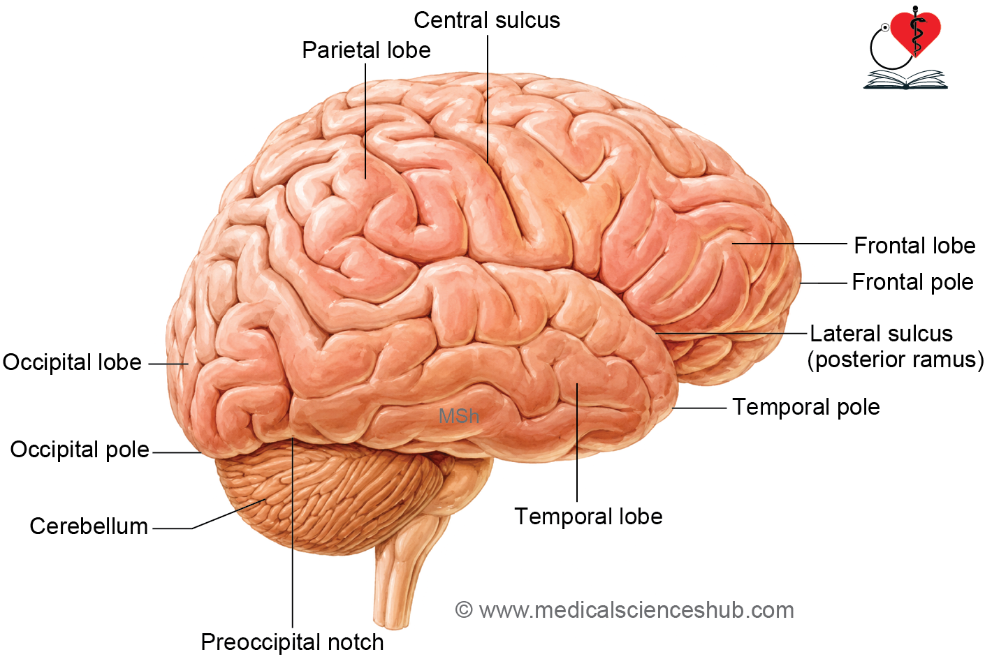
Borders of Cerebral Hemisphere
The surfaces of the cerebral hemisphere are demarcated by five borders:
- Superomedial border — Separates the superolateral surface from the medial surface.
- Inferolateral border — Separates the superolateral surface from the inferior surface (both its orbital and tentorial parts); features a small depression known as the preoccipital notch, located approximately 5 cm anterior to the occipital pole.
- Medial orbital border — Separates the medial surface from the orbital part of the inferior surface.
- Medial occipital border — Separates the medial surface of the occipital lobe from the tentorial part of the inferior surface.
- Inferomedial (hippocampal) border — Runs along the medial edge of the temporal lobe, separating the inferior surface from the medial surface; it lies superior to the cerebral peduncle.
Main Cerebral Sulci
- Lateral sulcus (Sylvian sulcus) — A deep sulcus consisting of a stem and three rami:
- The stem originates on the inferior surface, separating the orbital surface from the temporal pole, before reaching the superolateral surface.
- The anterior ramus (~2.5 cm) projects horizontally into the inferior frontal gyrus.
- The anterior ascending ramus (~2.5 cm) courses vertically upward within the inferior frontal gyrus.
- The posterior ramus (~7.5 cm) runs posteriorly and superiorly toward the inferior parietal lobule, with its posterior end turning sharply upward.
- Central sulcus (sulcus of Rolando) — Originates on the superomedial border approximately 1 cm posterior to the midpoint between the frontal and occipital poles. It courses inferiorly and anteriorly at an angle of approximately 70° toward the posterior ramus of the lateral sulcus; its upper end extends onto the medial surface.
- Parieto-occipital sulcus — Begins at the midpoint of the calcarine sulcus and runs superiorly and slightly posteriorly, intersecting the superomedial border approximately 5 cm anterior to the occipital pole; it extends a short distance onto the superolateral surface.
- Calcarine sulcus — Located exclusively on the medial surface. It originates just inferior to the posterior end (splenium) of the corpus callosum and runs posteriorly with a superiorly convex curve toward the occipital pole, extending briefly onto the superolateral surface.
Lobes of Cerebral Hemisphere
The cerebral hemisphere is divided into four lobes, defined by major sulci and anatomical landmarks:
- Frontal lobe — Located anterior to the central sulcus and superior to the posterior ramus of the lateral sulcus.
- Parietal lobe — Located posterior to the central sulcus and superior to a horizontal line drawn from the posterior ramus of the lateral sulcus; it is bounded posteriorly by an imaginary vertical line connecting the parieto-occipital sulcus to the preoccipital notch.
- Temporal lobe — Located inferior to the posterior ramus of the lateral sulcus and anterior to the imaginary vertical line joining the parieto-occipital sulcus and the preoccipital notch.
- Occipital lobe — Located entirely posterior to the imaginary vertical line connecting the parieto-occipital sulcus and the preoccipital notch.
Insula
Location and Visualization
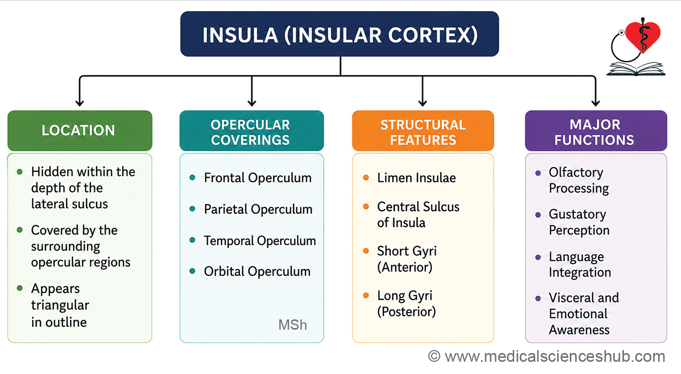
- The insula is a pyramidal, triangular elevation concealed deep within the floor of the lateral sulcus (Sylvian fissure); it is therefore considered the hidden or submerged portion of the cerebral cortex.
- It can be visualized by retracting the margins of the posterior ramus of the lateral sulcus to expose its surface.
- The inferior apex of the insula is termed the limen insulae, which is continuous with the anterior perforated substance.
Boundaries
- The insula is demarcated from the surrounding cortex on all sides by the circular sulcus (sulcus circularis), except at the limen insulae inferiorly.
- The regions of the cerebral hemisphere that overhang and conceal the insula are collectively termed the opercula (singular: operculum, meaning “lid”), which include:
- Frontal operculum
- Orbital operculum
- Frontoparietal operculum
- Temporal operculum
Structure
- The insula is divided into anterior and posterior regions by its own central sulcus (central insular sulcus):
- The anterior region bears three to four short convolutions known as the gyri breves (short gyri).
- The posterior region bears one to two longer convolutions known as the gyri longi (long gyri).
Functions
- The insula is involved in a broad range of higher and regulatory functions, including:
- Taste perception and visceral sensation
- Interoception and bodily homeostasis
- Autonomic regulation of cardiovascular and digestive activity
- Emotional awareness and empathy
- Self-awareness and integration of sensory and affective experiences
- Pain processing and modulation
Clinical Significance
- The insula plays a central role in interoception (conscious awareness of internal bodily states), emotional processing, taste perception, and regulation of autonomic functions such as heart rate and digestion.
- Lesions of the insula may result in loss of taste (ageusia), impaired emotional processing, altered pain perception, and dysregulation of autonomic responses.
Sulci and Gyri of Cerebral Hemisphere
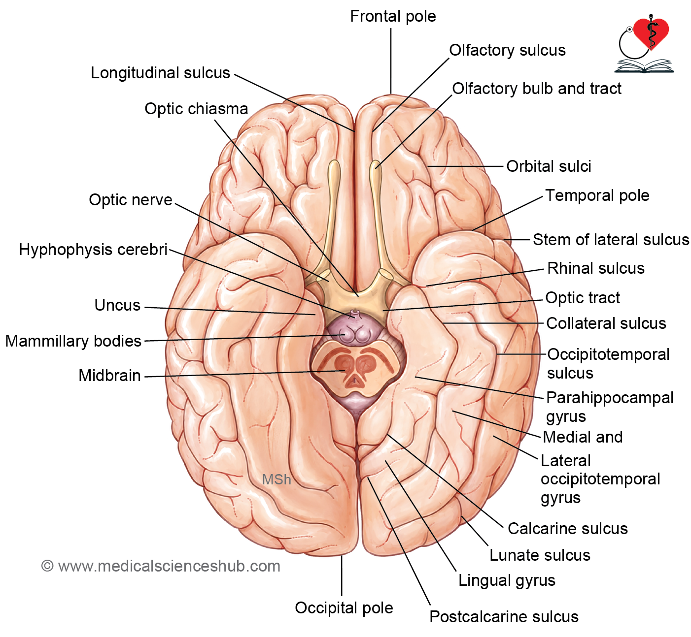
- The superolateral surface is divided into frontal, parietal, occipital, and temporal lobes by central sulcus, posterior ramus of the lateral sulcus, horizontal line extending from horizontal part of posterior ramus of lateral sulcus and a vertical line extending from parieto-occipital sulcus to the preoccipital notch (Fig. 14.5).
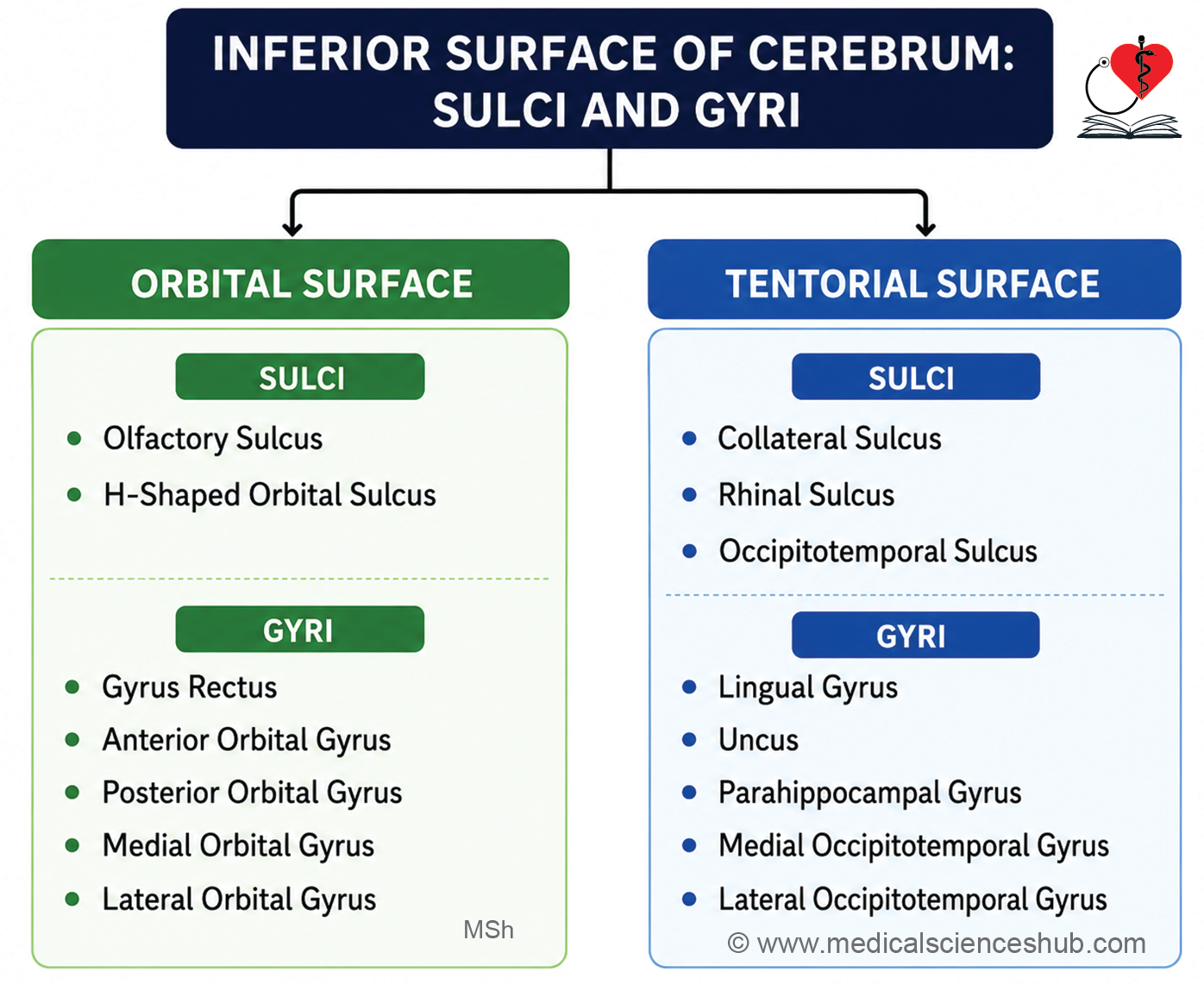
- Other than central, lateral, and parieto-occipital sulci, the superolateral, medial and inferior surfaces show other sulci and gyri (Table 14.1 to 14.3, Flowcharts 14.3 to 14.5).
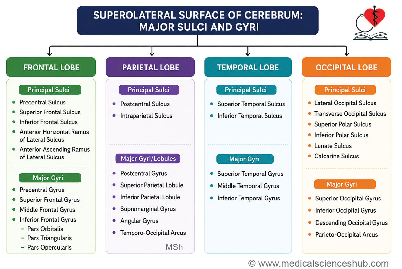
Table 12.1: Sulci and gyri on the superolateral surface of cerebral hemisphere
| Cerebral Lobe | Important Sulci | Major Gyri and Lobular Components |
|---|---|---|
| Frontal Lobe | Precentral sulcus, superior frontal sulcus, inferior frontal sulcus, anterior horizontal ramus of the lateral sulcus, anterior ascending ramus of the lateral sulcus | Precentral gyrus, superior frontal gyrus, middle frontal gyrus, inferior frontal gyrus, pars orbitalis, pars triangularis, pars opercularis |
| Parietal Lobe | Postcentral sulcus, intraparietal sulcus | Postcentral gyrus, superior parietal lobule, inferior parietal lobule, supramarginal gyrus, angular gyrus, arcus temporo-occipitalis |
| Temporal Lobe | Superior temporal sulcus, inferior temporal sulcus | Superior temporal gyrus, middle temporal gyrus, inferior temporal gyrus |
| Occipital Lobe | Transverse occipital sulcus, lateral occipital sulcus, lunate sulcus, superior and inferior polar sulci, calcarine sulcus | Arcus parieto-occipitalis, superior occipital gyrus, inferior occipital gyrus, gyrus descendens |
Table 12.2: Sulci and gyri on the medial surface
| Sulci on the Medial Surface | Related Gyri/Lobular Areas |
|---|---|
| Anterior paraolfactory sulcus | Paraterminal gyrus |
| Posterior paraolfactory sulcus | Paraolfactory gyrus |
| Cingulate sulcus | Medial frontal gyrus |
| Callosal sulcus | Paracentral lobule |
| Suprasplenial sulcus | Cingulate gyrus |
| Parieto-occipital sulcus | Cuneus |
| Calcarine sulcus | Precuneus |
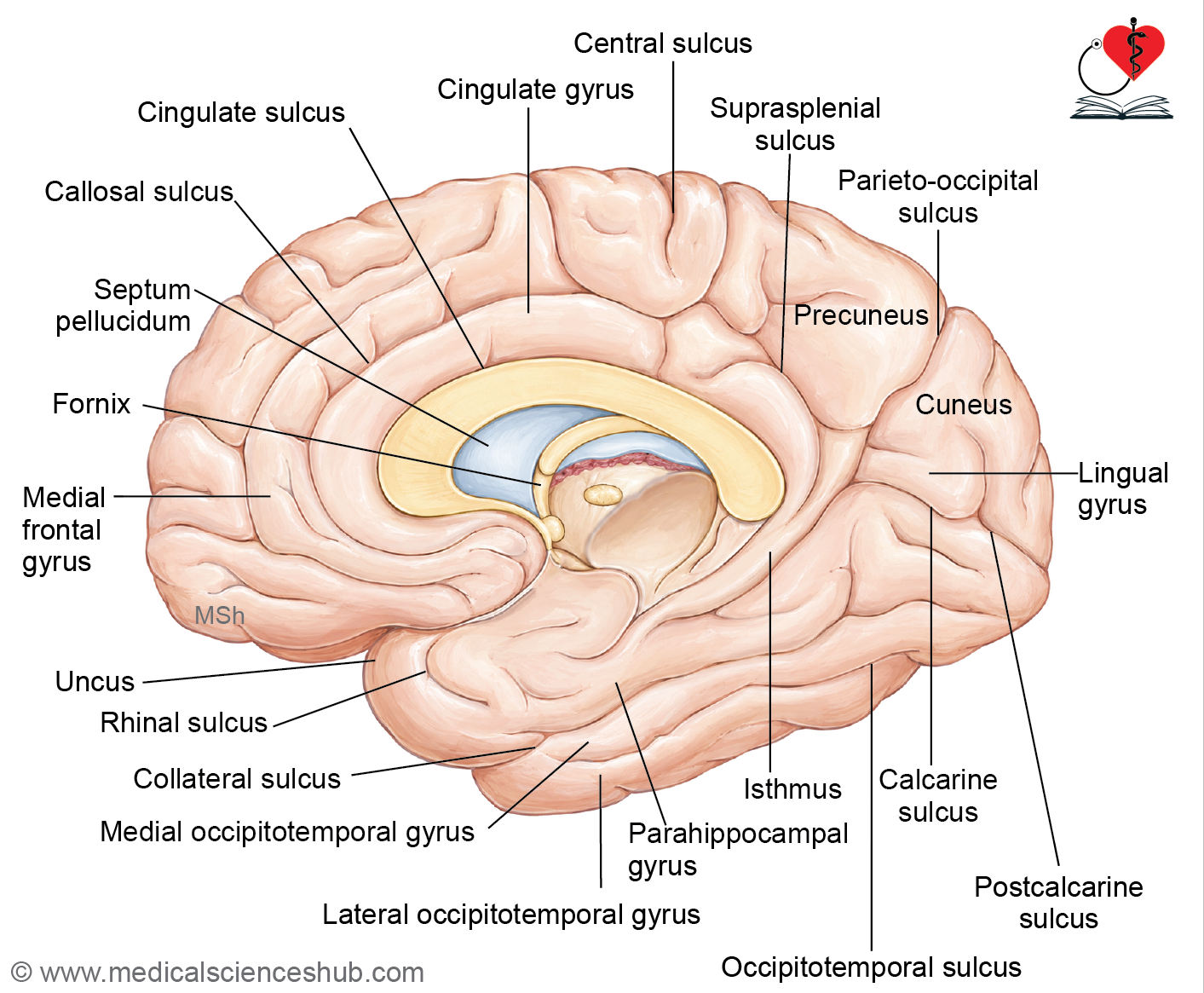
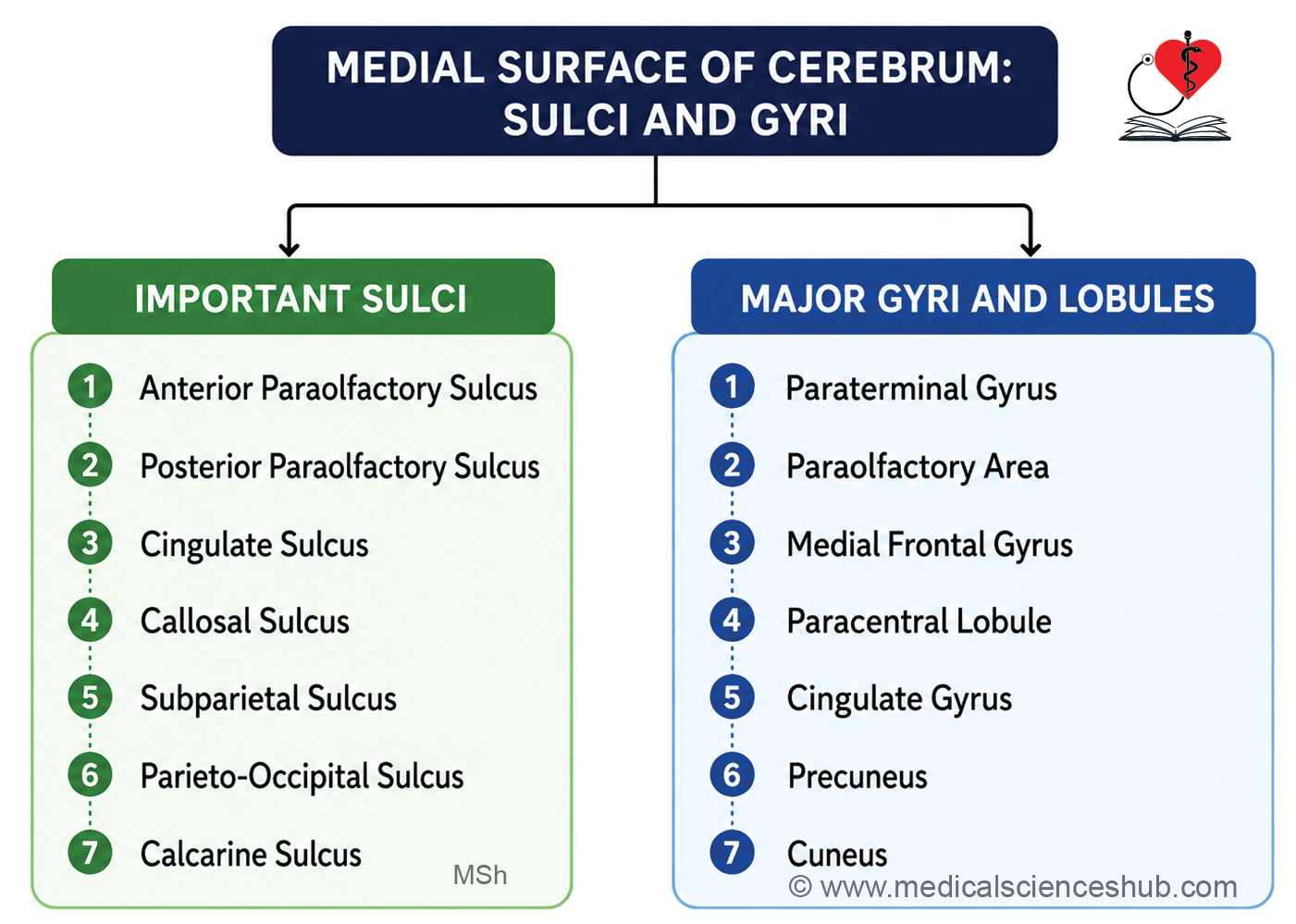
Table 12.3: Sulci and gyri on the inferior surface of cerebral hemisphere
| Region of Inferior Surface | Important Sulci | Related Gyri / Cortical Areas |
|---|---|---|
| Orbital Surface of Frontal Lobe | Olfactory sulcus, H-shaped orbital sulci | Gyrus rectus, anterior orbital gyrus, posterior orbital gyrus, medial orbital gyrus, lateral orbital gyrus |
| Tentorial Surface of Temporal and Occipital Lobes | Collateral sulcus, rhinal sulcus, occipitotemporal sulcus | Lingual gyrus, uncus, parahippocampal gyrus, medial occipitotemporal gyrus, lateral occipitotemporal gyrus |
Types of Cerebral Cortex
Based on phylogenetic (evolutionary) development, the cerebral cortex is classified into three types:
1. Archicortex (Archipallium)
- The archicortex is the most ancient division of the cerebral cortex in evolutionary terms.
- It comprises the hippocampus and associated parts of the rhinencephalon (the olfactory brain).
- Structurally, it is the simplest cortical type, consisting of only three neuronal layers.
- It is essential for memory formation, spatial navigation, and emotional processing.
- Lesions of the archicortex may result in memory impairment (amnesia), spatial disorientation, and emotional disturbances.
2. Paleocortex (Paleopallium)
- The paleocortex represents an evolutionarily intermediate stage of cortical development, more recently evolved than the archicortex but older than the neocortex.
- It includes the cingulate gyrus and regions associated with olfactory processing.
- It is involved in olfaction, emotional regulation, and the integration of sensory input with behavioral responses.
- Lesions may produce altered emotional responses, impaired sense of smell (anosmia), or behavioral changes.
3. Neocortex (Neopallium)
- The neocortex is the most recently evolved and by far the largest cortical division, constituting the majority of the human cerebral cortex.
- It is structurally the most complex, organized into six distinct neuronal layers.
- It is responsible for higher cognitive functions, sensory perception, and voluntary motor control.
- The neocortex is further subdivided into two functional types:
- Agranular cortex (motor cortex) — Characterized by a poorly developed granular layer (layer IV) and a prominent layer V; primarily involved in the initiation and control of voluntary movement.
- Granular cortex (sensory cortex) — Characterized by well-developed granular layers (layers II and IV); primarily involved in the processing of sensory information.
- Lesions of the neocortex produce a wide range of deficits depending on the region affected, including sensory loss, motor weakness, or cognitive impairment.
Histology of Cerebral Hemisphere
- The cerebrum is organized into an outer layer of grey matter, known as the cerebral cortex, and an underlying core of white matter.
- The cerebral cortex contains a dense population of neurons, synaptic connections, and neuronal processes.
Neurons of Cerebral Cortex
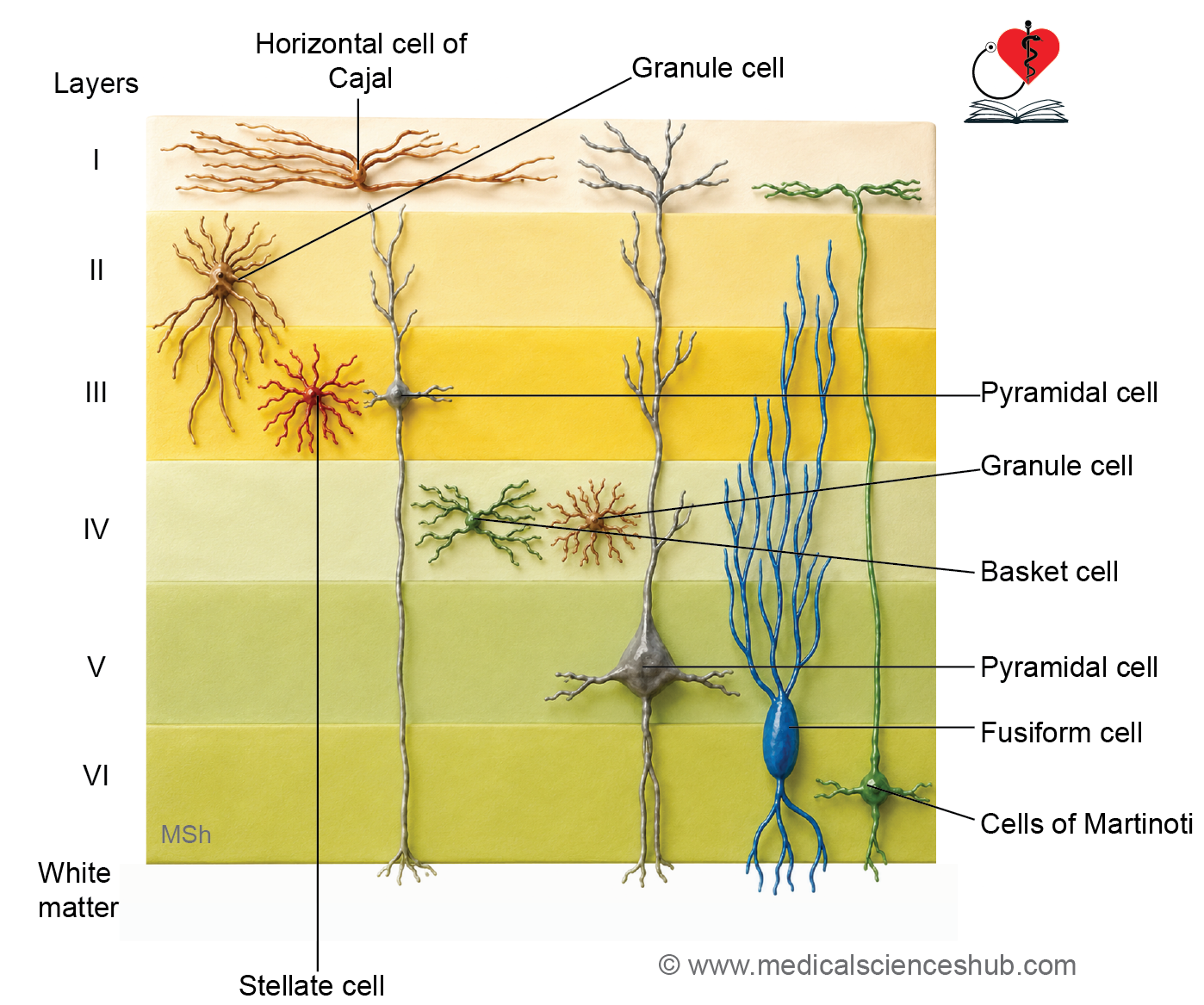
The cerebral cortex contains several morphologically and functionally distinct neuronal types:
Pyramidal Cells
- Pyramidal cells are the predominant neuron of the cerebral cortex, comprising approximately two-thirds of all cortical neurons.
- They are multipolar neurons with a characteristic triangular cell body, with the apex directed toward the cortical surface.
- Their processes include:
- Apical dendrites — arising from the apex, extending toward the cortical surface.
- Basal dendrites — arising from the base of the cell body.
- A single axon — arising from the base, projecting to other cortical or subcortical regions.
- They serve as the principal excitatory projection neurons of the cortex, transmitting signals to other brain regions and the spinal cord; they coordinate voluntary movement and higher cognitive functions.
- Lesions affecting pyramidal cells may produce motor deficits and cognitive dysfunction.
Stellate Cells (Granule Cells)
- Stellate neurons are small, star-shaped multipolar interneurons whose appearance resembles granules, hence the alternative name granular neurons.
- They function primarily as excitatory or inhibitory interneurons, processing incoming sensory signals and relaying information to pyramidal cells.
Cells of Martinotti
- These are small, triangular or polygonal neurons found predominantly in the external pyramidal layer of the cortex.
- Their axons project toward the cortical surface, where they regulate and inhibit pyramidal neuron activity, thereby controlling cortical excitability and preventing excessive neuronal firing.
- Dysfunction of Martinotti cells may contribute to cortical hyperexcitability and epilepsy.
Horizontal Cells of Cajal
- These are fusiform neurons oriented horizontally within the most superficial cortical layers.
- They are involved in regulating lateral communication between pyramidal neurons and contribute to cortical signal refinement.
Fusiform Cells
- Fusiform cells are spindle-shaped neurons with their long axes oriented perpendicular to the cortical surface.
- They are located in the deeper cortical layers and serve to connect the cortex with subcortical structures, facilitating signal transmission between cortical and deeper brain regions.
Layer of Cerebral Cortex
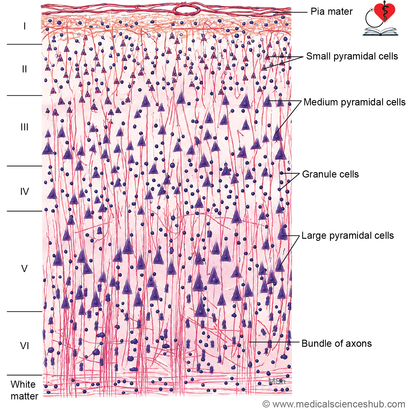
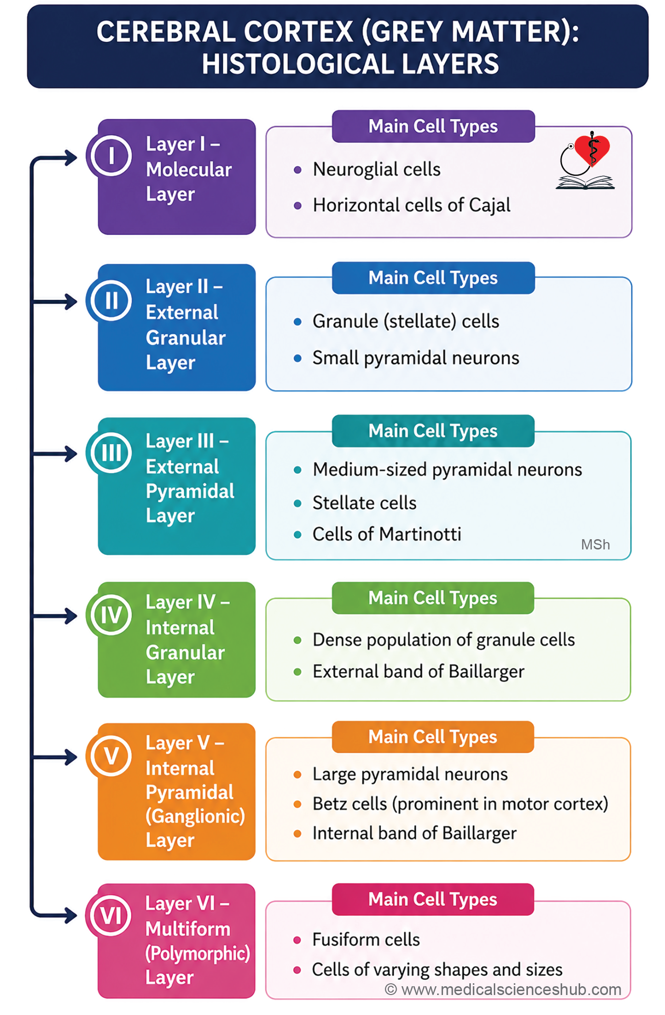
The neurons of the cerebral cortex are organized into six distinct layers, numbered from the surface inward:
Layer I — Molecular (Plexiform) Layer
- Situated immediately beneath the pia mater, this is the most superficial layer.
- It contains predominantly neuroglial cells, a sparse population of horizontal cells of Cajal, and a few capillaries.
- Neuroglial nuclei are identifiable histologically, but their cytoplasm is indistinguishable from the surrounding nerve fibers.
Layer II — External Granular Layer
- Also referred to as the small pyramidal cell layer, this layer contains two neuronal populations: small pyramidal cells and stellate (granule) cells.
Layer III — External Pyramidal Layer
- Also called the medium pyramidal cell layer, it contains predominantly medium-sized pyramidal cells, along with a few stellate cells and cells of Martinotti.
- Axons of the pyramidal cells in this layer contribute to association fibers (connecting ipsilateral cortical areas) and commissural fibers (connecting the two hemispheres).
Layer IV — Internal Granular Layer
- Composed of densely packed stellate (granule) cells, this layer also contains a prominent band of horizontally arranged myelinated fibers known as the external band of Baillarger.
Layer V — Internal Pyramidal (Ganglionic) Layer
- Contains large pyramidal cells; in the primary motor cortex, these are exceptionally large neurons known as Betz cells (first described by Ukrainian anatomist Vladimir Betz, 1834–1894), which give rise to the corticospinal (pyramidal) tract.
- A second band of horizontal myelinated fibers in this layer forms the internal band of Baillarger.
- Additional cell types include cells of Martinotti, fusiform cells, and scattered granule cells.
Layer VI — Multiform (Polymorphic) Layer
- The deepest cortical layer, containing predominantly fusiform cells oriented perpendicular to the cortical surface, along with scattered stellate cells and cells of Martinotti.
- Fusiform cells in this layer have dendrites arising from both poles and axons projecting into the underlying white matter.
White Matter
- Deep to the sixth cortical layer lies the white matter of the cerebral hemisphere.
- It consists of myelinated nerve fibers (axonal processes) and the small, round nuclei of neuroglial cells, which provide structural and metabolic support.
Functional Area of Cerebral Cortex
The cerebral cortex is organized into distinct regions based on their specialized functions, broadly classified into three categories:
- Motor Areas: Motor areas are responsible for initiating and controlling voluntary motor activity. They give rise to the corticospinal tract (projecting to spinal cord motor neurons) and the corticonuclear (corticobulbar) tract (projecting to cranial nerve nuclei in the brainstem).
- Sensory Areas: Sensory areas receive and process afferent (incoming) sensory information relayed via the thalamus and associated subthalamic nuclei. Different sensory areas are dedicated to specific modalities, including somatic sensation, vision, hearing, and taste.
- Association Areas: Association areas integrate and interpret information from both motor and sensory regions. They are responsible for higher-order functions including modification and refinement of motor activity, interpretation of sensory inputs, intellectual processing, emotional regulation, and behavioral responses.
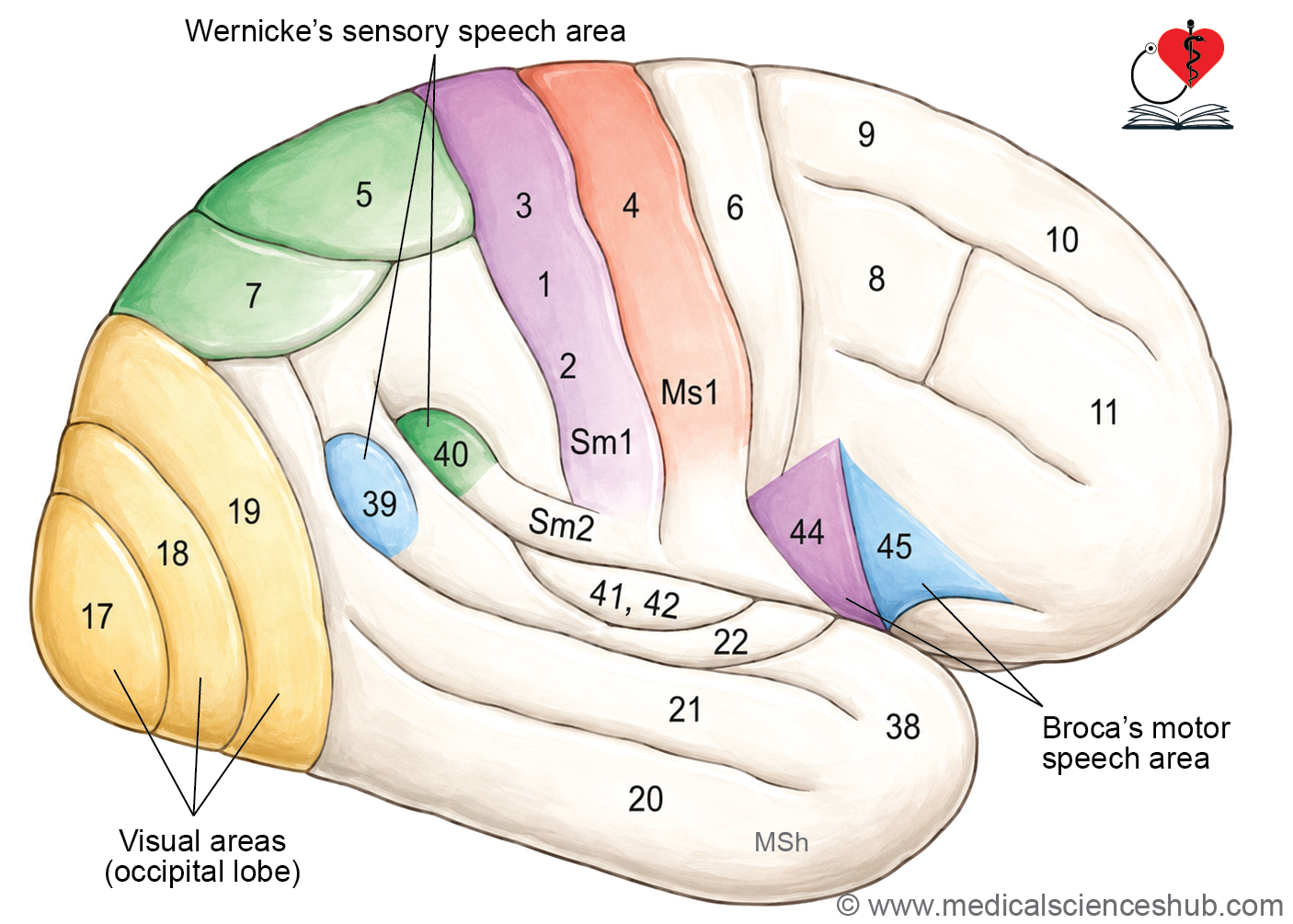
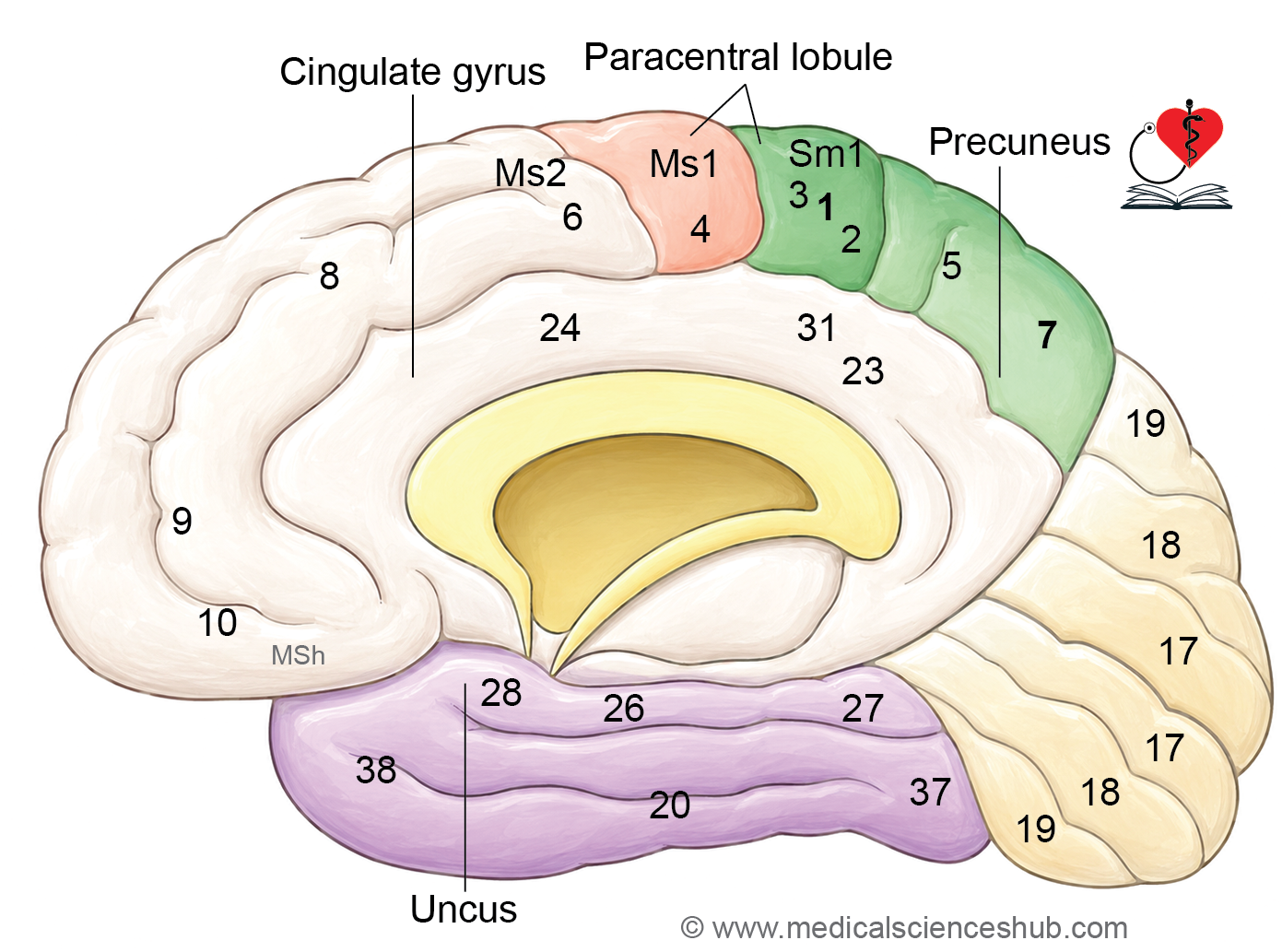
Brodmann Areas: The German neurologist Korbinian Brodmann (1868–1918) systematically mapped and numbered distinct cytoarchitectural regions of the cerebral cortex based on their microscopic neuronal organization. These numbered regions are collectively referred to as Brodmann areas and continue to serve as a standard reference framework for identifying and describing functional cortical territories.
Motor Areas
Primary Motor Area (area 4, Ms I)
Location
- Situated in the precentral gyrus and the anterior wall of the central sulcus on the superolateral surface, extending onto the anterior part of the paracentral lobule on the medial surface.
Function and Somatotopic Organization
- Controls voluntary motor activity of the contralateral (opposite) half of the body.
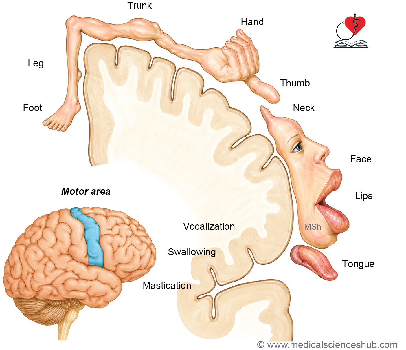
- The body is represented in an inverted (upside-down) topographic map known as the motor homunculus, organized from inferior to superior as follows:
- Pharynx and tongue → face → hand → arm → trunk → thigh (on the superolateral surface)
- Leg, foot, and perineum (on the medial surface within the paracentral lobule)
- The face representation is not inverted and remains in its expected inferior position.
- The size of each cortical representation reflects the degree of motor precision required, not the physical size of the body part; the hand and face therefore occupy disproportionately large areas.
Fiber Output
- Gives rise to corticospinal fibers (to spinal cord motor neurons), corticonuclear (corticobulbar) fibers (to cranial nerve nuclei), and frontopontine fibers (to the pons).
Clinical Significance
- A lesion of the primary motor cortex produces contralateral upper motor neuron (UMN) paralysis, clinically manifesting as hemiplegia (paralysis of the opposite half of the body).
Premotor Area (area 6 of Brodmann)
Location
- Located immediately anterior to the primary motor cortex, occupying the posterior portions of the superior, middle, and inferior frontal gyri on the lateral surface of the frontal lobe.
Functions
- Serves as the principal cortical component of the extrapyramidal system.
- Responsible for planning, programming, and coordinating complex voluntary movements, particularly those guided by sensory or visual cues.
- Maintains extensive connections with the primary motor cortex, basal ganglia, and cerebellum.
Clinical Significance
- Lesions of the premotor cortex impair the execution of skilled and sequential movements (motor apraxia) without causing frank muscle weakness or paralysis.
Supplementary Motor area (Ms II)
Location
- Situated on the medial surface of the hemisphere in the medial frontal gyrus, anterior to the paracentral lobule.
Functions
- Involved in the planning, initiation, and internal coordination of complex and bilateral movements, particularly those executed from memory without external sensory cues.
- Controls movements requiring sequential organization across both sides of the body.
Clinical Significance
- Lesions may result in difficulty initiating voluntary movements, impaired bilateral coordination, or transient speech arrest.
Motor Speech Area (area 44, 45)
Location
- Located in the inferior frontal gyrus of the dominant cerebral hemisphere (typically the left hemisphere), comprising:
- Pars opercularis — Brodmann area 44
- Pars triangularis — Brodmann area 45
- First described by French neurologist Pierre Paul Broca (1824–1880).
- In right-handed individuals (approximately 70% of the population), Broca’s area resides in the left hemisphere; in left-handed individuals (approximately 30%), it is typically located in the right hemisphere.
Functions
- Initiates and coordinates the motor programs required for articulate speech production, including coordinated movements of the lips, tongue, and larynx.
- Governs expressive language, including grammatical construction and speech fluency.
Clinical Significance
- A lesion of Broca’s area produces Broca’s (expressive) aphasia, characterized by slow, effortful, non-fluent speech with relatively preserved language comprehension.
- Affected individuals retain the ability to write words but cannot articulate them verbally.
- Importantly, there is no paralysis of the tongue, lips, or vocal cords, as the primary motor representations of these structures remain intact.
Frontal Eye Field (area 8)
Location
- Located in the posterior part of the middle frontal gyrus, immediately anterior to the facial representation area of the precentral gyrus.
Functions
- Controls voluntary conjugate eye movements directed toward the contralateral side.
- Involved in visual attention, voluntary gaze shifts, and tracking of moving objects.
Clinical Significance
- A unilateral lesion results in loss of voluntary horizontal conjugate gaze toward the opposite side; both eyes deviate toward the side of the lesion and cannot be voluntarily directed to the contralateral side.
Prefrontal Area (area 9, 10, 11, 12)
Location
- Comprises the portion of the frontal lobe located anterior to both the primary motor and premotor cortices.
Functions
- Mediates higher executive functions, including judgment, planning, decision-making, and goal-directed behavior.
- Regulates emotional responses, social and moral awareness, concentration, prospective thinking, and personality expression.
Clinical Significance
- Bilateral lesions of the prefrontal cortex produce marked personality changes, impaired judgment, reduced initiative, loss of concentration and orientation, and socially inappropriate behavior.
- These features are characteristic of frontal lobe injuries and certain neurodegenerative conditions such as frontotemporal dementia.
Table 12.4: Motor functional areas of cerebral cortex
| Functional Motor Area | Brodmann Areas | Anatomical Location | Major Functions | Clinical Effects of Lesions |
|---|---|---|---|---|
| Primary Motor Cortex (M1) | Area 4 | Located in the precentral gyrus, anterior wall of the central sulcus, and anterior part of the paracentral lobule | Responsible for execution of voluntary movements of the contralateral side of the body. Contains large pyramidal neurons contributing to the corticospinal tract. | Lesions produce upper motor neuron paralysis affecting the opposite half of the body, commonly presenting as contralateral hemiplegia. |
| Premotor Cortex | Area 6 | Situated anterior to the primary motor cortex within the posterior parts of the superior, middle, and inferior frontal gyri | Involved in planning, programming, and coordination of skilled voluntary movements. Functions as an important cortical component of the extrapyramidal system. | Damage results in impairment of learned skilled movements, often producing motor apraxia. |
| Supplementary Motor Area (M2) | Part of Area 6 | Medial surface of the superior frontal gyrus | Coordinates complex bilateral movements and sequential motor activities requiring planning and synchronization. | Lesions may cause difficulty initiating movements and impaired coordination of complex bilateral motor tasks. |
| Motor Speech Area (Broca Area) | Areas 44 and 45 | Located in the dominant hemisphere, usually the left inferior frontal gyrus. Area 45 occupies the pars triangularis, while Area 44 occupies the pars opercularis. | Controls motor programming necessary for speech production and expressive language. | Lesions produce motor aphasia (Broca aphasia) characterized by impaired speech production with relatively preserved comprehension. |
| Frontal Eye Field | Area 8 | Posterior part of the middle frontal gyrus | Controls voluntary conjugate eye movements and visual tracking of moving objects. | Damage causes loss of voluntary horizontal conjugate eye movements, with deviation of the eyes toward the side of the lesion. |
| Prefrontal Cortex | Areas 9, 10, 11, and 12 | Anterior part of the frontal lobe | Responsible for higher executive functions including judgment, planning, concentration, emotional regulation, social behavior, personality, and foresight. | Bilateral lesions may lead to impaired judgment, reduced concentration, loss of initiative, personality changes, emotional instability, and socially inappropriate behavior. |
Sensory and Association Areas
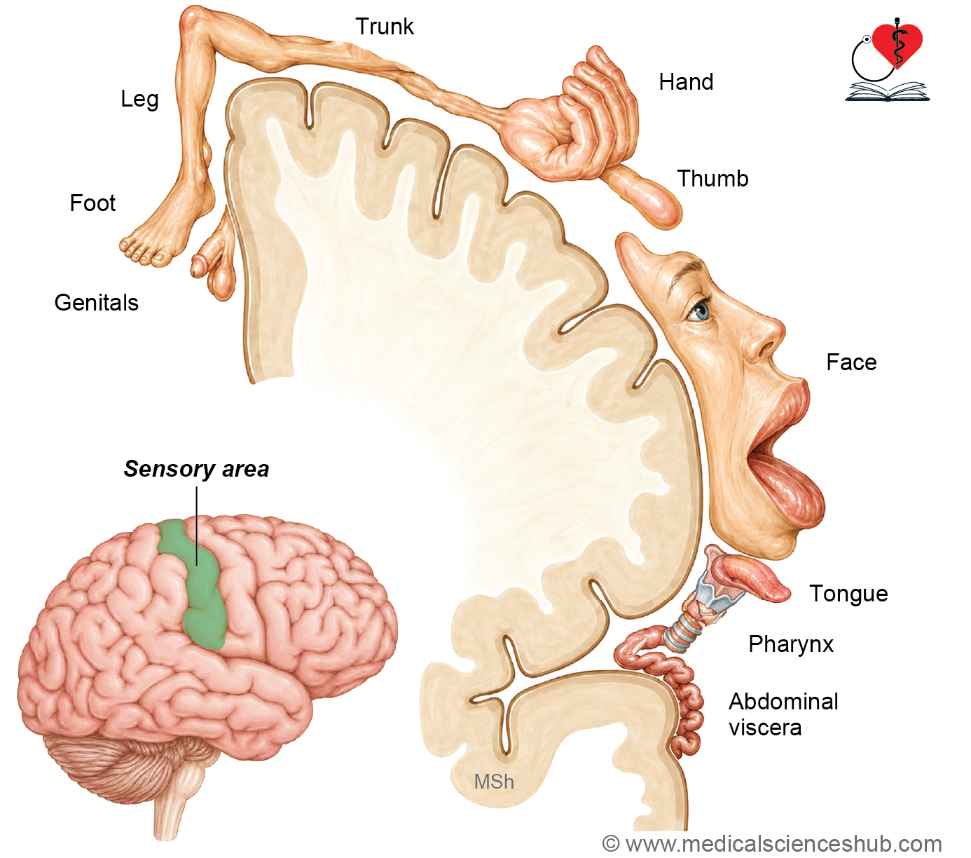
Primary Somatosensory Cortex — Sm I (Brodmann Areas 3, 1, and 2)
Location
- Situated in the postcentral gyrus on the superolateral surface, extending onto the posterior part of the paracentral lobule on the medial surface.
Somatotopic Organization
- Receives sensory input from the contralateral half of the body, represented in an inverted topographic map known as the sensory homunculus.
- Body regions requiring fine sensory discrimination — such as the tongue, lips, fingers, and thumb — occupy disproportionately large cortical areas.
Functions
- Processes both exteroceptive sensations (pain, touch, and temperature) and proprioceptive sensations (vibration, muscle tension, and joint position) from the opposite side of the body.
Clinical Significance
- Lesions result in contralateral loss or impairment of exteroceptive and proprioceptive sensations, along with reduced ability to localize stimuli and impaired tactile discrimination.
Secondary Somatosensory Cortex — Sm II
Location
- Located along the superior lip of the posterior ramus of the lateral sulcus, adjacent to the postcentral gyrus.
Functions
- Integrates and interprets complex sensory information, including bilateral touch and pain perception, contributing to overall body awareness.
- Uniquely, this area receives sensory representation from both sides of the body (bilateral representation).
Clinical Significance
- Lesions impair tactile recognition, sensory integration, and the ability to perceive stimuli from either side of the body.
Somatosensory Association Cortex (Brodmann Areas 5 and 7)
Location
- Occupies the superior parietal lobule, posterior to the primary somatosensory cortex.
Functions
- Integrates multimodal sensory information to enable perception of an object’s shape, size, texture, and roughness.
- Underlies the ability to recognize objects through touch alone, a capacity known as stereognosis.
Clinical Significance
- Lesions produce astereognosis — the inability to identify objects by touch with the eyes closed — as well as sensory neglect and impaired spatial awareness.
Wernicke’s Area — Sensory Speech Area (Brodmann Areas 22, 39, and 40)
Location
- Present exclusively in the dominant cerebral hemisphere (typically the left), comprising:
- Posterior superior temporal gyrus — area 22
- Angular gyrus of the inferior parietal lobule — area 39
- Supramarginal gyrus of the parietal lobe — area 40
Connectivity
- Wernicke’s area is connected to Broca’s area (motor speech area) via a white matter tract known as the arcuate fasciculus, forming the structural basis of the language network.
Functions
- Responsible for the comprehension and interpretation of language through both auditory and visual inputs, including the understanding of spoken and written words.
Clinical Significance
- Lesions produce Wernicke’s (receptive) aphasia, characterized by fluent but meaningless speech, severely impaired language comprehension, and difficulty understanding both spoken and written language.
Primary Auditory Cortex (Brodmann Areas 41 and 42)
Location
- Located on the superior (hidden) surface of the superior temporal gyrus, within the floor of the lateral sulcus; this region is anatomically referred to as the transverse temporal gyri of Heschl.
Functions
- Receives auditory input primarily from the contralateral ear via the auditory radiation projecting from the medial geniculate nucleus of the thalamus (with some ipsilateral input).
- Processes fundamental sound properties including pitch, loudness, and sound quality.
Clinical Significance
- Unilateral lesions cause only mild hearing impairment due to the bilateral projection of the auditory pathway; complete deafness requires bilateral cortical damage.
Secondary Auditory Cortex — Auditory Association Area (Brodmann Area 22)
Location
- Located on the lateral surface of the superior temporal gyrus, adjacent to and surrounding the primary auditory cortex.
Functions
- Correlates incoming auditory signals with past auditory experiences, enabling recognition and interpretation of complex sounds such as speech, music, and environmental noises.
Clinical Significance
- Lesions result in auditory agnosia (also termed word deafness or auditory verbal agnosia) — the inability to recognize or interpret heard sounds despite intact basic hearing.
- In the dominant hemisphere, area 22 forms an integral part of Wernicke’s area.
Primary Visual Cortex — Striate Cortex (Brodmann Area 17)
Location
- Located on both sides of the calcarine sulcus on the medial surface of the occipital lobe, extending slightly onto the superolateral surface around the occipital pole.
Structural Feature
- Contains a prominent band of myelinated fibers within layer IV known as the visual stria of Gennari (external band of Baillarger), which imparts a striped appearance to this cortex — hence the term striate cortex.
Functions
- Receives visual input from the temporal half of the ipsilateral retina and the nasal half of the contralateral retina, relayed via the lateral geniculate nucleus of the thalamus.
- Processes fundamental visual attributes including color, form, size, illumination, and transparency.
Clinical Significance
- Lesions produce contralateral homonymous hemianopia (loss of vision in the opposite visual field), characteristically with macular sparing due to the dual blood supply of the macular representation area.
Secondary Visual Cortex — Visual Association Area (Brodmann Areas 18 and 19)
Location
- Situated in the occipital lobe, surrounding the primary visual cortex (area 17).
Functions
- Processes complex visual information including object recognition, color discrimination, motion perception, and spatial localization, drawing on past visual experience.
Clinical Significance
- Lesions result in visual agnosia — the inability to recognize objects in the contralateral visual field despite intact basic vision.
Primary Gustatory Cortex — Taste Area (Brodmann Area 43)
Location
- Located in the inferior part of the postcentral gyrus at the junction of the parietal and insular cortices.
Functions
- Responsible for conscious perception and discrimination of taste modalities.
Clinical Significance
- Lesions may cause ageusia (loss of taste) or dysgeusia (distorted taste perception), impairing the ability to distinguish flavors.
Vestibular Cortex
Location
- Located in the posterior insula and the adjacent parietal operculum, receiving input from the vestibular nuclei of the brainstem.
Functions
- Processes information related to balance, spatial orientation, and head position, contributing to postural control and movement coordination.
Clinical Significance
- Lesions may produce vertigo, postural imbalance, nausea, and impaired movement coordination.
Primary Olfactory Cortex (Brodmann Area 28)
Location
- Situated in the uncus and the anterior parahippocampal gyrus of the temporal lobe, receiving direct input from the olfactory bulb via the olfactory tract.
Functions
- Mediates conscious perception and identification of odors, and links olfactory stimuli with memory and emotional responses.
Clinical Significance
- Lesions may cause anosmia (loss of smell) or dysosmia (distorted smell perception), affecting appetite, emotional associations, and the ability to detect environmental hazards such as smoke or gas.
Table 12.5: Sensory functional areas of cerebral cortex
| Sensory Functional Area | Brodmann Areas | Anatomical Location | Principal Functions | Effects of Lesions |
|---|---|---|---|---|
| Primary Somatosensory Cortex (SI) | Areas 3, 1, and 2 | Postcentral gyrus and posterior part of the paracentral lobule | Receives and interprets general sensory impulses from the opposite side of the body, including touch, pain, temperature, vibration, and proprioception | Loss or marked impairment of contralateral exteroceptive and proprioceptive sensations |
| Secondary Somatosensory Cortex (SII) | Part of sensory cortex adjacent to Area 43 | Superior lip of the posterior ramus of the lateral sulcus extending toward the postcentral gyrus | Integration and higher-order processing of somatic sensory information | Lesions may impair sensory discrimination and cortical interpretation of somatic sensations |
| Somatosensory Association Cortex | Areas 5 and 7 | Superior parietal lobule | Integrates sensory input for recognition of shape, size, texture, and spatial relationships; responsible for stereognosis | Damage results in astereognosis, where a familiar object cannot be identified by touch despite intact primary sensation |
| Sensory Speech Area (Wernicke Area) | Areas 22, 39, and 40 | Dominant cerebral hemisphere: posterior part of the superior temporal gyrus (Area 22), angular gyrus (Area 39), and supramarginal gyrus (Area 40) | Interpretation and comprehension of spoken and written language through auditory and visual inputs | Lesions cause sensory aphasia (Wernicke aphasia) characterized by impaired comprehension with fluent but often meaningless speech |
| Primary Auditory Cortex (A1) | Areas 41 and 42 | Transverse temporal gyri of Heschl within the superior temporal lobe | Perception of sound intensity, pitch, and quality | Unilateral lesions usually produce only mild hearing impairment because auditory pathways are bilaterally represented |
| Secondary Auditory Cortex | Area 22 | Lateral surface of the superior temporal gyrus | Interpretation and correlation of auditory stimuli, including spoken words | Lesions may produce word deafness, in which spoken language cannot be understood despite preserved hearing |
| Primary Visual Cortex | Area 17 | Medial surface of the occipital lobe along both banks of the calcarine sulcus | Receives visual impulses and mediates perception of form, color, size, brightness, and visual contrast | Lesions result in contralateral homonymous hemianopia |
| Visual Association Cortex | Areas 18 and 19 | Occipital cortex surrounding the primary visual area | Interpretation and recognition of visual information, including object identification and spatial analysis | Damage causes visual agnosia, in which visual perception is preserved but objects cannot be recognized |
| Gustatory Cortex | Area 43 | Inferior part of the parietal operculum near the insular region | Perception and interpretation of taste sensations | Lesions may impair taste perception (ageusia) |
| Vestibular Cortical Area | Not sharply localized | Region near the inferior part of the postcentral gyrus and adjacent insular cortex | Awareness of balance, head position, and vestibular sensations | Lesions may produce vertigo and disturbances of equilibrium |
| Primary Olfactory Cortex | Area 28 | Anterior part of the parahippocampal gyrus and uncus | Perception and processing of olfactory stimuli | Lesions may cause partial or complete loss of smell (anosmia) |
Cerebral Dominance
- The two cerebral hemispheres, while anatomically similar, are not functionally equivalent; each hemisphere is specialized for distinct cognitive and motor functions.
- In approximately 90% of individuals, the left cerebral hemisphere is the dominant hemisphere, primarily governing language, speech, and analytical processing.
- In most right-handed individuals, the left hemisphere is dominant; conversely, the right hemisphere tends to be dominant in a proportion of left-handed individuals.
- The non-dominant hemisphere (typically the right) specializes in complementary higher functions distinct from language.
Functional Specialization
- The dominant hemisphere (typically left) is responsible for:
- Language comprehension and production (spoken and written)
- Analytical and logical reasoning
- Mathematical processing
- Sequential and detail-oriented tasks
- The non-dominant hemisphere (typically right) is responsible for:
- Visuospatial processing and spatial awareness
- Facial recognition and interpretation of non-verbal cues
- Creative thinking, musical appreciation, and artistic processing
- Holistic and intuitive reasoning
- Emotional tone of language (prosody)
Anatomical Basis
- Cerebral dominance reflects both structural and functional asymmetry between the hemispheres.
- Key anatomical correlates in the dominant hemisphere include:
- Broca’s area (inferior frontal gyrus, areas 44 and 45) — governing speech production and expressive language.
- Wernicke’s area (posterior superior temporal gyrus and adjacent parietal regions) — governing language comprehension.
- Corresponding regions in the non-dominant hemisphere support non-verbal communication, spatial cognition, and emotional processing.
Functional Significance
- Cerebral dominance enables an efficient division of labor between the two hemispheres, facilitating the smooth coordination of complex cognitive, motor, and sensory functions.
- It forms the neurological basis for language acquisition, handedness, and higher-order problem-solving.
Clinical Neuroanatomy
- Disruption of cerebral dominance or damage to dominant hemisphere structures can result in:
- Aphasia — impaired language production or comprehension.
- Dyslexia — difficulty with reading and language processing, often associated with atypical hemispheric lateralization.
- Apraxia — difficulty executing learned, purposeful motor tasks.
- An understanding of cerebral dominance is essential in neurosurgical planning, stroke rehabilitation, and predicting functional outcomes following brain injury, enabling clinicians to tailor therapeutic interventions appropriately.
Important Questions
- Draw a well-labeled diagram showing sulci and gyri of the superolateral surface of the cerebral hemisphere.
- Draw a well-labeled diagram showing functional areas of the superolateral surface of the cerebral hemisphere.
- Draw a well-labeled diagram showing sulci and gyri of the inferior surface of the cerebral hemisphere.
- Write a short note on Broca’s area and Wernicke’s.
📝 Test Your Knowledge – Practice MCQs
Attempt the chapter MCQ quiz and assess your understanding of key concepts.
Click to see: Medical Disclaimer and Copyright Notice