
Competency
- AN63.1: Describe and demonstrate parts of boundaries and features of IIIrd, IVth, and lateral ventricles.
INTRODUCTION
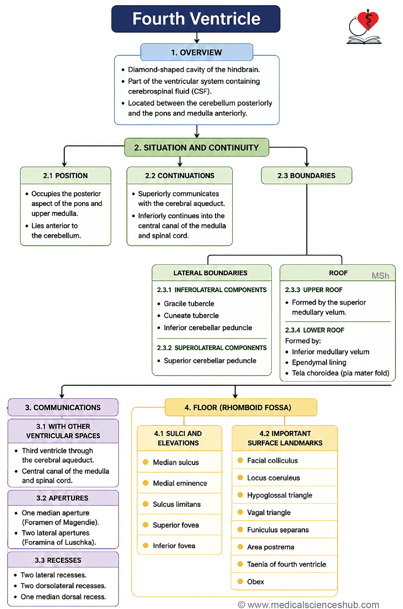
- The fourth ventricle is a tent-shaped cavity of the hindbrain, lined internally by ependymal cells.
- It contains cerebrospinal fluid (CSF) and the tela choroidea.
- The ventricle has openings that allow CSF to drain from the ventricular system into the subarachnoid space.
- Obstruction of these openings causes abnormal CSF accumulation within the ventricles, resulting in a condition known as internal hydrocephalus.
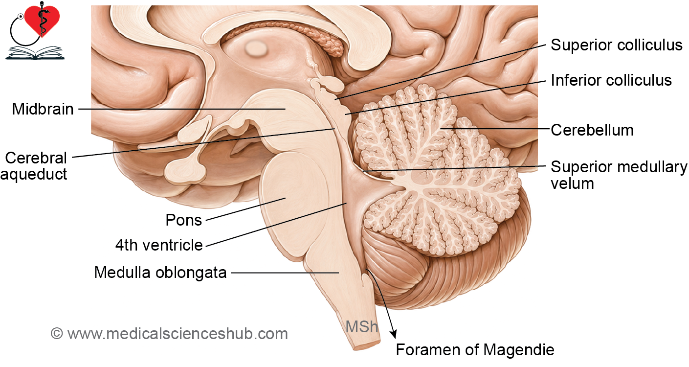
Location and Shape
- The fourth ventricle lies in the posterior cranial fossa, positioned between the cerebellum posteriorly and the pons and medulla oblongata anteriorly.
- Its cross-sectional shape is triangular, while its long axis has a rhomboid configuration.
Continuities
- Superiorly, it connects with the cerebral aqueduct of the midbrain.
- Inferiorly, it continues as the central canal of the closed part of the medulla oblongata.
Boundaries
The fourth ventricle has two lateral boundaries, one on each side, as follows:
- The inferolateral boundaries are formed by the gracile and cuneate tubercles and the inferior cerebellar peduncles.
- The superolateral boundaries are formed by the superior cerebellar peduncles.
Angles of 4th Ventricle
- The cavity has four angles: one superior, one inferior, and two lateral.
- At the superior angle, the ventricle opens into the cerebral aqueduct.
- At the inferior angle, it continues as the central canal of the medulla oblongata.
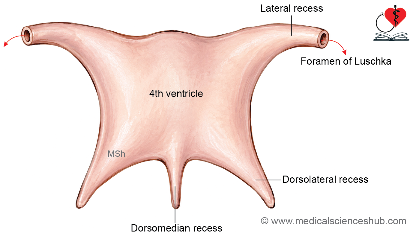
- At each lateral angle, the cavity extends as the lateral recess, which communicates with the pontine cistern via the foramen of Luschka.
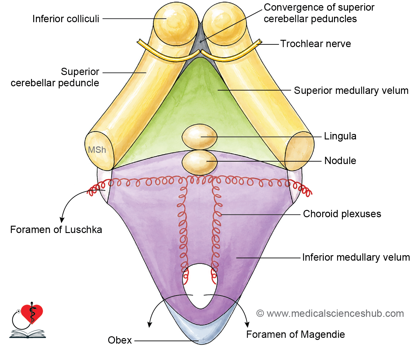
Roof of 4th Ventricle
- The roof (posterior wall) of the fourth ventricle is tent-shaped, consisting of an upper slope, a lower slope, and an apex.
Upper Part of the Roof
- It is formed by the superior cerebellar peduncles (one on each side) and the superior medullary velum, a thin triangular sheet of white matter that bridges the gap between the two peduncles.
- Inferiorly, the superior medullary velum is continuous with the vermis of the cerebellum.
Lower Part of the Roof
- It is formed by the inferior medullary velum, a thin, semilunar, non-neural sheet composed of two layers: an inner ependymal layer facing the ventricular cavity and an outer pial layer.
- Unlike the upper part, the inferior medullary velum contains no nervous tissue.
Foramen of Magendie
- The lower part of the inferior medullary velum contains a large midline opening, the foramen of Magendie (median aperture).
- Through this foramen, the fourth ventricle communicates directly with the cerebellomedullary cistern (cisterna magna), allowing CSF to drain into the subarachnoid space.
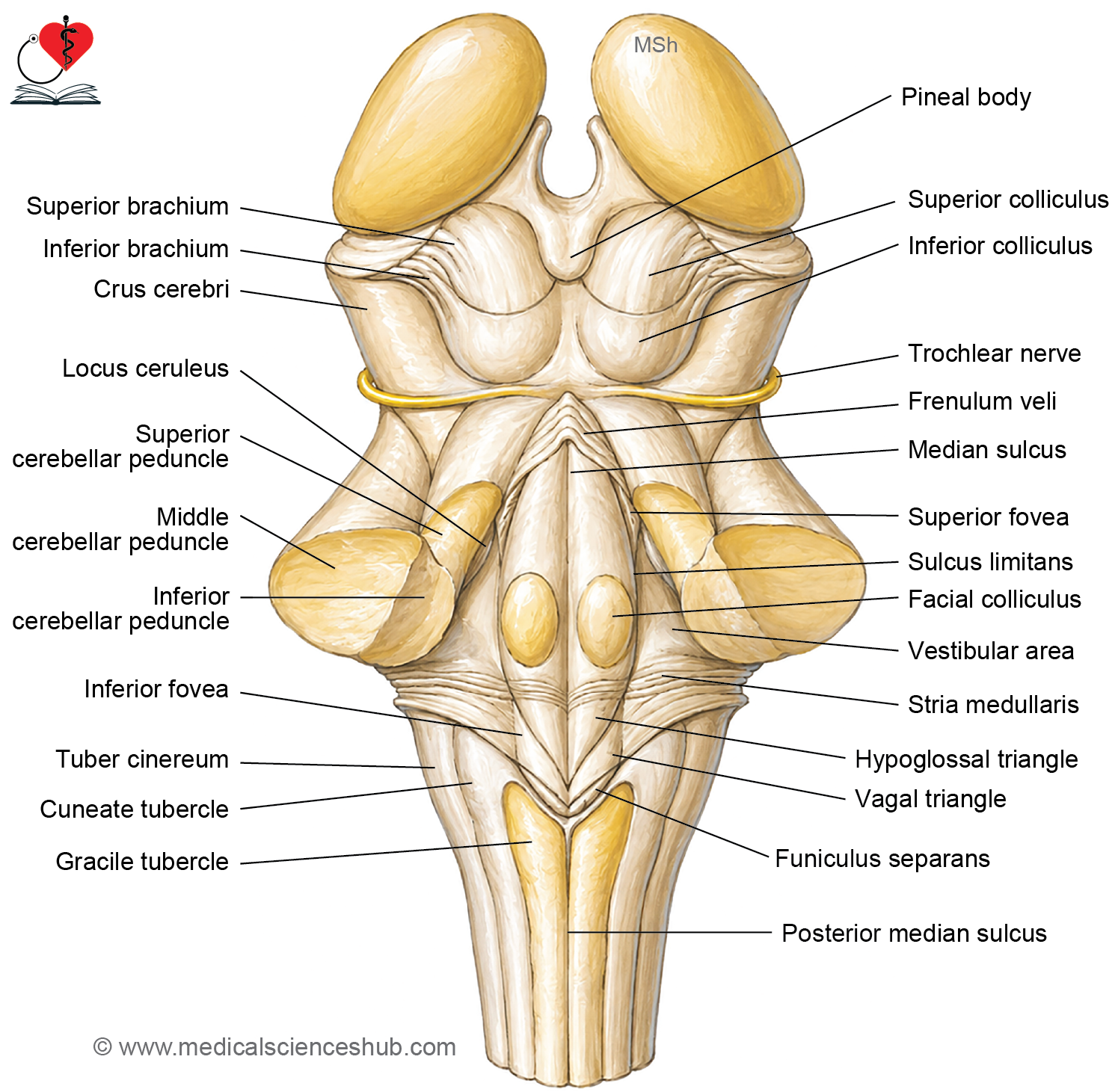
Floor of 4th Ventricle (Rhomboid fossa)
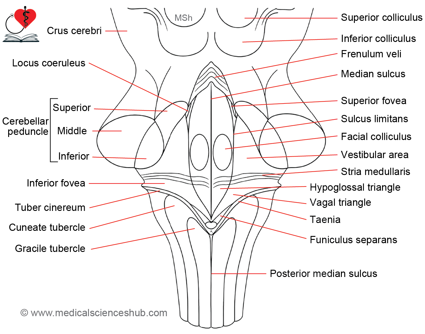
- The floor of the fourth ventricle is diamond-shaped, giving it the name rhomboid fossa, and forms its anterior (ventral) wall.
- It is formed by the posterior surfaces of the pons and the upper (open) part of the medulla oblongata.
- A shallow median sulcus divides the floor into two symmetrical halves.
- On either side of the median sulcus lies a longitudinal elevation called the median eminence, which is laterally bounded by the sulcus limitans.
Striae Medullares
- These are thin white fiber bundles running horizontally across the floor, representing axons of the arcuate nuclei.
- They emerge from the dorsal median sulcus, travel laterally, and reach the cerebellum via the middle cerebellar peduncle.
- They serve as a posterior landmark demarcating the pons from the medulla oblongata.
Sulcus Limitans
- The superior fovea is a small triangular depression at the upper end of the sulcus limitans.
- The inferior fovea is a corresponding depression at its lower end.
Facial Colliculus
- This is an oval elevation in the upper part of the median eminence, overlying the abducent nucleus.
- It is produced by fibers of the facial nerve looping around the abducent nucleus, forming the internal genu of the facial nerve.
- Lesions here can cause ipsilateral facial paralysis and lateral gaze palsy.
Locus Ceruleus and Substantia Ferruginea
- The locus ceruleus is a small, bluish-grey area in the upper part of the superior fovea, located in the pons.
- Its color is due to melanin-containing neurons of the underlying substantia ferruginea, which are predominantly noradrenergic (norepinephrine-secreting).
- These neurons regulate arousal, attention, the sleep-wake cycle, and stress responses.
- Degeneration of these neurons is associated with Parkinson’s disease, Alzheimer’s disease, and mood disorders.
Hypoglossal and Vagal Triangles
- In the lower part of the floor, the sulcus limitans runs obliquely toward the median sulcus, dividing the median eminence into two triangles.
- The hypoglossal triangle overlies the hypoglossal nucleus, which gives rise to the hypoglossal nerve (CN XII); the nearby nucleus intercalatus contributes to modulation of the vestibulocochlear reflex.
- The vagal triangle lies inferolateral to the hypoglossal triangle and overlies the dorsal nucleus of the vagus, which provides parasympathetic fibers to thoracic and abdominal viscera.
Area Postrema and Related Structures
- The funiculus separans is an oblique thickening of ependyma that separates the vagal triangle from the area postrema.
- The area postrema is a small, tongue-shaped, highly vascular region located between the funiculus separans superiorly and the gracile tubercle inferiorly.
- It lacks a blood–brain barrier and contains chemoreceptors, enabling it to detect emetic substances (such as chemotherapy drugs) in the bloodstream and trigger vomiting.
Additional Landmarks
- The taenia is a narrow fold of pia mater running along the inferolateral margin of the fourth ventricle, one on each side.
- The obex is the point where the two taeniae meet at the inferior angle of the fourth ventricle.
- The lower portion of the floor, due to its resemblance to a pen nib, is termed the calamus scriptorius.
Openings in the 4th Ventricle
- The fourth ventricle has five openings that allow CSF to move from the ventricular system (where it is produced) to the subarachnoid space (where it is absorbed).
- Obstruction at any of these sites can result in hydrocephalus.
The Five Openings
- Superiorly, the fourth ventricle communicates with the third ventricle via the cerebral aqueduct (of the midbrain).
- Inferiorly, it is continuous with the central canal of the medulla oblongata.
- The foramen of Magendie (median aperture) is a single midline opening in the lower part of the inferior medullary velum, through which the fourth ventricle communicates with the subarachnoid space.
- The foramina of Luschka (lateral apertures) are two openings, one on each side, located at the lateral recesses of the fourth ventricle, each communicating with the subarachnoid space (pontine cistern).
CLINICAL NEUROANATOMY
These five openings are the only pathways through which CSF can exit the ventricular system. Blockage at any point—whether at the cerebral aqueduct, foramen of Magendie, or foramina of Luschka—prevents normal CSF drainage and leads to its accumulation within the ventricles, causing internal hydrocephalus.
Recesses of 4th Ventricle
The fourth ventricle has five recesses, which are pouched extensions of its cavity.
The Five Recesses
- The lateral recesses (one on each side) extend laterally between the inferior cerebellar peduncle anteriorly and the flocculus of the cerebellum posteriorly.
- At their lateral ends, they open into the subarachnoid space via the foramina of Luschka, through which a portion of the choroid plexus protrudes.
- The dorsomedian recess (median dorsal recess) lies in the midline and extends posteriorly into the white matter of the cerebellum, between the nodule and the lingula.
- The dorsolateral recesses (lateral dorsal recesses, one on each side) project posteriorly above the inferior medullary velum, immediately lateral to the nodule.
Note: The terms dorsomedian recess and dorsolateral recess are the preferred anatomical designations for the median dorsal and lateral dorsal recesses, respectively.
Tela Choroidea and Choroid Plexuses of the Fourth Ventricle
Tela Choroidea
- The tela choroidea of the fourth ventricle is a double-layered fold of pia mater situated between the inferior vermis of the cerebellum and the lower part of the roof of the fourth ventricle.
- Its ventral layer is continuous inferiorly with the pia mater forming the roof of the fourth ventricle and, further below, with the pia mater covering the medulla oblongata.
- Its dorsal layer is continuous posteriorly with the pia mater covering the inferior surface of the cerebellum.
Choroid Plexus
- The choroid plexus of the fourth ventricle consists of capillary networks of blood vessels located between the two layers of the tela choroidea.
- It forms a T-shaped structure with two vertical (ventricular) limbs and one horizontal limb that extends into the lateral recesses, protruding through the foramina of Luschka.
- It is supplied by the choroidal branches of the posterior inferior cerebellar artery (PICA).
- The choroid plexus is responsible for CSF production and maintaining the blood–CSF barrier.
Note: A choroid plexus papilloma, though rare, can obstruct CSF flow and lead to hydrocephalus.
CLINICAL NEUROANATOMY
Medulloblastoma
- Medulloblastoma is the most common tumor arising in the region of the fourth ventricle and the most common malignant brain tumor in children; it rarely occurs in adults.
- It originates from poorly differentiated primitive neuroectodermal cells of the cerebellar vermis and is classified as a highly malignant neoplasm.
Signs and Symptoms
- Raised intracranial pressure (ICP): Blockage of the CSF outflow pathways of the fourth ventricle (median and lateral apertures) causes CSF accumulation, leading to headache, nausea, vomiting, and papilledema.
- Cerebellar dysfunction: Direct compression of the cerebellum results in ataxia, unsteady gait, and impaired fine motor coordination.
- Obstructive (internal) hydrocephalus: Progressive blockage of the ventricular outflow tracts leads to abnormal CSF accumulation within the ventricular system.
- Brainstem compression: Pressure on the brainstem affects vital centers, producing breathing and swallowing difficulties, tachycardia, nystagmus, diplopia, and other abnormal eye movements.
Treatment
- Obstructive hydrocephalus resulting from fourth ventricle blockage requires surgical intervention, typically involving surgical resection of the tumor along with CSF diversion (e.g., ventriculoperitoneal shunt) to restore normal CSF circulation.
Important Questions
- Describe the fourth ventricle.
- Draw a well labelled diagram of the floor of the fourth ventricle.
- Explain: Stria medullaris demarcates pons and medulla oblongata.
📝 Test Your Knowledge – Practice MCQs
Attempt the chapter MCQ quiz and assess your understanding of key concepts.
Click to see: Medical Disclaimer and Copyright Notice