
Competency
- AN63.1: Describe and demonstrate parts, boundaries and features of IIIrd, IVth, and lateral ventricles.
Introduction
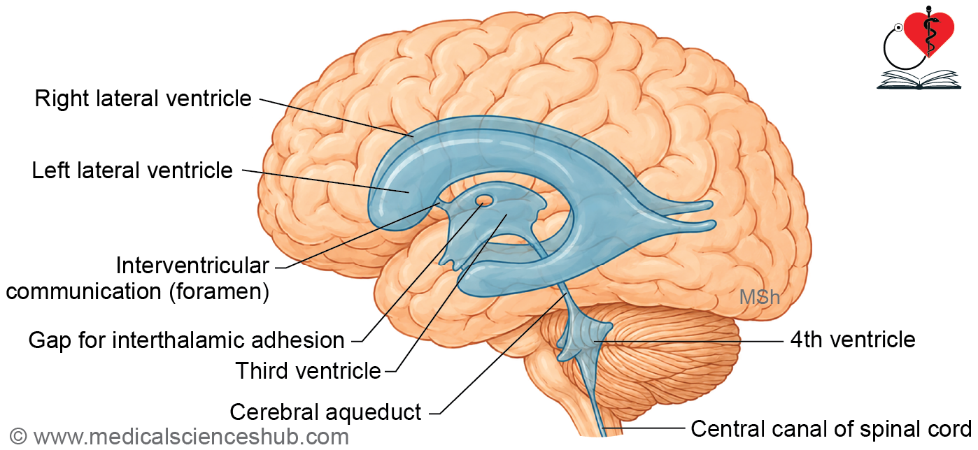
- The third ventricle is a narrow cavity of the diencephalon, containing cerebrospinal fluid (CSF).
- It connects with the lateral ventricles superiorly via the interventricular foramina (foramina of Monro) and with the fourth ventricle inferiorly via the cerebral aqueduct.
- Obstruction at any of these communication points leads to CSF accumulation and hydrocephalus, resulting in raised intracranial pressure (ICP).
Location
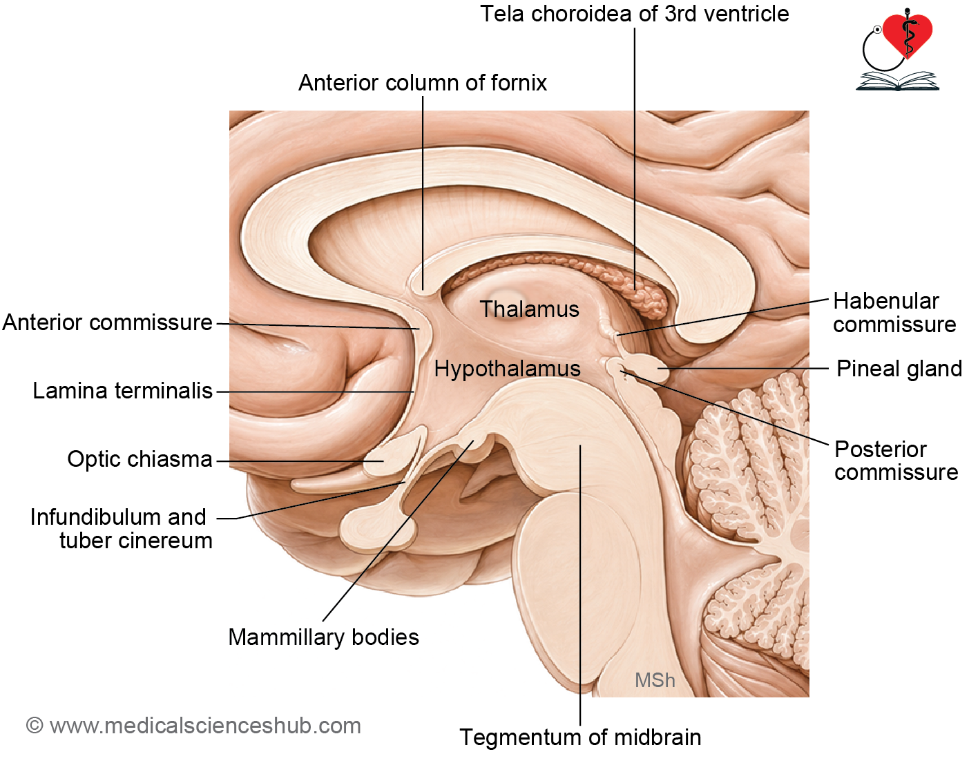
The third ventricle is a midline, slit-shaped cavity located between the two thalami laterally and within the hypothalamus inferiorly.
Extent
- The third ventricle extends from the lamina terminalis anteriorly to the cerebral aqueduct posteriorly.
- Its inner surface is lined by ependymal cells.
Contents
The third ventricle contains two structures:
- Cerebrospinal fluid (CSF), which circulates through and fills the cavity.
- The interthalamic adhesion (massa intermedia), a bridge of grey matter connecting the two thalami across the ventricular cavity, covered by an ependymal lining.
Communications
- The third ventricle communicates with the lateral ventricles on each side via the interventricular foramen of Monro.
- Posteriorly, it connects with the fourth ventricle through the cerebral aqueduct (of Sylvius).
Boundaries of the Third Ventricle
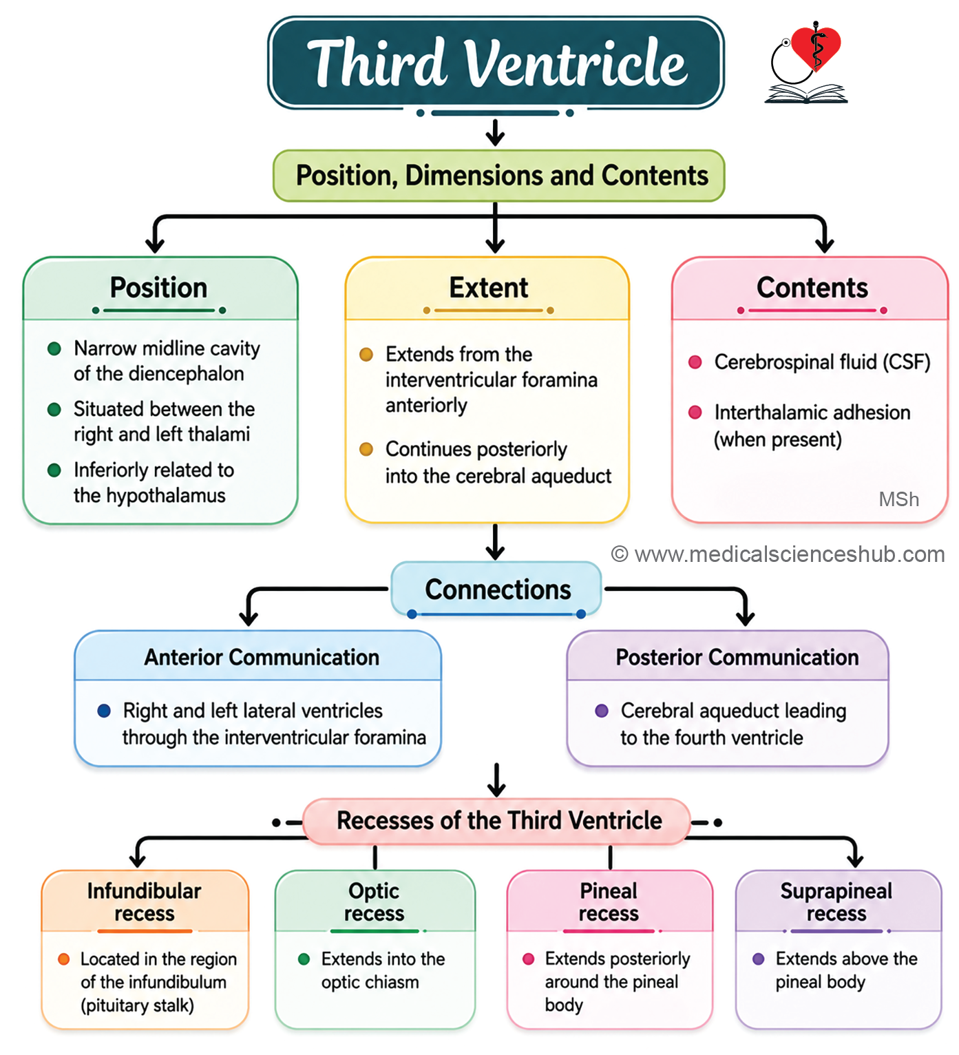
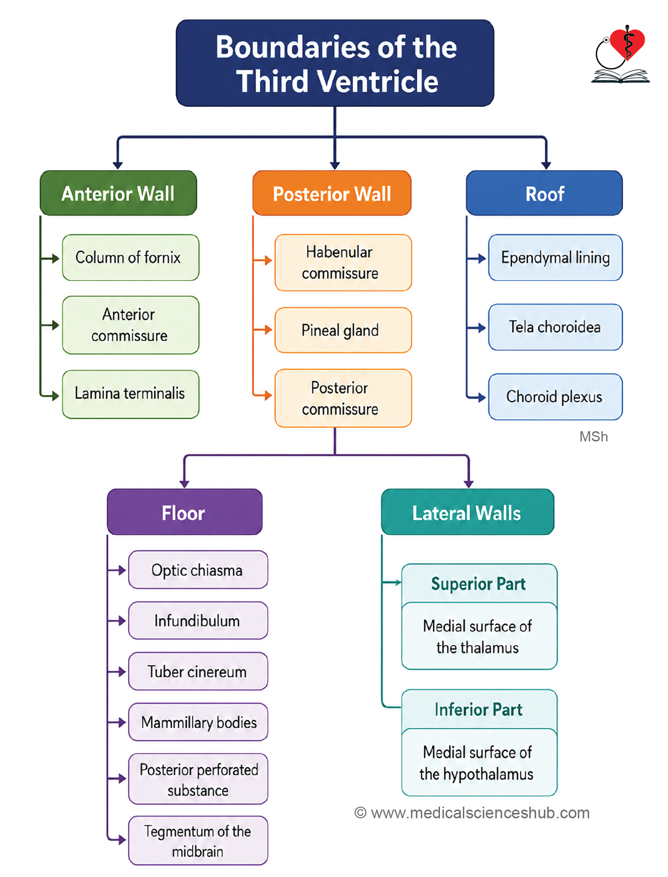
Anterior Wall (from above downward)
- Anterior column of the fornix
- Anterior commissure
- Lamina terminalis
Posterior Wall (from above downward)
- Habenular commissure
- Pineal gland
- Posterior commissure
Roof
- Formed by the ependymal lining stretching between the two thalami.
Floor (from anterior to posterior)
- Optic chiasma
- Infundibulum and tuber cinereum
- Mammillary bodies
- Posterior perforated substance
- Tegmentum of the midbrain
Note: Lesions involving the floor of the third ventricle can affect the nearby optic chiasma, hypothalamus, and pituitary gland, resulting in visual disturbances, hormonal imbalances, disrupted sleep and appetite regulation, and autonomic dysfunction.
Lateral Walls
- Each lateral wall is divided by the hypothalamic sulcus into two parts:
- A larger upper part, formed by the anterior two-thirds of the medial surface of the thalamus.
- A smaller lower part, formed by the hypothalamus.
Recesses of the Third Ventricle
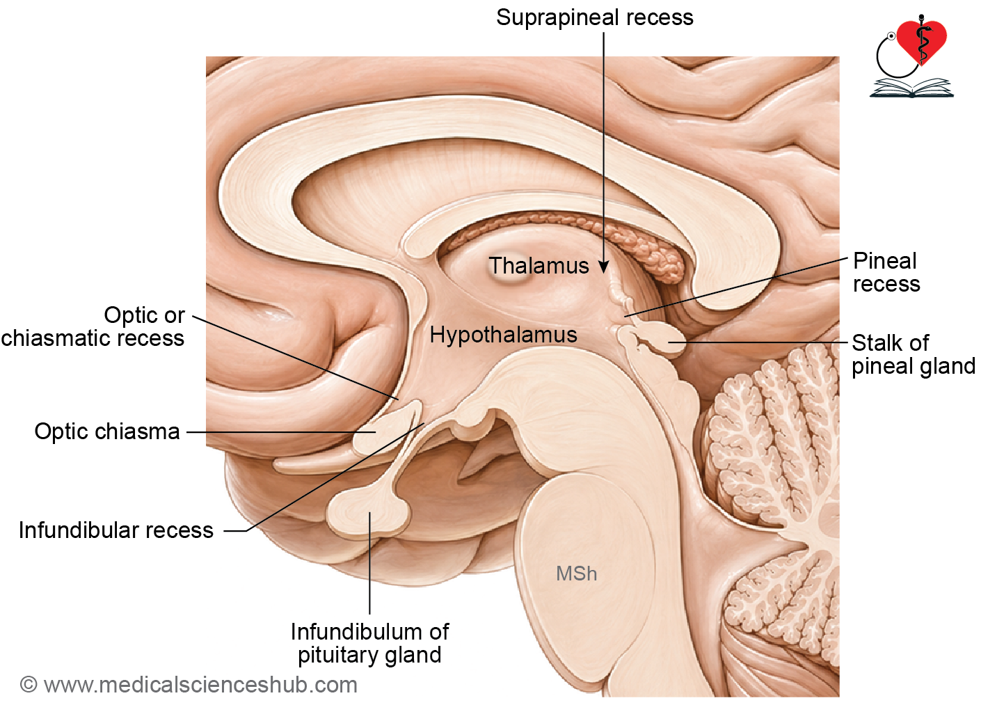
The cavity of the 3rd ventricle extends into the structures forming its boundaries. These extensions are called recesses. They are as follows:
- Infundibular recess — This recess projects inferiorly into the infundibulum, the stalk-like structure that connects the hypothalamus to the posterior pituitary gland.
- Optic (chiasmatic) recess — This recess extends anteriorly toward the optic chiasma, the X-shaped structure where the optic nerves partially decussate.
- Pineal recess — This recess projects posteriorly into the pineal stalk, at the base of the pineal gland.
- Suprapineal recess — This recess also extends posteriorly but lies superior to the pineal stalk, between it and the overlying tela choroidea.
Key Descriptions
Tela Choroidea and Choroid Plexus
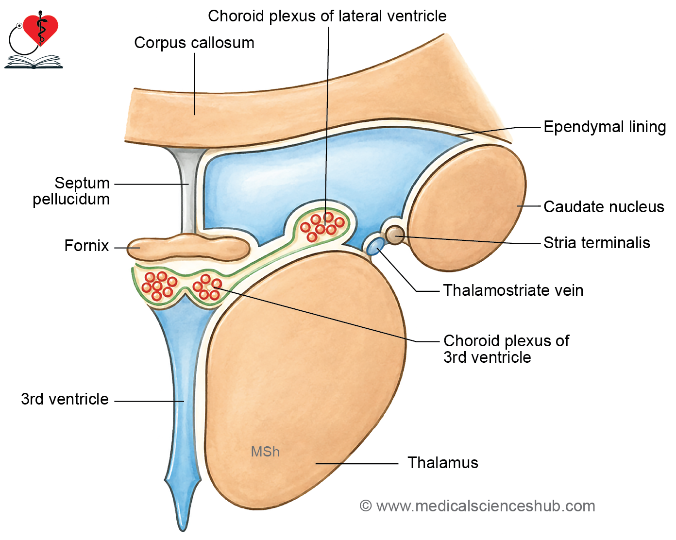
- The tela choroidea is a thin, vascular membrane where the pia mater and ependyma are directly apposed without any intervening neural tissue. (Tela is Latin for “woven.”) It consists of a thin layer of connective tissue richly supplied with blood vessels.
- It gives rise to the choroid plexuses within the ventricles, which are responsible for cerebrospinal fluid (CSF) production.
- The choroid plexus of the third ventricle hangs inferiorly from the tela choroidea as two longitudinal, finger-like projections oriented in an anteroposterior direction. These are continuous with the choroid plexus of the lateral ventricles.
- Infections, inflammation, or neoplasms involving the tela choroidea can disrupt CSF production or circulation, contributing to hydrocephalus or raised intracranial pressure.
Posterior Perforated Substance
- This is a small area of grey matter situated in the interpeduncular fossa, between the mammillary bodies anteriorly and the cerebral peduncles posteriorly. It is perforated by central (perforating) branches of the posterior cerebral artery.
- Deep to it lies a compact group of neurons known as the interpeduncular nucleus, which is considered part of the limbic system.
- Occlusion or injury to the perforating vessels in this region can produce brainstem or thalamic infarctions, presenting clinically with disturbances of consciousness, oculomotor dysfunction, or contralateral motor deficits.
Interventricular Foramen (Foramen of Monro)
- This is a narrow canal connecting each lateral ventricle to the third ventricle. Anteriorly, it is bounded by the anterior column of the fornix; posteriorly, by the anterior tubercle of the thalamus.
- It serves as the conduit through which CSF passes from the lateral ventricles into the third ventricle. Obstruction at this level results in unilateral or bilateral hydrocephalus with a consequent rise in intracranial pressure.
Lamina Terminalis
- The lamina terminalis is a thin midline membrane that forms the anterior wall of the third ventricle. It extends from the optic chiasma inferiorly to the rostrum of the corpus callosum superiorly.
- It participates in the regulation of body fluid homeostasis and contributes to hypothalamic osmosensory and thermoregulatory circuitry. It also transmits certain hypothalamic fiber projections.
- In cases of obstructive hydrocephalus, surgical fenestration of the lamina terminalis may be performed to re-establish CSF drainage.
Anterior Commissure
- The anterior commissure is a compact bundle of myelinated fibers that crosses the midline immediately anterior to the third ventricle. It interconnects the two temporal lobes and portions of the olfactory cortex on either side.
- It facilitates interhemispheric transmission of signals related to olfaction, episodic memory, and emotional processing.
- Lesions involving the anterior commissure can result in impaired olfactory perception, memory consolidation deficits, and reduced integration of affective responses across hemispheres.
CLINICAL NEUROANATOMY
Obstruction in 3rd ventricle
- A tumor or other lesion blocking the third ventricle interrupts normal CSF flow, causing it to accumulate in the ventricles proximal to the obstruction.
- In adults, where skull bones are fused, this leads to raised intracranial pressure (ICP), producing headache, vomiting, blurred vision, and confusion.
- In children, where skull sutures are not yet fused, CSF accumulation results in hydrocephalus, characterized by an enlarged head, bulging fontanelle, and delayed development.
Endoscopic third ventriculostomy (ETV)
- ETV is a surgical procedure used to treat obstructive hydrocephalus by restoring CSF circulation without implanting a permanent shunt.
- Using an endoscope introduced through a small cranial opening, the neurosurgeon creates a small perforation in the floor of the third ventricle, typically between the mammillary bodies and the infundibular recess.
- This opening allows CSF to bypass the obstruction and drain directly into the subarachnoid space, where it is absorbed naturally.
- ETV is particularly effective when obstruction occurs at the cerebral aqueduct (of Sylvius) or within the third ventricle itself, and it relieves symptoms of raised ICP including headache, vomiting, and visual disturbances.
Important Questions
- Describe the third ventricle under the following headings: Location, communications, recesses, and applied aspects.
📝 Test Your Knowledge – Practice MCQs
Attempt the chapter MCQ quiz and assess your understanding of key concepts.
Click to see: Medical Disclaimer and Copyright Notice