
Introduction
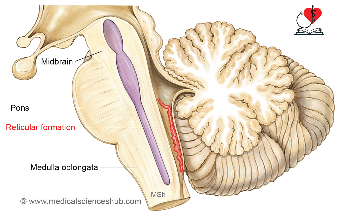
The reticular formation is a diffuse, poorly delineated mass of grey matter interspersed with nerve fibers that extends throughout the central core of the brainstem (reticulum = little net). It is an evolutionarily ancient structure with close connections to the olfactory and limbic systems, and together these three systems regulate emotions and body functions. Through its extensive connections with nearly all parts of the nervous system, the reticular formation influences movement, sensation, autonomic and endocrine functions, biological rhythms, and consciousness.
Functions
- Control of skeletal muscle activity — influences α and γ motor neurons via the reticulospinal and reticulobulbar tracts, regulating muscle tone, reflexes, and reciprocal inhibition (e.g., flexor contraction with simultaneous extensor relaxation). It also helps maintain antigravity muscle tone during standing, in coordination with the vestibular system. Brainstem respiratory centers are included within this system.
- Control of facial expression — regulates facial muscles associated with emotional expression. Symmetrical movements such as smiling and laughing involve bilateral reticular formation activity via a pathway distinct from corticobulbar fibers, which explains why symmetrical emotional facial expression can be preserved following certain strokes.
- Modulation of somatic and visceral sensation — positioned centrally, the reticular formation can enhance or inhibit all ascending sensory pathways; it plays a significant role in gating pain perception.
- Autonomic nervous system (ANS) control — via reticulobulbar and reticulospinal tracts, it relays higher regulatory signals from the cortex, hypothalamus, and subcortical nuclei to both sympathetic and parasympathetic outputs.
- Endocrine control — influences hormone secretion through connections with hypothalamic nuclei, thereby indirectly affecting the pituitary gland.
- Biological rhythms — connections with the hypothalamus are thought to contribute to the regulation of biological clocks.
- Reticular activating system — governs arousal and consciousness by relaying sensory signals to the cerebral cortex. Painful stimuli increase its activity and excite the cortex. Acetylcholine serves as a key excitatory neurotransmitter within this system.
Reticular Nuclei
The reticular formation contains more than 100 nuclei scattered throughout the tegmentum of the midbrain, pons, and medulla. Each subdivision has a distinct cytoarchitecture, specific connections, and defined functions. These nuclei are broadly classified according to four general roles: regulation of consciousness and cortical alertness, control of somatic motor movements, regulation of autonomic functions, and control of sensory transmission.
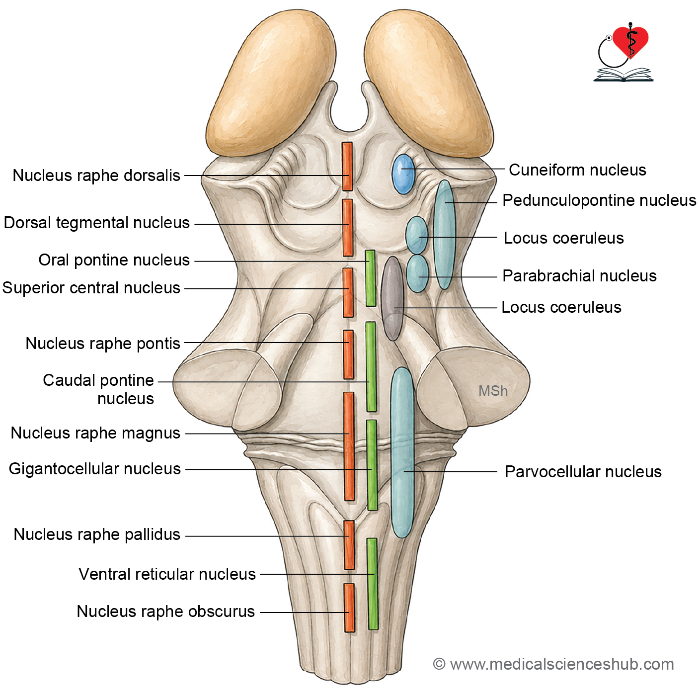
Anatomically, the reticular formation is organized into four longitudinal zones based on mediolateral position within the brainstem.
1. Median Zone (Midline Raphe Nuclei)
- Composed of medium-sized neurons arranged along the anterior brainstem, except where major fiber bundles cross.
- Raphe nuclei by region:
- Midbrain — dorsal tegmental nucleus, nucleus raphes dorsalis (nucleus linearis)
- Pons — nucleus raphes magnus, nucleus raphes pontis, superior central nucleus
- Medulla — nucleus raphes obscurus, nucleus raphes pallidus
- Many raphe neurons synthesize serotonin; those projecting to higher brain centers are associated with sleep regulation, while those projecting to the spinal trigeminal nucleus and dorsal horn of the spinal cord modulate nociceptive transmission.
2. Paramedian Zone (Paramedian Reticular Nuclei)
- Located lateral to the midline.
- Receives primarily uncrossed afferent fibers from the cerebral cortex, fastigial and dentate nuclei of the cerebellum, vestibular nuclei, and spinal cord.
- Projects efferent fibers to the vermis, uvula, and fastigial nucleus of the cerebellum.
- Functions in feedback systems involved in coordinating complex movement patterns.
3. Medial Zone (Magnocellular Reticular Formation)
- Also called the motor or effector zone; consists of large neurons.
- Main nuclei: ventral reticular nucleus, gigantocellular reticular nucleus, and pontine reticular nuclei (oral and caudal parts).
- Neurons have axons that bifurcate into long ascending and descending branches with collaterals:
- Ascending fibers — travel via the central tegmental tract to the hypothalamus (autonomic control) and thalamic intralaminar nuclei (arousal).
- Descending fibers — join reticulospinal tracts to control trunk and proximal limb muscles.
- Also gives rise to reticulobulbar fibers (modulating motor cranial nerve nuclei and sensory input) and reticulocerebellar fibers.
- The gigantocellular reticular nucleus, located in the caudal pons and rostral medulla, coordinates motor activity and integrates signals between the brainstem and cerebellum.
4. Lateral Zone (Parvicellular Reticular Formation)
- Also called the sensory or afferent zone; composed predominantly of small interneurons.
- Interneurons project locally to the medial zone; some terminate directly on cranial nerve motor nuclei.
- Present mainly in the pons and medulla, continuing into the intermediate grey matter of the spinal cord.
- Should not be confused with the intermediolateral horn of the spinal cord, which contains sympathetic preganglionic neurons.
- Key nuclei:
- Parvicellular nucleus — located medial to the spinal trigeminal nucleus and ventral to the vestibular nuclei; receives sensory input from the cerebrum, cranial nerves, cerebellum, and spinal cord, and relays processed information to the medial zone.
- Parabrachial nucleus — involved in visceral and limbic functions.
- Pedunculopontine nucleus — located at the rostral pons and caudal midbrain; projects to motor-related brain centers.
- Cuneiform nucleus — located in the midbrain; sends fibers to motor-related areas.
- Through its connections with the medial zone and reticulobulbar/reticulospinal tracts, the lateral zone modulates sensory input and supports alertness and motor coordination.
Nuclei Associated With The Reticular Formation
Several brainstem nuclei are closely linked to, but not considered part of, the reticular formation. These include the red nucleus, inferior olivary nucleus, and precerebellar reticular nuclei. The midbrain periaqueductal grey (PAG) also maintains extensive connections with the reticular formation.
Precerebellar Reticular Nuclei
- Comprise the paramedian reticular nucleus, pontine reticulotegmental nucleus, and lateral reticular nucleus.
- Function independently from other reticular nuclei.
- Project fibers to the cerebellum to assist in coordinating muscle activity.
Periaqueductal Grey Matter (PAG)
- Receives afferent input from the cerebral cortex, hypothalamus, limbic system, parabrachial nucleus, solitary nucleus, and ascending sensory pathways.
- Sends efferent output back to these same regions and to the medullary raphe nuclei.
- Plays an important role in autonomic and limbic functions, and is a key center for modulating pain perception.
Connections of the Reticular Formation
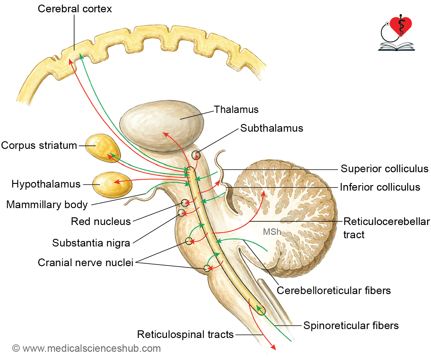
The reticular formation receives input from virtually all sensory and motor systems and, in turn, influences most of the body’s responses.
Afferent Fibers (Input)
- Spinal cord — via spinoreticular fibers and collaterals from all ascending tracts
- Visual system — via tectoreticular fibers
- Auditory system — via tectoreticular fibers
- Olfactory system — via descending fibers from the limbic system
- Cerebellum — via cerebelloreticular fibers
- Corpus striatum — via pallidotegmental fibers
- Cerebral cortex — via descending tracts
- Contralateral reticular formation and cranial nerve nuclei
Efferent Fibers (Output)
- Spinal cord — via reticulospinal tracts
- Brainstem — via reticulobulbar tract
- Cerebellum — via reticulocerebellar tract
- Red nucleus, substantia nigra, and tectum of the midbrain
- Ascending projections to the:
- Subthalamus
- Thalamus
- Hypothalamus
- Corpus striatum
- Cerebral cortex
The reticular nuclei influence both cortical and subcortical centers through polysynaptic pathways.
Functional Organization of the Reticular Formation
The reticular formation is functionally divided into two broad systems:
- Ascending reticular activating system (ARAS) — projects to higher brain centers to regulate arousal and consciousness.
- Descending reticular system (DRS) — projects to cranial nerve nuclei and the spinal cord to modulate motor and autonomic functions.
Functional Summary by Zone
- Median raphe nuclei — neurons projecting to higher brain centers are associated with sleep regulation.
- Paramedian reticular nuclei — via connections with the cerebral cortex, cerebellum, vestibular nuclei, and spinal cord, these nuclei participate in feedback systems that coordinate complex movement patterns.
- Medial reticular nuclei — influence the autonomic nervous system, level of arousal, and motor control of axial and proximal limb musculature.
- Lateral reticular nuclei — receive and integrate sensory information, then relay it to the medial reticular nuclei, which in turn modulate sensory afferent input and maintain alertness.
Reticulospinal Tracts
- Corticoreticular fibers terminate bilaterally in the medial reticular nuclei of the pons and medulla, modulating the activity of reticulobulbar and reticulospinal neurons.
- Medial reticulospinal tract (arising from the pons) — stimulates extensors and inhibits flexors of axial and proximal limb musculature.
- Lateral reticulospinal tract (arising from the medulla) — inhibits extensors and excites flexors of axial and proximal limb musculature.
Additional Functional Roles
- The medial reticular nuclei of the pons control conjugate horizontal eye movements elicited by head movements.
- The raphe nuclei suppress the transmission of nociceptive signals from first-order to second-order neurons, thereby reducing pain signal relay to higher brain centers.
- The brainstem reticular formation influences cranial nerve nuclei, coordinating reflex activity associated with cranial nerves.
- Regulation of the sleep-wake cycle — including waking, sustained wakefulness, and the transition to sleep — represents one of the most fundamental functions of the brainstem reticular formation.
Ascending Reticular Activating System (ARAS)
The ascending reticular activating system (ARAS), also called the reticular activating system (RAS), is responsible for maintaining wakefulness and alertness.
Connections
- Collateral fibers from all ascending sensory tracts of the spinal cord and cranial nerves project into the reticular formation.
- Sensory pathways — including the trigeminothalamic, spinothalamic, and spinoreticular tracts — send collaterals to the parvicellular nucleus of the lateral zone.
- The lateral zone projects to the medial zone, which relays signals via the central tegmental tract to the hypothalamus and thalamic intralaminar nuclei.
- Thalamocortical fibers then carry these signals to widespread cortical areas, sustaining wakefulness.
Functional Significance
- Continuous sensory input is essential for maintaining consciousness; stimuli such as pain, sudden sounds, unfamiliar smells, or unexpected events activate the ARAS and alert the cortex.
- Insufficient sensory stimulation leads to drowsiness.
- The ARAS coordinates the sleep-wake cycle and supports cortical activation during daily activities, emotional responses, and reactions to external stimuli.
CLINICAL NEUROANATOMY
- Coma — bilateral lesion of the ARAS results in coma, a state of unconsciousness in which even powerful sensory stimuli fail to produce arousal.
- Sleep — decreased ARAS activity induces sleep.
- Pharmacological suppression — general anesthetics and tranquilizing drugs suppress ARAS activity; notably, anesthetics may block conscious sensory awareness while auditory processing may partially persist.
- Severe ARAS damage — can result in permanent loss of consciousness, coma, or death.
Table 20.1: Zones of the reticular formation and their component nuclei
| Region | Median Group (Raphe Nuclei) | Medial Group | Lateral Group |
|---|---|---|---|
| Midbrain | • Nucleus raphe dorsalis • Dorsal tegmental nucleus | Not well defined | • Cuneiform nucleus • Parabrachial nucleus • Pedunculopontine nucleus |
| Pons | • Superior central nucleus | • Pontine reticulotegmental nucleus • Nucleus reticularis pontis oralis • Nucleus reticularis pontis caudalis | • Parabrachial nucleus • Pedunculopontine nucleus |
| Medulla Oblongata | • Nucleus raphe magnus • Nucleus raphe pallidus • Nucleus raphe obscurus | • Paramedian reticular nucleus • Gigantocellular nucleus • Ventral reticular nucleus | • Parvocellular reticular nucleus • Lateral reticular nucleus |
📝 Test Your Knowledge – Practice MCQs
Attempt the chapter MCQ quiz and assess your understanding of key concepts.
Click to see: Medical Disclaimer and Copyright Notice