
Competency
- AN71.2 Identify cartilage under the microscope and describe various types and structure-function correlation of the same.
Introduction
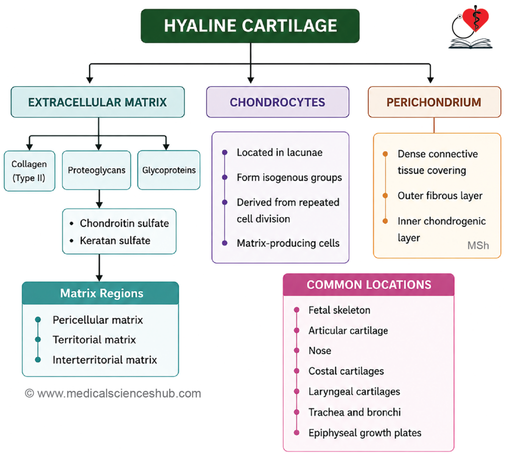
- Cartilage is a specialized connective tissue composed of chondrocytes (5%) embedded within an extracellular matrix (95%).
- Being avascular, cartilage relies on diffusion from surrounding tissues for nutrient delivery — facilitated by type II collagen fibers and glycosaminoglycans (GAGs) within the matrix.
- Based on matrix composition and mechanical properties, cartilage is classified into three types: hyaline cartilage, elastic cartilage, and fibrocartilage.
- Most cartilage types are enclosed by a fibrocellular sheath called the perichondrium, with the notable exception of fibrocartilage.
Hyaline Cartilage
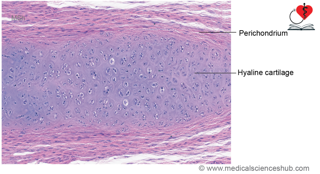
Hyaline cartilage is the most common type of cartilage in the human body. Its extracellular matrix appears smooth, homogeneous, and glass-like under the microscope, giving rise to the term hyaline (Greek: hyalos = glass).
Hyaline cartilage is found in:
- The fetal skeleton.
- Articular cartilage covering the ends of synovial joint surfaces.
- Cartilages of the nose.
- Costal cartilages connecting ribs to the sternum.
- Certain laryngeal cartilages, including the thyroid, cricoid, and arytenoid cartilages.
- The trachea and bronchi.
- Growing regions of developing bones, particularly the epiphyseal growth plates.
Histology of Hyaline Cartilage
- Hyaline cartilage is composed of:
- Chondrocytes (cartilage cells).
- Extracellular matrix (ground substance).
- Fine collagen fibers, predominantly type II collagen, which are not easily visible with routine staining.
Matrix
- The matrix is abundant and gives hyaline cartilage its characteristic glassy appearance.
- It contains small spaces called lacunae, which house the chondrocytes.
- Chondrocytes synthesize and maintain the matrix by producing proteoglycans, glycosaminoglycans, and collagen fibers.
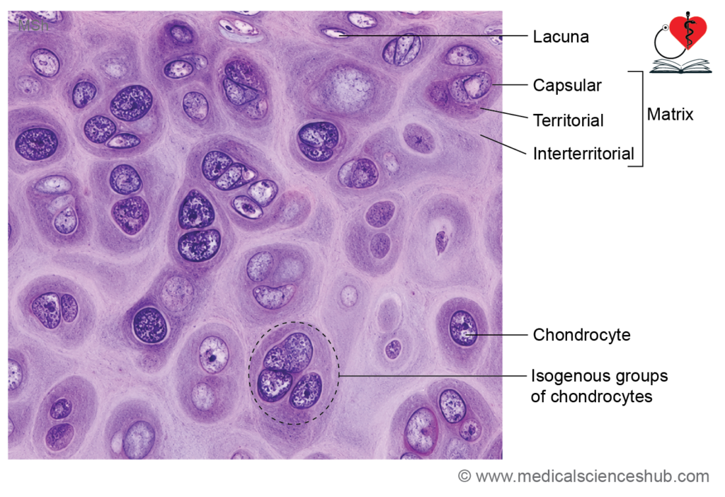
- Variations in proteoglycan concentration create distinct matrix regions:
- Pericellular (Capsular) Matrix
- Forms a narrow zone immediately surrounding each chondrocyte.
- Rich in sulfated proteoglycans and type VI collagen.
- Stains most intensely because of its high concentration of negatively charged molecules.
- Territorial Matrix
- Surrounds the pericellular matrix.
- Contains a lower concentration of proteoglycans than the pericellular matrix.
- Appears moderately basophilic and stains less intensely.
- Interterritorial Matrix
- Occupies the area between groups of chondrocytes.
- Contains relatively fewer proteoglycans and a greater proportion of collagen fibers.
- Stains more lightly than the territorial matrix.
Chondrocytes
- Chondrocytes are the mature cells of cartilage and are located within lacunae.
- During routine histological preparation, chondrocytes often appear separated from the lacunar wall by a clear space. This is an artifact caused by cellular shrinkage; in living tissue, the cell fills the lacuna.
- Chondrocytes originate from chondroblasts, which become enclosed within the matrix they produce and subsequently mature into chondrocytes.
- Chondrocytes can undergo mitotic division during cartilage growth.
Isogenous Groups (Cell Nests)
- Division of a single chondrocyte may produce clusters of daughter cells within the same region of matrix.
- These clusters are called isogenous groups or cell nests.
- They represent areas of active interstitial growth of cartilage.
Perichondrium
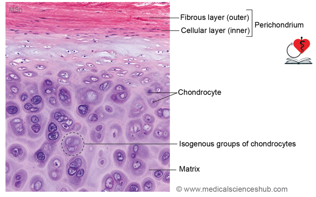
- Most hyaline cartilage is surrounded by a dense connective tissue covering known as the perichondrium.
- The perichondrium consists of two layers:
- Outer Fibrous Layer
- Composed of dense irregular connective tissue.
- Contains collagen fibers, fibroblasts, blood vessels, and nerves.
- Inner Cellular (Chondrogenic) Layer
- Contains progenitor cells that differentiate into chondroblasts and contribute to cartilage growth.
- Because cartilage is avascular, nutrients reach chondrocytes by diffusion from blood vessels present in the perichondrium.
Sites Lacking Perichondrium
- Articular cartilage.
- Areas where cartilage is in direct contact with bone.
Functions of Hyaline Cartilage
- Provides firm yet flexible support to various structures.
- Maintains the patency of the trachea and major bronchi, preventing airway collapse.
- Supports and protects thoracic organs through the costal cartilages.
- Forms a smooth, low-friction surface in synovial joints, facilitating efficient movement and reducing wear on articulating bones.
CLINICAL CORRELATION
Osteoarthritis
- It is the most common degenerative joint disorder and primarily affects articular cartilage.
- It occurs more frequently in older adults, particularly after 60 years of age, although it may develop earlier following joint injury or excessive mechanical stress.
- Common manifestations include chronic joint pain, stiffness, and reduced mobility, especially in weight-bearing joints such as the hip and knee.
- The disease is characterized by progressive loss of cartilage proteoglycans and degradation of the extracellular matrix.
- Chondrocytes produce increased levels of inflammatory mediators, including interleukin-1 (IL-1) and tumor necrosis factor-α (TNF-α), while synthesis of type II collagen decreases.
- There is no definitive cure; the condition generally progresses gradually with advancing age.
Elastic Cartilage
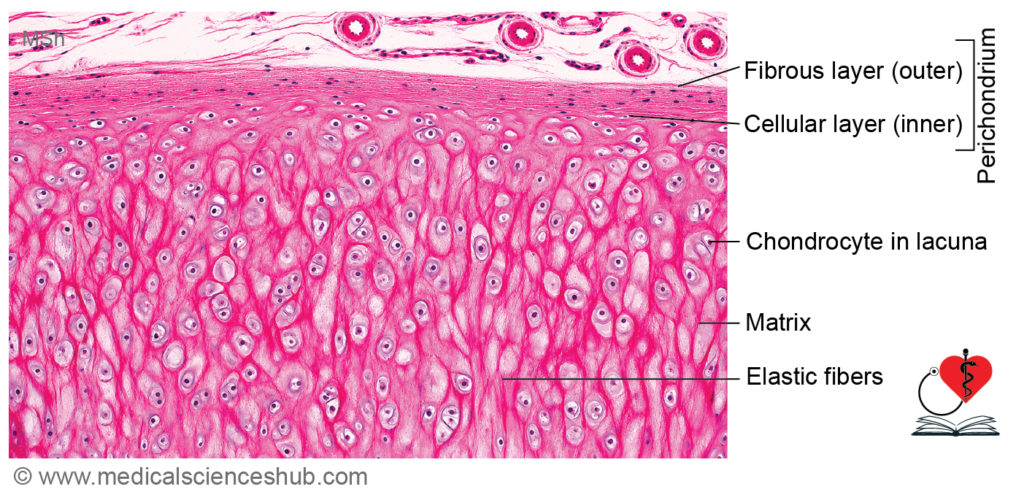
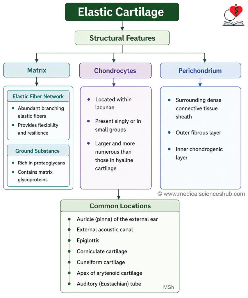
- Elastic cartilage is a specialized type of cartilage that contains abundant elastic fibers within its extracellular matrix, providing greater flexibility than hyaline cartilage.
- It is found in structures that require both support and elasticity, including:
- Auricle (pinna) of the external ear
- External acoustic meatus
- Auditory (Eustachian) tube
- Epiglottis
- Certain small laryngeal cartilages, such as the cuneiform and corniculate cartilages
Histology of Elastic Cartilage
Elastic cartilage is composed of:
- Chondrocytes
- Extracellular ground substance
- A dense network of elastic fibers
Fibers
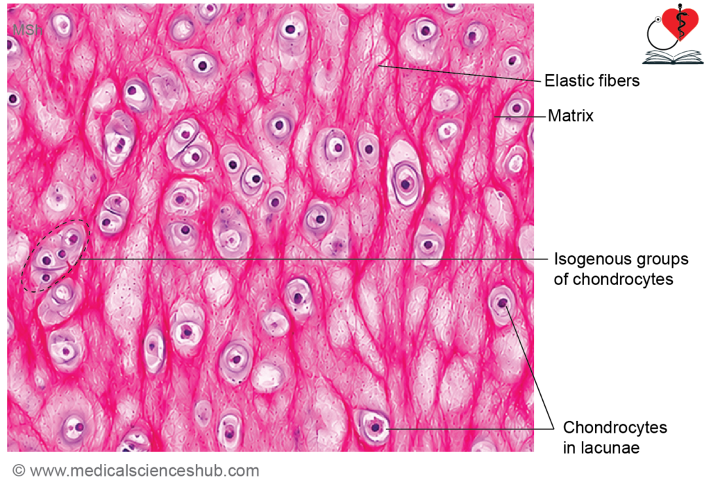
- The matrix contains numerous branching elastic fibers in addition to type II collagen fibrils.
- Elastic fibers form an interconnected network around the lacunae and are best demonstrated by special elastic tissue stains.
- The presence of abundant elastic fibers is the most important feature distinguishing elastic cartilage from hyaline cartilage.
Ground Substance
- The ground substance is similar to that of hyaline cartilage and contains proteoglycans, glycosaminoglycans, and water.
- It provides resilience and resistance to compressive forces.
Chondrocytes
- Chondrocytes lie within lacunae and are often larger and more numerous than those in hyaline cartilage.
- They may occur singly or in isogenous groups, reflecting interstitial growth.
- Chondrocytes maintain and renew the cartilage matrix.
Perichondrium
- Elastic cartilage is surrounded by a well-developed perichondrium.
- The perichondrium contains blood vessels that supply nutrients to the avascular cartilage through diffusion.
- It contributes to appositional growth and repair.
Identification Features
- Presence of numerous dark, branching elastic fibers within the matrix.
- Chondrocytes enclosed in distinct lacunae.
- A clearly visible perichondrium surrounding the cartilage.
Functions of Elastic Cartilage
- Provides flexible support while maintaining the shape of structures.
- Allows repeated bending and recoil without permanent deformation.
- Helps maintain the form and patency of structures such as the epiglottis and external ear.
Fibrocartilage
- Fibrocartilage is a specialized type of cartilage that combines the strength of dense connective tissue with the resilience of cartilage.
- It is found in areas subjected to high tensile and compressive forces, including:
- Intervertebral discs (annulus fibrosus)
- Pubic symphysis
- Articular discs and menisci of certain joints
- Sites of tendon or ligament attachment to bone
Histology of Fibrocartilage
Fibrocartilage consists of:
- Chondrocytes
- Extracellular matrix
- Abundant type I collagen fibers with smaller amounts of type II collagen
Fibers
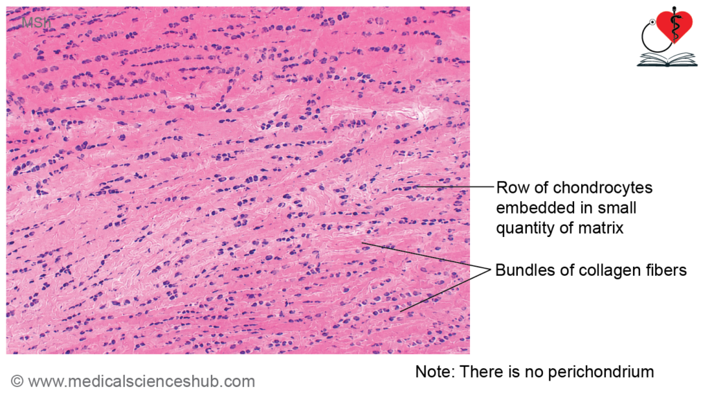
- Thick bundles of type I collagen are the most prominent component.
- Collagen fibers are arranged in parallel bundles, providing exceptional tensile strength.
- The abundance of collagen fibers is the principal feature distinguishing fibrocartilage from other cartilage types.
Chondrocytes
- Chondrocytes are located within lacunae and are typically arranged in rows between collagen bundles.
- They are fewer in number than those found in hyaline cartilage.
- The characteristic linear arrangement of chondrocytes is an important identifying feature.
Matrix
- The extracellular matrix contains proteoglycans and collagen fibers but is less abundant than in hyaline cartilage.
- Because of the dense collagen content, the matrix appears more fibrous and less homogeneous.
Identification Features
- Presence of thick bundles of type I collagen fibers.
- Chondrocytes lying in lacunae and arranged in rows between collagen bundles.
- Perichondrium is absent, unlike most hyaline and elastic cartilages.
Functions
- Provides strong resistance to compression, tension, and shear forces.
- Acts as a shock absorber in weight-bearing regions.
- Contributes to the stability and durability of joints and intervertebral discs.
CLINICAL CORRELATION
Achondroplasia/dwarfism
- Achondroplasia is the most common form of disproportionate dwarfism and is characterized by short limbs with a relatively normal trunk.
- It is caused by mutations in the FGFR3 (fibroblast growth factor receptor 3) gene.
- The mutation inhibits normal endochondral ossification, resulting in impaired longitudinal growth of bones.
- Most cases arise from new (de novo) mutations, although the disorder can be inherited in an autosomal dominant manner.
- Chondrosarcoma is a malignant tumor that produces cartilaginous matrix and commonly arises in the pelvis, ribs, shoulder girdle, or other axial skeletal structures.
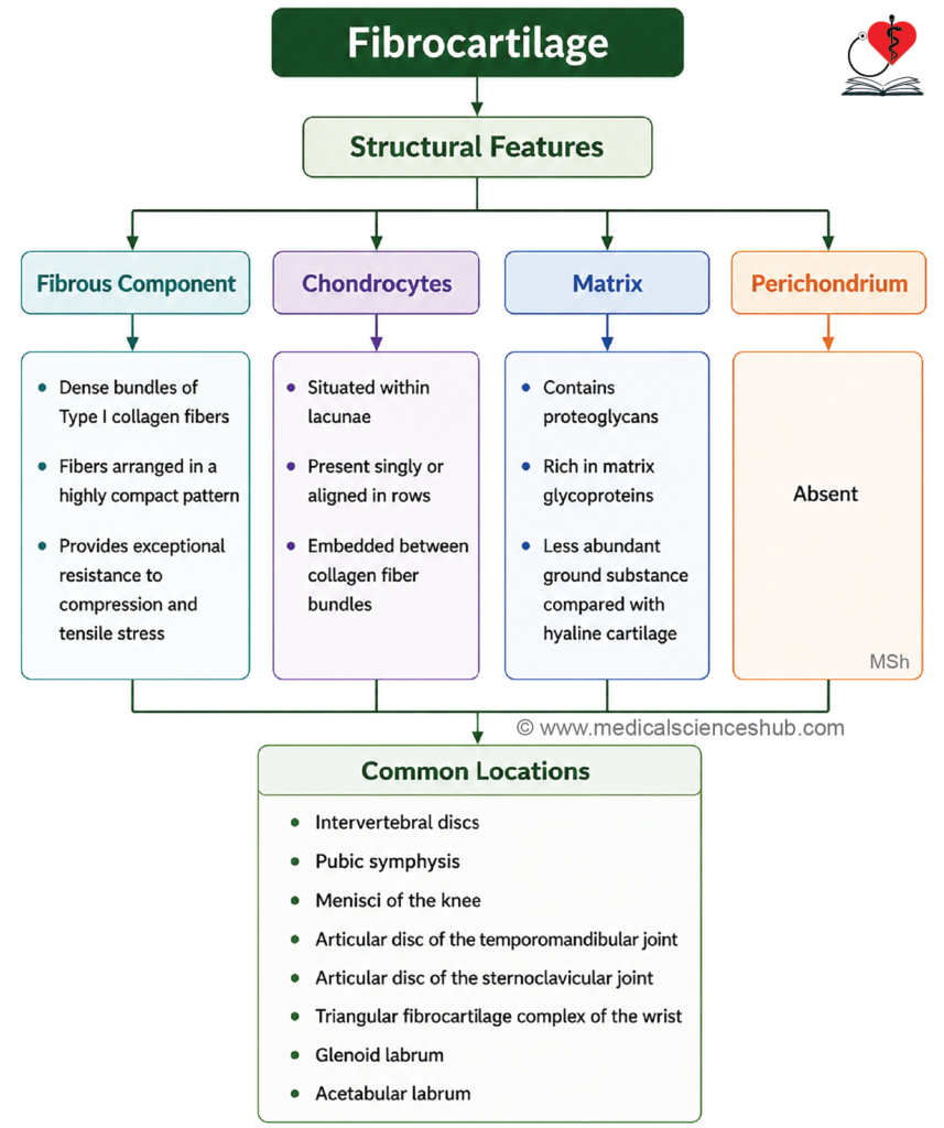
Table 5.1: Difference between hyaline, elastic, and fibrocartilages
| Feature | Hyaline Cartilage | Elastic Cartilage | Fibrocartilage |
|---|---|---|---|
| Cell Types | Contains chondroblasts and mature chondrocytes. | Contains chondroblasts and mature chondrocytes. | Predominantly contains chondrocytes with scattered fibroblasts. |
| Arrangement of Chondrocytes | Chondrocytes commonly occur in isogenous groups (cell nests). | Chondrocytes are present singly or in small isogenous groups. | Chondrocytes are usually arranged singly or in linear rows between collagen bundles. |
| Fibers | Matrix contains mainly type II collagen fibers. | Matrix contains abundant elastic fibers along with type II collagen. | Matrix contains dense bundles of type I collagen with smaller amounts of type II collagen. |
| Perichondrium | Usually present, except at articular surfaces and where cartilage contacts bone. | Present and well developed. | Absent. |
| Matrix Production | Matrix is synthesized primarily by chondrocytes. | Matrix is synthesized primarily by chondrocytes. | Matrix is produced mainly by chondrocytes, with fibroblasts contributing to collagen fiber formation. |
| Mechanical Property | Provides firm support and resistance to compression. | Provides flexibility while maintaining shape. | Provides high tensile strength and resistance to compression. |
| Common Locations | Articular surfaces, costal cartilages, nose, trachea, and larynx. | Auricle of ear, epiglottis, and auditory tube. | Intervertebral discs, pubic symphysis, and menisci. |
Important Questions
- Write a short note on histology of hyaline cartilage.
- Write a short note on histology of elastic cartilage.
- Write a short note on histology of white fibrocartilage.
- List the differences between hyaline cartilage, elastic cartilage, and fibrocartilage.
📝 Test Your Knowledge – Practice MCQs
Attempt the chapter MCQ quiz and assess your understanding of key concepts.
Click to see: Medical Disclaimer and Copyright Notice