
Competencies
- AN67.1 Describe and identify various types of muscle under the microscope.
- AN67.2 Classify muscle and describe the structure-function correlation of the same.
- AN67.3 Describe the ultrastructure of muscular tissue.
Introduction
- Muscular tissue possesses the specialized property of contractility, which enables cells to shorten and generate force. Although many body cells exhibit limited contractile ability, this property is most highly developed in muscle tissue.
- The cells of muscle tissue are called myocytes or muscle fibers because of their elongated shape.
- All muscle tissues originate primarily from the mesoderm during embryonic development.
- Important muscle cell terminology includes:
- Sarcolemma: the plasma membrane of a muscle cell.
- Sarcoplasm: the cytoplasm of a muscle cell.
- Sarcoplasmic reticulum: the specialized smooth endoplasmic reticulum involved in calcium storage and release.
- Sarcosomes: mitochondria present in muscle fibers.
- Histologically, muscles are classified into three types:
- Skeletal muscle
- Cardiac muscle
- Smooth muscle
- Muscles may also be categorized as:
- Striated muscles: skeletal and cardiac muscles.
- Non-striated muscles: smooth muscles.
- Striations result from the highly organized arrangement of contractile proteins within the sarcoplasm.
- Physiologically, muscles are classified as:
- Voluntary muscles: skeletal muscles, which are generally under conscious control.
- Involuntary muscles: cardiac and smooth muscles, which function without conscious control.
Skeletal Muscle
- Skeletal muscles are also known as striated muscles because they display transverse striations and are generally classified as voluntary muscles under conscious control.
- These muscles attach to the skeleton and are primarily responsible for body movements and locomotion.
- Visceral striated muscles resemble skeletal muscles microscopically but are located in structures such as the tongue, pharynx, diaphragm, and upper third of the esophagus.
- They play essential roles in speech, swallowing, breathing, and coughing.
Histology of Skeletal Muscle
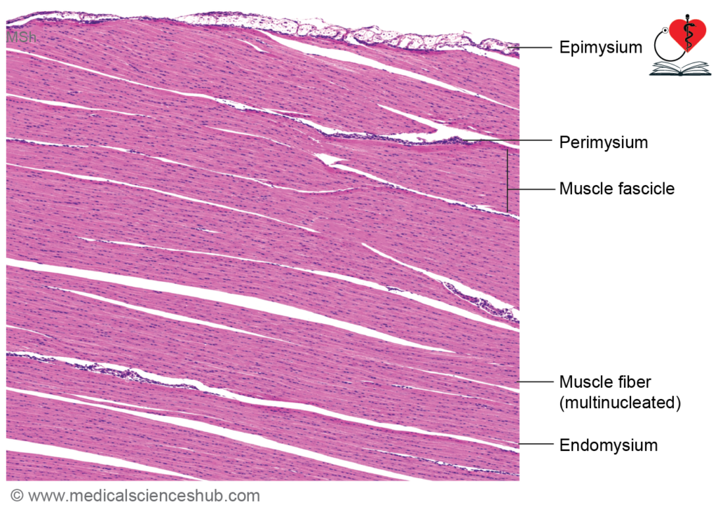
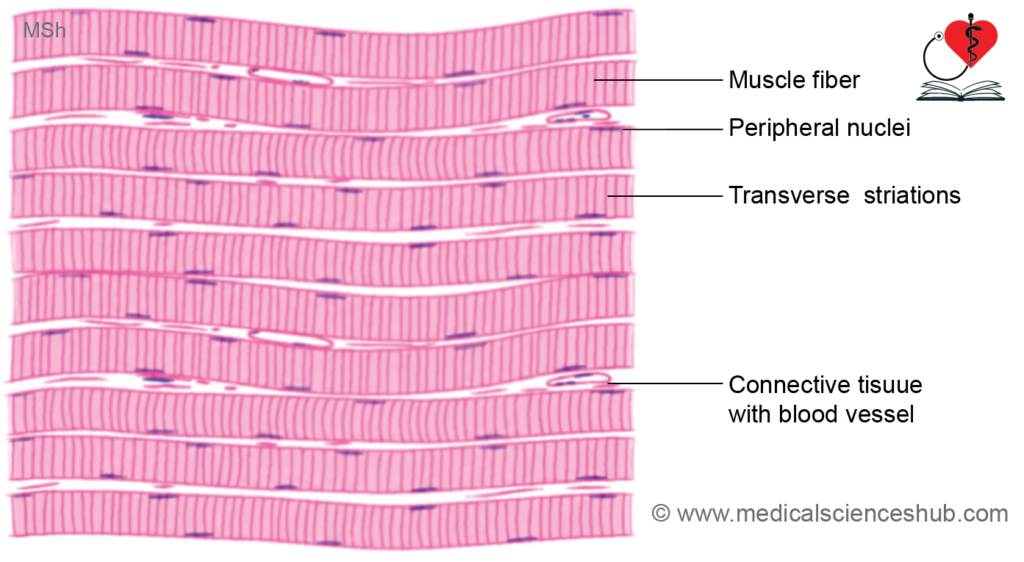
- Skeletal muscle is composed of elongated muscle cells known as muscle fibers or myocytes.
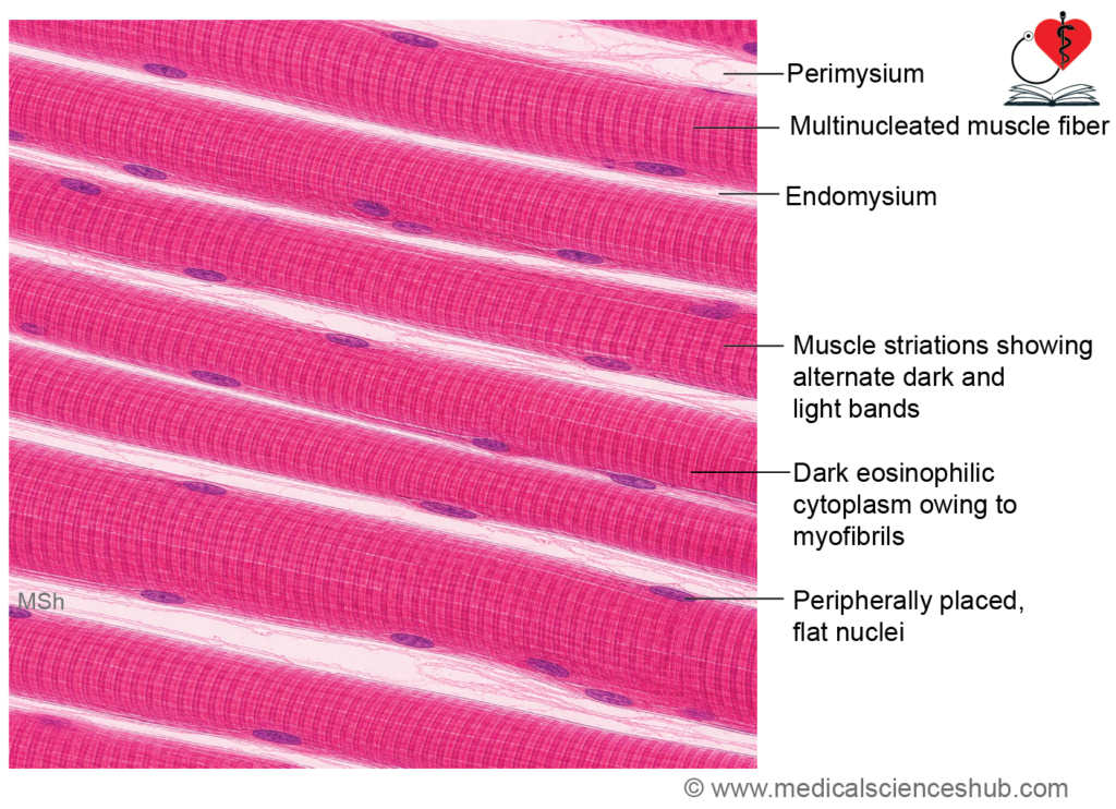
- Each muscle fiber is a long, cylindrical, multinucleated cell formed by the fusion of myoblasts during development.
- Muscle fibers are unbranched and may vary considerably in length, ranging from a few millimeters to several centimeters.
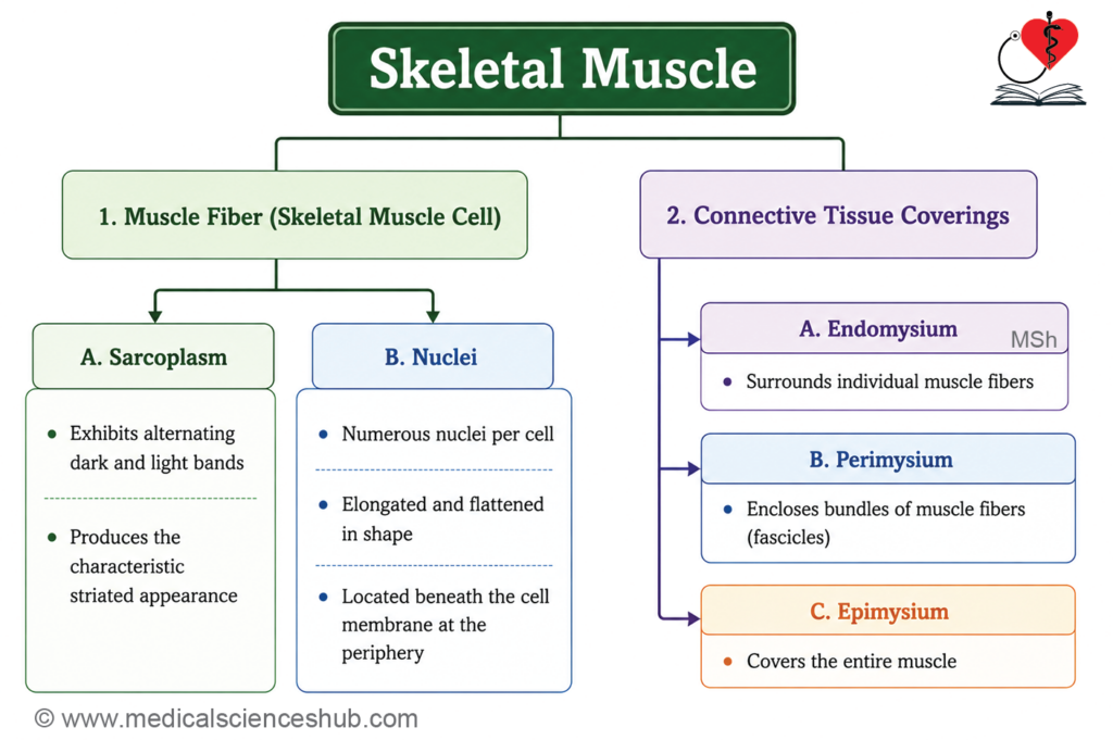
- The nuclei are flattened and lie just beneath the sarcolemma at the periphery of the cell, a characteristic feature of skeletal muscle.
- Unlike muscle fibers, connective tissue fibers are extracellular, non-living structures.
- In routine H&E-stained sections, skeletal muscle fibers exhibit prominent transverse striations due to the orderly arrangement of contractile proteins.
- Alternating dark A bands (anisotropic) and light I bands (isotropic) produce the striated appearance and differ in their behavior under polarized light.
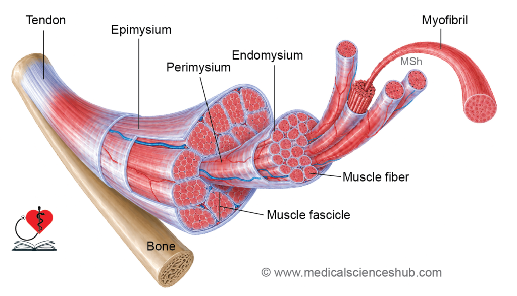
- The connective tissue framework of skeletal muscle consists of:
- Endomysium, which surrounds individual muscle fibers.
- Perimysium, which encloses bundles of muscle fibers called fascicles.
- Epimysium, which surrounds the entire muscle.
- Blood vessels and nerve fibers travel through all connective tissue layers, providing nourishment and innervation.
- The motor end plate (neuromuscular junction) is the specialized site where a motor neuron communicates with a muscle fiber to initiate contraction.
Ultrastructure of Skeletal Muscle
- Myofibrils are the principal contractile structures of skeletal muscle fibers and extend longitudinally throughout the cell.
- Each myofibril is composed of numerous myofilaments, which are organized into a highly ordered pattern.
- Myofilaments include:
- Thick filaments formed mainly by myosin II.
- Thin filaments composed of actin along with associated regulatory proteins.
- The precise arrangement of thick and thin filaments produces the characteristic striated appearance of skeletal muscle.
- Under light microscopy, alternating dark and light bands can be observed:
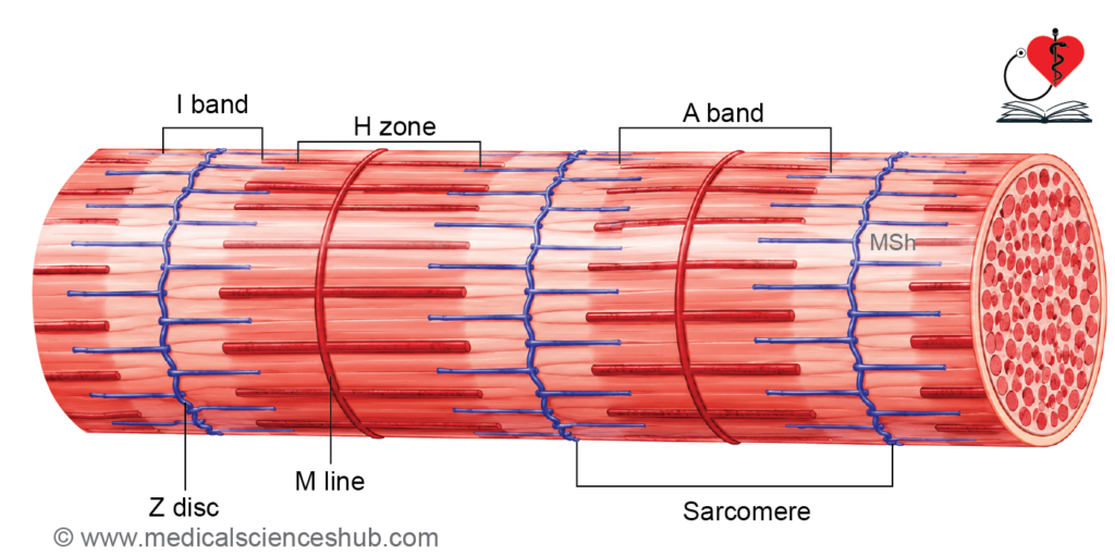
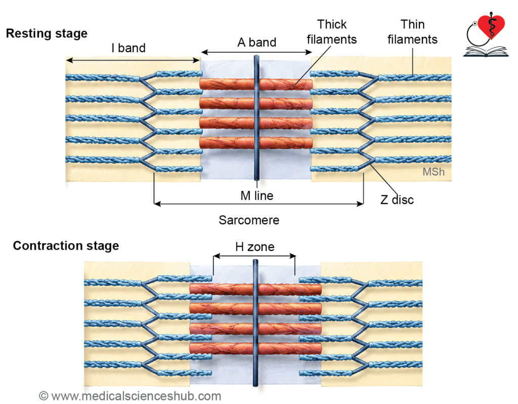
- A band (anisotropic band): Dark band containing the entire length of thick filaments and overlapping thin filaments. It appears dark under polarized light because it alters the passage of polarized light.
- I band (isotropic band): Light band containing only thin filaments. It appears lighter because polarized light passes through it with minimal alteration.
- A dark Z line (Z disc) is present at the center of each I band and serves as the attachment site for thin filaments.
- The central lighter region within the A band is the H zone, which contains only thick filaments.
- The M line, located at the center of the H zone, consists of proteins that stabilize and align thick filaments.
- A sarcomere is the structural and functional unit of a myofibril.
- Each sarcomere extends between two adjacent Z lines and measures approximately 2–3 μm in a relaxed muscle fiber.
- Repeated sarcomeres arranged end-to-end are responsible for muscle contraction and force generation.
Molecular Details of Myofibrils
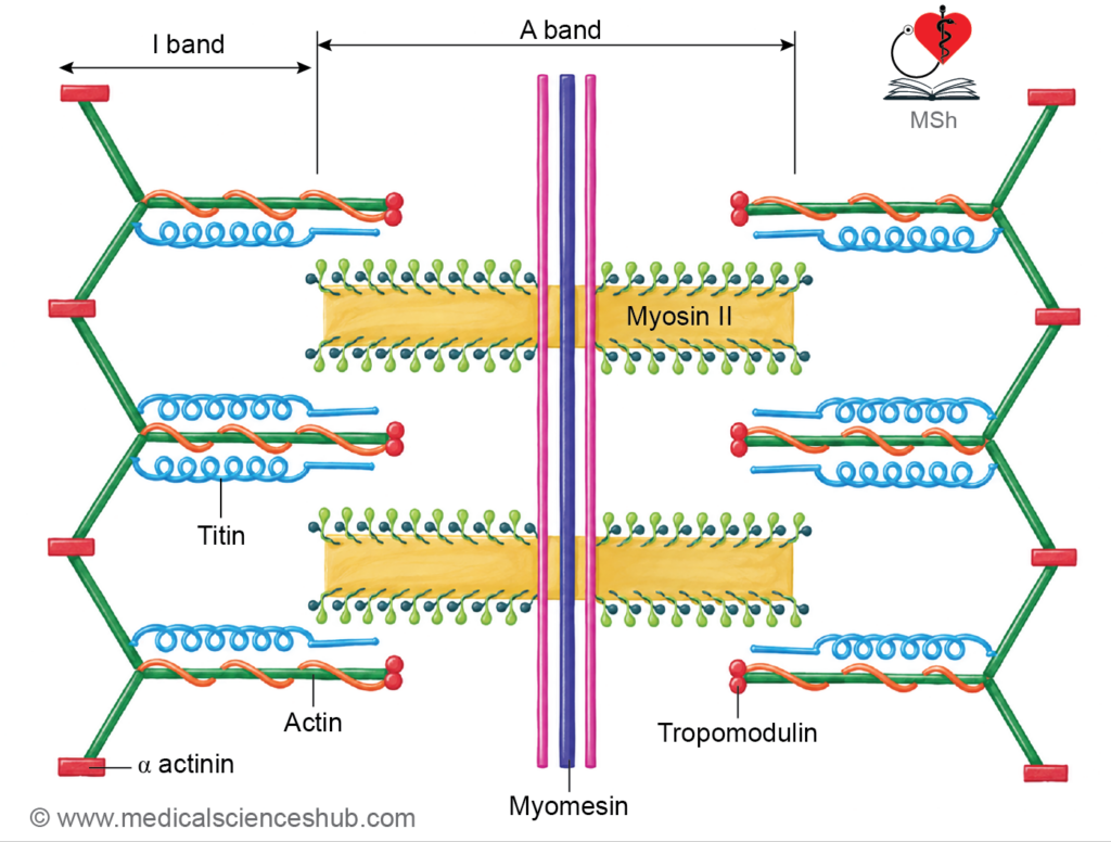
- The sarcomere, the contractile unit of skeletal muscle, contains two major filament systems: thin actin filaments and thick myosin filaments.
Thin Filament
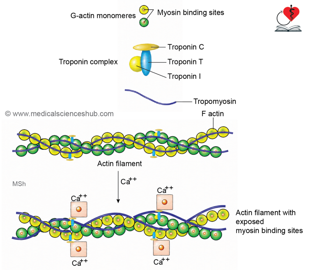
- Thin filaments measure approximately 5–7 nm in diameter and are composed of actin and associated regulatory proteins.
- Each filament contains a double-helical strand of F-actin, formed by polymerization of G-actin subunits.
- Every G-actin molecule possesses a myosin-binding site that participates in muscle contraction.
- Tropomyosin runs along the actin filament and covers the myosin-binding sites during muscle relaxation.
- Troponin regulates contraction and consists of three subunits:
- Troponin C (TnC): Binds calcium ions (Ca²⁺).
- Troponin T (TnT): Attaches troponin to tropomyosin.
- Troponin I (TnI): Inhibits actin-myosin interaction by binding to actin.
- Tropomodulin is located at the free end of the thin filament and regulates filament length.
- Nebulin acts as a molecular ruler that stabilizes and helps maintain the length of thin filaments.
- Dystrophin links the actin cytoskeleton to the muscle cell membrane and extracellular matrix. Deficiency of dystrophin causes Duchenne muscular dystrophy.
Thick Filament
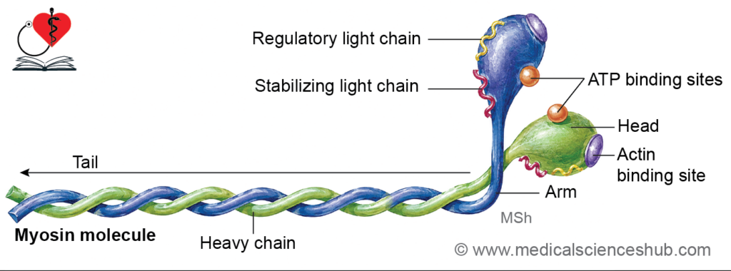
- Thick filaments are primarily composed of myosin II molecules.
- Each myosin II molecule contains:
- Two intertwined heavy chains.
- Four light chains (two essential and two regulatory light chains).
- Each heavy chain has:
- A long tail region.
- A globular head (S1 region) that functions as the motor domain.
- A neck/arm region (S2 region) connecting the head to the tail.
- Myosin heads possess binding sites for both ATP and actin, enabling force generation during contraction.
- Thick filaments contain a central bare zone, where myosin heads are absent.
- Adjacent thick filaments are interconnected at the M line by specialized proteins.
Accessory Proteins
- Several proteins provide structural support and maintain sarcomere organization:
- Titin: An elastic protein that connects thick filaments to the Z line and prevents excessive sarcomere stretching.
- α-Actinin: Anchors thin filaments to the Z line.
- Desmin: Links adjacent Z lines and helps maintain alignment of myofibrils within the muscle fiber.
Muscle Contraction
- Muscle contraction occurs through the interaction of thin actin filaments and thick myosin filaments within the sarcomere.
- During contraction:
- The overlap between actin and myosin filaments increases.
- The sarcomere shortens.
- The I band becomes narrower.
- The H zone decreases in width and may disappear in maximal contraction.
- The A band remains unchanged because the length of thick filaments does not alter.
- The widely accepted explanation for muscle contraction is the Sliding Filament Theory, which is based on the actomyosin cross-bridge cycle.
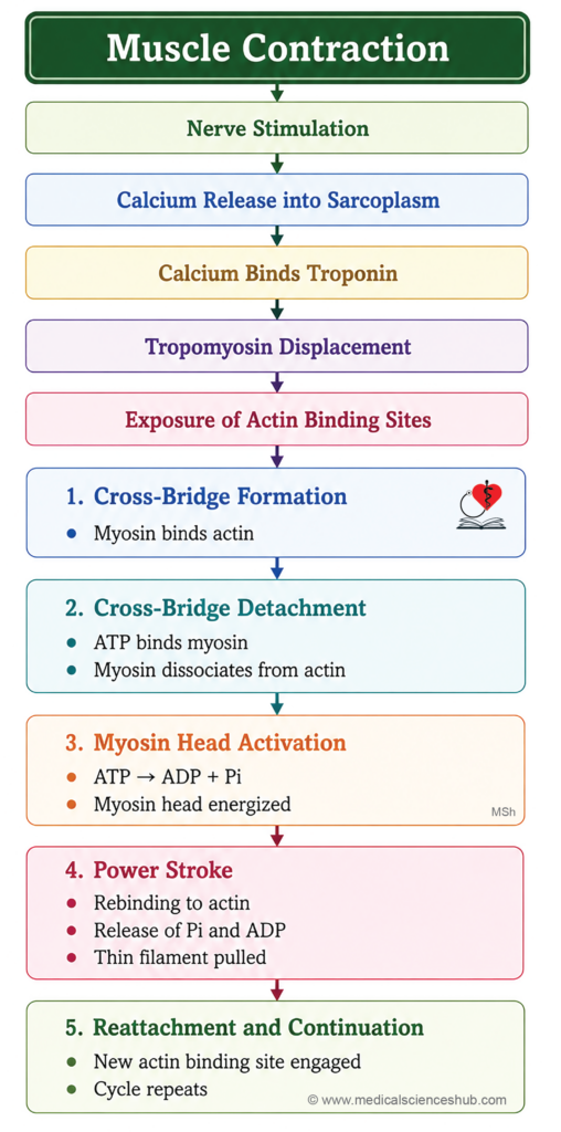
- In the relaxed state, tropomyosin covers the myosin-binding sites on actin, preventing interaction between actin and myosin.
- Nerve stimulation triggers the release of Ca²⁺ from the sarcoplasmic reticulum into the sarcoplasm.
- Calcium ions bind to troponin C, causing a conformational change that shifts tropomyosin away from the myosin-binding sites on actin.
- Exposure of these binding sites allows the actomyosin cross-bridge cycle to proceed through the following steps:
- Cross-bridge formation: Myosin heads attach to exposed binding sites on actin.
- Detachment: Binding of ATP to myosin decreases its affinity for actin, causing myosin to detach.
- Recovery stroke: Hydrolysis of ATP to ADP and inorganic phosphate (Pi) energizes the myosin head and moves it into a high-energy position.
- Power stroke: The energized myosin head binds to a new site on actin. Release of Pi, followed by ADP, causes the myosin head to pivot, pulling the thin filament toward the center of the sarcomere.
- Reattachment: The myosin head remains attached to actin until another ATP molecule binds, initiating a new cycle.
- Repeated cross-bridge cycling results in the sliding of thin filaments over thick filaments, producing shortening of the sarcomere and generation of muscular force.
Sarcoplasmic Reticulum and T-tubules
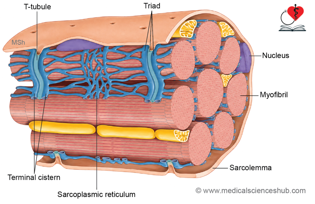
- The sarcoplasmic reticulum (SR) is a specialized form of smooth endoplasmic reticulum present within skeletal muscle fibers.
- It consists of an extensive network of interconnected longitudinal tubules that surround the myofibrils.
- The primary function of the sarcoplasmic reticulum is the storage, release, and reuptake of calcium ions (Ca²⁺), which are essential for muscle contraction and relaxation.
- The tubular network of the SR forms numerous anastomosing channels that facilitate rapid calcium movement throughout the muscle fiber.
Terminal Cisternae
- At specific locations near the junction of the A band and I band, the sarcoplasmic reticulum expands to form enlarged sac-like structures called terminal cisternae.
- Terminal cisternae serve as major intracellular reservoirs of calcium ions.
- During muscle excitation, these structures rapidly release calcium into the sarcoplasm, initiating contraction.
T-Tubules (Transverse Tubules)
- T-tubules are deep invaginations of the sarcolemma (muscle cell membrane).
- They extend transversely across the muscle fiber and penetrate deep into the sarcoplasm.
- Because T-tubules are continuous with the plasma membrane, their lumen contains extracellular fluid.
- In skeletal muscle, T-tubules are typically located at the junction of the A and I bands.
- Their strategic position allows rapid transmission of electrical signals from the cell surface to the interior of the muscle fiber.
Triad
- A triad is a specialized structural complex formed by:
- One central T-tubule, and
- Two adjacent terminal cisternae of the sarcoplasmic reticulum, one on each side.
- Triads are located at the A-I band junction in skeletal muscle fibers.
- This close anatomical relationship ensures efficient communication between membrane depolarization and calcium release.
Functions of T-Tubules and Triads
- T-tubules rapidly conduct the action potential from the sarcolemma to the deepest regions of the muscle fiber.
- Depolarization of the T-tubule membrane activates calcium-release channels in the adjacent terminal cisternae.
- This process results in the sudden release of Ca²⁺ into the sarcoplasm.
- Released calcium binds to troponin C, initiating the interaction between actin and myosin filaments and triggering muscle contraction.
- The triad system enables synchronized activation of myofibrils throughout the muscle fiber, ensuring rapid and efficient contraction.
- Following contraction, calcium ions are actively pumped back into the sarcoplasmic reticulum, allowing muscle relaxation and preparing the fiber for subsequent contractions.
Nerve Supply of Skeletal Muscle
- Skeletal muscle fibers receive motor innervation from α-motor neurons located in the anterior horn of the spinal cord.
- The axons of these neurons are large-diameter, myelinated nerve fibers that conduct impulses rapidly to muscle fibers.
- A motor unit consists of a single α-motor neuron and all the muscle fibers it supplies.
- Small motor units are found in muscles requiring precise movements, such as the extraocular muscles.
- Large motor units are characteristic of postural and powerful muscles, where fine control is less important.
- The neuromuscular junction (NMJ) or motor end plate is the specialized site of communication between a motor nerve terminal and a skeletal muscle fiber.
- Muscle spindles are sensory receptors located within skeletal muscles that monitor muscle length and stretch.
- They receive motor innervation from γ-motor neurons, whose myelinated fibers regulate spindle sensitivity.
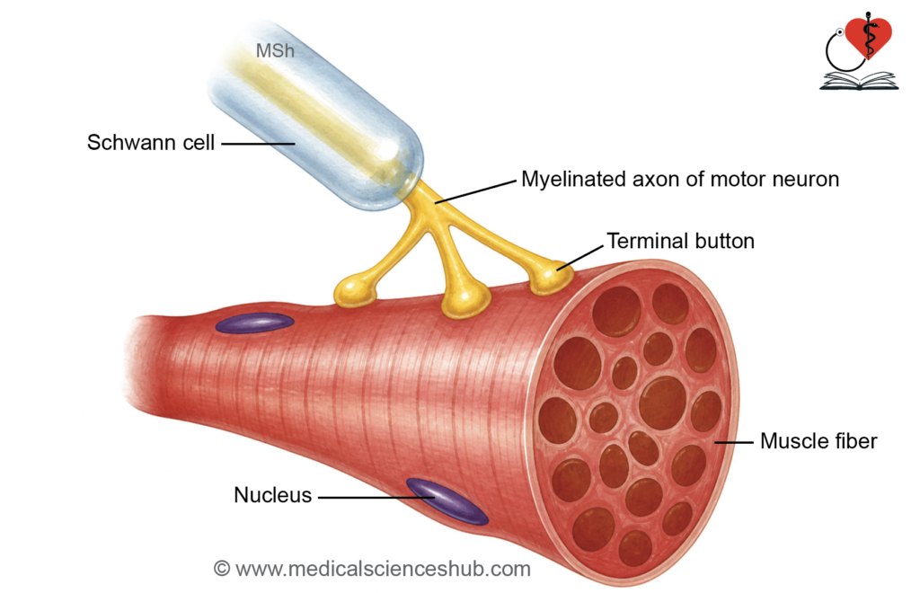
H4: Neuromuscular Junction/Motor End Plate
- Near the muscle fiber, the motor axon loses its myelin sheath and divides into several terminal branches.
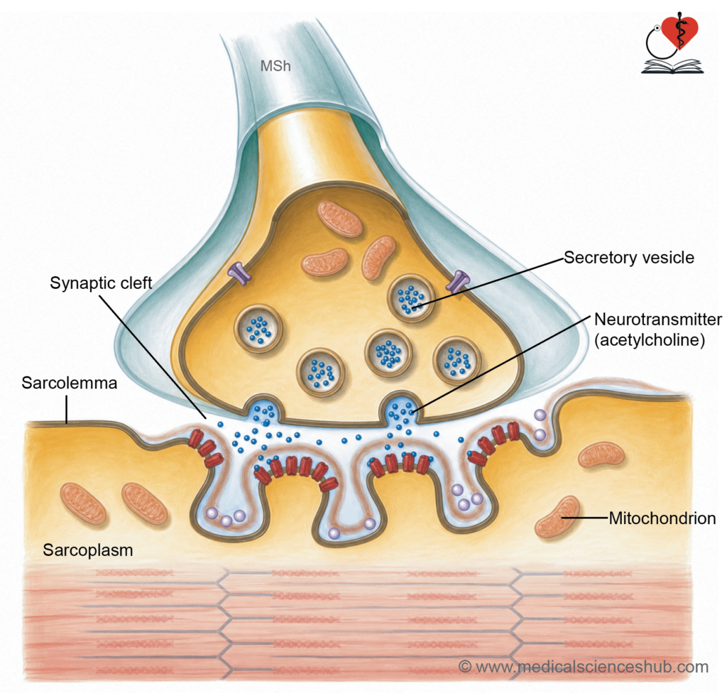
- Each branch ends in an expanded terminal bouton (terminal button) that lies against a specialized region of the sarcolemma called the motor end plate.
- Terminal boutons contain numerous mitochondria and synaptic vesicles filled with the neurotransmitter acetylcholine (ACh).
- The motor end plate exhibits deep junctional folds that increase surface area and contain a high density of acetylcholine receptors.
- A narrow synaptic cleft (approximately 40–100 nm wide) separates the nerve terminal from the muscle membrane.
- Released acetylcholine diffuses across the synaptic cleft and binds to receptors on the motor end plate, initiating muscle excitation.
- Acetylcholinesterase (AChE) rapidly breaks down acetylcholine, terminating the signal and preventing continuous stimulation of the muscle fiber.
CLINICAL CORRELATION
- Myasthenia gravis is an autoimmune disorder in which antibodies target acetylcholine receptors (AChRs) at the neuromuscular junction. Progressive loss of these receptors reduces neuromuscular transmission, leading to muscle weakness and fatigability. Many snake venoms contain neurotoxins that interfere with acetylcholine receptor function at the neuromuscular junction. Severe envenomation may cause paralysis of respiratory muscles, which can be life-threatening.
- The stretch reflex (myotatic reflex) is a monosynaptic reflex that helps maintain muscle tone and posture. It is elicited by tapping a muscle tendon, causing sudden stretching of muscle spindles. Sensory impulses from the spindles activate α-motor neurons, resulting in contraction of the same muscle. A common example is the biceps tendon reflex.
Muscle spindle
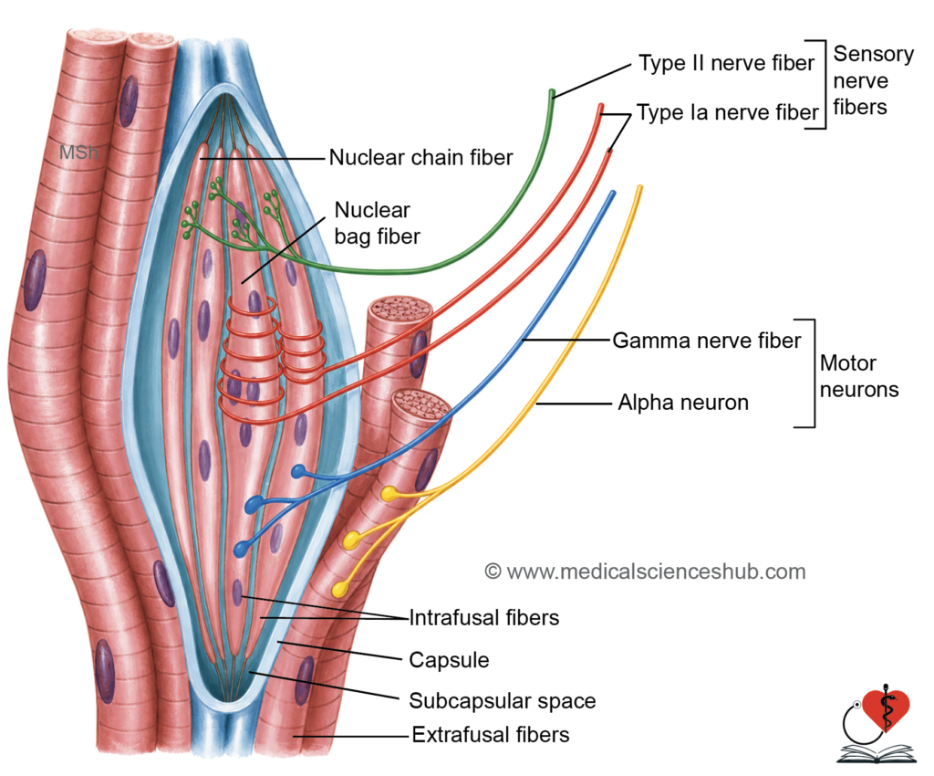
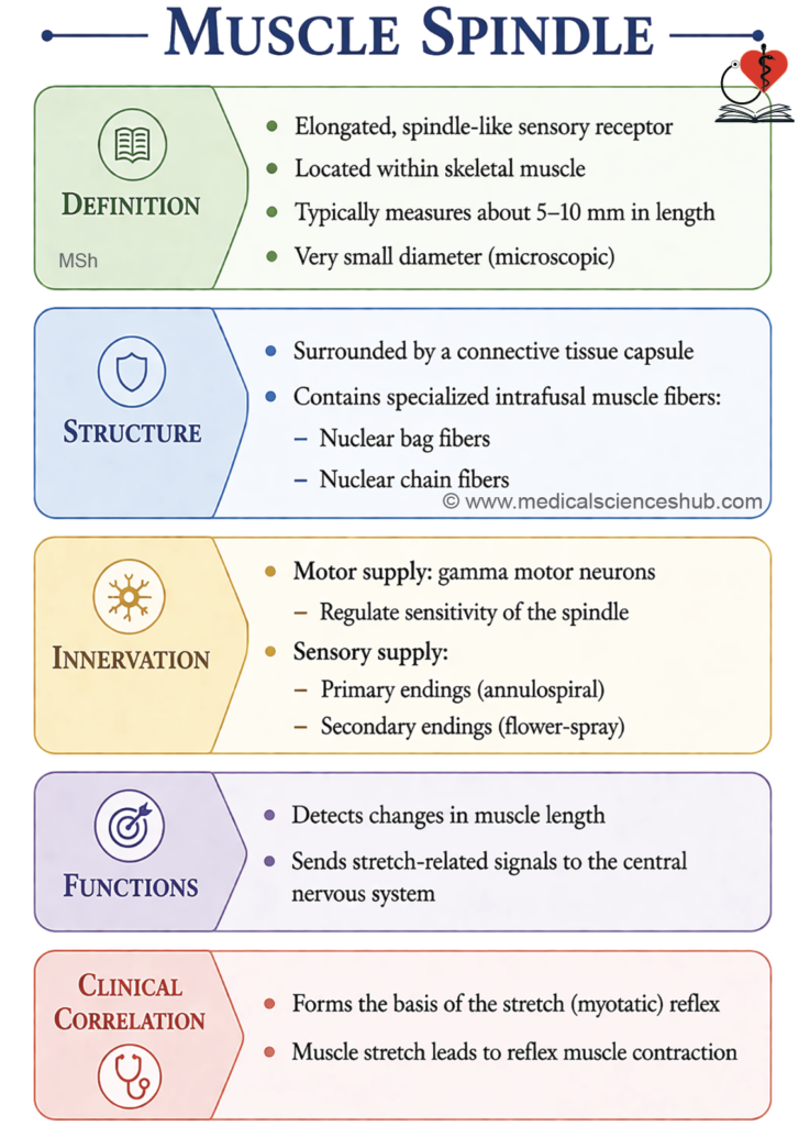
- The muscle spindle (neuromuscular spindle) is a specialized sensory receptor located within skeletal muscles that detects changes in muscle length and the rate of stretching.
- It is a spindle-shaped structure measuring approximately 5–10 mm in length and about 100 μm in diameter.
Structure
- Each muscle spindle is enclosed by a connective tissue capsule.
- Within the capsule are specialized intrafusal muscle fibers, sensory nerve endings, and motor nerve fibers.
- The main contractile muscle fibers outside the spindle are called extrafusal fibers and constitute the bulk of the muscle.
Types of Intrafusal Fibers
- Nuclear bag fibers contain numerous centrally clustered nuclei and are larger in diameter.
- Nuclear chain fibers contain nuclei arranged in a single longitudinal row.
- The ends of intrafusal fibers are attached to surrounding connective tissue and adjacent extrafusal muscle fibers.
Nerve Supply
- Intrafusal fibers receive motor innervation from γ-motor neurons, which regulate spindle sensitivity.
- Sensory information is carried to the central nervous system by type Ia and type II afferent fibers.
- Type Ia fibers form annulospiral endings around the central regions of both nuclear bag and nuclear chain fibers.
- Type II fibers form flower-spray endings, primarily on nuclear chain fibers and static nuclear bag fibers.
- In contrast, extrafusal fibers are supplied by α-motor neurons.
Functions
- Stretching of a muscle deforms the intrafusal fibers and activates sensory endings.
- Muscle spindles continuously provide information about muscle length and stretch to the central nervous system (CNS).
- This feedback helps regulate muscle tone, coordinate movement, and maintain posture through the stretch reflex.
Cardiac Muscle
- Cardiac muscle is a striated involuntary muscle found exclusively in the heart.
- Cardiac muscle fibers possess autorhythmicity, enabling spontaneous generation of impulses and rhythmic contraction without external stimulation.
- The fibers display prominent cross striations due to the organized arrangement of contractile proteins.
- Cardiac activity is regulated by the autonomic nervous system, with sympathetic stimulation increasing heart rate and force of contraction, while parasympathetic stimulation reduces heart rate.
- Adjacent cardiac myocytes are connected by intercalated discs, which provide mechanical and electrical coupling between cells.
Histology of Cardiac Muscle
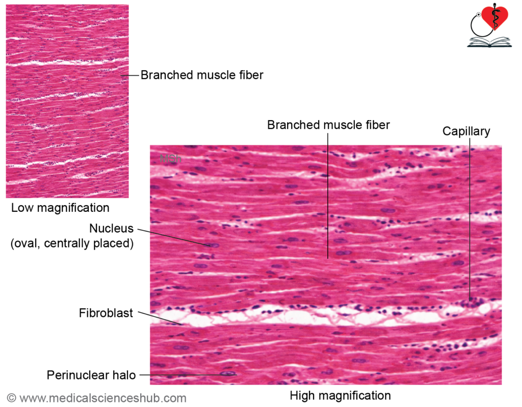
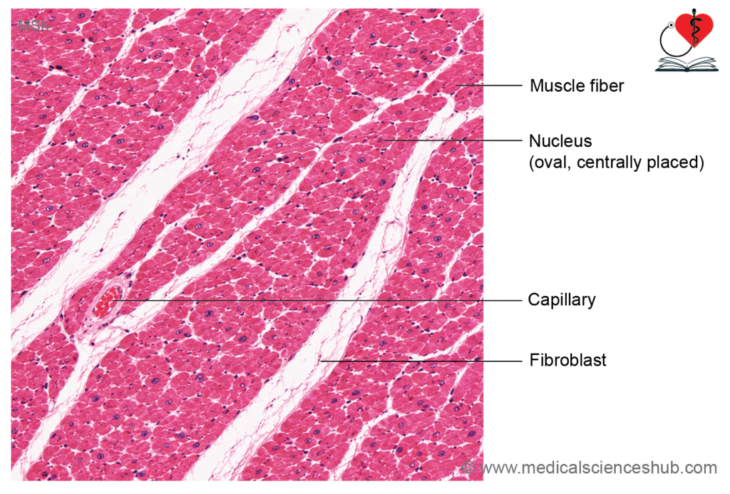
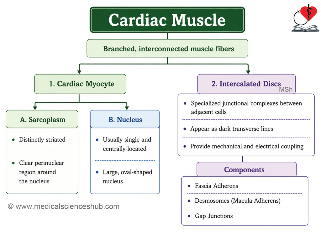
Cardiac muscle is composed of branching, interconnected muscle cells called cardiac myocytes, which form a functional network within the heart.
Cardiac Myocytes and Muscle Fibers
- Individual cardiac myocytes are typically 50–100 μm long and approximately 15 μm in diameter.
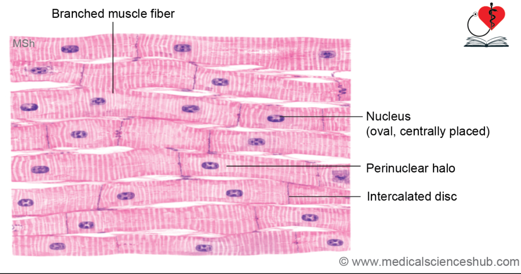
- Each cell usually contains a single centrally located oval nucleus, although occasional binucleate cells may be present.
- In contrast to skeletal muscle fibers, cardiac myocytes are shorter, branched, and connected end-to-end.
Cell Organelles
- A pale-staining perinuclear region (perinuclear halo) surrounds the nucleus and contains most cellular organelles.
- This region is relatively free of myofibrils.
- Cardiac myocytes contain abundant mitochondria, reflecting the high energy demand of continuous contraction.
- Other organelles include the Golgi apparatus, glycogen deposits, and lipofuscin granules, which may accumulate with age.
- Atrial myocytes contain secretory granules that store atrial natriuretic peptide (ANP) and brain natriuretic peptide (BNP), hormones involved in regulation of blood volume and blood pressure.
Intercalated Disc
- Adjacent cardiac myocytes are joined by specialized junctional complexes called intercalated discs.
- In routine H&E sections, intercalated discs appear as dark-staining transverse lines crossing the muscle fibers.
- They often exhibit a characteristic step-like arrangement.
- Intercalated discs contain specialized cell junctions that provide strong mechanical attachment and rapid electrical communication between neighboring cells.
Myofibrils and Striations
- Cardiac myocytes contain myofibrils arranged in a pattern similar to skeletal muscle.
- Consequently, they display A bands, I bands, Z lines, and H zones.
- However, myofibrils are less densely packed than in skeletal muscle, making striations less prominent.
- The greater volume of sarcoplasm accommodates numerous mitochondria required for sustained activity.
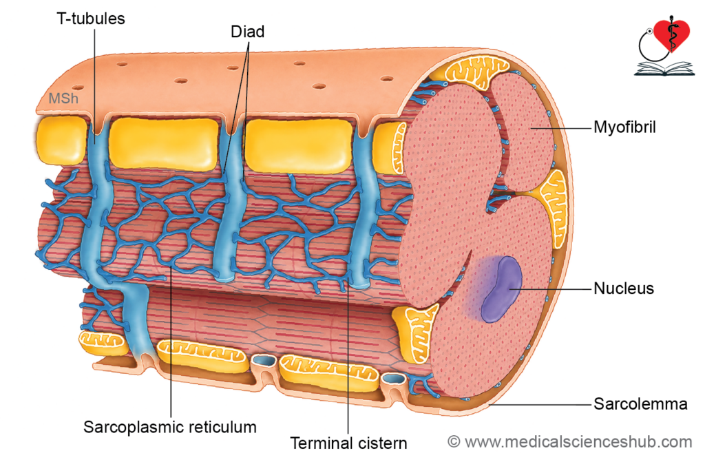
T-tubules and Sarcoplasmic Reticulum
- The sarcoplasmic reticulum in cardiac muscle is less extensive and less organized than that of skeletal muscle.
- T-tubules, which are invaginations of the sarcolemma, are located at the level of the Z lines.
- Each T-tubule is associated with a single terminal cisterna of the sarcoplasmic reticulum, forming a diad.
- This arrangement differs from skeletal muscle, where a triad consists of one T-tubule flanked by two terminal cisternae.
Intercalated disc
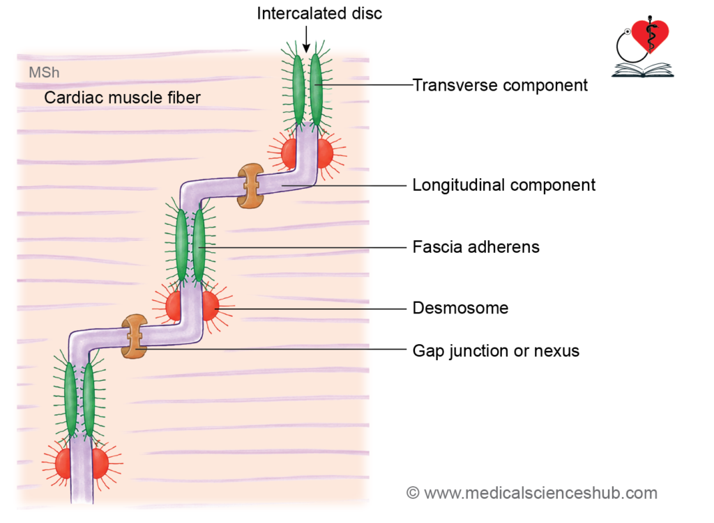
- Intercalated discs are specialized junctional complexes that connect adjacent cardiac myocytes, providing both mechanical attachment and electrical communication.
- In routine H&E-stained sections, they appear as dark-staining transverse lines crossing cardiac muscle fibers and serve as an important identifying feature of cardiac muscle.
- Intercalated discs consist of two structural components:
- Transverse component, oriented perpendicular to the myofibrils in a step-like pattern.
- Lateral (longitudinal) component, oriented parallel to the myofibrils and best visualized by electron microscopy.
- Three major types of cell junctions are present within intercalated discs:
Fascia Adherens
- Forms the largest part of the transverse component.
- Functions similarly to the zonula adherens of epithelial cells.
- Anchors adjacent cardiac myocytes and provides attachment sites for thin actin filaments of the terminal sarcomere.
- Contributes significantly to force transmission during contraction.
Desmosomes (Macula Adherens)
- Present in both transverse and lateral components.
- Provide strong mechanical adhesion between neighboring cardiac myocytes.
- Help prevent separation of cells during repetitive cardiac contractions.
Gap Junctions
- Located mainly in the lateral component.
- Create low-resistance channels that permit the passage of ions and small molecules between adjacent cells.
- Facilitate rapid spread of action potentials throughout the myocardium.
- Through the combined action of fascia adherens, desmosomes, and gap junctions, cardiac muscle functions as a functional (physiological) syncytium, allowing coordinated and synchronized contraction of the heart.
Smooth Muscle
- Smooth muscle is a non-striated, involuntary muscle controlled primarily by the autonomic nervous system.
- It is present in the walls of the gastrointestinal tract, blood vessels, respiratory passages, urinary tract, and arrector pili muscles of the skin.
Structure of Smooth Muscle
- Smooth muscle is composed of elongated, spindle-shaped cells with tapered ends.
- Cell size varies with location, ranging from about 15 μm in small blood vessels to 500 μm or more in the pregnant uterus.
H&E Staining
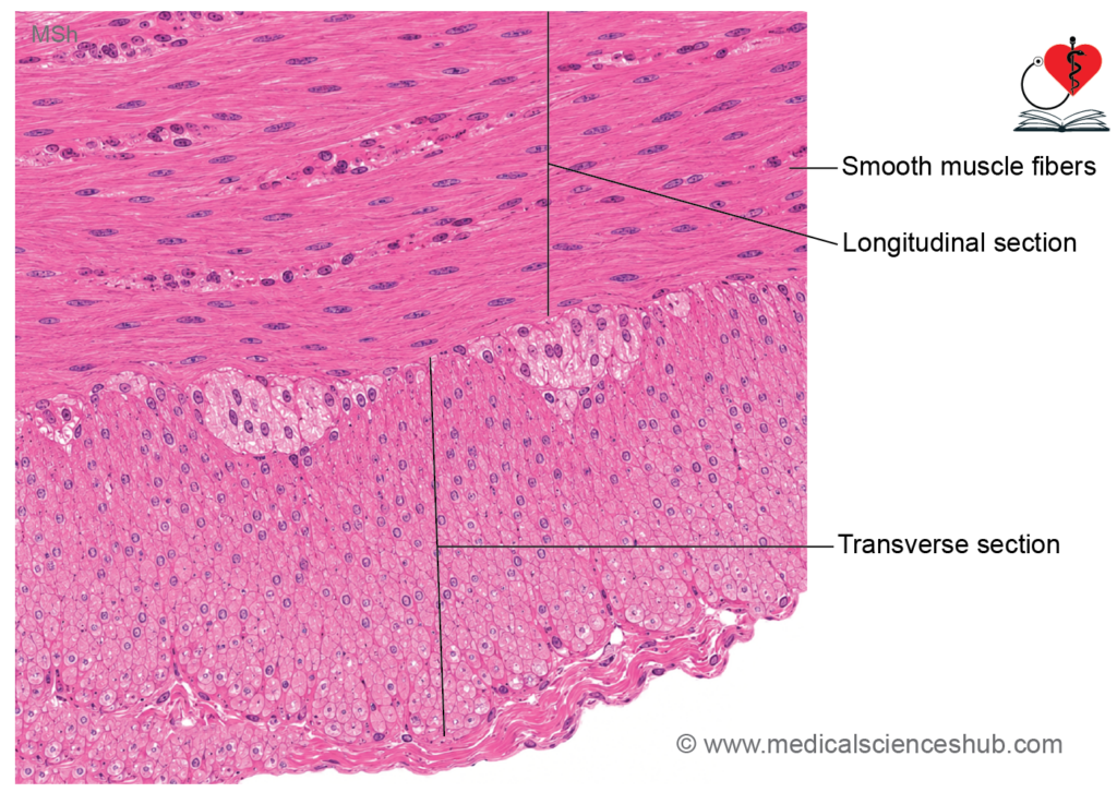
- In longitudinal sections, smooth muscle cells appear elongated with eosinophilic cytoplasm and a single, centrally located, elongated nucleus.
- The cells are arranged closely and parallel to one another, making individual cell boundaries difficult to distinguish.
- In transverse sections, cells appear circular or polygonal, with centrally placed nuclei visible only in sections passing through the cell center.
- Smooth muscle cells are commonly organized into bundles or layers.
- These bundles may be arranged longitudinally, circularly, or obliquely depending on the function of the organ.
- Within a bundle, the thick central portion of one cell lies adjacent to the tapered end of neighboring cells, producing a compact and efficient arrangement.
Electron Microscopy of Smooth Muscle
- Each smooth muscle cell is enclosed by a plasma membrane and is externally surrounded by a basal lamina and a delicate network of reticular fibers.
- Adjacent smooth muscle cells communicate through gap junctions, which facilitate the spread of electrical signals and promote coordinated contraction.
- Unlike skeletal muscle fibers, smooth muscle cells lack T-tubules.
- The contractile apparatus consists of thin actin filaments and thick myosin filaments.
- These filaments are arranged in an irregular, non-sarcomeric pattern rather than in orderly striated units. As a result, smooth muscle does not exhibit visible cross-striations under light microscopy.
- The cytoplasm contains specialized anchoring structures called dense bodies, which function similarly to Z discs of skeletal muscle. Dense bodies are connected to the sarcolemma by intermediate filaments and serve as attachment sites for actin filaments.
- During contraction, actin filaments slide over myosin filaments through an ATP-dependent mechanism. This sliding action pulls dense bodies closer together, resulting in shortening of the smooth muscle cell.
- The arrangement of contractile filaments allows smooth muscle cells to contract efficiently over a wide range of lengths while maintaining force generation.
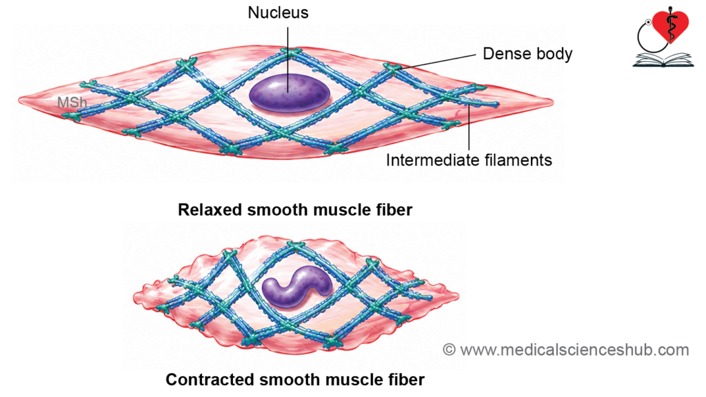
Table 7.1: Differences between skeletal, cardiac and smooth muscles
| Feature | Skeletal Muscle | Cardiac Muscle | Smooth Muscle |
|---|---|---|---|
| Muscle cells | Long, cylindrical, unbranched fibers; may extend several centimeters in length | Short, branched cylindrical cells measuring approximately 50–100 μm | Small, spindle-shaped cells measuring about 20–500 μm |
| Nuclei | Multiple nuclei located at the cell periphery | Usually a single centrally placed nucleus | Single elongated nucleus located centrally |
| Striations | Distinct cross-striations are present | Striations are present but less prominent | Striations are absent |
| Sarcoplasmic reticulum | Highly developed and well organized | Less extensive than in skeletal muscle | Poorly developed |
| T-tubules | Present at the A–I junction | Present at the Z line | Absent |
| Cell junctions | Specialized cell junctions are absent | Intercalated discs connect adjacent cells | Gap junctions commonly connect neighboring cells |
| Control | Voluntary | Involuntary | Involuntary |
| Innervation | Supplied by somatic motor nerves | Regulated by the autonomic nervous system | Regulated by the autonomic nervous system |
| Mitotic activity | Absent under normal conditions | Virtually absent in adult cardiac muscle | Limited mitotic activity may occur |
| Regenerative capacity | Minimal | Negligible | Relatively good regenerative ability |
| Typical locations | Attached to the skeleton; also present in the tongue, pharynx, upper esophagus, and diaphragm | Forms the myocardium of the heart and extends into the roots of major vessels | Found in the walls of blood vessels, hollow viscera, and various internal organs |
Important Questions
- Write a short note on intercalated disc.
- List the differences between red fibers and white fibres.
- Write a short note on histology of cardiac muscle.
- List the differences between skeletal, cardiac and smooth muscles.
📝 Test Your Knowledge – Practice MCQs
Attempt the chapter MCQ quiz and assess your understanding of key concepts.
Click to see: Medical Disclaimer and Copyright Notice